Embed Size (px)
Citation preview
Submitted 25 November 2015Accepted 14 April 2017Published 31 May 2017
Corresponding authorsRory M. Welsh,[email protected] L. Vega Thurber, [email protected]
Academic editorMauricio Rodriguez-Lanetty
Additional Information andDeclarations can be found onpage 17
DOI 10.7717/peerj.3315
Copyright2017 Welsh et al.
Distributed underCreative Commons CC-BY 4.0
OPEN ACCESS
Alien vs. predator: bacterial challengealters coral microbiomes unlesscontrolled by HalobacteriovoraxpredatorsRory M. Welsh1,*, Stephanie M. Rosales1, Jesse R. Zaneveld1,2,Jérôme P. Payet1, Ryan McMinds1, Steven L. Hubbs1 andRebecca L. Vega Thurber1,*
1Department of Microbiology, Oregon State University, Corvallis, OR, USA2Department of Biological Sciences, University of Washington Bothell, Bothell, WA, USA*These authors contributed equally to this work.
ABSTRACTCoral microbiomes are known to play important roles in organismal health, response toenvironmental stress, and resistance to disease. The coral microbiome contains diverseassemblages of resident bacteria, ranging from defensive and metabolic symbionts toopportunistic bacteria that may turn harmful in compromised hosts. However, little isknown about how these bacterial interactions influence the mechanism and controlsof overall structure, stability, and function of the microbiome. We sought to test howcoral microbiome dynamics were affected by interactions between two bacteria: Vibriocoralliilyticus, a known temperature-dependent pathogen of some corals, andHalobac-teriovorax, a unique bacterial predator of Vibrio and other gram-negative bacteria.We challenged reef-building coral with V. coralliilyticus in the presence or absence ofHalobacteriovorax predators, and monitored microbial community dynamics with 16SrRNA gene profiling time-series. Vibrio coralliilyticus inoculation increased the meanrelative abundance of Vibrios by greater than 35% from the 4 to 8 hour time point, butnot in the 24 & 32 hour time points. However, strong secondary effects of the Vibriochallenge were also observed for the rest of the microbiome such as increased richness(observed species), and reduced stability (increased beta-diversity). Moreover, after thetransient increase inVibrios, two lineages of bacteria (Rhodobacterales andCytophagales)increased in coral tissues, suggesting that V. coralliilyticus challenge opens niche spacefor these known opportunists. Rhodobacterales increased from 6.99% (±0.05 SEM)to a maximum mean relative abundance of 48.75% (±0.14 SEM) in the final timepoint and Cytophagales from <0.001% to 3.656%. Halobacteriovorax predators arecommonly present at low-abundance on coral surfaces. Based on the keystone roleof predators in many ecosystems, we hypothesized that Halobacteriovorax predatorsmight help protect corals by consuming foreign or ‘‘alien’’ gram negative bacteria.Halobacteriovorax inoculation also altered the microbiome but to a lesser degreethan V. coralliilyticus, and Halobacteriovorax were never detected after inoculation.Simultaneous challenge with both V. coralliilyticus and predatory Halobacteriovoraxeliminated the increase in V. coralliilyticus, ameliorated changes to the rest of the coralmicrobiome, and prevented the secondary blooms of opportunistic Rhodobacterales
How to cite this article Welsh et al. (2017), Alien vs. predator: bacterial challenge alters coral microbiomes unless controlled by Halobac-teriovorax predators. PeerJ 5:e3315; DOI 10.7717/peerj.3315
and Cytophagales seen in the V. coralliilyticus challenge. These data suggest that, undercertain circumstances, host-associated bacterial predators may mitigate the ability ofother bacteria to destabilize the microbiome.
Subjects Bioinformatics, Ecology, Marine Biology, MicrobiologyKeywords BALOs, Halobacteriovorax, Vibrio coralliilyticus, Microbiome, Bacterial challenge
INTRODUCTIONCoral reefs have experienced sharp declines in coral cover from environmental factors(De’ath et al., 2012), temperature induced bleaching (Fitt & Warner, 1995), and disease(Bourne et al., 2009; Burge et al., 2014), with some areas of the Caribbean experiencing asmuch as 80% coral loss over the past several decades (Gardner et al., 2003). While manystudies have identified microbial consortia that increase in diseased corals (e.g., Gignoux-Wolfsohn & Vollmer, 2015), etiological agents are generally unknown for the majority ofcoral diseases (Mouchka, Hewson & Harvell, 2010) and others are defined in broad terms aspolymicrobial disease (Cooney et al., 2002). Vibrio coralliilyticus is a well described modelbacterium for the study of interactions between corals, the environment, and pathogenicbacteria (Ben-Haim et al., 2003). Several V. coralliilyticus virulence factors are temperature-dependent and upregulated above 27 ◦C (Kimes et al., 2012), and it has been suggestedthat host tissue invasion can only occur above this threshold (Vidal-Dupiol et al., 2011).Given the continuous rise in sea surface temperatures due to global climate change (Hoegh-Guldberg et al., 2007), and the projected increased variability of temperature extremes, it islikely that the incidence of infections by V. coralliilyticus and other temperature-dependentpathogens will increase (Maynard et al., 2015). Bacterial communities of diseased corals arealso known to have large numbers of opportunistic pathogens and secondary colonizers(Gignoux-Wolfsohn & Vollmer, 2015). It has been hypothesized that the majority of coraldisease may be the result of normally-benign coral microbionts that become opportunisticpathogens during physiological stress to the host (Lesser et al., 2007a). Thus, the linkagesbetween infection by a primary foreign agents and secondary opportunistic infectionsremain an area of active exploration.
Corals also formmutualistic and commensal partnerships with diverse microorganisms,ranging from endosymbiotic photosynthetic dinoflagellates (Symbiodinium spp.), toconsortia of archaea, fungi, and bacteria. Although the role of Symbiodinium in the coralholobiont is well studied, the exact roles of each member of the bacterial portion of theholobiont remains far from clear. Experiments and metagenomic analyses have providedsome insights into the roles of individual members of the coral microbiome (e.g., Wegleyet al., 2007). It has been suggested that some of these bacteria provide direct benefits to thecoral host, such as nitrogen fixation by symbiotic Cyanobacteria in Montastraea cavernosa(Lesser et al., 2007b), or ammonia oxidation by archaea (Beman et al., 2007). Other bacteria,particularly those in the coral surface mucus layer, are thought to provide a first line ofdefense against potentially invading foreign bacteria. Mucosal bacteria are thought toprotect the host by several mechanisms, including production of antibiotics (Ritchie,
Welsh et al. (2017), PeerJ, DOI 10.7717/peerj.3315 2/22
2006), secretion of chemical compounds that inhibit pathogen metabolism (Rypien, Ward& Azam, 2010), or competition for necessary resources and niche space (Ritchie & Smith,1997). Increasingly, viruses and phages are recognized as also playing a regulatory role inthe holobiont by controlling microbial populations (Barr et al., 2013; Soffer, Zaneveld &Thurber, 2014; Nguyen-Kim et al., 2014).
We have recently described how the predatory bacteria Halobacteriovorax, also likelyinfluences the diversity and dynamics of the microbial community in the coral surfacemucus layer through consumption of a broad range of bacterial prey (Welsh et al., 2016).Halobacteriovorax spp. are small, highly motile predatory bacteria that exhibit a biphasiclifestyle and prey exclusively on gram negative bacteria, including known coral pathogens(Williams, Falkler & Shay, 1980; Welsh et al., 2016). Halobacteriovorax are the marinecomponent of a group of delta-proteobacteria known as Bdellovibrio and like organisms(BALOs). In free-living attack phase, BALOs actively seek out prey in order to attach,burrow inside, and restructure their host cell into a rounded bdelloplast. This kills theirprey and provides BALOs with an osmotically stable structure free from competitionto utilize prey resources for growth and replication. A new generation of attack-phasepredators then bursts forth from the bdelloplast to seek new hosts.
Bacterial predators in the coral microbiome could be a type of top-down control,that directly alters the structure and function of the coral microbiome as demonstratedin other aquatic systems by bacterivorous predators (see reviews by Jürgens & Matz,2002; Pernthaler, 2005; Matz & Kjelleberg, 2005). For example, we highlighted potentialinteractions of Halobacteriovorax and other members of the coral holobiont usingco-occurrence network analysis of an in-field experimental time series of three coralgenera, across three years, several treatments, and range of temperature conditions. Thesenetworks showed that Halobacteriovorax are core members of the coral microbiome,present in >78% of samples from three coral genera Porites, Agarica, and Siderastrea (Welshet al., 2016; Zaneveld et al., 2016). We also showed that isolated strains of coral-associatedHalobacteriovorax prey upon known coral pathogens in cultured settings. Such antagonismsbetween predators and prey in the holobiont may have variable effects on the microbiome,such that they could be occlusive to pathogens or disruptive to the coral microbiome itself.Here we examine how a specific bacterial predator (Halobacteriovorax), a foreign bacterium(V. coralliilyticus), and a coral host (M. cavernosa) interact to affect the complex system ofthe coral microbiome in a laboratory-based system.While V. coralliilyticus is not associatedwith causing disease in M. cavernosa, our study still provides baseline experiments forpredator and prey interactions on a coral host.
METHODSBacterial strains, growth conditions, and prey range assaysV. coralliilyticus are known to be naturally abundant (Wilson et al., 2013) and infect corals(Ushijima et al., 2014; Tout et al., 2015). Although, V. coralliilyticus has not been shownto cause disease in the coral M. cavernosa, we believe that V. coralliilyticus serves as aninteresting model to investigate the interactions and effects of bacterial predation on a
Welsh et al. (2017), PeerJ, DOI 10.7717/peerj.3315 3/22
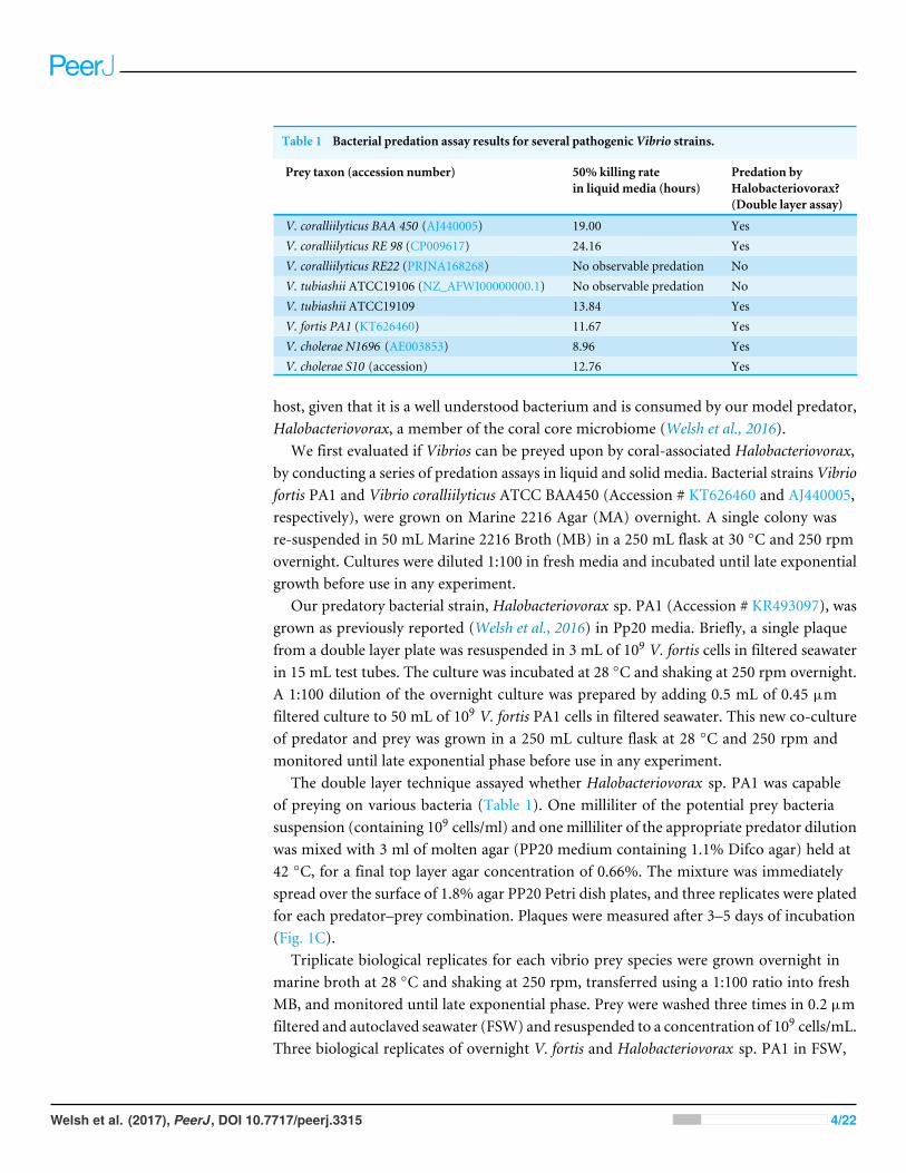
Table 1 Bacterial predation assay results for several pathogenic Vibrio strains.
Prey taxon (accession number) 50% killing ratein liquid media (hours)
Predation byHalobacteriovorax?(Double layer assay)
V. coralliilyticus BAA 450 (AJ440005) 19.00 YesV. coralliilyticus RE 98 (CP009617) 24.16 YesV. coralliilyticus RE22 (PRJNA168268) No observable predation NoV. tubiashii ATCC19106 (NZ_AFWI00000000.1) No observable predation NoV. tubiashii ATCC19109 13.84 YesV. fortis PA1 (KT626460) 11.67 YesV. cholerae N1696 (AE003853) 8.96 YesV. cholerae S10 (accession) 12.76 Yes
host, given that it is a well understood bacterium and is consumed by our model predator,Halobacteriovorax, a member of the coral core microbiome (Welsh et al., 2016).
We first evaluated if Vibrios can be preyed upon by coral-associated Halobacteriovorax,by conducting a series of predation assays in liquid and solid media. Bacterial strains Vibriofortis PA1 and Vibrio coralliilyticus ATCC BAA450 (Accession # KT626460 and AJ440005,respectively), were grown on Marine 2216 Agar (MA) overnight. A single colony wasre-suspended in 50 mL Marine 2216 Broth (MB) in a 250 mL flask at 30 ◦C and 250 rpmovernight. Cultures were diluted 1:100 in fresh media and incubated until late exponentialgrowth before use in any experiment.
Our predatory bacterial strain, Halobacteriovorax sp. PA1 (Accession # KR493097), wasgrown as previously reported (Welsh et al., 2016) in Pp20 media. Briefly, a single plaquefrom a double layer plate was resuspended in 3 mL of 109 V. fortis cells in filtered seawaterin 15 mL test tubes. The culture was incubated at 28 ◦C and shaking at 250 rpm overnight.A 1:100 dilution of the overnight culture was prepared by adding 0.5 mL of 0.45 µmfiltered culture to 50 mL of 109 V. fortis PA1 cells in filtered seawater. This new co-cultureof predator and prey was grown in a 250 mL culture flask at 28 ◦C and 250 rpm andmonitored until late exponential phase before use in any experiment.
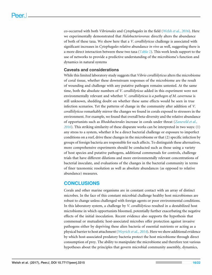
The double layer technique assayed whether Halobacteriovorax sp. PA1 was capableof preying on various bacteria (Table 1). One milliliter of the potential prey bacteriasuspension (containing 109 cells/ml) and one milliliter of the appropriate predator dilutionwas mixed with 3 ml of molten agar (PP20 medium containing 1.1% Difco agar) held at42 ◦C, for a final top layer agar concentration of 0.66%. The mixture was immediatelyspread over the surface of 1.8% agar PP20 Petri dish plates, and three replicates were platedfor each predator–prey combination. Plaques were measured after 3–5 days of incubation(Fig. 1C).
Triplicate biological replicates for each vibrio prey species were grown overnight inmarine broth at 28 ◦C and shaking at 250 rpm, transferred using a 1:100 ratio into freshMB, and monitored until late exponential phase. Prey were washed three times in 0.2 µmfiltered and autoclaved seawater (FSW) and resuspended to a concentration of 109 cells/mL.Three biological replicates of overnight V. fortis and Halobacteriovorax sp. PA1 in FSW,
Welsh et al. (2017), PeerJ, DOI 10.7717/peerj.3315 4/22
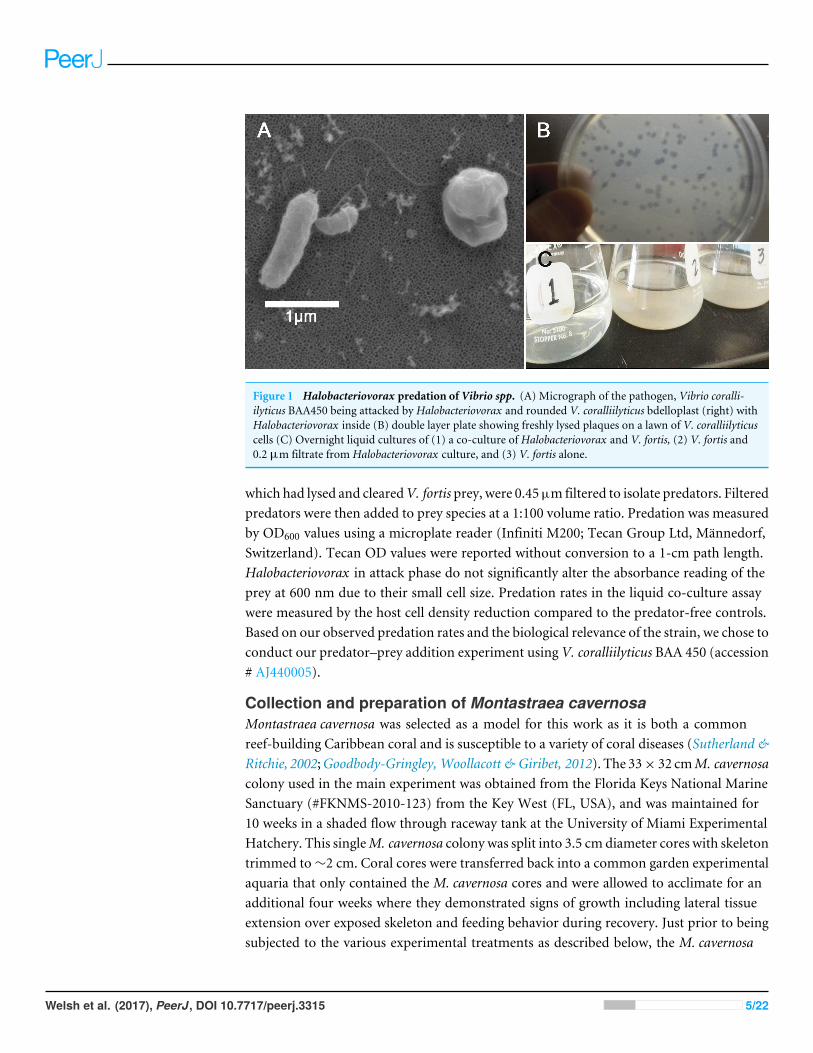
Figure 1 Halobacteriovorax predation of Vibrio spp. (A) Micrograph of the pathogen, Vibrio coralli-ilyticus BAA450 being attacked by Halobacteriovorax and rounded V. coralliilyticus bdelloplast (right) withHalobacteriovorax inside (B) double layer plate showing freshly lysed plaques on a lawn of V. coralliilyticuscells (C) Overnight liquid cultures of (1) a co-culture of Halobacteriovorax and V. fortis, (2) V. fortis and0.2 µm filtrate from Halobacteriovorax culture, and (3) V. fortis alone.
which had lysed and clearedV. fortis prey, were 0.45µmfiltered to isolate predators. Filteredpredators were then added to prey species at a 1:100 volume ratio. Predation was measuredby OD600 values using a microplate reader (Infiniti M200; Tecan Group Ltd, Männedorf,Switzerland). Tecan OD values were reported without conversion to a 1-cm path length.Halobacteriovorax in attack phase do not significantly alter the absorbance reading of theprey at 600 nm due to their small cell size. Predation rates in the liquid co-culture assaywere measured by the host cell density reduction compared to the predator-free controls.Based on our observed predation rates and the biological relevance of the strain, we chose toconduct our predator–prey addition experiment using V. coralliilyticus BAA 450 (accession# AJ440005).
Collection and preparation of Montastraea cavernosaMontastraea cavernosa was selected as a model for this work as it is both a commonreef-building Caribbean coral and is susceptible to a variety of coral diseases (Sutherland &Ritchie, 2002;Goodbody-Gringley, Woollacott & Giribet, 2012). The 33× 32 cmM. cavernosacolony used in the main experiment was obtained from the Florida Keys National MarineSanctuary (#FKNMS-2010-123) from the Key West (FL, USA), and was maintained for10 weeks in a shaded flow through raceway tank at the University of Miami ExperimentalHatchery. This singleM. cavernosa colony was split into 3.5 cm diameter cores with skeletontrimmed to∼2 cm. Coral cores were transferred back into a common garden experimentalaquaria that only contained the M. cavernosa cores and were allowed to acclimate for anadditional four weeks where they demonstrated signs of growth including lateral tissueextension over exposed skeleton and feeding behavior during recovery. Just prior to beingsubjected to the various experimental treatments as described below, the M. cavernosa
Welsh et al. (2017), PeerJ, DOI 10.7717/peerj.3315 5/22
cores were then transferred to a second common garden at FIU which was composed of arecirculating seawater tank that only contained theM. cavernosa cores. All coral cores weredistributed randomly into new 40L recirculating treatment tanks and only used once inthe experiment. Seawater for the experiment was obtained from the University of MiamiExperimental Hatchery (sand and UV-filtered seawater pumped in from Biscayne Bay).
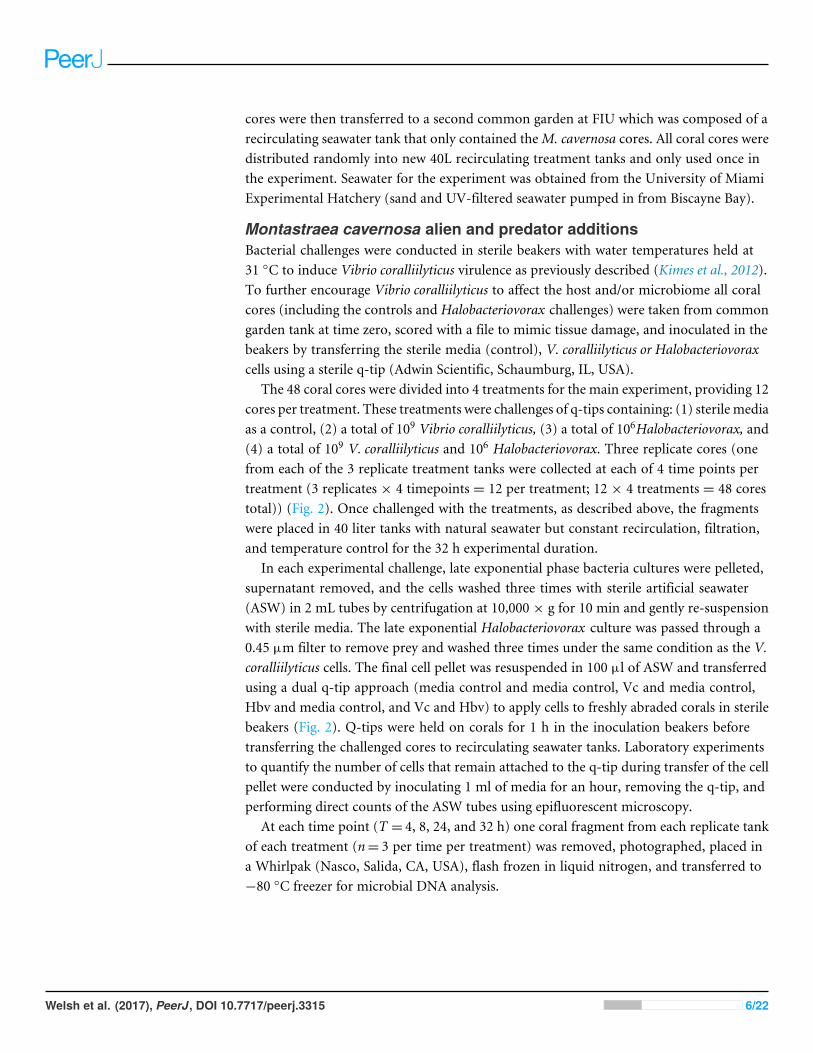
Montastraea cavernosa alien and predator additionsBacterial challenges were conducted in sterile beakers with water temperatures held at31 ◦C to induce Vibrio coralliilyticus virulence as previously described (Kimes et al., 2012).To further encourage Vibrio coralliilyticus to affect the host and/or microbiome all coralcores (including the controls and Halobacteriovorax challenges) were taken from commongarden tank at time zero, scored with a file to mimic tissue damage, and inoculated in thebeakers by transferring the sterile media (control), V. coralliilyticus or Halobacteriovoraxcells using a sterile q-tip (Adwin Scientific, Schaumburg, IL, USA).
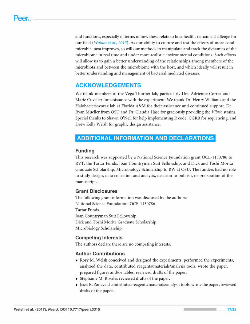
The 48 coral cores were divided into 4 treatments for the main experiment, providing 12cores per treatment. These treatments were challenges of q-tips containing: (1) sterile mediaas a control, (2) a total of 109 Vibrio coralliilyticus, (3) a total of 106Halobacteriovorax, and(4) a total of 109 V. coralliilyticus and 106 Halobacteriovorax. Three replicate cores (onefrom each of the 3 replicate treatment tanks were collected at each of 4 time points pertreatment (3 replicates × 4 timepoints = 12 per treatment; 12 × 4 treatments = 48 corestotal)) (Fig. 2). Once challenged with the treatments, as described above, the fragmentswere placed in 40 liter tanks with natural seawater but constant recirculation, filtration,and temperature control for the 32 h experimental duration.
In each experimental challenge, late exponential phase bacteria cultures were pelleted,supernatant removed, and the cells washed three times with sterile artificial seawater(ASW) in 2 mL tubes by centrifugation at 10,000 × g for 10 min and gently re-suspensionwith sterile media. The late exponential Halobacteriovorax culture was passed through a0.45 µm filter to remove prey and washed three times under the same condition as the V.coralliilyticus cells. The final cell pellet was resuspended in 100 µl of ASW and transferredusing a dual q-tip approach (media control and media control, Vc and media control,Hbv and media control, and Vc and Hbv) to apply cells to freshly abraded corals in sterilebeakers (Fig. 2). Q-tips were held on corals for 1 h in the inoculation beakers beforetransferring the challenged cores to recirculating seawater tanks. Laboratory experimentsto quantify the number of cells that remain attached to the q-tip during transfer of the cellpellet were conducted by inoculating 1 ml of media for an hour, removing the q-tip, andperforming direct counts of the ASW tubes using epifluorescent microscopy.
At each time point (T = 4, 8, 24, and 32 h) one coral fragment from each replicate tankof each treatment (n= 3 per time per treatment) was removed, photographed, placed ina Whirlpak (Nasco, Salida, CA, USA), flash frozen in liquid nitrogen, and transferred to−80 ◦C freezer for microbial DNA analysis.
Welsh et al. (2017), PeerJ, DOI 10.7717/peerj.3315 6/22
Control
109 V. coralliilyticus
109 V. coralliilyticus +106 Halobacteriovorax sp. PA1
106 Halobacteriovorax sp. PA1
Treatments(n=12 per treatment)
Coral Microbiome Extraction and Sequencing Protocol
Acclimated coral cores in common garden
Coral cores innoculated in beakers for one hour
Cores removed, rinsed, and frozen in liquid nitrogenT = 4, 8, 24, and 32
Coral tissue removed in quadrants from each core
Only inoculation quadrant used for PCR amplification of 16S V4 region, and Illumina MiSeq amplicon libraries
Experimental Design
1
2
4
DNA extracted from each quadrant3
Figure 2 Montastraea cavernosamicrobiomemanipulation experimental design detailing collectionand inoculation of coral cores, treatment tanks and replication, sample preservation, tissue removal,DNA extraction, andmicrobiome sample processing.
Montastraea cavernosa microbiome DNA extraction, sequencing, andquality controlFrom each core one quadrant of the coral tissue layer was removed using a dental tooland transferred into separate microcentrifuge tubes (4 per core) containing 500 µl of TESBuffer (10 mM Tris–HCl [pH 7.5], 1 mM EDTA, and 100 mMNaCl). A 1.5 mL microtubepestle was used to homogenize the tissue before adding 400 µl of TES buffer with lysozyme(Epicentre; final: 10 Uµl−1), followed by incubation at 37 ◦C for 30min. A 200µl aliquot ofhomogenized sample was used for DNA extraction with the Power Soil DNA extraction kit(MoBio Laboratories, Carlsbad, CA, USA); the remainder was stored at−20 ◦C. Microbialamplicon libraries were generated using 515F and 806R primers to the V4 region of the16S rRNA gene with Schloss sequencing adapters (Kozich et al., 2013). AccuStart II PCRToughMix (Gaithersburg, MD, USA) and the following thermocycling conditions wereused for amplification:1 cycle of 94 ◦C for 3 min; 35 cycles of 94 ◦C for 30 s, 50 ◦C for30 s, and 72 ◦C for 60 s; and 1 cycle of 72 ◦C for 10 min were used for amplification. Eachsample underwent triplicate reactions that were pooled and cleaned using the PromegaWizard SV Gel and PCR Clean-Up System (Madison, WI, USA). The samples were thenquantified using a Qubit dsDNA HS kit (Invitrogen, OR, USA) before being pooled in anequimolar ratio. The amplicon purity and length was checked on an Agilent Bioanalyzer2100 prior to sequencing on a MiSeq Illumina sequencing platform at the Oregon StateUniversity’s Center for Genome Research and Biocomputing (CGRB) Core Laboratories.
Welsh et al. (2017), PeerJ, DOI 10.7717/peerj.3315 7/22
Quality control and selection of operational taxonomic units (OTUs) was performedusing QIIME (v.1.8) (Caporaso et al., 2010). Sequences with quality scores less than amean of 35 were removed. Sequences were clustered into (OTUs) at a 97% 16S rRNAgene identity threshold using USEARCH 6.1.54 (Edgar, 2010) and the subsampled open-reference OTU-picking protocol in QIIME v.1.8 (Rideout et al., 2014), using greengenes13_8 as the reference (McDonald et al., 2012). Chimeric sequences were removed withQIIME’s wrapper of the UCHIME software (Edgar et al., 2011). Singleton OTUs wereremoved. The OTUs were assigned taxonomic classification using the QIIME wrapper tothe UCLUST software package (Edgar, 2010). OTUs that were classified as chloroplast,eukaryotic or mitochondria were filtered out of the dataset.
Statistical analysisTo avoid artifacts due to uneven sampling depth during comparisons of alpha and betadiversity, all samples were rarified (randomly subsampled) to equal sequencing depth. Afterquality control steps, the least sequenced sample had 11,716 reads; this valuewas thus chosenas the rarefaction depth. For alpha diversity (richness), total observed OTUs and Chao1diversity statistics (Chao, 1984) were calculated in QIIME. The significance of differencesin alpha diversity across treatments was determined in QIIME using nonparametrict -tests with 999 Monte Carlo permutations. For beta diversity analysis, weighted UniFracdistances (Lozupone & Knight, 2005) were calculated in QIIME. Distances within samplesin each treatment category were summarized and tested for significance using MonteCarlo Permutation tests (make_distance boxplots.py, non-parametric p-value, n= 999permutations). To account for multiple comparisons between treatments, Bonferroni-corrected p-values are reported for both alpha- and beta-diversity analyses in the textand figures.
To analyze how order-level taxa responded to bacterial challenge, a generalized linearmodel (GLM) was fitted with the R package DESeq2 (Love, Huber & Anders, 2014). TheGLM design specified time point, treatment, and their interaction as factors. For thisanalysis, the order level OTU table was pre-filtered in QIIME where we excluded all taxapresent in fewer than 6 samples. To test for the effect of treatment, a full model wascompared to a reduced model fit using only time as a factor, and likelihood ratio tests(LRT) were performed to assess taxon (Table 2). Post hoc Wald tests were performed onthe full model object to identify the specific treatments responsible for driving changes inthese taxa. To control the rate of false positives due to multiple comparisons, differentiallyabundant taxa were identified as taxa with Benjamin-Hochberg FDR q-values less than 0.05.
RESULTSHalobacteriovorax sp. PA1 can prey on multiple Vibrio speciesStrain and species level differences in susceptibility to predation were detected among someof the Vibrio species (Table 1). For example, among the V. coralliilyticus strains, BAA 450was most susceptible to predation in the liquid assay while strains RE22 and RE98 wereless susceptible (Table 1). In the double layer plate assay, Halobacteriovorax sp. PA1 wascapable of killing prey and forming plaques on all Vibrio spp. except V. coralliilyticus RE22
Welsh et al. (2017), PeerJ, DOI 10.7717/peerj.3315 8/22
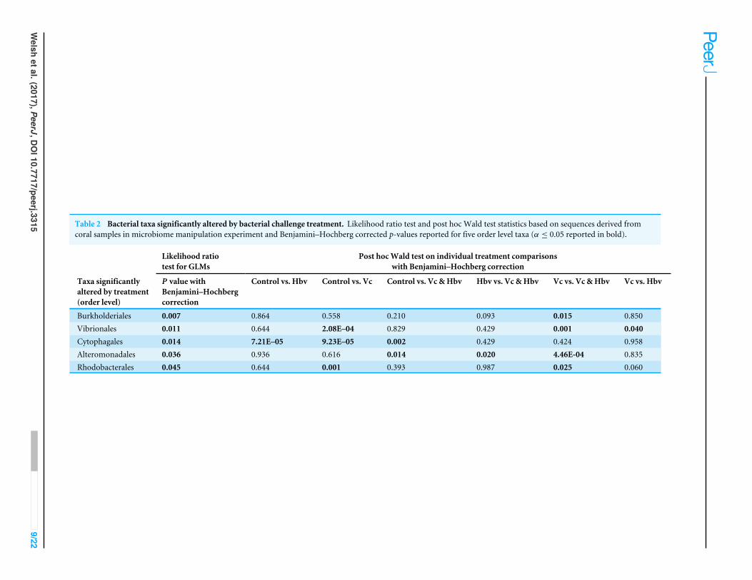
Table 2 Bacterial taxa significantly altered by bacterial challenge treatment. Likelihood ratio test and post hoc Wald test statistics based on sequences derived fromcoral samples in microbiome manipulation experiment and Benjamini–Hochberg corrected p-values reported for five order level taxa (α ≤ 0.05 reported in bold).
Likelihood ratiotest for GLMs
Post hocWald test on individual treatment comparisonswith Benjamini–Hochberg correction
Taxa significantlyaltered by treatment(order level)
P value withBenjamini–Hochbergcorrection
Control vs. Hbv Control vs. Vc Control vs. Vc &Hbv Hbv vs. Vc &Hbv Vc vs. Vc &Hbv Vc vs. Hbv
Burkholderiales 0.007 0.864 0.558 0.210 0.093 0.015 0.850Vibrionales 0.011 0.644 2.08E–04 0.829 0.429 0.001 0.040Cytophagales 0.014 7.21E–05 9.23E–05 0.002 0.429 0.424 0.958Alteromonadales 0.036 0.936 0.616 0.014 0.020 4.46E-04 0.835Rhodobacterales 0.045 0.644 0.001 0.393 0.987 0.025 0.060
Welsh
etal.(2017),PeerJ,DOI10.7717/peerj.3315
9/22
A B
Figure 3 Impact of bacterial challenge on coral associated microbial diversity. (A) Mean alpha diversity(observed species) plotted for each treatment, and (B) mean beta diversity (Weighted UniFrac distance)by treatment. The asterisks indicate Bonferroni-corrected p values <0.05 for the nonparametric t -test be-tween treatments. In both cases while addition of the pathogen alone increased diversity, predator addi-tion counteracted this effect.
and V. tubiashii ATCC19106 (Table 1; Fig. 1C). Halobacteriovorax predation rates (50%killing) ranged from 8.96 h in V. cholerae N1696 to 24.16 h in V. coralliilyticus RE98. Theprey rate for V. coralliilyticus BAA 450, a model coral pathogen, was 19.00 h.
Vibrio coralliilyticus treated corals experienced altered coralmicrobiome α- and β-diversityGiven the observation thatHalobacteriovorax sp. PA1 was capable of killing V. coralliilyticusBAA 450 in co-culture, we conducted an in situ challenge experiment that directlyinoculated M. cavernosa corals with this foreign bacterium in the presence and absence ofthe predator (Fig. 2). Corals were inoculated using a swab transfer method and laboratoryexperiments indicate that an average of 6.02×10⁸ (±6.63×10⁷) total V. coralliilyticus cellswere transferred using the swab transfer method (Table S1). No significant differenceswere observed in tissue loss or bleaching among the treatments at any of the time points(data not shown). We quantified relative microbial changes using 16S rRNA sequencing ofcorals in each treatment at 4, 8, 16, and 32 h post inoculation. After quality filtering of theexperimental microbiomes, 4,464,765 reads remained with an average of 85,860± 112,003reads per samples. After rarefaction the mean number of observed OTUs across all sampleswas 197 (Fig. 3A).
Vibrio coralliilyticus challenge increased microbial alpha diversity in the tissues of M.cavernosa corals. Corals challenged with V. coralliilyticus showed significantly increased
Welsh et al. (2017), PeerJ, DOI 10.7717/peerj.3315 10/22
richness relative to controls (Vibrio mean = 259.767 ± 14.196; control mean = 178.167±47.398) asmeasured by Chao1 and observed species diversity metrics (p= 0.048; Fig. 3A).However, when M. cavernosa samples were co-inoculated with both V. coralliilyticus andHalobacteriovorax, species richness returned to lower levels (mean = 163.058 ± 36.772)that were similar to control conditions, but distinct from the V. coralliilyticus treatment(p= 0.018; Fig. 3A). Changes in alpha diversity occurred early in the experiment, andnormalized for all treatments, except the V. coralliilyticus treatment which graduallyincreased over the remaining time points by 48% (Table S2). No significant differencesin evenness were observed between treatments. Further no significant differences inα-diversity were found between tanks or time points for either the Chao1 or observedspecies metrics.
Weighted UniFrac distances (β-diversity) were also significantly different between thecontrol treatment and the V. coralliilyticus treatment (p= 0.012) (Fig. 3B). In a similarpattern to α-diversity, the Halobacteriovorax and V. coralliilyticus combination treatmentreturned β-diversity to control levels and were not significantly different than the othertreatments (Fig. 3B). The Halobacteriovorax sp. PA1 alone did not significantly changeβ-diversity, and no significant differences were found between tanks or time points forβ-diversity metrics.
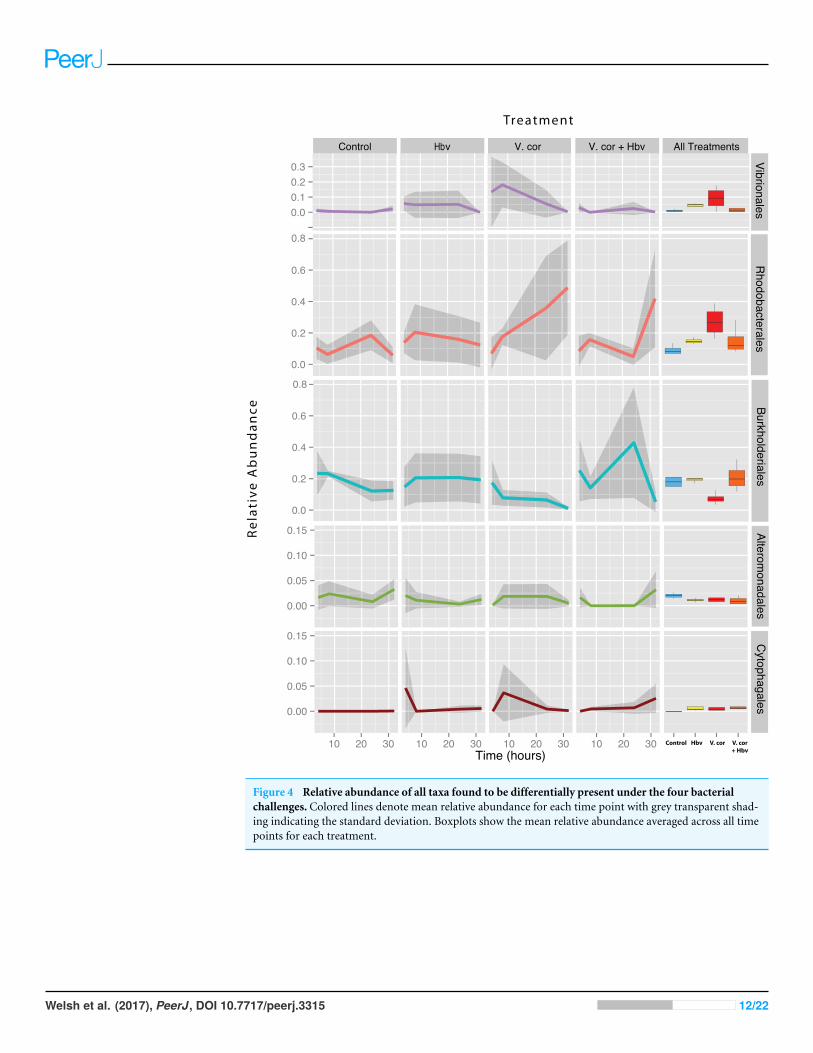
Vibrio coralliilyticus inoculation increases opportunists likeRhodobacterales and Cytophagales unless controlled by predatorsDespite the inoculations lack of any visual signs of pathogenesis, we still foundsignificant differences in the relative abundance of 16S rRNA genes to both Vibrioand other taxa associated sequences in the tissue microbiomes of the challenged corals(Fig. 4). To test if inoculation treatments caused any differences among bacterial orders,a generalized linear model was constructed using DESeq2 (Love, Huber & Anders, 2014).Significant differences were detected across treatments in Vibrionales, Rhodobacterales,Alteromonadales, Cytophagales, and Burkholderiales (Benjamini–Hochberg correctedp= 0.014, 0.045, 0.036, 0.011 and 0.007, respectively) taxa. To identify specific pair-wisedifferences in the relative abundance of these taxa across all the individual treatments(Control, Vcor alone, Hbv alone, Hbv + Vcor) we used Wald post hoc tests (Table 2).
As expected, addition of V. coralliilyticus resulted in increased Vibrionales affiliatedsequence mean relative abundances in coral tissues (Fig. 4 purple lines). In the V.coralliilyticus treatment, the mean relative abundance of Vibrionales increased over 35%from the 4 to 8 h time point. Yet corals challenged with V. coralliilyticus in the presence ofthe predator Halobacteriovorax had an 84.74% reduction in Vibrionales compared to theV. coralliilyticus alone treatment. The combined treatment had a mean Vibrionales relativeabundance of 1.43%, similar to the controls at 0.98%, while the V. coralliilyticus alonetreatment had a mean 9.38% Vibrionales relative abundance across the whole experiment(Table 2; Fig. 4 box plots).
The V. coralliilyticus challenge also reported significantly higher relative abundance oftwo groups of taxa,Cytophagales and Rhodobacterales, compared to controls. Addition ofV.coralliilyticus to corals increased the abundance of Rhodobacterales affiliated sequences in
Welsh et al. (2017), PeerJ, DOI 10.7717/peerj.3315 11/22
Control Hbv V. cor V. cor + Hbv All Treatments
Rel
ativ
e A
bu
nd
ance
Treatment
Control Hbv V. cor V. cor + Hbv
Alterom
onadalesC
ytophagales
10 20 30 10 20 30 10 20 30 10 20 30Time (hours)
0.0
0.1
0.2
0.3
0.0
0.2
0.4
0.6
0.0
0.2
0.4
0.6
0.8
Rhodobacterales
Burkholderiales
Vibrionales
0.8
0.00
0.05
0.10
0.15
0.00
0.05
0.10
0.15
Figure 4 Relative abundance of all taxa found to be differentially present under the four bacterialchallenges. Colored lines denote mean relative abundance for each time point with grey transparent shad-ing indicating the standard deviation. Boxplots show the mean relative abundance averaged across all timepoints for each treatment.
Welsh et al. (2017), PeerJ, DOI 10.7717/peerj.3315 12/22
tissues to an even greater extent than V. coralliilyticus itself, and this increase persisted afterVibrio sequence abundances fell at later time points (Fig. 4 red lines). Similarly, patternsof increased Rhodobacteraceae-affiliated sequences (at the coral tissue loss disease front) inwhite syndrome affected corals have recently been reported (Pollock et al., 2017).
Rhodobacterales sequences increased throughout the experiment in the V. coralliilyticusaddition treatment, nearly doubling at each time point starting from 6.99% (±0.05SEM) relative abundance to a maximum mean value of 48.75% (± 0.14 SEM) inthe final time point. However, during joint inoculation of Halobacteriovorax and V.coralliilyticus, Rhodobacterales showed no significant differences vs. controls. Similarly,there were no differences in the abundance of Rhodobacterales vs. controls in the treatmentwhere Halobacteriovorax was added alone (Table 2). Cytophagales affiliated sequencesalso increased by several orders of magnitude, from <0.001% to 3.656%, early in theV. coralliilyticus addition experiment (Fig. 4 brown lines).
The two other taxa that significantly changed, but did so in different patterns, wereBurkholderiales and Alteromonadales (Fig. 4 blue and green lines, respectively). The meanrelative abundance for the order Burkholderiales was lowest (8.21%) in the V. coralliilyticustreatment (Fig. 4 boxplots) and was significantly lower (p= 0.015) in the V. coralliilyticusversus the combined Halobacteriovorax and V. coralliilyticus treatment that had a relativeabundance of 21.86% (Table 2; Fig. 4 box plots). Alteromonadales were 40.47% moreabundant in the controls than the combined Halobacteriovorax and V. coralliilyticustreatment (p= 0.014) (Table 2; Fig. 4 box plots).
DISCUSSIONTracking microbial community dynamics after different bacterialchallengesHigh-throughput sequencing allows researchers to more easily document membershipdynamics and community topology, yet we often lack the ability to confirm causalrelationships among them. Manipulative studies are necessary to link cause and effect.While some host-microbe models can be more readily manipulated (e.g., mouse gut, squidlight organ, and rhizosphere), there remain considerable methodological barriers for manysystems, especially those for which gnotobiotic (germ-free) host animals are not available.Here we used individual and combinatorial bacterial challenges to a coral host in order toask three specific questions: (1) How can inoculation of a foreign bacteria (V. coralliilyticus)alter the microbiome of a compromised host in a laboratory setting (2) Can predators ofthis taxa prevent or ameliorate the downstream effects of the alien challenges, and (3) Doesthe predator itself affect the coral microbiome?
Vibrio coralliilyticus is a known disease-causing pathogen of corals worldwide (Ben-haim, Zicherman-keren & Rosenberg, 2003; Wilson et al., 2013) and has been documentedto induce bleaching and tissue loss in some species of corals (Ben-Haim et al., 1999;Ben-haim, Zicherman-keren & Rosenberg, 2003). Furthermore, experimental evidence hasdemonstrated that under increased thermal stress V. coralliilyticus concentrations risedramatically in corals (Tout et al., 2015). However, the changes, if any, that V. coralliilyticus
Welsh et al. (2017), PeerJ, DOI 10.7717/peerj.3315 13/22
challenge causes to the microbial communities normally present in corals was previouslyunknown. V. coralliilyticus represents an alien or potential pathogen in this study, as it hasnot been previously shown to be a member of Montastraea cavernosa microbiome, andit remains unknown if it can infect certain Caribbean corals like Montastraea cavernosa.Never the less, determining how bacterial challenge can alter the normal flora of a host mayprovide insight into whether mutualists are lost and additional antagonisms arise duringan infection cycle and thus contribute to secondary negative effects on animal hosts.
Here we show that a V. coralliilyticus inoculation not only changes its own relativeabundance in the system (as would be expected) but also alters the microbiome in variousways, including increases in alpha and beta diversity (Fig. 3). However, when these coralswere challenged with the V. coralliilyticus in the presence of the predator, these effectswere diminished and resulted in almost no changes in the normal coral microbiome.Furthermore, addition of just the bacterial predator did not change the community ina similar fashion to the V. coralliilyticus challenge, suggesting that exposure to differentforeign bacterial taxa (as opposed to any gram negative bacteria) will likely elicit variabledownstream responses in the microbiome, unlike taxa that core members.
Addition of V. coralliilyticus led to an increase in relative abundance in a known groupof opportunists of corals, the Rhodobacterales (Fig. 4). The increase in Rhodobacteralespersisted at later time-points, even after the abundance of V. coralliilyticus had declined inthe tissues. Rhodobacterales sequence abundances have been linked to disease outbreaks inwhite plague diseased Siderastrea siderea andDiploria strigosa corals (Cárdenas et al., 2012).Also, sequences from the family Rhodobacteraceae have been shown to increase by 4-foldsat the lesion front of corals with white syndrome (Pollock et al., 2017). Rhodobacteralesare fast growing taxa, capable of quickly responding to increasing availability of aminoacids (Mayali et al., 2014), and could be responding to resources made available fromcells damaged by V. coralliilyticus. Such a mechanism would explain associations betweenRhodobacterales and many stressed or diseased corals. While the present study cannotdistinguish whether these secondary Vibrio-induced Rhodobacterales are harmful to corals,the experimental framework used here could test this question in the future.
More broadly, V. coralliilyticus challenged samples showed a wider variety of bacteriasequences within the tissues (Fig. 3A). It is likely these opportunist species gained accessand/or established within the tissue shortly after Vibrio coralliilyticus inoculation, as theincreases in observed species persisted for the duration of the experiment (Table S2).However, the disproportionate impact of the V. coralliilyticus was not observed in sampleschallenged with the predator Halobacteriovorax suggesting that this is not a generalizableresponse to the wounding and addition of any kind of bacteria.
Halobacteriovorax as a possible top down control of opportunistsWe have previously cultivated Halobacteriovorax from multiple-species of corals, andused long-term microbial time series data to show that, despite its low abundance, it isa core member of the microbiome of several coral genera (Welsh et al., 2016). Here weused bacterial challenge experiments to demonstrate that Halobacteriovorax can protectits coral host by consuming its prey V. coralliilyticus. We found that the application of
Welsh et al. (2017), PeerJ, DOI 10.7717/peerj.3315 14/22
Halobacteriovorax at the same time as V. coralliilyticus can prevent detectable changesin the relative abundance of V. coralliilyticus in M. cavernosa coral tissue after challenge.The Halobacteriovorax alone treatment showed higher variance in the mean relativeabundance of Vibrionales than the controls or combined treatment (Fig. 4), but the meanwas not significantly different than the controls. Co-inoculations of this predator with V.coralliilyticus showed no significant differences in the abundance of Vibrionales in coraltissues versus control inoculations at any time in the course of the experiment (Fig. 4 purplelines). Thus it is likely that these predators consumed the Vibrio immediately or at thepoint of inoculation, and therefore provided a biotic barrier to the host tissues. The abilityofHalobacteriovorax to mitigate inoculation, if added hours or days after a V. coralliilyticuschallenge, remains unknown. At the same time the generality of this effect of the Vibrio andthe predatory Halobacteriovorax remains untested, but could be evaluated using similarmethods to those we describe here. For example, additional types of pathogens that showclear infection signs upon addition or the use of more commensal strains of bacteria ascontrols would strengthen support for this hypothesis.
Phage have already been shown to be effective against V. coralliilyticus (Cohen et al.,2013), and likely play a role in controlling natural populations of V. coralliilyticus inthe environment, which is similar to what has been suggested for phage and V. cholerae(Faruque et al., 2005). Phages also provide an antimicrobial function in the mucus layerof corals (Barr et al., 2013; Barr et al., 2015) and are often considered the main top-downcontrol mechanism of bacteria in some systems. However in certain circumstances,Halobacteriovorax predation has been shown to be a more dominant factor in bacterialmortality than viral lysis (Williams et al., 2015). In addition, predatory bacteria are thoughtto play a major role in controlling pathogenic Vibrio in seawater and shellfish (Richardset al., 2012). In our study we show predatory Halobacteriovorax sp. PA1 is effectiveagainst V. coralliilyticus BAA 450 and other Vibrio strains, offering further support tothe hypothesis that bacterial predators are likely to play a role in controlling populations inthe environment. In a similar fashion to phages,Halobacteriovorax thusmediates top-downcontrol of pathogens by preventing initial invasion of the host.
Microbiome manipulation validates previous network analysispredictionsA small but growing body of research suggests Halobacteriovorax naturally occur andregularly interact with members of the coral microbiome. For example, a previousmetagenomic study of P. astreoides from Panama reported that sequences similar topredatory Halobacteriovorax were among the most commonly identified bacterialannotations in the coral microbiome (Wegley et al., 2007). Furthermore, we foundHalobacteriovorax was present in ∼80% of samples collected approximately monthlyfrom three genera of Caribbean corals across a three-year time span. Network analysisof 198 of these samples detected intriguing co-occurrences between these predators andother taxa (Welsh et al., 2016). Here in the bacterial challenge study, we validated severalof the co-occurrence patterns detected in our network analysis. For example, in ournetworks from Agaricia corals, Bdellovibrionales (the order ofHalobacteriovorax) positively
Welsh et al. (2017), PeerJ, DOI 10.7717/peerj.3315 15/22
co-occurred with both Vibrionales and Cytophagales in the field (Welsh et al., 2016). Herewe experimentally demonstrated that Halobacteriovorax directly alters the abundanceof both of these taxa. We show here that a V. coralliilyticus challenge is associated withsignificant increases in Cytophagales relative abundance in vivo as well, suggesting there isa more direct interaction between these two taxa (Table 2). This work lends support to theuse of networks to provide a predictive understanding of the microbiome’s function anddynamics in natural systems
Caveats and considerationsWhile this limited laboratory study suggests that Vibrio coralliilyticus alters the microbiomeof coral tissue, whether these downstream responses of the microbiome are the resultof wounding and challenge with any putative pathogen remains untested. At the sametime, both the absolute numbers of V. coralliilyticus added in this experiment were notenvironmentally relevant and whether V. coralliilyticus is a pathogen of M. cavernosa isstill unknown, shedding doubt on whether these same effects would be seen in trueinfection scenarios. Yet the patterns of change in the community after addition of V.coralliilyticus remarkably mirror the changes we found in corals exposed to stressors in theenvironment. For example, we found that overall beta-diversity and the relative abundanceof opportunists such as Rhodobacterales increase in corals under threat (Zaneveld et al.,2016). This striking similarity of these disparate works can be interpreted in two ways: (1)any stress to a system, whether it be a direct bacterial challenge or exposure to imperfectconditions on a reef, drive these changes in the microbiome or that (2) specific infection bygroups of foreign bacteria are responsible for such affects. To distinguish these alternatives,more comprehensive experiments should be conducted such as those using a varietyof host species and putative pathogens, additional commensals for controls, challengetrials that have different dilutions and more environmentally relevant concentrations ofbacterial inoculate, and evaluations of the changes in the bacterial community in termsof finer taxonomic resolution as well as absolute abundances (as opposed to relativeabundance) measures.
CONCLUSIONSCorals and other marine organisms are in constant contact with an array of distinctmicrobes. In the face of this constant microbial challenge healthy host microbiomes arerobust to change unless challenged with foreign agents or poor environmental conditions.In this laboratory system, a challenge by V. coralliilyticus resulted in a destabilized hostmicrobiome in which opportunists bloomed, potentially further exacerbating the negativeeffects of the initial inoculation. Recent evidence also supports the hypothesis thatcommensal or mutualistic host-associated microbes offer protection against invasivepathogens either by depriving these alien bacteria of essential nutrients or acting as aphysical barrier to host attachment (Weyrich et al., 2014). Here we show additional evidenceby which host-associated predatory bacteria protect the host microbiome through directconsumption of prey. The ability to manipulate the microbiome and therefore test varioushypotheses about the principles that govern microbial community assembly, dynamics,
Welsh et al. (2017), PeerJ, DOI 10.7717/peerj.3315 16/22
and functions, especially in terms of how these relate to host health, remain a challenge forour field (Waldor et al., 2015). As our ability to culture and test the effects of more coralmicrobial taxa improves, so will our methods to manipulate and track the dynamics of themicrobiome in real time and under more realistic environmental conditions. Such effortswill allow us to gain a better understanding of the relationships among members of themicrobiota and between the microbiome with the host, and which ideally will result inbetter understanding and management of bacterial mediated diseases.
ACKNOWLEDGEMENTSWe thank members of the Vega Thurber lab, particularly Drs. Adrienne Correa andMarie Cuvelier for assistance with the experiment. We thank Dr. Henry Williams and theHalobacteriovorax lab at Florida A&M for their assistance and continued support. Dr.Ryan Mueller from OSU and Dr. Claudia Häse for graciously providing the Vibrio strains.Special thanks to Shawn O’Neil for help implementing R code, CGRB for sequencing, andDrew Kelly Welsh for graphic design assistance.
ADDITIONAL INFORMATION AND DECLARATIONS
FundingThis research was supported by a National Science Foundation grant OCE-1130786 toRVT, the Tartar Funds, Joan Countryman Suit Fellowship, and Dick and Toshi MoritaGraduate Scholarship, Microbiology Scholarship to RW at OSU. The funders had no rolein study design, data collection and analysis, decision to publish, or preparation of themanuscript.
Grant DisclosuresThe following grant information was disclosed by the authors:National Science Foundation: OCE-1130786.Tartar Funds.Joan Countryman Suit Fellowship.Dick and Toshi Morita Graduate Scholarship.Microbiology Scholarship.
Competing InterestsThe authors declare there are no competing interests.
Author Contributions• Rory M. Welsh conceived and designed the experiments, performed the experiments,analyzed the data, contributed reagents/materials/analysis tools, wrote the paper,prepared figures and/or tables, reviewed drafts of the paper.• Stephanie M. Rosales reviewed drafts of the paper.• Jesse R. Zaneveld contributed reagents/materials/analysis tools, wrote the paper, revieweddrafts of the paper.
Welsh et al. (2017), PeerJ, DOI 10.7717/peerj.3315 17/22
• Jérôme P. Payet analyzed the data.• RyanMcMinds analyzed the data, contributed reagents/materials/analysis tools, revieweddrafts of the paper.• Steven L. Hubbs performed the experiments.• Rebecca L. Vega Thurber conceived and designed the experiments, analyzed the data,contributed reagents/materials/analysis tools, wrote the paper, reviewed drafts of thepaper.
Field Study PermissionsThe following information was supplied relating to field study approvals (i.e., approvingbody and any reference numbers):
The Florida Keys National Marine Sanctuary and NOAA coral nursery in Key Westapproved permits (#FKNMS-2010-123) and provided coral specimen for this work.
Data AvailabilityThe following information was supplied regarding data availability:
The mapping file, biom otu table, source code and tree file can be found in theSupplemental Information.
Supplemental InformationSupplemental information for this article can be found online at http://dx.doi.org/10.7717/peerj.3315#supplemental-information.
REFERENCESBarr JJ, Auro R, FurlanM,Whiteson KL, ErbML, Pogliano J, Stotl A, Wolkowicz
R, Cutting AS, Doran KS, Salamon P, Youle M, Rohwer F. 2013. Bacteriophageadhering to mucus provide a non-host-derived immunity. Proceedings of the NationalAcademy of Sciences of the United States of America 110:10771–10776DOI 10.1073/pnas.1305923110.
Barr JJ, Auro R, Sam-Soon N, Kassegne S, Peters G, Bonilla N, HatayM,MourtadaS, Bailey B, Youle M, Felts B, Baljon A, Nulton J, Salamon P, Rohwer F. 2015.Subdiffusive motion of bacteriophage in mucosal surfaces increases the frequency ofbacterial encounters. Proceedings of the National Academy of Sciences of of the UnitedStates of America 112:201508355.
Beman JM, Roberts KJ, Wegley L, Rohwer F, Francis CA. 2007. Distribution anddiversity of archaeal ammonia monooxygenase genes associated with corals. Appliedand Environmental Microbiology 73:5642–5647 DOI 10.1128/AEM.00461-07.
Ben-Haim Y, Banim E, Kushmaro A, Loya Y, Rosenberg E. 1999. Inhibition of pho-tosynthesis and bleaching of zooxanthellae by the coral pathogen Vibrio shiloi.Environmental Microbiology 1:223–229 DOI 10.1046/j.1462-2920.1999.00027.x.
Ben-Haim Y, Thompson FL, Thompson CC, Cnockaert MC, Hoste B, Swings J, Rosen-berg E. 2003. Vibrio coralliilyticus sp. nov., a temperature-dependent pathogen of thecoral Pocillopora damicornis. International Journal of Systematic and EvolutionaryMicrobiology 53:309–315 DOI 10.1099/ijs.0.02402-0.
Welsh et al. (2017), PeerJ, DOI 10.7717/peerj.3315 18/22

Ben-haim Y, Zicherman-kerenM, Rosenberg E. 2003. Temperature-regulated bleachingand lysis of the coral Pocillopora damicornis by the novel pathogen Vibrio coralliilyti-cus. Applied and Environmental Microbiology 69:4236–4241DOI 10.1128/AEM.69.7.4236-4242.2003.
Bourne DG, GarrenM,Work TM, Rosenberg E, Smith GW, Harvell CD. 2009.Microbial disease and the coral holobiont. Trends in Microbiology 17:554–562DOI 10.1016/j.tim.2009.09.004.
Burge C, Mark Eakin C, Friedman CS, Froelich B, Hershberger PK, Hofmann EE,Petes LE, Prager KC,Weil E, Willis BL, Ford SE, Harvell CD. 2014. Climate changeinfluences on marine infectious diseases: implications for management and society.Annual Review of Marine Science 6:249–277DOI 10.1146/annurev-marine-010213-135029.
Caporaso JG, Kuczynski J, Stombaugh J, Bittinger K, Bushman FD, Costello EK, FiererN, Peña AG, Goodrich JK, Gordon JI, Huttley GA, Kelley ST, Knights D, Koenig JE,Ley RE, Lozupone CA, McDonald D, Muegge BD, PirrungM, Reeder J, SevinskyJR, Turnbaugh PJ, WaltersWA,Widmann J, Yatsunenko T, Zaneveld J, KnightR. 2010. QIIME allows analysis of high-throughput community sequencing data.Nature Methods 7:335–336 DOI 10.1038/nmeth.f.303.
Cárdenas A, Rodriguez-R LM, Pizarro V, Cadavid LF, Arévalo-Ferro C. 2012. Shifts inbacterial communities of two Caribbean reef-building coral species affected by whiteplague disease. The ISME Journal 6:502–512 DOI 10.1038/ismej.2011.123.
Chao A. 1984. Nonparametric estimation of the number of classes in a population.Scandinavian Journal of Statistics 11:265–270.
Cohen Y, Pollock F, Rosenberg E, Bourne DG. 2013. Phage therapy treatment of thecoral pathogen Vibrio coralliilyticus.Microbiologyopen 2:64–74 DOI 10.1002/mbo3.52.
Cooney RP, Pantos O, Tissier MDAL, Barer MR, O’Donnell AG, Bythell JC. 2002.Characterization of the bacterial consortium associated with black band diseasein coral using molecular microbiological techniques. Environmental Microbiology4:401–413 DOI 10.1046/j.1462-2920.2002.00308.x.
De’ath G, Fabricius KE, Sweatman H, PuotinenM. 2012. The 27-year decline of coralcover on the Great Barrier Reef and its causes. Proceedings of the National Academy ofSciences of the United States of America 109:17995–17999DOI 10.1073/pnas.1208909109.
Edgar RC. 2010. Search and clustering orders of magnitude faster than BLAST. Bioinfor-matics 26:2460–2461 DOI 10.1093/bioinformatics/btq461.
Edgar RC, Haas BJ, Clemente JC, Quince C, Knight R. 2011. UCHIME improvessensitivity and speed of chimera detection. Bioinformatics 27:2194–2200DOI 10.1093/bioinformatics/btr381.
Faruque SM, Naser I Bin, IslamMJ, Faruque ASG, Ghosh AN, Nair GB, Sack DA,Mekalanos JJ. 2005. Seasonal epidemics of cholera inversely correlate with the preva-lence of environmental cholera phages. Proceedings of the National Academy of Sci-ences of the United States of America 102:1702–1707 DOI 10.1073/pnas.0408992102.
Welsh et al. (2017), PeerJ, DOI 10.7717/peerj.3315 19/22
Fitt WK,Warner ME. 1995. Bleaching patterns of four species of Caribbean reef corals.The Biological Bulletin 189:298–307 DOI 10.2307/1542147.
Gardner TA, Côté IM, Gill JA, Grant A,Watkinson AR. 2003. Long-term region-widedeclines in Caribbean corals. Science 301:958–960 DOI 10.1126/science.1086050.
Gignoux-Wolfsohn SA, Vollmer SV. 2015. Identification of candidate coral pathogenson white band disease-infected staghorn coral. PLOS ONE 10:e0134416DOI 10.1371/journal.pone.0134416.
Goodbody-Gringley G,Woollacott RM, Giribet G. 2012. Population structure andconnectivity in the Atlantic scleractinian coralMontastraea cavernosa (Linnaeus,1767).Marine Ecology 33:32–48 DOI 10.1111/j.1439-0485.2011.00452.x.
Hoegh-Guldberg O, Anthony K, Berkelmans R, Dove S, Fabricus K, Lough J, MarshallP, van OppenMJH, Negri A,Willis B. 2007. Vulnerability of reef-building coralson the Great Barrier Reef to climate change. In: Johnson JE, Marshall PA, eds. GreatBarrier Reef and climate change: a vulnerability assessment. Townsville: Great BarrierReef Marine Park Authority, 272–307.
Jürgens K, Matz C. 2002. Predation as a shaping force for the phenotypic and genotypiccomposition of planktonic bacteria. Antonie van Leeuwenhoek, International Journalof General and Molecular Microbiology 81:413–434 DOI 10.1023/A:1020505204959.
Kimes NE, Grim CJ, JohnsonWR, Hasan NA, Tall BD, Kothary MH, Kiss H, Munk AC,Tapia R, Green L, Detter C, Bruce DC, Brettin TS, Colwell RR, Morris PJ. 2012.Temperature regulation of virulence factors in the pathogen Vibrio coralliilyticus. TheISME Journal 6:835–846 DOI 10.1038/ismej.2011.154.
Kozich JJ, Westcott SL, Baxter NT, Highlander SK, Schloss PD. 2013. Development ofa dual-index sequencing strategy and curation pipeline for analyzing amplicon se-quence data on the MiSeq Illumina sequencing platform. Applied and EnvironmentalMicrobiology 79:5112–5120 DOI 10.1128/AEM.01043-13.
Lesser MP, Bythell JC, Gates RD, Johnstone RW, Hoegh-Guldberg O. 2007a. Are in-fectious diseases really killing corals? Alternative interpretations of the experimentaland ecological data. Journal of Experimental Marine Biology and Ecology 346:36–44DOI 10.1016/j.jembe.2007.02.015.
Lesser M, Falcón L, Rodríguez-Román A, Enríquez S, Hoegh-Guldberg O, Iglesias-Prieto R. 2007b. Nitrogen fixation by symbiotic cyanobacteria provides a source ofnitrogen for the scleractinian coralMontastraea cavernosa.Marine Ecology ProgressSeries 346:143–152 DOI 10.3354/meps07008.
LoveMI, HuberW, Anders S. 2014.Moderated estimation of fold change and dispersionfor RNA-seq data with DESeq2. Genome Biology 15:550DOI 10.1186/s13059-014-0550-8.
Lozupone C, Knight R. 2005. UniFrac: a new phylogenetic method for comparingmicrobial communities. Applied and Environmental Microbiology 71:8228–8235DOI 10.1128/AEM.71.12.8228-8235.2005.
Matz C, Kjelleberg S. 2005. Off the hook–how bacteria survive protozoan grazing. Trendsin Microbiology 13:302–307 DOI 10.1016/j.tim.2005.05.009.
Welsh et al. (2017), PeerJ, DOI 10.7717/peerj.3315 20/22
Mayali X,Weber PK, Mabery S, Pett-Ridge J. 2014. Phylogenetic patterns in the micro-bial response to resource availability: amino acid incorporation in San Francisco Bay.PLOS ONE 9:e95842 DOI 10.1371/journal.pone.0095842.
Maynard J, Van Hooidonk R, Eakin CM, PuotinenM, GarrenM,Williams G, HeronSF, Lamb J, Weil E, Willis B, Harvell CD. 2015. Projections of climate conditionsthat increase coral disease susceptibility and pathogen abundance and virulence.Nature Climate Change 5:688–694 DOI 10.1038/nclimate2625.
McDonald D, Price MN, Goodrich J, Nawrocki EP, DeSantis TZ, Probst A, AndersenGL, Knight R, Hugenholtz P. 2012. An improved Greengenes taxonomy withexplicit ranks for ecological and evolutionary analyses of bacteria and archaea. TheISME Journal 6:610–618 DOI 10.1038/ismej.2011.139.
MouchkaME, Hewson I, Harvell CD. 2010. Coral-associated bacterial assemblages:current knowledge and the potential for climate-driven impacts. Integrative andComparative Biology 50:662–674 DOI 10.1093/icb/icq061.
Nguyen-KimH, Bouvier T, Bouvier C, Doan-Nhu H, Nguyen-Ngoc L, Rochelle-NewallE, Baudoux A-C, Desnues C, Reynaud S, Ferrier-Pages C, Bettarel Y. 2014.Highoccurrence of viruses in the mucus layer of scleractinian corals. EnvironmentalMicrobiology Reports 6:675–682 DOI 10.1111/1758-2229.12185.
Pernthaler J. 2005. Predation on prokaryotes in the water column and its ecologicalimplications. Nature Reviews Microbiology 3:537–546 DOI 10.1038/nrmicro1180.
Pollock FJ, Wada N, Torda G,Willis BL, Bourne DG. 2017.White syndrome-affectedcorals have a distinct microbiome at disease lesion fronts. Applied and EnvironmentalMicrobiology 83:e02799-16 DOI 10.1128/AEM.02799-16.
Richards GP, Fay JP, Dickens KA, Parent MA, Soroka DS, Boyd EF. 2012. Predatorybacteria as natural modulators of Vibrio parahaemolyticus and Vibrio vulnificusin seawater and oysters. Applied and Environmental Microbiology 78:7455–7466DOI 10.1128/AEM.01594-12.
Rideout JR, He Y, Navas-Molina JA,WaltersWA, Ursell LK, Gibbons SM, Chase J,McDonald D, Gonzalez A, Robbins-Pianka A, Clemente JC, Gilbert JA, HuseSM, Zhou H-W, Knight R, Caporaso JG. 2014. Subsampled open-referenceclustering creates consistent, comprehensive OTU definitions and scales to billionsof sequences. PeerJ 2:e545 DOI 10.7717/peerj.545.
Ritchie K. 2006. Regulation of microbial populations by coral surface mucusand mucus-associated bacteria.Marine Ecology Progress Series 322:1–14DOI 10.3354/meps322001.
Ritchie KB, Smith GW. 1997. Microbial communities of coral surface mucopolysaccha-ride layers. In: Coral health and disease. Berlin, Heidelberg: Springer, 259–264.
Rypien KL,Ward JR, Azam F. 2010. Antagonistic interactions among coral-associatedbacteria. Environmental Microbiology 12:28–39DOI 10.1111/j.1462-2920.2009.02027.x.
Soffer N, Zaneveld J, Thurber R. 2014. Phage-bacteria network analysis and itsimplication for the understanding of coral disease. Environmental Microbiology17:1203–1218.
Welsh et al. (2017), PeerJ, DOI 10.7717/peerj.3315 21/22
Sutherland KP, Ritchie KB. 2002.White pox disease of the caribbean elkhorn coral.Coral Health and Disease 30:289–300.
Tout J, Siboni N, Messer LF, GarrenM, Stocker R,Webster NS, Ralph PJ, SeymourJR. 2015. Increased seawater temperature increases the abundance and altersthe structure of natural Vibrio populations associated with the coral Pocilloporadamicornis. Frontiers in Microbiology 6:432 DOI 10.3389/fmicb.2015.00432.
Ushijima B, Videau P, Burger AH, Shore-Maggio A, Runyon CM, SudekM, AebyGS, Callahan SM. 2014. Vibrio coralliilyticus strain OCN008 is an etiological agentof acute Montipora white syndrome. Applied and Environmental Microbiology80:2102–2109 DOI 10.1128/AEM.03463-13.
Vidal-Dupiol J, Ladrière O, Meistertzheim A-L, Fouré L, AdjeroudM,Mitta G. 2011.Physiological responses of the scleractinian coral Pocillopora damicornis to bacterialstress from Vibrio coralliilyticus. The Journal of Experimental Biology 214:1533–1545DOI 10.1242/jeb.053165.
WaldorMK, Tyson G, Borenstein E, OchmanH,Moeller A, Finlay BB, Kong HH,Gordon JI, Nelson KE, Dabbagh K, Smith H. 2015.Where next for microbiomeresearch? PLOS Biology 13:e1002050 DOI 10.1371/journal.pbio.1002050.
Wegley L, Edwards R, Rodriguez-Brito B, Liu H, Rohwer F. 2007.Metagenomicanalysis of the microbial community associated with the coral Porites astreoides.Environmental Microbiology 9:2707–2719 DOI 10.1111/j.1462-2920.2007.01383.x.
Welsh RM, Zaneveld JR, Rosales SM, Payet JP, Burkepile DE, Thurber RV. 2016.Bacterial predation in a marine host-associated microbiome. The ISME Journal10(6):1540–1544.
Weyrich LS, Feaga HA, Park J, Muse SJ, Safi CY, Rolin OY, Young SE, Harvill ET. 2014.Resident microbiota affect Bordetella pertussis infectious dose and host specificity.The Journal of Infectious Diseases 209:913–921 DOI 10.1093/infdis/jit597.
Williams HN, FalklerWA, Shay DE. 1980. Incidence of marine bdellovibrios lyticagainst Vibrio parahaemolyticus in Chesapeake Bay. Applied and EnvironmentalMicrobiology 40:970–972.
Williams HN, Lymperopoulou DS, Athar R, Chauhan A, Dickerson TL, Chen H, LawsE, Berhane T-K, Flowers AR, Bradley N, Young S. 2015.Halobacteriovorax, anunderestimated predator on bacteria: potential impact relative to viruses on bacterialmortality. The ISME Journal 10(2):491–499.
Wilson B, Muirhead A, Bazanella M, Huete-Stauffer C, Vezzulli L, Bourne DG. 2013.An improved detection and quantification method for the coral pathogen Vibriocoralliilyticus. PLOS ONE 8:e81800 DOI 10.1371/journal.pone.0081800.
Zaneveld JR, Burkepile DE, Shantz AA, Pritchard CE, McMinds R, Payet JP, Welsh RM,Correa AMS, Lemoine NP, Rosales SM, Fuchs C, Vega Thurber R. 2016. Overfish-ing and nutrient pollution interact with temperature to disrupt coral reefs down tomicrobial scales. Nature Communications 7:11833 DOI 10.1038/ncomms11833.
Welsh et al. (2017), PeerJ, DOI 10.7717/peerj.3315 22/22


![[Savage Worlds] Alien vs Predator](https://img.pdfslide.net/doc/110x75/55cf9d27550346d033ac7422/savage-worlds-alien-vs-predator.jpg)