Embed Size (px)
Citation preview
11
Amomi Fructus Rotundus
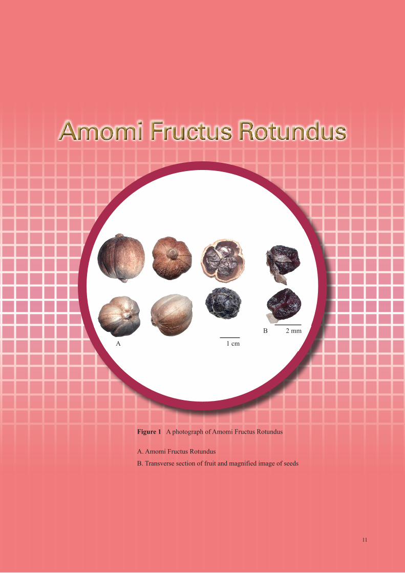
Figure 1 A photograph of Amomi Fructus Rotundus
A. Amomi Fructus Rotundus
B. Transverse section of fruit and magnified image of seeds
A
B
1 cm
2 mm
11
12
1. NAMES
Official Name: Amomi Fructus Rotundus
Chinese Name:
Chinese Phonetic Name: Doukou
2. SOURCE
Amomi Fructus Rotundus is the dried ripe fruit of Amomum compactum Soland ex Maton
(Zingiberaceae). The fruit-spike is collected mostly in July and August when the fruit is nearly ripe
and turns yellow but indehiscent, persistent perianth and fruit stalk removed, then dried under the sun
to obtain Amomi Fructus Rotundus.
3. DESCRIPTION
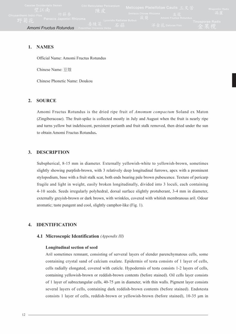
Subspherical, 8-15 mm in diameter. Externally yellowish-white to yellowish-brown, sometimes
slightly showing purplish-brown, with 3 relatively deep longitudinal furrows, apex with a prominent
stylopodium, base with a fruit stalk scar, both ends bearing pale brown pubescence. Texture of pericarp
fragile and light in weight, easily broken longitudinally, divided into 3 loculi, each containing
4-10 seeds. Seeds irregularly polyhedral, dorsal surface slightly protuberant, 3-4 mm in diameter,
externally greyish-brown or dark brown, with wrinkles, covered with whitish membranous aril. Odour
aromatic; taste pungent and cool, slightly camphor-like (Fig. 1).
4. IDENTIFICATION
4.1 Microscopic Identification (Appendix III)
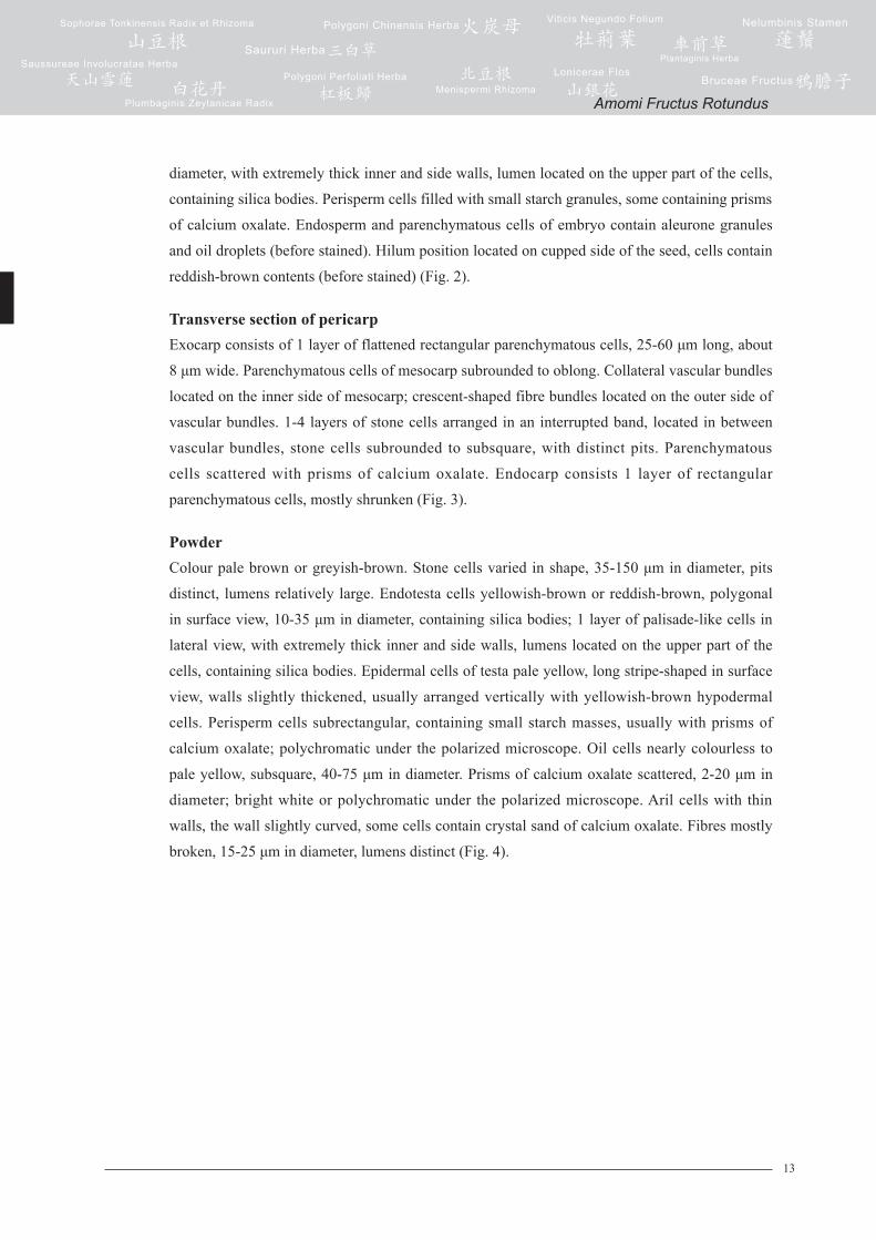
Longitudinal section of seedAril sometimes remnant, consisting of serveral layers of slender parenchymatous cells, some
containing crystal sand of calcium oxalate. Epidermis of testa consists of 1 layer of cells,
cells radially elongated, covered with cuticle. Hypodermis of testa consists 1-2 layers of cells,
containing yellowish-brown or reddish-brown contents (before stained). Oil cells layer consists
of 1 layer of subrectangular cells, 40-75 μm in diameter, with thin walls. Pigment layer consists
several layers of cells, containing dark reddish-brown contents (before stained). Endotesta
consists 1 layer of cells, reddish-brown or yellowish-brown (before stained), 10-35 μm in
Amomi Fructus Rotundus
13
diameter, with extremely thick inner and side walls, lumen located on the upper part of the cells,
containing silica bodies. Perisperm cells filled with small starch granules, some containing prisms
of calcium oxalate. Endosperm and parenchymatous cells of embryo contain aleurone granules
and oil droplets (before stained). Hilum position located on cupped side of the seed, cells contain
reddish-brown contents (before stained) (Fig. 2).
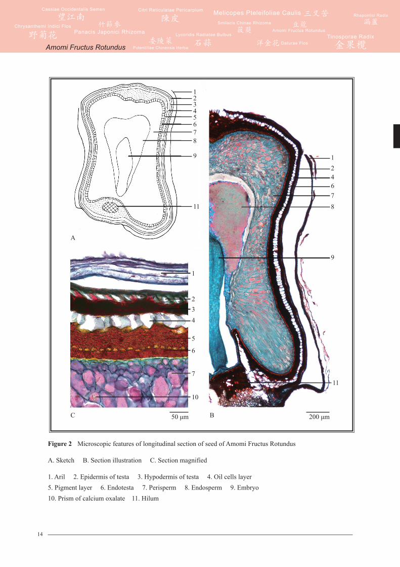
Transverse section of pericarpExocarp consists of 1 layer of flattened rectangular parenchymatous cells, 25-60 μm long, about
8 μm wide. Parenchymatous cells of mesocarp subrounded to oblong. Collateral vascular bundles
located on the inner side of mesocarp; crescent-shaped fibre bundles located on the outer side of
vascular bundles. 1-4 layers of stone cells arranged in an interrupted band, located in between
vascular bundles, stone cells subrounded to subsquare, with distinct pits. Parenchymatous
cells scattered with prisms of calcium oxalate. Endocarp consists 1 layer of rectangular
parenchymatous cells, mostly shrunken (Fig. 3).
PowderColour pale brown or greyish-brown. Stone cells varied in shape, 35-150 μm in diameter, pits
distinct, lumens relatively large. Endotesta cells yellowish-brown or reddish-brown, polygonal
in surface view, 10-35 μm in diameter, containing silica bodies; 1 layer of palisade-like cells in
lateral view, with extremely thick inner and side walls, lumens located on the upper part of the
cells, containing silica bodies. Epidermal cells of testa pale yellow, long stripe-shaped in surface
view, walls slightly thickened, usually arranged vertically with yellowish-brown hypodermal
cells. Perisperm cells subrectangular, containing small starch masses, usually with prisms of
calcium oxalate; polychromatic under the polarized microscope. Oil cells nearly colourless to
pale yellow, subsquare, 40-75 μm in diameter. Prisms of calcium oxalate scattered, 2-20 μm in
diameter; bright white or polychromatic under the polarized microscope. Aril cells with thin
walls, the wall slightly curved, some cells contain crystal sand of calcium oxalate. Fibres mostly
broken, 15-25 μm in diameter, lumens distinct (Fig. 4).
Amomi Fructus Rotundus
14
Figure 2 Microscopic features of longitudinal section of seed of Amomi Fructus Rotundus
A. Sketch B. Section illustration C. Section magnified
1. Aril 2. Epidermis of testa 3. Hypodermis of testa 4. Oil cells layer 5. Pigment layer 6. Endotesta 7. Perisperm 8. Endosperm 9. Embryo 10. Prism of calcium oxalate 11. Hilum
1234
11
5678
9
1
4
6
7
10
23
5
9
8
76421
11
C B 200 μm50 μm
A
Amomi Fructus Rotundus
15
Figure 3 Microscopic features of transverse section of pericarp of Amomi Fructus Rotundus
A. Sketch B. Section illustration C. Prisms of calcium oxalate in mesocarp
1. Exocarp 2. Mesocarp 3. Fibre bundle 4. Vascular bundle5. Stone cell 6. Endocarp 7. Prism of calcium oxalate
1
A
B
73
456
2
1
27
3
456
200 μm
7
C 50 μm
Amomi Fructus Rotundus
16
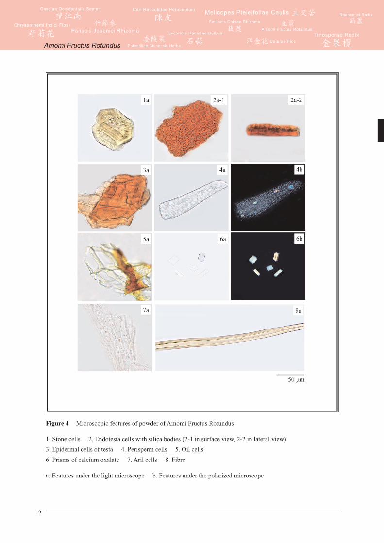
Figure 4 Microscopic features of powder of Amomi Fructus Rotundus
1. Stone cells 2. Endotesta cells with silica bodies (2-1 in surface view, 2-2 in lateral view)3. Epidermal cells of testa 4. Perisperm cells 5. Oil cells 6. Prisms of calcium oxalate 7. Aril cells 8. Fibre
a. Features under the light microscope b. Features under the polarized microscope
2a-1 2a-21a
3a
5a
7a 8a
6a 6b
4a 4b
50 μm
Amomi Fructus Rotundus
17
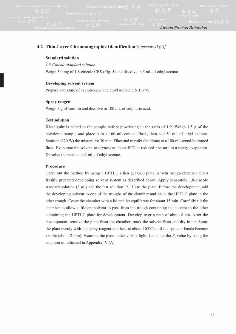
4.2 Thin-Layer Chromatographic Identification [Appendix IV(A)]
Standard solution1,8-Cineole standard solution
Weigh 5.0 mg of 1,8-cineole CRS (Fig. 5) and dissolve in 5 mL of ethyl acetate.
Developing solvent systemPrepare a mixture of cyclohexane and ethyl acetate (19:1, v/v).
Spray reagentWeigh 5 g of vanillin and dissolve in 100 mL of sulphuric acid.
Test solutionKieselguhr is added to the sample before powdering in the ratio of 1:2. Weigh 1.5 g of the
powdered sample and place it in a 100-mL conical flask, then add 50 mL of ethyl acetate.
Sonicate (320 W) the mixture for 30 min. Filter and transfer the filtrate to a 100-mL round-bottomed
flask. Evaporate the solvent to dryness at about 40ºC at reduced pressure in a rotary evaporator.
Dissolve the residue in 2 mL of ethyl acetate.
ProcedureCarry out the method by using a HPTLC silica gel G60 plate, a twin trough chamber and a
freshly prepared developing solvent system as described above. Apply separately 1,8-cineole
standard solution (3 μL) and the test solution (2 μL) to the plate. Before the development, add
the developing solvent to one of the troughs of the chamber and place the HPTLC plate in the
other trough. Cover the chamber with a lid and let equilibrate for about 15 min. Carefully tilt the
chamber to allow sufficient solvent to pass from the trough containing the solvent to the other
containing the HPTLC plate for development. Develop over a path of about 8 cm. After the
development, remove the plate from the chamber, mark the solvent front and dry in air. Spray
the plate evenly with the spray reagent and heat at about 105ºC until the spots or bands become
visible (about 2 min). Examine the plate under visible light. Calculate the Rf value by using the
equation as indicated in Appendix IV (A).
Amomi Fructus Rotundus
18
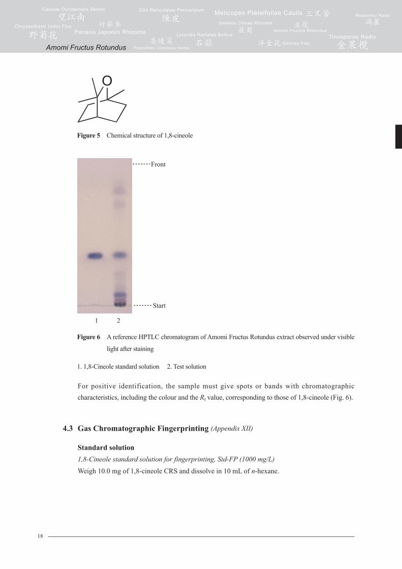
Figure 6 A reference HPTLC chromatogram of Amomi Fructus Rotundus extract observed under visible
light after staining
1. 1,8-Cineole standard solution 2. Test solution
For positive identification, the sample must give spots or bands with chromatographic characteristics, including the colour and the Rf value, corresponding to those of 1,8-cineole (Fig. 6).
4.3 Gas Chromatographic Fingerprinting (Appendix XII)
Standard solution1,8-Cineole standard solution for fingerprinting, Std-FP (1000 mg/L)
Weigh 10.0 mg of 1,8-cineole CRS and dissolve in 10 mL of n-hexane.
Front
1 2
Start
Figure 5 Chemical structure of 1,8-cineole
Amomi Fructus Rotundus
19
Test solutionKieselguhr is added to the sample before powdering in the ratio of 1:2. Weigh 1.5 g of the
powdered sample and place it in a 500-mL round-bottomed flask, then add 200 mL of water.
Connect the round-bottomed flask to a volatile oil determination tube, then add 20 mL of water
and 3 mL of n-hexane. Connect to the condenser and heat the flask gently until boiling for 2 h.
Cool down and allow to stand until two layers can be separated. Transfer the n-hexane layer to
a 25-mL volumetric flask. Transfer the aqueous layer to a 100-mL separating funnel. Rinse the
volatile oil determination tube with 5 mL of n-hexane. Transfer the solution to the separating
funnel and extract with aqueous layer. Collect the n-hexane extract and filter through the funnel
containing 1 g of anhydrous sodium sulphate. Transfer the filtrate to a 25-mL volumetric
flask. Extract the aqueous layer with 5 mL of n-hexane. Collect the n-hexane extract and filter
through the funnel containing 1 g of anhydrous sodium sulphate. Transfer the filtrate to a 25-mL
volumetric flask. Wash the filter funnel with 5 mL of n-hexane. Combine the n-hexane extracts
and make up to the mark with n-hexane. Filter through a 0.45-μm nylon filter.
Chromatographic systemThe gas chromatograph is equipped with a FID and a capillary column (DB-1701, 0.32 mm × 30 m)
of which the internal wall is covered with (14%-cyanopropyl-phenyl)-methylpolysiloxane in a
layer about 0.25 µm thick. The injection temperature is at 210ºC. The detector temperature is
at 230ºC. The split injection mode at a ratio of 20:1 is used. Programme the chromatographic
system as follows (Table 1) –
Table 1 Chromatographic system conditions
System suitability requirementsPerform at least five replicate injections, each using 1 µL of 1,8-cineole Std-FP. The requirements of the system suitability parameters are as follows: the RSD of the peak area of 1,8-cineole should not be more than 5.0%; the RSD of the retention time of 1,8-cineole peak should not be more than 2.0%; the column efficiency determined from 1,8-cineole peak should not be less than 100000 theoretical plates.
The R value between peak 4 and the closest peak in the chromatogram of the test solution should not be less than 1.5 (Fig. 7).
Time(min)
Temperature (ºC)
Rate (ºC/min)
0 – 3 40 –
3 – 39 40 → 220 5
Amomi Fructus Rotundus
20
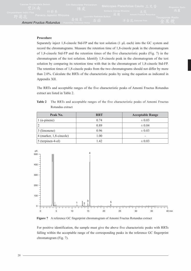
ProcedureSeparately inject 1,8-cineole Std-FP and the test solution (1 μL each) into the GC system and record the chromatograms. Measure the retention time of 1,8-cineole peak in the chromatogram of 1,8-cineole Std-FP and the retention times of the five characteristic peaks (Fig. 7) in the chromatogram of the test solution. Identify 1,8-cineole peak in the chromatogram of the test solution by comparing its retention time with that in the chromatogram of 1,8-cineole Std-FP. The retention times of 1,8-cineole peaks from the two chromatograms should not differ by more than 2.0%. Calculate the RRTs of the characteristic peaks by using the equation as indicated in Appendix XII.
The RRTs and acceptable ranges of the five characteristic peaks of Amomi Fructus Rotundus extract are listed in Table 2.
Table 2 The RRTs and acceptable ranges of the five characteristic peaks of Amomi Fructus Rotundus extract
Figure 7 A reference GC fingerprint chromatogram of Amomi Fructus Rotundus extract
For positive identification, the sample must give the above five characteristic peaks with RRTs falling within the acceptable range of the corresponding peaks in the reference GC fingerprint chromatogram (Fig. 7).
Peak No. RRT Acceptable Range1 (α-pinene) 0.74 ± 0.032 0.89 ± 0.043 (limonene) 0.96 ± 0.034 (marker, 1,8-cineole) 1.00 -5 (terpinen-4-ol) 1.42 ± 0.03
Amomi Fructus Rotundus
21
5. TESTS
5.1 Heavy Metals (Appendix V): meet the requirements.
5.2 Pesticide Residues (Appendix VI): meet the requirements.
5.3 Mycotoxins (Appendix VII): meet the requirements.
5.4 Foreign Matter (Appendix VIII): not more than 2.0%.
5.5 Ash (Appendix IX)
Total ash: not more than 11.0%.
Acid-insoluble ash: not more than 2.0%.
5.6 Water Content (Appendix X)
Toluene distillation method: not more than 12.0%.
6. EXTRACTIVES (Appendix XI)
Water-soluble extractives (hot extraction method): not less than 12.0%.
Ethanol-soluble extractives (hot extraction method): not less than 5.0%.
7. ASSAY
7.1 Assay of 1,8-Cineole
Carry out the method as directed in Appendix IV (C).
Standard solution1,8-Cineole standard stock solution, Std-Stock (5000 mg/L)
Weigh accurately 50.0 mg of 1,8-cineole CRS and dissolve in 10 mL of n-hexane.
1,8-Cineole standard solution for assay, Std-AS
Measure accurately the volume of the 1,8-cineole Std-Stock, dilute with n-hexane to produce a
series of solutions of 200, 500, 1000, 2000, 2500 mg/L for 1,8-cineole.
Amomi Fructus Rotundus
22
Test solutionKieselguhr is added to the sample before powdering in the ratio of 1:2. Weigh accurately 1.5 g of
the powdered sample and place it in a 500-mL round-bottomed flask, then add 200 mL of water.
Connect the round-bottomed flask to a volatile oil determination tube, then add 20 mL of water
and 3 mL of n-hexane. Connect to the condenser and heat the flask gently until boiling for 2 h.
Cool down and allow to stand until two layers can be separated. Transfer the n-hexane layer to
a 25-mL volumetric flask. Transfer the aqueous layer to a 100-mL separating funnel. Rinse the
volatile oil determination tube with 5 mL of n-hexane. Transfer the solution to the separating
funnel and extract with aqueous layer. Collect the n-hexane extract and filter through the funnel
containing 1 g of anhydrous sodium sulphate. Transfer the filtrate to a 25-mL volumetric
flask. Extract the aqueous layer with 5 mL of n-hexane. Collect the n-hexane extract and filter
through the funnel containing 1 g of anhydrous sodium sulphate. Transfer the filtrate to a 25-mL
volumetric flask. Wash the filter funnel with 5 mL of n-hexane. Combine the n-hexane extracts
and make up to the mark with n-hexane. Filter through a 0.45-μm nylon filter.
Chromatographic systemThe gas chromatograph is equipped with a FID and a capillary column (DB-1701, 0.32 mm × 30 m)
of which the internal wall is covered with (14%-cyanopropyl-phenyl)-methylpolysiloxane in a
layer about 0.25 µm thick. The injection temperature is at 210ºC. The detector temperature is
at 230ºC. The split injection mode at a ratio of 20:1 is used. Programme the chromatographic
system as follows (Table 3) –
Table 3 Chromatographic system conditions
Time (min)
Temperature (ºC)
Rate (ºC/min)
0 – 3 40 -
3 – 39 40 → 220 5
System suitability requirementsPerform at least five replicate injections, each using 1 µL of 1,8-cineole Std-AS (1000 mg/L).
The requirements of the system suitability parameters are as follows: the RSD of the peak area
of 1,8-cineole should not be more than 5.0%; the RSD of the retention time of 1,8-cineole peak
should not be more than 2.0%; the column efficiency determined from 1,8-cineole peak should
not be less than 100000 theoretical plates.
The R value between 1,8-cineole peak and the closest peak in the chromatogram of the test
solution should not be less than 1.5.
Amomi Fructus Rotundus
23
Calibration curveInject a series of 1,8-cineole Std-AS (1 μL each) into the GC system and record the
chromatograms. Plot the peak areas of 1,8-cineole against the corresponding concentrations of
1,8-cineole Std-AS. Obtain the slope, y-intercept and the r2 value from the 5-point calibration
curve.
ProcedureInject 1 μL of the test solution into the GC system and record the chromatogram. Identify
1,8-cineole peak in the chromatogram of the test solution by comparing its retention time with
that in the chromatogram of 1,8-cineole Std-AS. The retention times of 1,8-cineole peaks from
the two chromatograms should not differ by more than 5.0%. Measure the peak area and calculate
the concentration (in milligram per litre) of 1,8-cineole in the test solution, and calculate the
percentage content of 1,8-cineole in the sample by using the equations as indicated in Appendix
IV (B).
LimitsThe sample contains not less than 3.0% of 1,8-cineole (C10H18O), calculated with reference to the
dried substance.
7.2 Assay of Volatile Oil
Weigh accurately 30 g of the powdered sample and place it in a 1000-mL round-bottomed
flask. Add 500 mL of water and a few glass beads, shake and mix well. Carry out the method as
directed in Appendix XIII (Method A).
LimitsThe sample contains not less than 4.0% (v/w) of volatile oil.
Amomi Fructus Rotundus


![INDEX [botanicalink.com]botanicalink.com/SGHCatalogue.pdf · Fructus Capsici Hot Pepper/Chilli Fructus Chaenomelis Floweringquince Fruit Fructus Corni Dogwood Fruit/Asiatic cornelian](https://img.pdfslide.net/doc/110x75/5cdf42a688c9938b288e092b/index-fructus-capsici-hot-pepperchilli-fructus-chaenomelis-floweringquince.jpg)