Embed Size (px)
Citation preview
Proc. Natl. Acad. Sci. USAVol. 85, pp. 4804-4808, July 1988Genetics
Amplified human MYC oncogenes localized to replicatingsubmicroscopic circular DNA molecules
(oncogene amplification/amplification mechanisms/replication origins/tumor prognosis)
DANIEL D. VON HOFF*, DONALD R. NEEDHAM-VANDEVANTER*, JENNIFER YUCELt, BRADFORD E. WINDLEt,AND GEOFFREY M. WAHLttGene Expression Laboratory, The Salk Institute for Biological Studies, P.O. Box 85800, San Diego, CA 92138; and *Cancer Therapy and Research Center ofSouth Texas, University of Texas Health Science Center, 7703 Floyd Curl Drive, San Antonio, TX 78284
Communicated by Donald Helinski, February 29, 1988 (received for review September 28, 1987)
ABSTRACT Amplification of genes can sometimes bedetected by molecular hybridization but not by cytogeneticmethods, suggesting that in some cases the units of amplifica-tion may be too small to be detected by light microscopy. Theexperiments reported here investigate whether submicroscopicamplification units are present in early passages of the humantumor cell lines HL-60 and COLO 320. The results show thatsuch cells do contain submicroscopic, extrachromosomal, su-percoiled circular molecules harboring MYC genes. The mol-ecules in HL-60 are -250 kilobase pairs (kbp), while those inCOLO 320 are 120-160 kbp. The extrachromosomal moleculesin HL-60 are shown to replicate semiconservatively and ap-proximately once in one cell cycle. We propose that thesesubmicroscopic elements are precursors of double-minutechromosomes, the usual extrachromosomal manifestation ofgene amplification, since both are structurally similar andreplicate autonomously.
The large number of oncogenes and drug-resistance genessubject to amplification indicates that many, possibly most,genomic regions are capable ofundergoing regional increasesin copy number (see refs. 1-3 for review). Cytogeneticanalyses of many independently isolated cell lines containingdifferent amplified genes show that amplified sequenceslocalize to two types of abnormal chromosomal structures,referred to as double-minute chromosomes (DMs) and ex-panded chromosomal regions (also called homogeneouslystaining or abnormally banding regions). DMs are acentricextrachromosomal elements, which replicate approximatelyonce per cell cycle and are believed to be circular (4, 5).Expanded chromosomal regions consist of amplificationunits arranged as both inverted and direct repeats (6-9). Sizeestimates for individual amplified units of different chromo-somal regions range from 250 to >1000 kilobase pairs (kbp)(1-3).While DMs and expanded chromosomal regions are gen-
erally visible by light microscopy, a submicroscopic extra-chromosomal structure not previously known, which issupercoiled, circular, and replicates autonomously, has re-cently been implicated in mammalian gene amplification (10).Subsequent studies showed that these elements are precur-sors of DMs (11). Recently, -650-kbp elements were foundin methotrexate-resistant human cells in which only a minor-ity of cells contain DMs or expanded chromosomal regions(12).
It is possible that submicroscopic amplification productsmay be peculiar to cases involving drug selections in whichchromosomal damage results from drug treatment or to thoseinvolving chromosomal destabilization caused by gene trans-
fer. The experiments reported here were initiated to deter-mine whether submicroscopic elements are also generated inthe amplification ofendogenous cellular oncogenes where theselective pressures for gene amplification were imposedwithin the tumor environment in vivo. The cell line HL-60was chosen for our initial studies because previous workestablished that MYC amplification existed in the tumor cellsprior to their establishment as a cell line (13).Two observations suggested that the visible cytogenetic
anomalies found in the HL-60 and COLO 320 neuroendocrinetumor cells analyzed here may be formed from precursorsthat are <1000 kbp and, hence, would not be visible by lightmicroscopy. First, the DMs observed in single cells in theseand other cell lines are usually variable in size and aresometimes barely detectable by light microscopy (14, 15).Second, estimation of genetic complexity showed that theamplified units in HL-60 and COLO 320 cells containing DMsor homogeneously staining regions (HSRs) are, respectively,--95 and ="300 kbp (16). The results reported here show thatsome early passages of HL-60 promyelocytic leukemia cellsand some subclones of COLO 320 (DM) neuroendocrinetumor cells do indeed harbor submicroscopic circular extra-chromosomal elements.
MATERIALS AND METHODSCell Lines. An early passage (passage 36) of HL-60 pro-
myelocytic leukemia cells (16) was kindly provided by S.Collins (Fred Hutchinson Cancer Center), and HL-60 latepassage (>200 passages) was obtained from B. Sefton (SalkInstitute). COLO 320 cells (17) containing DMs (passage 15)and HSRs (passage 11) were obtained from the AmericanType Culture Collection, and Raji cells [ref. 18; containingthe 178-kbp circular Epstein-Barr virus (EBV)] were pro-vided by C. Mulder (University of Massachusetts, Wor-cester). All human cell lines were grown in RPMI 1640medium/10% fetal calf serum. Single cell subclones ofCOLO320 DM and HSR cells were obtained by limiting dilution.T5S1-3 and C5R500 are Chinese hamster ovary (CHO) celllines containing -50 copies per cell of a transfected, ampli-fied CAD gene (CAD, carbamoyl-phosphate synthetase,aspartate transcarbamylase, dihydroorotase) (10, 19). InT5S1-3, the CAD genes are amplified intrachromosomally,while in C5R500 they localize to extrachromosomal super-coiled -250-kbp circular molecules (10). Both lines are grownin Dulbecco's modified Eagle's medium supplemented with10% dialyzed fetal calf serum, 1 x nonessential amino acids(GIBCO), and 500 ,uM N-phosphonacetyl-L-aspartate (a
Abbreviations: DHFR, dihydrofolate reductase; CAD, the multi-functional protein that catalyzes the first three steps ofde novo UMPbiosynthesis; DM, double-minute chromosome; HSR, homogene-ously staining region; rDNA, ribosomal RNA-encoding DNA; EBV,Epstein-Barr virus; CHO, Chinese hamster ovary.
4804
The publication costs of this article were defrayed in part by page chargepayment. This article must therefore be hereby marked "advertisement"in accordance with 18 U.S.C. §1734 solely to indicate this fact.
Proc. Natl. Acad. Sci. USA 85 (1988) 4805
specific inhibitor of CAD). B5-4 is a Syrian hamster cell linecontaining -200 intrachromosomally amplified CAD genes(20), many of which are presented as inverted repeats (6).
Isolation and Analysis of Large Circular DNA from Mam-malian Cells. Circular DNA exceeding 100 kbp was isolatedby alkaline lysis (10) and then either applied (21) directly ontoNitrocellulose (BA85; Schleicher & Schuell) through a dotblot apparatus (Minifold I; Schleicher & Schuell) or analyzedby electrophoresis through agarose gels under conditionsdesigned to fractionate supercoiled circular DNA accordingto molecular weight (10). Blots were hybridized as described(21).
Replication ofMYC Episomes in HL-60 Cells. Exponential-phase suspension cultures of early passage HL-60 cells wereused. The cells were labeled with 3 ,uM BrdUrd plus 3 AuMthymidine for either 0.5, 1, or -2 generations as determineddirectly by cell counting. Thymidine was essential to reducethe toxicity of BrdUrd to HL-60 cells. Even in the presenceof equimolar thymidine and BrdUrd, growth retardation wasobserved after one generation. Replication ofMYC episomeswas then determined either from total high molecular weightDNA prepared by standard procedures (22), or from epi-some-enriched DNA prepared by the alkaline lysis method(10). DNA from 1-5 x 10⁷ cells prepared by either methodwas sheared by 10 passages through a 27-gauge needle andthen sedimented to equilibrium in 5-ml gradients (45,000 rpmin a Beckman Vti65 rotor at 15'C for 16 hr) of CsCl added toan initial density of 1.75 g/ml. Each gradient was fractionatedinto 24 200-plI aliquots, and the refractive index ofevery otherfraction was determined. A 100-,ul portion of each fractionwas then denatured by adding NaOH to 0.3 M and heating at650C for 15 min, neutralized by adding an equal volume of 2M NH4OAc, and the samples when then applied to a dot blot
100
80
0
C
60-
40-
20-
'I I40-I
apparatus (Schleicher & Schuell), hybridized with the nick-translated probes indicated, and washed under stringentconditions as described (21). The dots were excised with apaper punch and the cpm per fraction was determined in aliquid scintillation counter.
RESULTS
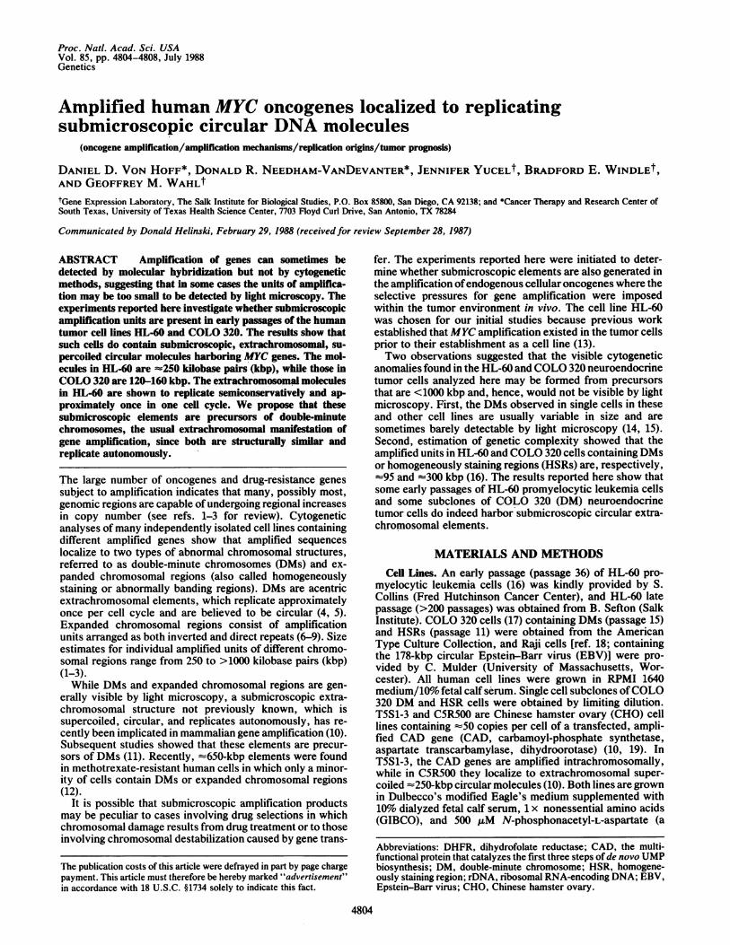
Submicroscopic Elements Containing MYC Genes in HL-60Revealed by Alkaline Lysis. Lysis of mammalian cells at pH12.45, followed by neutralization and phenol extraction athigh salt concentrations, enables isolation of supercoiledDNA >100 kbp in the supernatant, while chromosomal DNApartitions at the phenol-aqueous interface. To determinewhether MYC sequences might reside in circular elementsrecoverable by this method, duplicate DNA samples fromearly passage HL-60 cells were prepared by either thealkaline lysis method or by a NaDodSO4 lysis method thatenables the recovery of total cellular DNA. Fig. 1 shows thatin six independent experiments, 8-52% of the total MYCsequences are recovered in the alkaline lysate supernatant.By contrast, only 1% ofthe MYC sequences are recovered inthe alkaline lysate supernatant from late-passage HL-60 cellsin which the MYC sequences are amplified intrachromosom-ally (6). However, recovery of MYC DNA in the extrachro-mosomal fraction of early passage HL-60 cells obtained fromdifferent laboratories is variable (data not shown). The sameprocedure applied to the isolation of the 178-kbp circularEBV genome from Raji cells (18) and the 250-kbp circularCAD episome from C5R500 CHO cells (10) produced yieldsof 31-100% and 31-54%, respectively. Fig. 1 also shows thatthe recovery of the circular mitochondrial DNA from HL-60cells is only about half that from Raji cells, suggesting that it
I Ir
Cell line Ie Raji - | IC5R500IT5S1-31B5-41 T4IL60 early-0i IHL60 latelProbe IEBV IMitol Ribo| I I CAD- - I I C-myc IMitoiRibol C-myc I
k- Colo 320 HSR-4lIC-myc Mito Ribol
FIG. 1. Circular MYC sequences in HL-60 cells revealed by alkaline lysis. Approximately 10' cells of the indicated cell lines were dividedinto two equal aliquots and the DNA from one half was isolated by the NaDodSO4/phenol method (i.e., total DNA), while that from the otherhalf was isolated by alkaline lysis (i.e., circular DNA). Equal amounts of DNA isolated by either method were then applied to nitrocellulosefilters through a dot blot apparatus (Minifold I; Schleicher& Schuell) and hybridized (21) with probes specific forMYC sequences, mitochondrialDNA (Mito), rDNA (Ribo), or the CAD gene. The dots were excised with a paper punch and the cpm of probe bound was determined in a liquidscintillation counter. The percentage of the total sequences hybridized to each probe in the alkaline lysate was then calculated by dividing thecpm in the alkaline lysate by the cpm in the total DNA. Each dot represents the value determined in an independent experiment.
Genetics: Von Hoff et aL
0 0S:
Proc. Natl. Acad. Sci. USA 85 (1988)
is more difficult to obtain intact circularDNA from the HL-60cells. By contrast, the recovery of naturally highly repeatedchromosomal sequences such as ribosomal RNA-encodingDNA (rDNA) or chromosomally amplified CAD genes (10,19) is generally <3%. Therefore, the recovery of MYCsequences from early passage HL-60 cells that contain few orno DMs is in the same range as the isolation of 178- to 250-kbpsubmicroscopic circular elements in Raji and CHO cells andis far higher than the recovery of repeated chromosomalsequences.The data presented above are compatible with the idea that
some of the amplified MYC genes reside in submicroscopiccircular elements in the early passage HL-60 cells used forthese experiments. However, some amplified MYC genes insome sublines of later passages of HL-60 cells are present ininverted repeats within expanded chromosomal regions (i.e.,HSRs; see ref. 6), suggesting that the molecules detected arenot circular, but rather are renatured inverted repeats gen-erated by the alkaline lysis procedure. This possibility iseliminated since late passage HL-60 and COLO 320 (HSR)cell lines that contain amplified MYC genes in invertedduplications (6) was only 1-3%, as was the recovery ofamplified CAD genes present, in part, as inverted duplica-tions in the Syrian hamster cell line B5-4 (ref. 20; see Fig. 1).The recovery of CAD sequences in this case was alsobetween 1% and 3%. In addition, only 1% of the dihydrofo-late reductase (DHFR) sequences from a mouse cell linecontaining amplified DHFR sequences in DMs fractionatedinto the alkaline lysate supernatant (J. Ruiz and G.M.W.,unpublished data; cell line R50 provided by T. Tlsty, Uni-versity of North Carolina). These results show that therecovery ofMYC sequences from early passage HL-60 cellsby the alkaline lysis method is significantly higher than therecovery ofintrachromosomally amplified sequences that arearranged as inverted repeats or amplified sequences con-tained in DMs and strongly indicates that some of the MYCsequences are contained in submicroscopic extrachromo-somal circular elements in the HL-60 cells analyzed here.MYC Is Amplified in =250-kbp Supercoiled Circular Mol-
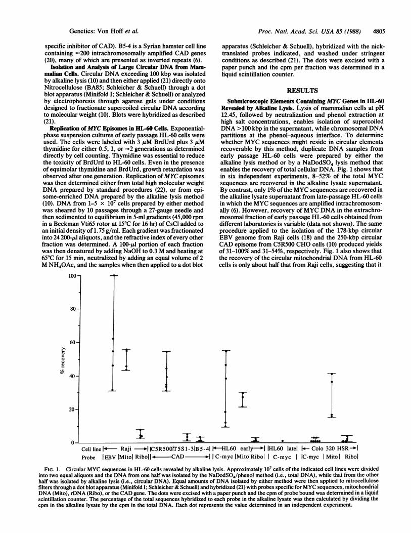
ecules in Early Passage HL-60 Cells. If the MYC sequences inearly passage HL-60 cells are supercoiled circles less than-750 kbp, then they should be detectable by electrophoresisof alkaline lysates through agarose gels under conditions thatfractionate supercoiled circular DNA (10) according to thelogarithm of molecular weight (e.g., 7.5/cm; 0.7% agarose).Under these conditions, some nicked circular molecules aretrapped at the well, and some circular and linear DNAmolecules migrate near the dye front (e.g., see ref. 23).Electrophoresis of alkaline lysates derived from early pas-sage HL-60 cells revealed three intense bands that hybridizewith a MYC probe: one band was at the well of the gel,possibly reflecting very large circles, or circles impaled byagarose threads (e.g., see ref. 24); one band was at theposition of a 250-kbp supercoiled circular molecule; and onemigrated at the dye front, corresponding to open circular orsheared DNA (Fig. 2, lanes 1, 2, and 5). Importantly, thealkaline lysates prepared from Raji and CHO C5R500 cellsthat provided 178-kbp (EBV) and 250-kbp (CAD) supercoiledcircular size standards, revealed an analogous pattern ofthree bands. An alkaline lysate prepared from a COLO 320(HSR) cell line containing approximately the same level ofMYC amplification as the early passage HL-60 cells revealedonly faint hybridization at the position of fragmented DNA(Fig. 2B, lane 7). These data show that some of the c-mycsequences in early passage HL-60 cells reside in -250 kbpsupercoiled circular molecules (i.e., MYC episomes).MYC Episomes in HL-60 Cells Replicate Semiconservatively
and Approximately Once in One Cell Cycle. If the MYCepisomes are analogs of the previously described CADepisomes, which are precursors of DMs (10, 11), then they
ASIZE
1 2 3 4 (kbp)
j~~jw m
0 _.1250 SC-178 SC
am-50 SC
I Si
B
5 6 7 8
inw
-50 LOC
SIZE(kbp)
_-250 SC
FIG. 2. Characterization of circular molecules in HL-60 andCOLO 320 cells by gel electrophoresis. Alkaline lysates wereprepared from i07 cells from the indicated cell lines, fractionated byelectrophoresis on agarose gels at high voltage (7.5 V/cm) to separatesupercoiled circular molecules by molecular weight (10, 23), trans-ferred to nitrocellulose, and hybridized with probes specific forMYC(lanes 1, 5, 6, and 7), EBV (lane 2), orCAD (lanes 3, 4, and 8). Lanes:1 and 5, alkaline lysates HL-60 cells; 2, from Raji cells containing the178-kbp EBV episome; 3 and 8, from C5R500 CHO cells that containthe 250-kbp CAD episome; 4, the 50-kbp CAD cosmid (22); 6,alkaline lysates from COLO 320 (DM) clone 10; 7, from COLO 320(HSR). Hybridizations were done with 2 x 10⁶ cpm per ml of eachprobe with 10%6 (wt/vol) dextran sulfate, overnight, with standardincubation and stringent washing procedures (21). Autoradiographywas for 24-48 hr at -700C with two Lightning Plus intensifyingscreens and Kodak XR-5 film. The MYC episome in early passageHL-60 cells (lanes 1 and 5) is clearly different in size from thatdetected in COLO 320 clone 10 (asterisk). SC, L, and OC, super-coiled, linear, and open circular DNA, respectively.
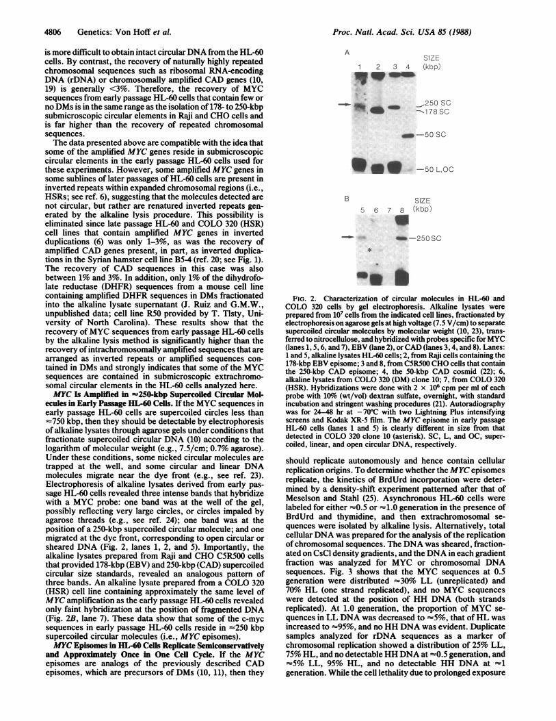
should replicate autonomously and hence contain cellularreplication origins. To determine whether the MYC episomesreplicate, the kinetics of BrdUrd incorporation were deter-mined by a density-shift experiment patterned after that ofMeselson and Stahl (25). Asynchronous HL-60 cells werelabeled for either =0.5 or =1.0 generation in the presence ofBrdUrd and thymidine, and then extrachromosomal se-quences were isolated by alkaline lysis. Alternatively, totalcellular DNA was prepared for the analysis of the replicationof chromosomal sequences. The DNA was sheared, fraction-ated on CsCl density gradients, and the DNA in each gradientfraction was analyzed for MYC or chromosomal DNAsequences. Fig. 3 shows that the MYC sequences at 0.5generation were distributed z30% LL (unreplicated) and70%o HL (one strand replicated), and no MYC sequenceswere detected at the position of HH DNA (both strandsreplicated). At 1.0 generation, the proportion of MYC se-quences in LL DNA was decreased to =5%, that ofHL wasincreased to =95%, and no HH DNA was evident. Duplicatesamples analyzed for rDNA sequences as a marker ofchromosomal replication showed a distribution of 25% LL,75% HL, and no detectable HH DNA at =0.5 generation, and=5% LL, 95% HL, and no detectable HH DNA at ~1generation. While the cell lethality due to prolonged exposure
4806 Genetics: Von Hoff et al.
Proc. Natl. Acad. Sci. USA 85 (1988) 4807
(
0
0
I-
20
0
100
80'
60
40.
20-
0
HH LH
DNA Density
FIG. 3. MYC episomes in HL-60 cells replicate semiconserva-tively and approximately once in one cell cycle. HL-60 cells weregrown for either -0.5 (A) or -1 (B) generation in our equimolarmixture of thymidine and BrdUrd. Total or circular DNA was thenisolated by the NaDodSO4/phenol or alkaline lysis methods,sheared, and centrifuged to equilibrium on CsCI gradients. Thegradients were fractionated, applied to nitrocellulose through aMinifold 1 dot blotter, and hybridized with nick-translated (-2 x 10⁶cpm per ml) probes specific for MYC or rDNA. After stringentwashing, the cpm in each fraction was determined by counting in aliquid scintillation counter. Results similar to those shown wereobtained in three independent experiments. The yield of rDNAsequences in the alkaline lysate was undetectable in the experimentshown, indicating that there was negligible contamination of thecircular DNA with chromosomal sequences. n, MYC; o, rDNA.
to BrdUrd prevented accurate determination of BrdUrdincorporation after a second round of replication, we notethat a similar pattern of incorporation was seen for rDNA andMYC sequences after -2 generations of labeling (data notshown). These data indicate that BrdUrd is incorporated intoMYC episomes in a manner consistent with semiconservativereplication, but the MYC episomes appear to replicate laterthan the rDNA sequences, on the average. Since no HHDNA was observed at either 0.5 or 1 generation, and theamount of LL DNA decreased as the incubation timeapproached one cell doubling, the data also indicate thatmost, if not all, of them replicated only once during the singlecell cycle analyzed.MYC Episomes in COLO 320 Ceils. The results described
above encouraged us to analyze COLO 320 (DM) cells forsubmicroscopic MYC episomes. Analysis of an unclonedpopulation obtained directly from the American Type CultureCollection failed to reveal any MYC episomes in alkalinelysates derived from >107 cells. However, the COLO 320(DM) cells had grown for an undefined period of time in vivoand then were passaged in culture before being frozen. Byanalogy with our observation that submicroscopic episomesincrease in size over time during in vitro culture (11), ifsubmicroscopic episomes had been present initially, mostwould have increased in size to form DMs by the time we
obtained the cells. Therefore, we prepared seven indepen-dent single cell subclones of the COLO 320 (DM) and COLO320 (HSR) cell lines. We found that each of the seven DMsubclones contained very similar levels ofMYC amplification(29 + 6 copies per haploid genome), in agreement withpublished values (26). However, there was considerablevariation in the median number and size of DMs in eachsubclone (median, 0-27 DMs per cell). No HSR subclonecontained DMs, and the average -fold amplification ofMYCgenes was virtually identical to that in the DM subclones (31+ 10 copies per haploid genome). All DM subclones wereanalyzed for the presence ofMYC episomes, but only two ofthem were found to contain such molecules. Subclone 10 hadthe lowest median number of DMs, while subclone 3 had thehighest median number, but in the latter the vast majority ofDMs were very small. Gel analysis ofan alkaline lysate of 107cells from COLO 320 (DM) subclone 10 reveals a faint bandofMYC hybridization at the position ofa supercoiled circularmolecule with a size of 120 kbp and a very intense band ofhybridization corresponding to nicked and/or linearizedepisomes. The yield of extrachromosomal MYC sequencesfrom COLO 320 DM is higher than from HL-60, as isexpected, since COLO 320 DM has twice the MYC copynumber ofHL-60. The size ofthe MYCepisome in COLO 320(DM) clone 3 is -160 kbp (data not shown).No evidence of MYC episomes was found in any COLO
320 HSR subclone as indicated by the lack of MYC hybrid-ization at the position of supercoiled molecules or at the dyefront (Fig. 2, compare lanes 6 and 7). These results indicatethat MYC episomes and expanded chromosomal regionscontaining amplified MYC sequences are rarely, if ever,found in the same cell.
DISCUSSIONThe results presented here document the existence of sub-microscopic circular extrachromosomal molecules harboringamplified MYC genes in two human tumor cell lines derivedfrom patients who had not undergone chemotherapy orradiation therapy (16, 17). Since other results indicate thatsuch molecules can be precursors of DMs (11), and DMscontaining MYC genes in HL-60 cells were observed previ-ously in tumor cells not subjected to in vitro culturing (16), wepropose that the submicroscopic elements were generated invivo under the conditions that presumably select for in-creased expression ofMYC genes. Furthermore, the submi-croscopic molecules containing amplified MYC genes weredetected only in some early passages of HL-60 cells and insome subclones of COLO 320 (DM), but not in the initialuncloned population of COLO 320 (DM) cells, in COLO 320(HSR) cells, or in late passage HL-60 cells that have intra-chromosomal amplification ofMYC genes present in part asinverted repeats (6). These results are similar to those weobtained in a gene transfer model system (10, 11), leading usto propose that the submicroscopic MYC molecules repre-sent an early stage of gene amplification in these two tumorcell lines and that they are precursors ofDMs. However, ourdata cannot address the issue of whether these elementsrepresent the initial products of gene amplification, since thetumor cells grew for an undocumented number ofgenerationsin vivo prior to their establishment as a cell line. These resultsand others concerning CAD (10, 11) and DHFR (12) geneamplification strongly indicate that the formation of submi-croscopic elements may be a general early event in theamplification process, independent of the type of selectivepressure imposed. In light of statistical analyses indicatingthat amplification of specific oncogenes can provide signifi-cant indicators of a poor prognosis in human breast cancerand neuroblastoma (27, 28), it will be important to determinewhether patients whose tumor cells are at an early stage ofthe
ADS10 J
B
Genetics: Von Hoff et A
Proc. Natl. Acad. Sci. USA 85 (1988)
amplification process as manifested by the presence ofextrachromosomal submicroscopic elements have betterprognoses than those at later stages in which amplification ischaracterized by microscopic cytogenetic anomalies.The idea that submicroscopic elements are early products
of gene amplification and may be precursors of both DMs andintrachromosomal units of gene amplification is supported byobservations from other systems. For example, early stagesof methotrexate resistance due to DHFR gene amplificationhave been described in which only a small percentage of thecells contained DMs (29), implying that those without DMscontained elements below the resolution of light microscopy.In addition, DMs within a single cell are often highly variablein size (e.g., see ref. 4, 13, and 14), suggesting that small DMscould be precursors of larger ones. The frequent observationthat cell lines containing DMs in vivo are replaced by cell linescontaining the same sequences amplified intrachromosom-ally after passage in vitro has generally been interpreted interms of chromosomal integration of DMs, followed byovergrowth of DM-containing cells by those with intrachro-mosomal amplification under in vitro growth conditions (30).Recently, we have shown that submicroscopic extrachromo-somal elements are precursors of DMs and that these ele-ments can reintegrate into chromosomes and subsequentlyamplify from the new site (11).The sizes we have determined for the submicroscopic
episomes in HL-60 (250 kbp) and two subclones of COLO 320cells (120-160 kbp) are significantly different from thosereported previously in which in-gel renaturation was used toobtain estimates of size of the amplified region (HL-60, -95kbp; COLO 320, ='300 kbp; see ref. 13). Note that the sizeestimates made here are based on the physical parameter ofgel migration, while measurements made by in-gel renatur-ation estimate "genetic complexity." Since the physical sizewe determined for the MYC episomes in HL-60 is about twicethat of the genetic complexity estimated by in-gel renatura-tion, it is possible that each episome contains a duplication(or higher multimer) of each amplified unit. Interestingly, amechanism proposed by Passananti et al. (7) to explain MYCamplification in HL-60 and COLO 320 cells involves achromosomal excision to generate replicating circular mole-cules containing an inverted repeat of MYC sequences. Thephysical size of such molecules should, therefore, be approx-imately double that of their genetic complexity. While ourdata are compatible with some of the predictions of thismodel, it remains to be determined whether MYC episomescontain an inverted repeat and whether they are formed byexcision. In the absence of such information, and the proofthat the first product of amplification is a circular moleculewith the properties described above, additional models suchas those invoking rereplication-recombination (31) or recom-bination of replication intermediates (32) are also compatiblewith the data and cannot be excluded.The fact that the previous estimates for the size of the
amplified unit in COLO 320 cells far exceed those made heresuggest that sequences unlinked to the MYC gene, andtherefore not present in MYC episomes, are amplified inthese cells. Consistent with this idea, we have found nocorrelation between the number of MYC copies and thenumber and sizes of DMs in any of the subclones analyzed,suggesting that most of the DMs may not harbor MYCamplification regions.
It has been suggested that functional chromosomal sub-structures such as replication domains comprise each unit ofamplification because large expanses ofDNA are coamplifiedwith the selected gene (1, 3). We have now described threeinstances in which the early products of amplification arewithin the size range expected for mammalian replicons (33).Importantly, in the two cases analyzed for replication, both
were shown to replicate in a manner similar to that ofchromosomal DNA sequences. These results indicate thatone or more functional DNA replication origin must residewithin each submicroscopic element, and they furtherstrengthen the view that in some cases the unit of amplifi-cation may comprise part or all of the unit of replication (1,3). The ability to isolate and analyze the submicroscopicelements should provide a means for elucidating the mech-anisms of their formation at the molecular level and forobtaining functional mammalian replication origins.We thank M. L. DeRose for guidance on many procedures, C.
Moore for assistance with cytogenetic analyses, J. Meinkoth and S.Carroll for helpful suggestions concerning the manuscript, and B.Draper (MacGraphics). This work was supported by grants toG.M.W. from the National Institutes of Health/National Institute ofGeneral Medical Sciences and the G. Harold and Leila Y. MathersCharitable Foundation, and to D.D.V.H. from the National Foun-dation for Cancer Research and the American Cancer Society.B.E.W. is a postdoctoral fellow supported by the Anna Fuller Fund.
1. Stark, G. R. & Wahl, G. M. (1984) Annu. Rev. Biochem. 53, 447-491.2. Alitalo, K. & Schwab, M. (1986) Adv. Cancer Res. 47, 235-238.3. Hamlin, J. L., Milbrandt, J. D., Heintz, N. H. & Azizkahn, J. C. (1984)
Int. Rev. Cytol. 90, 31-82.4. Cowell, J. K. (1982) Annu. Rev. Genet. 16, 21-59.5. Hamkalo, B., Farnham, P. J., Johnston, R. & Schimke, R. T. (1985)
Proc. Natl. Acad. Sci. USA 82, 1126-1130.6. Ford, M. & Fried, M. (1986) Cell 45, 425-430.7. Passananti, C., Davies, B., Ford, M. & Fried, M. (1987) EMBO J. 6,
1697-1703.8. Saito, I. & Stark, G. R. (1986) Proc. Natl. Acad. Sci. USA 83, 8664-8668.9. Looney, J. & Hamlin, J. (1987) Mol. Cell. Biol. 7, 569-577.
10. Carroll, S. M., Gaudray, P., De Rose, M. L., Emery, J. F., Meinkoth,J. L., Nakkim, E., Subler, M., Von Hoff, D. D. & Wahl, G. M. (1987)Mol. Cell. Biol. 7, 1740-1750.
11. Carroll, S., DeRose, M. L., Gaudray, P., Moore, C., Needham-VanDe-vanter, D. R., Von Hoff, D. D. & Wahl, G. M. (1988) Mol. Cell. Biol.,in press.
12. Maurer, B. T., Lai, E., Hamkalo, B. A., Hood, L. & Attardi, G. (1987)Nature (London) 327, 434-437.
13. Collins, S. J., Gallo, R. C. & Gallagher, R. E. (1977) Nature (London)270, 347-349.
14. Meinkoth, J., Killary, A. N., Fournier, R. E. K. & Wahl, G. M. (1987)Mol. Cell. Biol. 7, 1415-1424.
15. Trent, J., Meltzer, P., Rosenblum, M., Harsh, G., Kinzler, K., Marshal,R., Feinberg, A. & Vogelstein, B. (1986) Proc. Natl. Acad. Sci. USA 83,470-473.
16. Kinzler, K. W., Zehnbauer, B. A., Brodeur, G. M., Seeger, R. L.,Trent, J. M., Meltzer, P. J. & Vogelstein, B. (1986) Proc. Natl. Acad.Sci. USA 83, 1031-1035.
17. Quinn, L. A., Moore, G. E., Morgan, R. T. & Woods, L. K. (1979)Cancer Res. 39, 4914-4924.
18. Adams, A. & Lindahl, T. (1975) Proc. Natl. Acad. Sci. USA 72, 1477-1481.
19. Wahl, G. M., Robert de Saint Vincent, B. & De Rose, M. L. (1984)Nature (London) 307, 516-520.
20. Ardeshir, F., Giulloto, E., Zieg, J., Brison, O., Liao, W. S. L. & Stark,G. R. (1983) Mol. Cell. Biol. 3, 2076-2088.
21. Meinkoth, J. & Wahl, G. M. (1984) Anal. Biochem. 138, 267-284.22. Robert de Saint Vincent, B., Delbruck, S., Eckhart, W., Meinkoth, J.,
Vitto, L. & Wahl, G. M. (1981) Cell 27, 267-277.23. Casse, F., Boucher, C., Julliot, J. S., Michel, M. & Denarie, J. (1979) J.
Gen. Microbiol. 113, 229-242.24. Schindler, C. W., Krolewski, J. J. & Rush, M. G. (1982) Plasmid 7, 263-
270.25. Meselson, M. & Stahl, F. W. (1957) Proc. Natl. Acad. Sci. USA 44,671-
687.26. Alitalo, K., Schwab, M., Varmus, H. E. & Bishop, J. M. (1983) Proc.
Natl. Acad. Sci. USA 80, 1701-1711.27. Slamon, D. J., Clark, G. M., Wong, S. F., Levin, W. J., Ullrich, A. &
McGuire, W. L. (1987) Science 235, 177-182.28. Brodeur, G. M., Seeger, R. C., Schwab, M., Varmus, H. E. & Bishop,
J. M. (1984) Science 24, 1121-1124.29. Kaufman, R. J. & Schimke, R. T. (1981) Mol. Cell. Biol. 1, 1069-1076.30. Cowell, J. K. (1980) Cytogenet. Cell Genet. 27, 2-7.31. Schimke, R. T., Sherwood, S. W., Hill, A. B. & Johnston, R. N. (1986)
Proc. Natl. Acad. Sci. USA 83, 2157-2161.32. Wahl, G. M., Carroll, S. M., Gaudray, P., De Rose, M. L., Emery, J. &
Von Hoff, D. D. (1987) in Proceedings of the Workshop on the Role ofDNA Amplyfication in Tumor Initiation and Promotion, eds. Schlehofer,J. R. & zurHausen, H. (Lippincott, Philadelphia), pp. 45-57.
33. Hand, R. (1978) Cell 15, 317-325.
4808 Genetics: Von Hoff et al.