Embed Size (px)
Citation preview
SHORT REPORT ABSTRACT: In this study we describe an autosomal dominant distal mus-cular dystrophy in a small Austrian family. The myopathy started in earlyadulthood with a slowly progressive weakness of the muscles of the anteriortibial compartment, followed by the long finger extensors and sternocleido-mastoids in some family members. Other muscles were spared. Histopa-thology showed fiber size variation and autophagic vacuoles. This diseasepattern is similar to Laing distal myopathy, which has been described pre-viously in only one other family.
© 2000 John Wiley & Sons, Inc. Muscle Nerve 23: 1876–1879, 2000
AN AUTOSOMAL DOMINANT EARLY ADULT-ONSETDISTAL MUSCULAR DYSTROPHY
FRITZ ZIMPRICH, MD, PhD,1 ATBIN DJAMSHIDIAN, MD, 1
JOHANNES A. HAINFELLNER, MD, 2 HERBERT BUDKA, MD, 2 and
JOSEF ZEITLHOFER, MD, PhD 1
1 Universitatsklinik fur Neurologie, Allgemeines Krankenhaus der Stadt Wien,Wahringer Gurtel 18-20, 1090 Vienna, Austria2 Institute of Neurology, University of Vienna, Austria
Accepted 12 June 2000
Distal muscular dystrophies are a heterogeneousgroup of inherited muscle disorders with a preferen-tial and often selective involvement of distal muscles.Since their first systematic description by Welanderin the 1950s, at least five distinct types have beendelineated on clinical grounds, mode of inheritance,and genetic linkage.2,12,13 Here we present a familywith an early adult-onset distal myopathy that clini-cally most closely resembles the myopathy describedby Laing et al.10 This is a rare distal myopathy, whichhas, to our knowledge, been observed in only asingle kindred.10,12
CASE REPORTS
Family. Our initial patient from this Austrian fam-ily was a 34-year-old man (case 1) referred to ourclinic after a 20-year history of progressive distal my-opathy. His 66-year-old mother (case 2) and his onlysibling, a 30-year-old sister (case 3), suffered from asimilar but somewhat milder myopathy. Neither the
patient’s father, his 5-year-old niece, nor any othermore distant relatives were affected. The extent towhich the deceased grandparents showed a similarmyopathy is not known. The family’s remainingmedical history was unremarkable.
Clinical Description and Electrophysiology. Case 1.This man, the only member of his family to seekmedical help, was initially seen by us in 1998 (thenaged 33 years), although he had first come to a spe-cialist’s attention at the age of 19 years when inves-tigations, including a muscle biopsy, were per-formed. He was born healthy and developednormally until his midteens. At that time he noticedbilateral weakness of toe and ankle dorsiflexion,which slowly progressed over the years. In his late 20she also developed a slowly progressive weakness ofthe finger extensors, but other muscle groups re-mained unaffected.
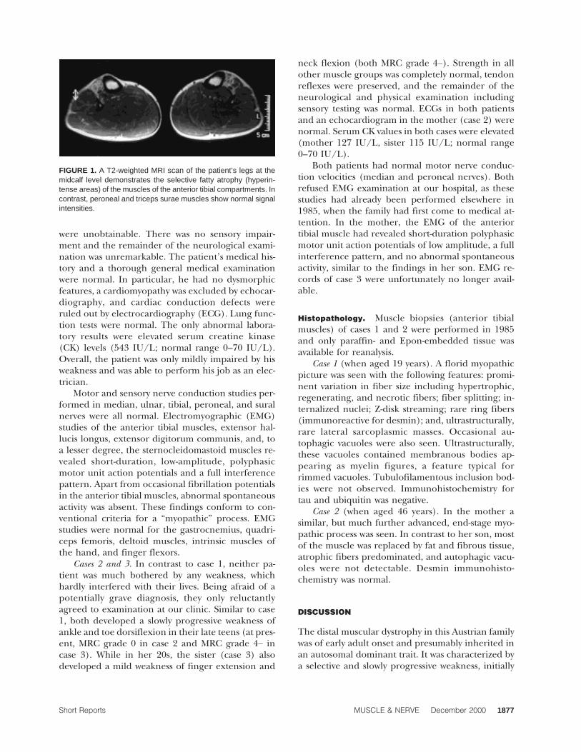
At presentation, a selective atrophy of the ante-rior tibial compartment was evident (Fig. 1), in con-trast to an otherwise athletic habitus. On examina-tion, he had bilateral foot-drop and weakness of toeextension (both MRC grade 2) and weak finger ex-tension (MRC grade 4). The muscles of the face,neck, and upper and lower limb girdles, as well asthe flexors and intrinsic muscles of the hands andfeet, had full strength. Tendon reflexes in all limbs
Abbreviations: CK, creatine kinase; MRC, Medical Research Council;ECG, electrocardiography; EMG, electromyography; MRI, magnetic reso-nance imaging; TMD, tibial muscular dystrophy; WDM, Welander distalmyopathyKey words: autosomal dominant distal myopathy; differential diagnosis;electrophysiology; histopathology; muscular dystrophyCorrespondence to: F. Zimprich; e-mail: [email protected]
© 2000 John Wiley & Sons, Inc.
1876 Short Reports MUSCLE & NERVE December 2000
were unobtainable. There was no sensory impair-ment and the remainder of the neurological exami-nation was unremarkable. The patient’s medical his-tory and a thorough general medical examinationwere normal. In particular, he had no dysmorphicfeatures, a cardiomyopathy was excluded by echocar-diography, and cardiac conduction defects wereruled out by electrocardiography (ECG). Lung func-tion tests were normal. The only abnormal labora-tory results were elevated serum creatine kinase(CK) levels (543 IU/L; normal range 0–70 IU/L).Overall, the patient was only mildly impaired by hisweakness and was able to perform his job as an elec-trician.
Motor and sensory nerve conduction studies per-formed in median, ulnar, tibial, peroneal, and suralnerves were all normal. Electromyographic (EMG)studies of the anterior tibial muscles, extensor hal-lucis longus, extensor digitorum communis, and, toa lesser degree, the sternocleidomastoid muscles re-vealed short-duration, low-amplitude, polyphasicmotor unit action potentials and a full interferencepattern. Apart from occasional fibrillation potentialsin the anterior tibial muscles, abnormal spontaneousactivity was absent. These findings conform to con-ventional criteria for a “myopathic” process. EMGstudies were normal for the gastrocnemius, quadri-ceps femoris, deltoid muscles, intrinsic muscles ofthe hand, and finger flexors.
Cases 2 and 3. In contrast to case 1, neither pa-tient was much bothered by any weakness, whichhardly interfered with their lives. Being afraid of apotentially grave diagnosis, they only reluctantlyagreed to examination at our clinic. Similar to case1, both developed a slowly progressive weakness ofankle and toe dorsiflexion in their late teens (at pres-ent, MRC grade 0 in case 2 and MRC grade 4− incase 3). While in her 20s, the sister (case 3) alsodeveloped a mild weakness of finger extension and
neck flexion (both MRC grade 4−). Strength in allother muscle groups was completely normal, tendonreflexes were preserved, and the remainder of theneurological and physical examination includingsensory testing was normal. ECGs in both patientsand an echocardiogram in the mother (case 2) werenormal. Serum CK values in both cases were elevated(mother 127 IU/L, sister 115 IU/L; normal range0–70 IU/L).
Both patients had normal motor nerve conduc-tion velocities (median and peroneal nerves). Bothrefused EMG examination at our hospital, as thesestudies had already been performed elsewhere in1985, when the family had first come to medical at-tention. In the mother, the EMG of the anteriortibial muscle had revealed short-duration polyphasicmotor unit action potentials of low amplitude, a fullinterference pattern, and no abnormal spontaneousactivity, similar to the findings in her son. EMG re-cords of case 3 were unfortunately no longer avail-able.
Histopathology. Muscle biopsies (anterior tibialmuscles) of cases 1 and 2 were performed in 1985and only paraffin- and Epon-embedded tissue wasavailable for reanalysis.
Case 1 (when aged 19 years). A florid myopathicpicture was seen with the following features: promi-nent variation in fiber size including hypertrophic,regenerating, and necrotic fibers; fiber splitting; in-ternalized nuclei; Z-disk streaming; rare ring fibers(immunoreactive for desmin); and, ultrastructurally,rare lateral sarcoplasmic masses. Occasional au-tophagic vacuoles were also seen. Ultrastructurally,these vacuoles contained membranous bodies ap-pearing as myelin figures, a feature typical forrimmed vacuoles. Tubulofilamentous inclusion bod-ies were not observed. Immunohistochemistry fortau and ubiquitin was negative.
Case 2 (when aged 46 years). In the mother asimilar, but much further advanced, end-stage myo-pathic process was seen. In contrast to her son, mostof the muscle was replaced by fat and fibrous tissue,atrophic fibers predominated, and autophagic vacu-oles were not detectable. Desmin immunohisto-chemistry was normal.
DISCUSSION
The distal muscular dystrophy in this Austrian familywas of early adult onset and presumably inherited inan autosomal dominant trait. It was characterized bya selective and slowly progressive weakness, initially
FIGURE 1. A T2-weighted MRI scan of the patient’s legs at themidcalf level demonstrates the selective fatty atrophy (hyperin-tense areas) of the muscles of the anterior tibial compartments. Incontrast, peroneal and triceps surae muscles show normal signalintensities.
Short Reports MUSCLE & NERVE December 2000 1877
only of the muscles of the anterior tibial compart-ment, but in some members later involving the longextensors of the fingers and the sternocleidomas-toids. Other distal and proximal muscles were sparedand cardiomyopathy or cardiac conduction defectswere absent, indicating the specificity of the under-lying pathophysiological process for certain skeletalmuscles. Serum CK levels were mildly elevated.
Histopathological findings included variations infiber size and autophagic vacuoles and — probablyas an nonspecific sign of myofibrillary pathology —rare desmin immunoreactive ring fibers, as well asoccasional lateral sarcoplasmic masses. These histo-logical changes are similar to those found in otherdistal hereditary myopathies and presumably repre-sent a relatively nonspecific pattern of degenera-tion.2,12
A recently suggested classification of hereditarydistal myopathies is based on clinical manifestation,mode of inheritance, and genetic linkage.2,12 Thegreatest similarity of our cases is to the myopathydescribed by Laing et al.10 This is also an early adult-onset autosomal dominant distal myopathy and hasso far been reported in only one English/Australianfamily, in which it was linked to chromosome 14q11.In this kindred it also started with weakness of theankle extensors, followed after several years by fingerextensors and sternocleidomastoids. In contrast tothe family studied here, some of Laing’s patientseventually developed a mild weakness of certainlimb-girdle muscles (hip and shoulder abductorsand external rotators) associated with a mild degreeof incapacity at an older age. Serum CK levels werealso slightly elevated, but in contrast to our cases norimmed or autophagic vacuoles were found atmuscle biopsy (of vastus lateralis). In autosomaldominant tibial muscular dystrophy (TMD) —linked to chromosome 2q31 — symptoms are usuallyrestricted to mild weakness of ankle extension; only1 of 150 patients showed involvement of the brachio-radial muscle, and none had involvement of the longfinger extensors.9,15–17 In contrast to our patients,the onset of this disease is only in older adulthood.An allelic variant of TMD, which is also of late adultonset, exhibits more extensive distal and later proxi-mal severe muscle weakness.11 Welander autosomaldominant distal myopathy (WDM), recently linkedto chromosome 2p13, is different from the myopathyin the presently studied family, because it usuallystarts in late adulthood, beginning with weakness ofthe finger extensors and intrinsic hand muscles, andonly later involving the anterior tibial muscles.1,5,6
Other forms of hereditary distal myopathies, such asthose described by Nonaka, can be separated from
the disorder in the presently studied family by theirclinical presentation and mode of inheritance.3,4,14
The recently identified desmin myopathies, causedby various mutations in the desmin filamentous net-work, often start with an unspecific distal weakness,but eventually show proximal and cardiac muscle in-volvement in most cases. The clinical appearanceand the histological findings in this disorder areclearly different from the myopathy in the presentfamily.7,8
In conclusion, lacking a clear genetic definitionit is still difficult to classify distal myopathies un-equivocally on clinical grounds alone. The myopathypresented here shows most similarities to the rareLaing myopathy, although minor differences werealso present in that limb-girdle muscles were not af-fected in the present family. Detailed descriptions ofsuch disease variants is essential for an understand-ing of the phenotypic diversity of distal myopathiesand thus recognition of distinct clinical entities.Moreover, the collection and pooling of such rarecases is a prerequisite for future genetic studies.
REFERENCES
1. Ahlberg G, Borg K, Edstrom L, Anvret M. Welander distalmyopathy is not linked to other defined distal myopathy geneloci. Neuromuscul Disord 1997;7:256–260.
2. Barohn RJ, Amato AA, Griggs RC. Overview of distal myopa-thies: from the clinical to the molecular. Neuromuscul Disord1998;8:309–316.
3. Barohn RJ, Miller RG, Griggs RC. Autosomal recessive distaldystrophy. Neurology 1991;41:1365–1370.
4. Bejaoui K, Hirabayashi K, Hentati F, Haines JL, Ben HC, BelalS, Miller RG, McKenna YD, Weissenbach J, Rowland LP,Griggs RC, Munsat TL, Ben Hamida M, Arahata K, Brown RH.Linkage of Miyoshi myopathy (distal autosomal recessive mus-cular dystrophy) locus to chromosome 2p12-14. Neurology1995;45:768–772.
5. Borg K, Ahlberg G, Anvret M, Edstrom L. Welander distalmyopathy — an overview. Neuromuscul Disord 1998; 8:115–118.
6. Borg K, Ahlberg G, Borg J, Edstrom L. Welander’s distal my-opathy: clinical, neurophysiological and muscle biopsy obser-vations in young and middle aged adults with early symptoms.J Neurol Neurosurg Psychiatry 1991;54:494–498.
7. Dalakas CD, Park KJ, Semino-Mora C, Lee HS, Sivakumar K,Goldfarb LG. Desmin myopathy, a skeletal myopathy with car-diomyopathy caused by mutations in the desmin gene. N EnglJ Med 2000;342:770–780.
8. Goebel HH. Desmin-related neuromuscular disorders.Muscle Nerve 1995;18:1306–1320.
9. Haravuori H, Makela BP, Udd B, Partanen J, Pulkkinen L,Somer H, Peltonen L. Assignment of the tibial muscular dys-trophy locus to chromosome 2q31. Am J Hum Genet 1998;62:620–626.
10. Laing NG, Laing BA, Meredith C, Wilton SD, Robbins P, Hon-eyman K, Dorosz S, Kozman H, Mastaglia FL, Kakulas BA.Autosomal dominant distal myopathy: linkage to chromo-some 14. Am J Hum Genet 1995;56:422–427.
1878 Short Reports MUSCLE & NERVE December 2000
11. Markesbery WR, Griggs RC, Leach RP, Lapham LW. Lateonset hereditary distal myopathy. Neurology 1974;24:127–134.
12. Mastaglia FL, Laing NG. Distal myopathies: clinical and mo-lecular diagnosis and classification. J Neurol Neurosurg Psy-chiatry 1999;67:703.
13. Nonaka I. Distal myopathies. Curr Opin Neurol 1999;12:493–499.
14. Nonaka I, Murakami N, Suzuki Y, Kawai M. Distal myopathywith rimmed vacuoles. Neuromuscul Disord 1998;8:333–337.
15. Partanen J, Laulumaa V, Paljarvi L, Partanen K, Naukkarinen
A. Late onset foot-drop muscular dystrophy with rimmedvacuoles. J Neurol Sci 1994;125:158–167.
16. Udd B, Haravuori H, Kalimo H, Partanen J, Pulkkinen L,Paetau A, Peltonen L, Somer H. Tibial muscular dystrophy —from clinical description to linkage on chromosome 2q31.Neuromuscul Disord 1998;8:327–332.
17. Udd B, Partanen J, Halonen P, Falck B, Hakamies L, HeikkilaH, Ingo S, Kalimo H, Kaariainen H, Laulumaa V, Paljarvi L,Rapola J, Reunanen M, Sonninen V, Somer H. Tibial muscu-lar dystrophy. Late adult-onset distal myopathy in 66 Finnishpatients. Arch Neurol 1993;50:604–608.
Short Reports MUSCLE & NERVE December 2000 1879