Embed Size (px)
Citation preview

© 2018 Mascolo et al. This work is published and licensed by Dove Medical Press Limited. The full terms of this license are available at https://www.dovepress.com/terms. php and incorporate the Creative Commons Attribution – Non Commercial (unported, v3.0) License (http://creativecommons.org/licenses/by-nc/3.0/). By accessing the work
you hereby accept the Terms. Non-commercial uses of the work are permitted without any further permission from Dove Medical Press Limited, provided the work is properly attributed. For permission for commercial use of this work, please see paragraphs 4.2 and 5 of our Terms (https://www.dovepress.com/terms.php).
Clinical, Cosmetic and Investigational Dentistry 2018:10 171–177
Clinical, Cosmetic and Investigational Dentistry Dovepress
submit your manuscript | www.dovepress.com
Dovepress 171
C a s e r e p o rt
open access to scientific and medical research
Open Access Full Text Article
http://dx.doi.org/10.2147/CCIDE.S170670
an ectodermal dysplasia patient treated with a small diameter implant supporting a single crown
andrea Mascolo,1 elio Boschetti,1 Dennis Flanagan2
1european Institute for Medical studies, H.e.I. Graduate school, Malta; 2private practice, Willimantic, Ct, Usa
Abstract: Ectodermal dysplasia (EDD) is a developmental disorder that affects the skin, hair,
and teeth among other organs generated in the ectoderm. Dental implants have been used to
successfully treat partial edentulism in EDD patients, but the success rate is much lower for
these patients. The report herein is a successful case of a single mini, small diameter, implant
used to support a single crown of a mandibular right second premolar. A review of implant
treatment in EDD patients is included.
Keywords: dental implant, mini implant, occlusal, load, osseointegration
IntroductionDental implants can be used to successfully restore partial and complete edentulism.
There are patients who present with systemic disorders that may affect outcomes.
Ectodermal dysplasia (EDD) is one of these.1,2 There have been reports on successful
dental implant treatment in patients with EDD, but the success rate is much lower for
EDD patients.3,4 This is a case report of a successful single crown supported by a small
diameter, mini, implant in a patient with EDD.
EDDEDD is a large group of disorders with developmental abnormalities in ectodermal
structures that include skin, hair, nails, teeth, and glands.1,2 Among a variety of signs
and symptoms of EDD are dry hypoplastic skin, sparse scalp and body hair, dystro-
phic fingernails, deficient sweat glands, and oligodontia.1,2 These patients exhibit two
or more affected structures that develop from ectoderm.1,2 Teeth affected by EDD is
one of the criteria for distinguishing between affected and unaffected individuals in
a clinical examination. There are over 120 syndromes in which oligodontia is one of
the associated findings.4,5 The majority of these are hereditary. The most commonly
associated with missing teeth are Down syndrome and EDD.6 Oligodontia is defined
as the absence of more than six teeth, excluding third molars.2,6–9 There are more than
200 types of EDD, but the causative gene is known in only 60 types.7–9 This disorder
has an incidence of 1:100,000 births and is more common among boys.1,2,7–9 The
phenotypical outcome is the result of an autosomal dominant, autosomal recessive,
or X-linked genetic expression.9–13 The most common form is the X-linked, hypohi-
drotic form and constitutes about 80% of those affected.14–16 Edentulous sites are often
hypoplastic with sharp narrow atrophic ridges.17,18 Early diagnosis of EDD is essential
Correspondence: Dennis FlanaganMain st Willimantic, Willimantic, Ct 06226, Usaemail [email protected]
Journal name: Clinical, Cosmetic and Investigational DentistryArticle Designation: Case reportYear: 2018Volume: 10Running head verso: Mascolo et alRunning head recto: Implant treatment in ectodermal dysplasia patientDOI: http://dx.doi.org/10.2147/CCIDE.S170670
C
linic
al, C
osm
etic
and
Inve
stig
atio
nal D
entis
try
dow
nloa
ded
from
http
s://w
ww
.dov
epre
ss.c
om/ b
y 17
2.84
.227
.224
on
11-J
an-2
019
For
per
sona
l use
onl
y.
Powered by TCPDF (www.tcpdf.org)
1 / 1
Clinical, Cosmetic and Investigational Dentistry 2018:10submit your manuscript | www.dovepress.com
Dovepress
Dovepress
172
Mascolo et al
for appropriate treatment. To address all aspects of EDD,
multidisciplinary treatment planning may be important from
childhood to adulthood.19–26
Dental implant treatment of patients with EDDThe dentition and smile are crucial for the oral function and
overall facial esthetics.27,28 Generally, patients desire dental
treatment for functional and esthetic reasons. Oral reha-
bilitation of EDD patients in the past has involved partial
or complete removable prostheses supported by tissue or
teeth. Dental implant-supported prostheses has provided an
additional treatment option for these patients.29
Wong et al studied the oral health and related quality
of life (QL) in patients with EDD.25 They targeted young
individuals aged 11–15 years. These patients lacked a mean
of 8.9 teeth and reported this to have a considerable QL
impact. The majority (88%) reported functional limitations
and emotional well-being issues.25 Locker et al studied QL in
36 Canadian children who were missing a mean of 6.8 teeth;
75% reported functional and psychosocial impacts “often”
or “every day/almost every day”.26
Small diameter implants (SDIs) can be considered for
the retention of removable dentures and the support of fixed
prostheses especially in narrow ridges. Patients treated with
SDIs for removable prosthetic retention had more satisfaction
with their rehabilitation than those treated with conventional
nonimplant-supported prostheses.30 The treating clinicians
reported better retention, masticatory efficiency, and comfort
with a positive impact on their QL.30
Some reports in literature described the use of mini
implants for prosthetic rehabilitation of children and in
children affected by EDD.31–33 The early insertion of dental
implants is recommended in children with severe hypodon-
tia.34,35 Buser et al at an International Team of Oral Implan-
tology Consensus Conference 2000 described EDD as a
condition which may affect oral tissues by increasing their
susceptibility to other diseases by interfering with healing
and increasing the risk of implant failure.36 Guckes et al, in a
prospective study done on 51 patients with EDD, aged 8–68
years, placed implants in anterior mandibles and anterior max-
illa.37 The implant survival rates were 91% in the mandible and
76% in the maxilla.37 A literature review by Yap and Klineberg
concluded that implant survival rates vary between 88.5% and
97.6% in patients with EDD and between 90% and 100% in
patients with tooth agenesis.38 Implants placed in adolescents
with EDD do not have a significant effect on craniofacial
growth, but there is a higher risk of implant failure.38,39
Case reportA 20-year-old female affected by EDD presented with a chief
complaint of a painful, mobile, and carious mandibular right
second deciduous molar. Extraoral examination revealed
hypotonicity of the perioral musculature. Her hair was thin and
sparse. Her skin was dry, prone to rashes and easily sunburned.
A medical consultation was made but no genetic testing was
obtained on this patient, so a specific diagnosis type could not
be made. The oral examination disclosed carious deciduous
teeth and oligodontia (Figures 1–3). On the right side, missing
permanent teeth were the maxillary second and third molars,
second premolar, central and lateral incisors, left premolars,
and second and third molars. In the mandible missing teeth
were the right third molar, second premolar, left third molar
and second premolar, and central incisors. Eleven teeth were
absent from agenesis. The mandibular incisors were incom-
pletely developed (Figure 2). Multiple carious deciduous teeth
were deemed hopeless (Figures 1 and 2). After a discussion
and informed consent was obtained from the patient to have
the case details and any accompanying images published,
implant treatment was decided upon.
Figure 1 Maxilla of the ectodermal dysplasia patient.
Figure 2 Mandible of the ectodermal dysplasia patient.
C
linic
al, C
osm
etic
and
Inve
stig
atio
nal D
entis
try
dow
nloa
ded
from
http
s://w
ww
.dov
epre
ss.c
om/ b
y 17
2.84
.227
.224
on
11-J
an-2
019
For
per
sona
l use
onl
y.
Powered by TCPDF (www.tcpdf.org)
1 / 1
Clinical, Cosmetic and Investigational Dentistry 2018:10 submit your manuscript | www.dovepress.com
Dovepress
Dovepress
173
Implant treatment in ectodermal dysplasia patient
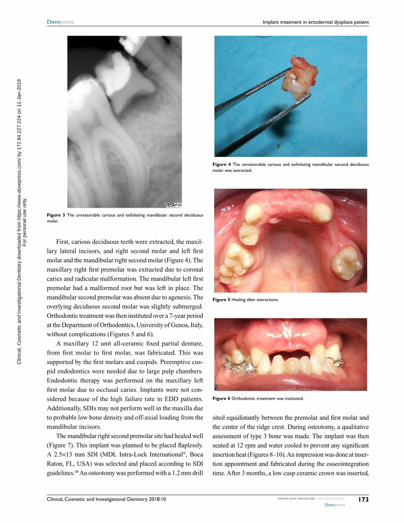
First, carious deciduous teeth were extracted, the maxil-
lary lateral incisors, and right second molar and left first
molar and the mandibular right second molar (Figure 4). The
maxillary right first premolar was extracted due to coronal
caries and radicular malformation. The mandibular left first
premolar had a malformed root but was left in place. The
mandibular second premolar was absent due to agenesis. The
overlying deciduous second molar was slightly submerged.
Orthodontic treatment was then instituted over a 7-year period
at the Department of Orthodontics, University of Genoa, Italy,
without complications (Figures 5 and 6).
A maxillary 12 unit all-ceramic fixed partial denture,
from first molar to first molar, was fabricated. This was
supported by the first molars and cuspids. Preemptive cus-
pid endodontics were needed due to large pulp chambers.
Endodontic therapy was performed on the maxillary left
first molar due to occlusal caries. Implants were not con-
sidered because of the high failure rate in EDD patients.
Additionally, SDIs may not perform well in the maxilla due
to probable low bone density and off-axial loading from the
mandibular incisors.
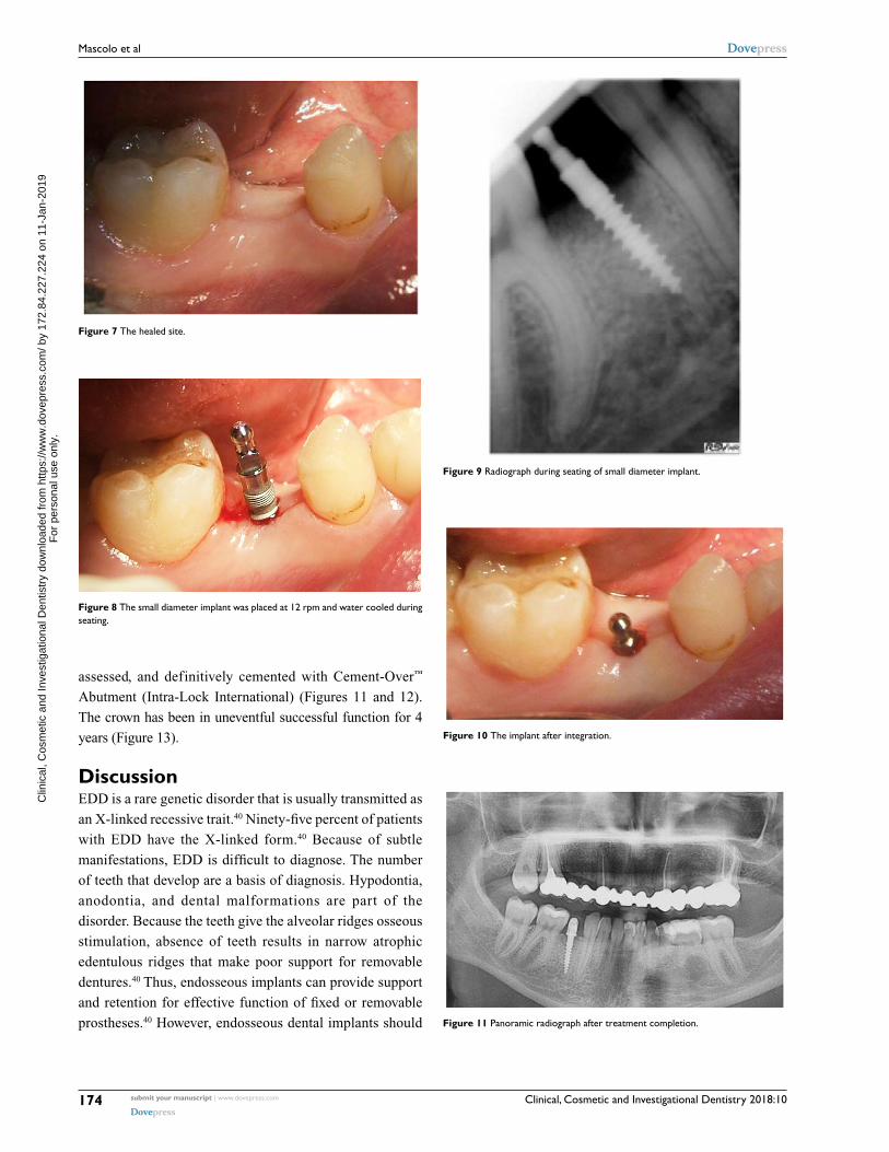
The mandibular right second premolar site had healed well
(Figure 7). This implant was planned to be placed flaplessly.
A 2.5×13 mm SDI (MDL Intra-Lock International®, Boca
Raton, FL, USA) was selected and placed according to SDI
guidelines.48 An osteotomy was performed with a 1.2 mm drill
sited equidistantly between the premolar and first molar and
the center of the ridge crest. During osteotomy, a qualitative
assessment of type 3 bone was made. The implant was then
seated at 12 rpm and water cooled to prevent any significant
insertion heat (Figures 8–10). An impression was done at inser-
tion appointment and fabricated during the osseointegration
time. After 3 months, a low cusp ceramic crown was inserted,
Figure 3 the unrestorable carious and exfoliating mandibular second deciduous molar.
Figure 4 the unrestorable carious and exfoliating mandibular second deciduous molar was extracted.
Figure 5 Healing after extractions.
Figure 6 orthodontic treatment was instituted.
C
linic
al, C
osm
etic
and
Inve
stig
atio
nal D
entis
try
dow
nloa
ded
from
http
s://w
ww
.dov
epre
ss.c
om/ b
y 17
2.84
.227
.224
on
11-J
an-2
019
For
per
sona
l use
onl
y.
Powered by TCPDF (www.tcpdf.org)
1 / 1
Clinical, Cosmetic and Investigational Dentistry 2018:10submit your manuscript | www.dovepress.com
Dovepress
Dovepress
174
Mascolo et al
Figure 7 the healed site.
Figure 8 the small diameter implant was placed at 12 rpm and water cooled during seating.
Figure 9 radiograph during seating of small diameter implant.
Figure 10 the implant after integration.
Figure 11 panoramic radiograph after treatment completion.
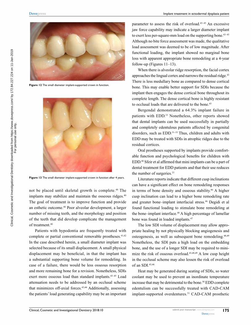
assessed, and definitively cemented with Cement-Over™
Abutment (Intra-Lock International) (Figures 11 and 12).
The crown has been in uneventful successful function for 4
years (Figure 13).
DiscussionEDD is a rare genetic disorder that is usually transmitted as
an X-linked recessive trait.40 Ninety-five percent of patients
with EDD have the X-linked form.40 Because of subtle
manifestations, EDD is difficult to diagnose. The number
of teeth that develop are a basis of diagnosis. Hypodontia,
anodontia, and dental malformations are part of the
disorder. Because the teeth give the alveolar ridges osseous
stimulation, absence of teeth results in narrow atrophic
edentulous ridges that make poor support for removable
dentures.40 Thus, endosseous implants can provide support
and retention for effective function of fixed or removable
prostheses.40 However, endosseous dental implants should
C
linic
al, C
osm
etic
and
Inve
stig
atio
nal D
entis
try
dow
nloa
ded
from
http
s://w
ww
.dov
epre
ss.c
om/ b
y 17
2.84
.227
.224
on
11-J
an-2
019
For
per
sona
l use
onl
y.
Powered by TCPDF (www.tcpdf.org)
1 / 1
Clinical, Cosmetic and Investigational Dentistry 2018:10 submit your manuscript | www.dovepress.com
Dovepress
Dovepress
175
Implant treatment in ectodermal dysplasia patient
not be placed until skeletal growth is complete.40 The
implants may stabilize and maintain the osseous ridges.40
The goal of treatment is to improve function and provide
an esthetic outcome.40 Poor alveolar development, a larger
number of missing teeth, and the morphology and position
of the teeth that did develop complicate the management
of treatment.40
Patients with hypodontia are frequently treated with
complete or partial conventional removable prostheses.41,42
In the case described herein, a small diameter implant was
selected because of its small displacement. A small physical
displacement may be beneficial, in that the implant has
a substantial supporting bone volume for remodeling. In
case of a failure, there would be less osseous resorption
and more remaining bone for a revision. Nonetheless, SDIs
exert more osseous load than standard implants.43–45 Load
attenuation needs to be addressed by an occlusal scheme
that minimizes off-axial forces.43,44 Additionally, assessing
the patients’ load generating capability may be an important
parameter to assess the risk of overload.43–45 An excessive
jaw force capability may indicate a larger diameter implant
to exert less per-square-mm load on the supporting bone.43–45
Although no bite force assessment was made, the qualitative
load assessment was deemed to be of low magnitude. After
functional loading, the implant showed no marginal bone
loss with apparent appropriate bone remodeling at a 4-year
follow-up (Figures 11–13).
When there is alveolar ridge resorption, the facial cortex
approaches the lingual cortex and narrows the residual ridge.43
There is less medullary bone as compared to dense cortical
bone. This may enable better support for SDIs because the
implant then engages the dense cortical bone throughout its
complete length. The dense cortical bone is highly resistant
to occlusal loads that are delivered to the bone.43
Bergendal demonstrated a 64.3% implant failure in
patients with EDD.23 Nonetheless, other reports showed
that dental implants can be used successfully in partially
and completely edentulous patients affected by congenital
disorders, such as EDD.31–33 Thus, children and adults with
EDD may be treated with SDIs in atrophic ridges due to the
residual cortices.
Oral prostheses supported by implants provide comfort-
able function and psychological benefits for children with
EDD.40 Sfeir et al affirmed that mini implants can be a part of
dental treatment for EDD patients and that their use reduces
the number of surgeries.33
Literature reports indicate that different cusp inclinations
can have a significant effect on bone remodeling responses
in terms of bone density and osseous stability.46 A higher
cusp inclination can lead to a higher bone remodeling rate
and greater bone–implant interfacial stress.46 Degidi et al
found functional loading to stimulate bone remodeling at
the bone–implant interface.48 A high percentage of lamellar
bone was found in loaded implants.47
The low SDI volume of displacement may allow appro-
priate healing by not physically blocking angiogenesis and
osteogenesis, as well as subsequent bone remodeling.48,49
Nonetheless, the SDI puts a high load on the embedding
bone, and the use of a longer SDI may be required to mini-
mize the risk of osseous overload.43,48,49 A low cusp height
in the occlusal scheme may also lessen the risk of overload
of an SDI.43,46
Heat may be generated during seating of SDIs, so water
coolant may be used to prevent an inordinate temperature
increase that may be detrimental to the bone.50 EDD complete
edentulism can be successfully treated with CAD-CAM
implant-supported overdentures.51 CAD-CAM prosthetic
Figure 12 the small diameter implant-supported crown in function.
Figure 13 the small diameter implant-supported crown in function after 4 years.
C
linic
al, C
osm
etic
and
Inve
stig
atio
nal D
entis
try
dow
nloa
ded
from
http
s://w
ww
.dov
epre
ss.c
om/ b
y 17
2.84
.227
.224
on
11-J
an-2
019
For
per
sona
l use
onl
y.
Powered by TCPDF (www.tcpdf.org)
1 / 1

Clinical, Cosmetic and Investigational Dentistry 2018:10submit your manuscript | www.dovepress.com
Dovepress
Dovepress
176
Mascolo et al
fabrication is a definitive technical advance for accuracy,
and occlusal loads may be better controlled.51
The small percutaneous exposure of SDIs may
dramatically reduce the incidence of peri-implant mucositis
and peri-implantitis.44 No marginal bone loss was seen with
this patient in the postoperative 4 years. Implants may not
be appropriate in children who have no complete osseous
growth due to drifting and displacement of the bones during
maturation.45
ConclusionsSDIs may be a minimally invasive approach for support in
fixed prosthetics in selected patients with EDD. Preoperative
site evaluation and evaluation of the patient’s disorder and an
appropriate occlusal scheme that minimizes off-axial loads
is required. This is a case report and is of a low credibility
level. In the future, a meta-analysis of case reports and case
series may elucidate this topic for appropriate treatment of
these individuals.
DisclosureThe authors report no conflicts of interest in this work.
References 1. Salinas CF, Jorgenson RJ, Wright JT, Digiovanna JJ, Fete MD. 2008
International Conference on Ectodermal Dysplasias Classification: conference report. Am J Med Genet A. 2009;149A(9):1958–1969.
2. Irvine AD. Towards a unified classification of the ectodermal dys-plasias: opportunities outweigh challenges. Am J Med Genet A. 2009;149A(9):1970–1972.
3. Worsaae N, Jensen BN, Holm B, Holsko J. Treatment of severe hypodontia-oligodontia--an interdisciplinary concept. Int J Oral Maxil-lofac Surg. 2007;36(6):473–480.
4. Talo T, Acun Kaya F. The effects of ectodermal dysplasia on periodontal tissues. J Int Dent Med Res. 2009;2(2):53–57.
5. Bani M, Tezkirecioglu AM, Akal N, Tuzuner T. Ectodermal dysplasia with anodontia: a report of two cases. Eur J Dent. 2010;4(2):215–222.
6. Ulm MR, Kratochwil A, Ulm B, Solar P, Aro G, Bernaschek G. Three-dimensional ultrasound evaluation of fetal tooth germs. Ultrasound Obstet Gynecol. 1998;12(4):240–243.
7. Itin PH. Rationale and background as basis for a new classification of the ectodermal dysplasias. Am J Med Genet A. 2009;149A(9):1973–1976.
8. Visinoni AF, Lisboa-Costa T, Pagnan NA, Chautard-Freire-Maia EA. Ectodermal dysplasias: clinical and molecular review. Am J Med Genet A. 2009;149A(9):1980–2002.
9. Bergendal B. Oligodontia ectodermal dysplasia--on signs, symptoms, genetics, and outcomes of dental treatment. Swed Dent J Suppl. 2010 (205):7–8.
10. Aydinbelge M, Gumus HO, Sekerci AE, Demetoğlu U, Etoz OA. Implants in children with hypohidrotic ectodermal dysplasia: an alter-native approach to esthetic management: case report and review of the literature. Pediatr Dent. 2013;35(5):441–446.
11. Kere J, Srivastava AK, Montonen O, et al. X-linked anhidrotic (hypo-hidrotic) ectodermal dysplasia is caused by mutation in a novel trans-membrane protein. Nat Genet. 1996;13(4):409–416.
12. Ziada H, Holland T. Ectodermal dysplasia: a case report. J Ir Dent Assoc. 1997;43(4):127–129.
13. Sakai VT, Oliveira TM, Pessan JP, Santos CF, Machado MA. Alternative oral rehabilitation of children with hypodontia and conical tooth shape: a clinical report. Quintessence Int. 2006;37(9):725–730.
14. Huang C, Yang Q, Ke T, et al. A novel de novo frame-shift mutation of the EDA gene in a Chinese Han family with hypohidrotic ectodermal dysplasia. J Hum Genet. 2006;51(12):1133–1137.
15. Lexner MO, Bardow A, Hertz JM, Nielsen LISA, Kreiborg S. Anomalies of tooth formation in hypohidrotic ectodermal dysplasia. Int J Paediatr Dent. 2007;17(1):10–18.
16. Gruber J, Kreitzberg G. Ectodermal dysplasia. A seven-year case report. N Y State Dent J. 2006;72(6):28–31.
17. Glavina D, Majstorovic M, Lulic-Dukic O, Juric H. Hypohidrotic ecto-dermal dysplasia: dental features and carrier detection. Coll Anthropol. 2001;25(1):303–310.
18. Freiman A, Borsuk D, Barankin B, Sperber GH, Krafchik B. Dental manifestations of dermatologic conditions. J Am Acad Dermatol. 2009;60(2):289–298.
19. Clauss F, Manière MC, Obry F, et al. Dento-craniofacial phenotypes and underlying molecular mechanisms in hypohidrotic ectodermal dysplasia (HED): a review. J Dent Res. 2008;87(12):1089–1099.
20. Bergendal B, Bergendal T, Hallonsten AL, Koch G, Kurol J, Kvint S. A multidisciplinary approach to oral rehabilitation with osseointegrated implants in children and adolescents with multiple aplasia. Eur J Orthod. 1996;18(2):119–129.
21. Hobson RS, Carter NE, Gillgrass TJ, et al. The interdisciplinary manage-ment of hypodontia: the relationship between an interdisciplinary team and the general dental practitioner. Br Dent J. 2003;194(9):479–482.
22. Kotsiomiti E, Kassa D, Kapari D. Oligodontia and associated char-acteristics: assessment in view of prosthodontic rehabilitation. Eur J Prosthodont Restor Dent. 2007;15(2):55–60.
23. Bergendal B. When should we extract deciduous teeth and place implants in young individuals with tooth agenesis? J Oral Rehabil. 2008;35(Suppl 1): 55–63.
24. Gotfredsen K, Carlsson GE, Jokstad A, et al. Implants and/or teeth: consensus statements and recommendations. J Oral Rehabil. 2008;35(Suppl 1):2–8.
25. Wong AT, Mcmillan AS, Mcgrath C. Oral health-related quality of life and severe hypodontia. J Oral Rehabil. 2006;33(12):869–873.
26. Locker D, Jokovic A, Prakash P, Tompson B. Oral health-related quality of life of children with oligodontia. Int J Paediatr Dent. 2010;20(1):8–14.
27. Somani A, Newton JT, Dunne S, Gilbert DB. The impact of visible dental decay on social judgements: comparison of the effects of location and extent of lesion. Int Dent J. 2010;60(3):5–8.
28. Havens DC, Mcnamara JA, Sigler LM, Baccetti T. The role of the posed smile in overall facial esthetics. Angle Orthod. 2010;80(2): 322–328.
29. Johnson EL, Roberts MW, Guckes AD, Bailey LJ, Phillips CL, Wright JT. Analysis of craniofacial development in children with hypohidrotic ectodermal dysplasia. Am J Med Genet. 2002;112(4):327–334.
30. Preoteasa E, Marin M, Imre M, Lerner H, Preoteasa CT. Patients’ satisfaction with conventional dentures and mini implant anchored overdentures. Rev Med Chir Soc Med Nat Iasi. 2012;116(1):310–316.
31. Güler N, Cildir S, Iseri U, Sandalli N, Dilek O. Hypohidrotic ectodermal dysplasia with bilateral impacted teeth at the coronoid process: a case rehabilitated with mini dental implants. Oral Surg Oral Med Oral Pathol Oral Radiol Endod. 2005;99(5):E34–E38.
32. Giannetti L, Murri dello Diago A, Vecci F, Consolo U. Mini-implants in growing patients: a case report. Pediatr Dent. 2010;32(3):239–244.
33. Sfeir E, Nassif N, Moukarzel C. Use of mini dental implants in ectoder-mal dysplasia children: follow-up of three cases. Eur J Paediatr Dent. 2014;15(2 Suppl):207–212.
34. Kramer FJ, Baethge C, Tschernitschek H. Implants in children with ectodermal dysplasia: a case report and literature review. Clin Oral Implants Res. 2007;18(1):140–146.
35. Sweeney IP, Ferguson JW, Heggie AA, Lucas JO. Treatment outcomes for adolescent ectodermal dysplasia patients treated with dental implants. Int J Paediatr Dent. 2005;15(4):241–248.
C
linic
al, C
osm
etic
and
Inve
stig
atio
nal D
entis
try
dow
nloa
ded
from
http
s://w
ww
.dov
epre
ss.c
om/ b
y 17
2.84
.227
.224
on
11-J
an-2
019
For
per
sona
l use
onl
y.
Powered by TCPDF (www.tcpdf.org)
1 / 1
Clinical, Cosmetic and Investigational Dentistry 2018:10 submit your manuscript | www.dovepress.com
Dovepress
Dovepress
Clinical, Cosmetic and Investigational Dentistry
Publish your work in this journal
Submit your manuscript here: https://www.dovepress.com/clinical-cosmetic-and-investigational-dentistry-journal
Clinical, Cosmetic and Investigational Dentistry is an international, peer-reviewed, open access, online journal focusing on the latest clini-cal and experimental research in dentistry with specific emphasis on cosmetic interventions. Innovative developments in dental materials, techniques and devices that improve outcomes and patient satisfac-
tion and preference will be highlighted. The manuscript management system is completely online and includes a very quick and fair peer- review system, which is all easy to use. Visit http://www.dovepress. com/testimonials.php to read real quotes from published authors.
Dovepress
177
Implant treatment in ectodermal dysplasia patient
36. Buser D, von Arx T, Ten Bruggenkate C, Weingart D. Basic surgical prin-ciples with ITI implants. Clin Oral Implants Res. 2000;11(Suppl):59–68.
37. Guckes AD, Scurria MS, King TS, Mccarthy GR, Brahim JS. Prospective clinical trial of dental implants in persons with ectodermal dysplasia. J Prosthet Dent. 2002;88(1):21–25.
38. Yap AK, Klineberg I. Dental implants in patients with ectodermal dysplasia and tooth agenesis: a critical review of the literature. Int J Prosthodont. 2009;22(3):268–276.
39. Singer SL, Henry PJ, Liddelow G, Rosenberg I. Long-term follow-up of implant treatment for oligodontia in an actively growing individual: a clinical report. J Prosthet Dent. 2012;108(5):279–285.
40. Knobloch LA, Larsen PE, Saponaro PC, L’Homme-Langlois E. Early implant placement for a patient with ectodermal dysplasia: Thirteen years of clinical care. J Prosthet Dent. 2018;119(5):702–709.
41. Paul ST, Tandon S, Kiran M. Prosthetic rehabilitation of a child with induced anodontia. J Clin Pediatr Dent. 1995;20(1):5–8.
42. Pettit S, Campbell PR. Ectrodactyly-ectodermal dysplasia-clefting syndrome: the oral hygiene management of a patient with EEC. Spec Care Dentist. 2010;30(6):250–254.
43. Bulut E, Guler AU, Sen Tunc E, Telcioglu NT. Oral rehabilitation with endosseous implants in a child with ectodermal dysplasia: a case report. Eur J Paediatr Dent. 2010;11(3):149–152.
44. Flanagan D, Mascolo A. The mini dental implant in fixed and removable prosthetics: a review. J Oral Implantol. 2011;37:123–132.
45. Flanagan D, Ilies H, O’Brien B, Mcmanus A, Larrow B. Jaw bite force measurement device. J Oral Implantol. 2012;38(4):361–364.
46. Flanagan D. Bite force and dental implant treatment: a short review. Med Devices. 2017;10:141–148.
47. Rungsiyakull C, Rungsiyakull P, Li Q, Li W, Swain M. Effects of occlusal inclination and loading on mandibular bone remodeling: a finite element study. Int J Oral Maxillofac Implants. 2011;26(3): 527–537.
48. Degidi M, Scarano A, Piattelli M, Perrotti V, Piattelli A. Bone remod-eling in immediately loaded and unloaded titanium dental implants: a histologic and histomorphometric study in humans. J Oral Implantol. 2005;31(1):18–24.
49. Serra G, Morais LS, Elias CN, et al. Sequential bone healing of immediately loaded mini-implants. Am J Orthod Dentofacial Orthop. 2008;134(1):44–52.
50. Flanagan D. Heat generated during seating of dental implant fixtures. J Oral Implantol. 2014;40(2):174–181.
51. Chrcanovic BR. Dental implants in patients with ectodermal dys-plasia: A systematic review. J Cranio Maxilloofac Surg. Epub 2018 May 21.
52. Alsayed HD, Alqahtani NM, Alzayer YM, Morton D, Levon JA, Baba NZ. Prosthodontic rehabilitation with monolithic, multichromatic CAD-CAM complete overdentures in an adolescent patient with ectodermal dysplasia: A clinical report. J Prosthet Dent. 2018;119(6):873–878.
C
linic
al, C
osm
etic
and
Inve
stig
atio
nal D
entis
try
dow
nloa
ded
from
http
s://w
ww
.dov
epre
ss.c
om/ b
y 17
2.84
.227
.224
on
11-J
an-2
019
For
per
sona
l use
onl
y.
Powered by TCPDF (www.tcpdf.org)
1 / 1