Embed Size (px)
Citation preview
Case Report
Ocimum Scientific Publishers .01. Submit Manuscript
OCIMUM
Shahban SA et al. Open J Surg Case Rep 2018(1): 04-08.
https://doi.org/10.33513/OJSC/1801-04
Open Journal of Surgery Case Reports
An Unusual Presentation of a Bicondylar Tibial Plateau Fracture, Managed Without Surgery and Early Full Mobilisation
Shafiq A Shahban, Ahmed Isam Saad* and Panos Makrides
Department of Trauma and Orthopaedics, Heartlands Hospital, Birmingham, United Kingdom
Received: 29 August 2018Accepted: 11 November 2018Version of Record Online: 19 November 2018
Citation
Shahban SA, Saad AI, Makrides P (2018) An Unusual Presentation of a Bicondylar Tibial Plateau Fracture, Managed Without Surgery and Early Full Mobilisation. Open J Surg Case Rep 2018(1): 04-08.
Correspondence should be addressed toAhmed Isam Saad, UKE-mail: [email protected]
Copyright
Copyright © 2018 Ahmed Isam Saad et al. This is an open access article distributed under the Creative Commons Attribution License which permits unrestricted use, distribution, and reproduction in any medium, provided the original author and work is properly cited.
AbstractIntroduction: Tibial plateau fractures are often sustained following high energy trauma and as a result are likely to require surgical stabilisation. In rare circumstances these injuries can be either difficult, or missed on initial radiographs. Computer tomography and/or Magnetic resonance imaging can be key not only for diagnosis for also for surgical planning.
Of course, non-operative treatment is an option and should be considered in fracture patterns which have little or no displacement. And with or without surgery, early intervention to assist in knee mobilisation is key to prevent stiffness and muscle atrophy.
Case report: We present a case a 51-year-old gentleman who after high energy axial loading of his left knee initially presented atypically of a tibial plateau fracture. Despite a later diagnosis of a Schatzker 5 tibial plateau plateau fracture was able to mobilise fully, without developing fracture displacement. We present the successful non-operative management of this case and report how and why we managed this injury in this manner.
Discussion: Atypical presentations of tibial plateau fractures can catch clinicians out, and so it is important to have a high index of suspicion of these injuries. There is literature to support non-operative management of plateau fractures, but this is by no means in the majority opinion.
Conclusion: For the patients managed without surgery, close monitoring of the fracture fragments with serial radiographs along with early range of movement exercises is key for successful management.
KeywordsAtypical Presentation; Early Mobilisation; Non-Operative Treatment; Tibial Plateau Fractures
IntroductionTibial plateau fractures are becoming increasingly common most often affecting individuals between the ages of 40 and 60 years and comprise approximately 1% of all fractures [1]. They can present in many different ways and subsequently this can pose challenges for even the experienced orthopaedic surgeons. In this report we present a case of an unusual presentation of a Schatzker 5 tibial plateau fracture, which was later managed, successfully, without operative intervention.
Open Journal of Surgery Case Reports https://doi.org/10.33513/OJSC/1801-04
Ocimum Scientific Publishers .02. Submit Manuscript
Over the years, there has been a vast amount of understanding of the different types of injuries involved in the tibial plateau, and this ultimately led to several enhanced and developed surgical techniques to appropriately manage these fractures. The basic principles of treatment range from restoring the congruity of the articular surfaces, reduction of the anatomic alignment of the lower limb as well as preventing postoperative pain and arthritis [2].
Orthopaedic surgeons can choose to manage tibial plateau fractures operatively or non-operatively depending on various factors such as displacement, depression of the tibial plateau, comminution, or associated ligamentous or meniscal injury. Usually this is guided by clinical examination and, more recently, evaluation using various imaging modalities. Tibial plateau fractures can be difficult to see on a plain film and their reliability for detection as well as management has always been questioned. With the advent of Computer Tomography (CT) and Magnetic Resonance Imaging (MRI), there are often the go to imaging modalities of choice for fracture classification and subsequent management [3].
We present a case of a tibial plateau fracture that was primarily undetectable on initial plain radiographs. Subsequent ultrasound and MRI scans were required to help diagnose the injury and later decide further management.
Case ReportWe present a case of PC, a 51-year-old male, who suffered a motor bike accident driving at a speed of approximately 30 miles per hour, when he was side swept to his left by a car driving at a similar speed. PC lost his balance and hit his left foot onto the floor, bringing his motorbike to a halt. He describes his left knee being forced into a fully extended position, with the foot in neutral. He did not fall off the motor bike, however due to the impact of the injury, he experienced severe pain and was unable to weight bear on his left lower limb.
In the emergency department, his primary complaint was that of left calf swelling, with a reduced range of knee movement. Proximal and distal pulses were intact and there was normal sensation throughout the entire limb. Ankle and hip joint examinations were unremarkable.
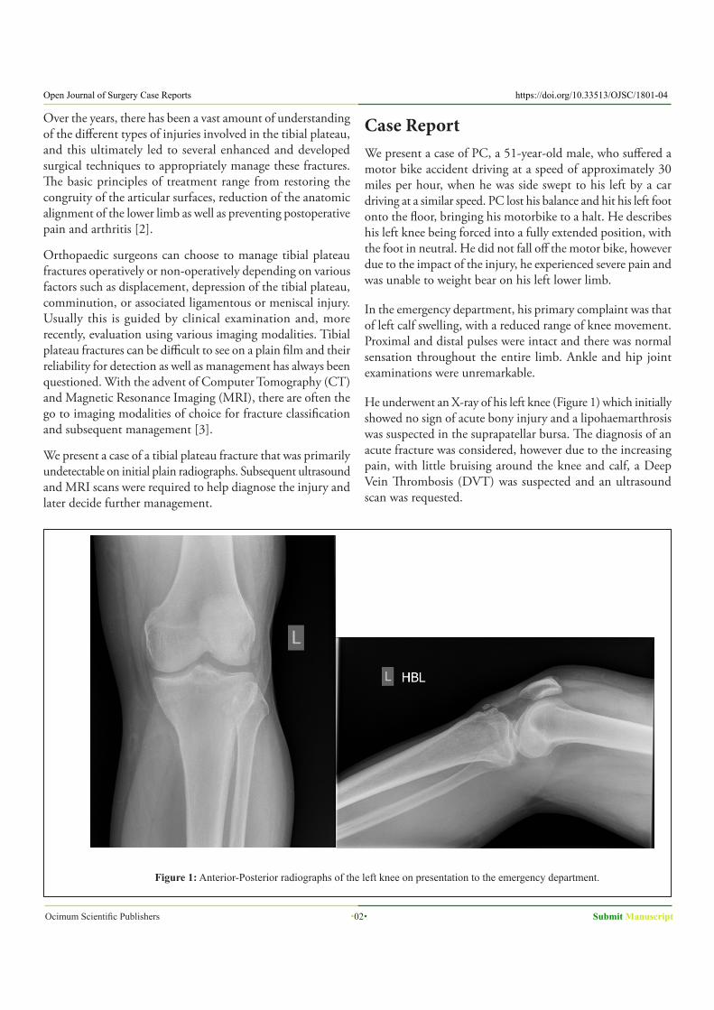
He underwent an X-ray of his left knee (Figure 1) which initially showed no sign of acute bony injury and a lipohaemarthrosis was suspected in the suprapatellar bursa. The diagnosis of an acute fracture was considered, however due to the increasing pain, with little bruising around the knee and calf, a Deep Vein Thrombosis (DVT) was suspected and an ultrasound scan was requested.
Figure 1: Anterior-Posterior radiographs of the left knee on presentation to the emergency department.
Submit Manuscript .03. Ocimum Scientific Publishers
https://doi.org/10.33513/OJSC/1801-04 Open Journal of Surgery Case Reports
Owing to the unresolved symptoms and unexpected negative results on the initial investigations carried out, a MRI scan of the left knee was requested to rule out possibility of an acute ligamentous injury.
Prior to the MRI scan, with the use of ice and strict high elevation, the following day, the patient was seen mobilising, partially weight bearing on his left leg with the aid of crutches. Due to his ability to weight bear and now resolving pain levels, the suspicion of a fracture was now even less common.
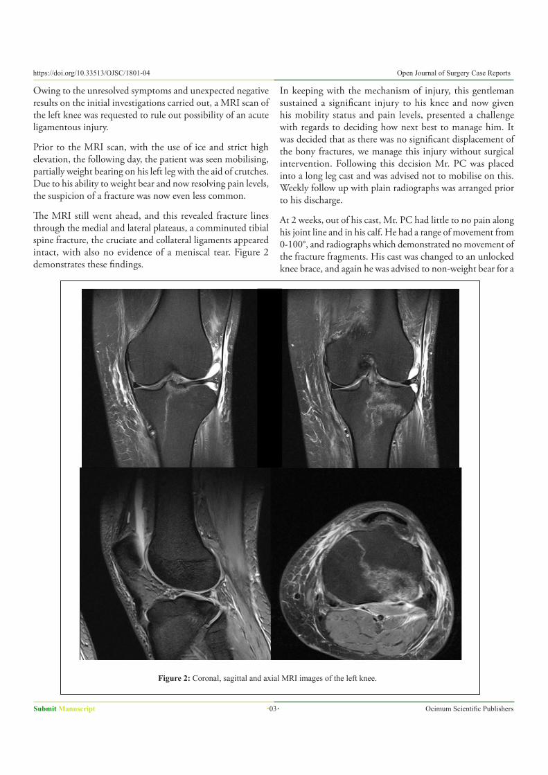
The MRI still went ahead, and this revealed fracture lines through the medial and lateral plateaus, a comminuted tibial spine fracture, the cruciate and collateral ligaments appeared intact, with also no evidence of a meniscal tear. Figure 2 demonstrates these findings.
In keeping with the mechanism of injury, this gentleman sustained a significant injury to his knee and now given his mobility status and pain levels, presented a challenge with regards to deciding how next best to manage him. It was decided that as there was no significant displacement of the bony fractures, we manage this injury without surgical intervention. Following this decision Mr. PC was placed into a long leg cast and was advised not to mobilise on this. Weekly follow up with plain radiographs was arranged prior to his discharge.
At 2 weeks, out of his cast, Mr. PC had little to no pain along his joint line and in his calf. He had a range of movement from 0-100°, and radiographs which demonstrated no movement of the fracture fragments. His cast was changed to an unlocked knee brace, and again he was advised to non-weight bear for a
Figure 2: Coronal, sagittal and axial MRI images of the left knee.

further 4 weeks. At 6 weeks Mr. PC had a range of movement from 0-120°, could fully weight bear and had satisfactory X-rays (Figure 3). The knee appeared to be stable clinically with no evidence of ligament laxity.
Figure 3: Mr. PC was successfully managed non-operatively for his Schatzker 5 tibial plateau fracture, and remains happy with his knee.
DiscussionTibial Plateau Fractures (TPF) can present in many different ways, following high or low energy trauma, and should be considered as complex injuries which, often, requires surgical intervention. Typically, patients with a traumatic TPF presents, as you would expect, with an acutely painful knee and being unable to weight bear - 85% of these fracture types involve only the lateral aspect of the tibia [4,5].
A plain radiograph is usually the initial investigation of choice when there is a high suspicion of a TPF. Standard anteroposterior and lateral plain films have been shown to be 79% sensitive for fractures of the lateral tibial plateau and adding oblique knee views to the radiographic examination increases sensitivity to 85%. The possible reasons why these fractures are difficult to visualise can be due to the fact that the fracture may be lying in an oblique plane and the tibial plateau surface slopes inferiorly from anterior to posterior. This makes it difficult to see because the fracture may not lie parallel to the X-ray beam [6]. Equally, undisplaced, or minimally displaced, fractures may be difficult to pick up on plain radiograph.
Even after an X-ray diagnosis of a TPF, the use of a CT scan
and occasionally MRI scan, has become routine practice for definition of the fracture outline and for surgical planning [7]. In one study by Holt et al., MRI was considered to be the imaging method of choice. MRI scans were found to be
more accurate in determining the classification of the fracture, in the identification of previously ‘occult’ fracture lines, and in accurate measurement of displacement and depression of the fracture fragments [8].
As in this case, Mr. PC, on initial radiographs, demonstrated signs of a lipohaemarthrosis, suggestive of an intraarticular knee fracture. However, with his significant calf swelling which superseded and knee pain, it was this which prompted the ultrasound scan to rule out the possibility of a DVT. It was the lipohaemarthrosis, which warranted further investigation. A retrospective analysis of MRI scans of patients with a TPF found joint effusions in all patients, 41% of whom had lipohaemarthrosis [9,10].
When considering treatment options, many classifications have been demonstrated to guide surgical fixation of these fractures and the Schatzker classification, developed in 1979, is often used. This classification describes the fractures on plain radiographs as a combination of medial and lateral condyle, primarily in the coronal plane [10].
Other classifications systems require the use of CT and/or MRI images to guide diagnosis and management, however, many studies have shown moderate intra and inter-observer
Open Journal of Surgery Case Reports https://doi.org/10.33513/OJSC/1801-04
Ocimum Scientific Publishers .04. Submit Manuscript
reliability dependence. One study suggested that the use of 2D and 3D modalities typically improves reliability estimates, however, a more detailed assessment of fracture patterns and morphology, with information on surgical fixation is desirable for predicting outcomes as well as serving as a guide to clinical decision making [11].
Operative intervention is often employed to help to manage these types of fractures. This can range from a single sided percutaneous approach to a dual approach with bony and soft tissue reconstruction. The option to treat non-operatively of course is always there and is only utilised in unwell/unfit patient for surgery or in fracture patterns where there is little or no displacement of the fracture without an associated soft tissue injury [2]. If this option is employed, then it is important to closely watch these fractures to ensure that fracture displacement does not occur in the ensuing weeks.
ConclusionTibial plateau fractures are common injuries and, as in this case, can be an injury missed on initial plain radiographs. This case highlights the importance of amalgamating the history, mechanism of injury and examination findings which can then help to plan the need for further imaging as appropriate. With or without surgery, it is imperative that the range of motion of the knee is encouraged sooner rather than later.
References1. Rademakers MV, Kerkhoffs GM, Sierevelt IN, Raaymakers
EL, Marti RK (2007) Operative Treatment of 109 Tibial Plateau Fractures: Five- to 27-Year Follow-up-results. J Orthop Trauma 21: 5-10.
2. Rohra N, Suri HS, Gangrade K (2016) Functional and Radiological Outcome of Schatzker type V and VI Tibial Plateau Fracture Treatment with Dual Plates with Minimum 3 years follow-up: A Prospective Study. J Clin Diagn Res 10: 5-10.
3. Malik S, Rosenberg N (2017) Fracture, Tibial Plateau. StatPearls Publishing, Florida, USA.
4. Gray SD, Kaplan PA, Dussault RG, Omary RA, Campbell SE, et al. (1997) Acute knee trauma: how many plain film views are necessary for the initial examination? Skeletal Radiol 26: 298-302.
5. Schwartz DT (2009) Ten most commonly missed radiographic findings in the ED. In: ACEP Scientific Assembly (ed.). Boston Convention and Exhibition Center, USA.
6. https://accessemergencymedicine.mhmedical.com/content.aspx?bookid=434§ionid=41825448.
7. Spirvulis P, Frazer A, Waring A (2001) Same-day X-ray reporting is not needed in well-supervised emergency departments. Emerg Med (Fremantle) 13: 194-197.
8. Holt MD, Williams LA, Dent CM (1995) MRI in the management of tibial plateau fractures. Injury 26: 595-599.
9. Colletti P, Greenberg H, Terk MR (1996) MR findings in patients with acute tibial plateau fractures. Comput Med Imaging Graph 20: 389-394.
10. Schatzker J, McBroom R, Bruce D (1979) The tibial plateau fracture. The Toronto experience 1968-1975. Clin Orthop Relat Res 138: 94-104.
11. Millar SC, Arnold JB, Thewlis D, Fraysse F, Solomon LB (2018) A systematic literature review of tibial plateau fractures: What classifications are used and how reliable and useful are they? Injury 49: 473-490.
https://doi.org/10.33513/OJSC/1801-04 Open Journal of Surgery Case Reports
Submit Manuscript .05. Ocimum Scientific Publishers