Embed Size (px)
Citation preview
Erin H. Kimmerle,1 Ph.D.; Lyle W. Konigsberg,2 Ph.D.; Richard L. Jantz,3 Ph.D.;and Jose Pablo Baraybar,4 M.Sc.
Analysis of Age-at-Death Estimation Throughthe Use of Pubic Symphyseal Data*
ABSTRACT: The question of whether age parameters derived from an American population will reliably estimate age-at-death for East Europeanskeletal populations is important since the ability to accurately estimate an individual’s age-at-death hinges on what standard is used. A referencesample of identified individuals with known ages-at-death from the regions of the Former Yugoslavia (n = 861) is used to determine the age structureof victims and serves as the prior in the Bayesian analysis. Pubic symphyseal data in the manners of Todd (Am J Phys Anthropol, 3 [1920], 285;Am J Phys Anthropol, 4 [1921], 1) and Suchey-Brooks (Am J Phys Anthropol, 80 [1986], 167) were collected for n = 296 Balkan males and femalesand for n = 2078 American males and females. An analysis of deviance is calculated using an improvement chi-square to test for population variationin the aging processes of American and East European populations using proportional odds probit regression. When males and females are treatedseparately, there is a significant association among females and the population (df = 1, chi-square likelihood ratio = 15.071, p = 0.001). New age esti-mates for Balkan populations are provided and are based on the calculated age distribution from the Gompertz-Makeham hazard analysis and theages-of-transition. To estimate the age-at-death for an individual, the highest posterior density regions for each symphyseal phase are provided.
KEYWORDS: forensic science, Balkans, age estimation, pubic symphysis, Bayesian statistics
American standards developed for determining the skeletal ages-at-death from pubic symphyseal morphology began with T. W.Todd in 1920. Todd first created standards for American Whitemales (1) and later for White females and Black males and females(2). Since his initial work, many studies have been conducted onthe accuracy and application of his method for various populations.Most notable are the modifications to his technique by Brooks,Katz, and Suchey, beginning with Brooks (3). According to Katzand Suchey (4) the problem with the original Todd method is thatit overestimates the age-at-death for most individuals, particularlythose under the age of 40 years; does not account for individualvariation; and does not accurately age older individuals. Theseresearchers, along with many others, e.g., Gillet (5), Galera et al.(6), and Kemkes-Grottenthaler (7), found that the modified six-phase system is a more accurate estimator than the original Toddsystem. Consequently, the Suchey-Brooks system (4,8) is currentlyused throughout the world as the standard for estimating age fromthe pubic symphysis.
The modifications made to the Todd method did not end thecontroversial issues surrounding age estimation in terms of variationbetween the sexes or among different populations, inter-observererror, or method reliability. Originally, Todd noted that there wereno racial differences in the aging process of the pubic symphysisand only minor differences between males and females. The
primary difference he noted was that females appeared to age ‘‘fas-ter’’ or ‘‘older’’ than their male counterparts due to trauma resultingfrom pregnancy and childbirth. He argued that females age two tothree years later in phases III and VI in regard to development ofthe ventral aspect of the pubic symphysis, but appear two to threeyears younger in respect to dorsal flattening. Since Todd’s work,other researchers have found similar differences between males andfemales. The review by Kemkes-Grottenthaler (7) cites numerousstudies that report female age estimation is less reliable than thatfor males. All of these studies attribute such differences to preg-nancy and childbirth. For example, Gilbert and McKern (9) arguedthat the female pubic symphyseal face appears about 10 years olderthan those of their male counterparts. More recently, Hoppa (10)looked into this issue using actual data on parity but found no rela-tionship with parity and aging. In contrast, he found that femalepubic symphyses appeared younger than males after the age of 40years in several different populations, and that they did not exhibitgreater variation than males. Hoppa attributed this finding to differ-ences in the aging processes of the samples used and points outthat one reference sample does not work for estimating age in allpopulations. Most differences noted between males and femaleshave been observed for those over the age of 40. Kemkes-Grottent-haler (7) point out that apparent population differences may alsobe the result of extrinsic factors such as material culture (i.e.,health care access, nutrition, or activity) which may affect bonedensity or degeneration, particularly among older aged individualswhich may also explain differences between older males andfemales. It is most likely that the skeletal remains of females(including the pubic symphysis) may appear older in some popula-tions due to nutritional differences, bone mineral density, andosteoporosis.
The question of whether there are substantial ethnic or popula-tion differences in the morphological aging process of the pubicsymphysis has remained open to debate. This question is importantsince our ability to accurately estimate age among different popula-tions hinges on the issue of what standard to use. The problem of
1Department of Anthropology, University of South Florida, 4202 E. Fow-ler Avenue SOC 107, Tampa, FL 33620.
2Department of Anthropology, University of Illinois, Urbana–Champaign,109 Davenport Hall, 607 South Matthews Ave. Urbana, IL 61801.
3Department of Anthropology, The University of Tennessee, 250 SouthStadium Hall, Knoxville, TN 37996.
4Peruvian Forensic Anthropology Team (EPAF), Av. Mello Franco #341,Jesus Maria Lima, Peru.
*Presented at the 56th Annual Meeting of the American Academy ofForensic Sciences, Dallas, TX, February 18, 2004.
Received 17 Feb. 2007; and in revised form 14 Oct. 2007; accepted 4Nov. 2007.
J Forensic Sci, May 2008, Vol. 53, No. 3doi: 10.1111/j.1556-4029.2008.00711.x
Available online at: www.blackwell-synergy.com
558 � 2008 American Academy of Forensic Sciences
whether one standard will work on populations that differ in timeor space is important for successful age estimation and individualidentification. However, age parameters biased in the direction ofthe reference sample, age mimicry, have resulted in unreliable ageestimators (11,12). In the forensic context, the accuracy and reli-ability of all methods must be demonstrated to the scientific com-munity, as well as the court. Therefore, the guidelines establishedby the court for the scientific presentation of evidence must be metfor each method or technique used. Across populations and a vari-ety of legal contexts, the evidentiary rules for the admissibility ofevidence vary. However, the rules set out in the American Federalcase law, Daubert v. Merrell Dow Pharmaceuticals, 509 US 5791993, offer conservative scientific standards by which most interna-tional courts should apply similar reasoning (13).
The study presented here investigates age estimation based onthe pubic symphysis using the Suchey-Brooks six-phase system.This calibration is based on a set of Bayesian statistical methodsand utilizes the Gompertz-Makeham model (14,15). Using this sta-tistical approach allows for estimation in the level of accuracy andprecision. New point estimates for calculating individual ages-at-death are provided that can be used in the field at the time ofautopsy ⁄examination or may be applied to cases retroactively. Thisstudy is based on the prior work of Kimmerle (15).
Materials
A reference sample comprised of individuals with known ages-at-death (15–98 years) from Kosovo, BiH, and Croatia (n = 861)was used to determine the age structure of genocide victims in theBayesian analysis. The Balkan skeletal data came from a subset ofthis reference sample. Balkan male (n = 212) and female (n = 84)pubic symphyses were scored in the manner of Suchey-Brooks(4,8). Data from this sample were collected by Kimmerle. Permis-sion to use this data was given by the ICTY to UT who enteredinto a working relationship with the expressed goal of sharing dataand results that would aid OTP in their investigations as well asother agencies working on human identification in the region. Anessential component of this effort was the publication of scientificfindings to ensure the admissibility of any new method or revisedbiological parameters for existing methods in court.
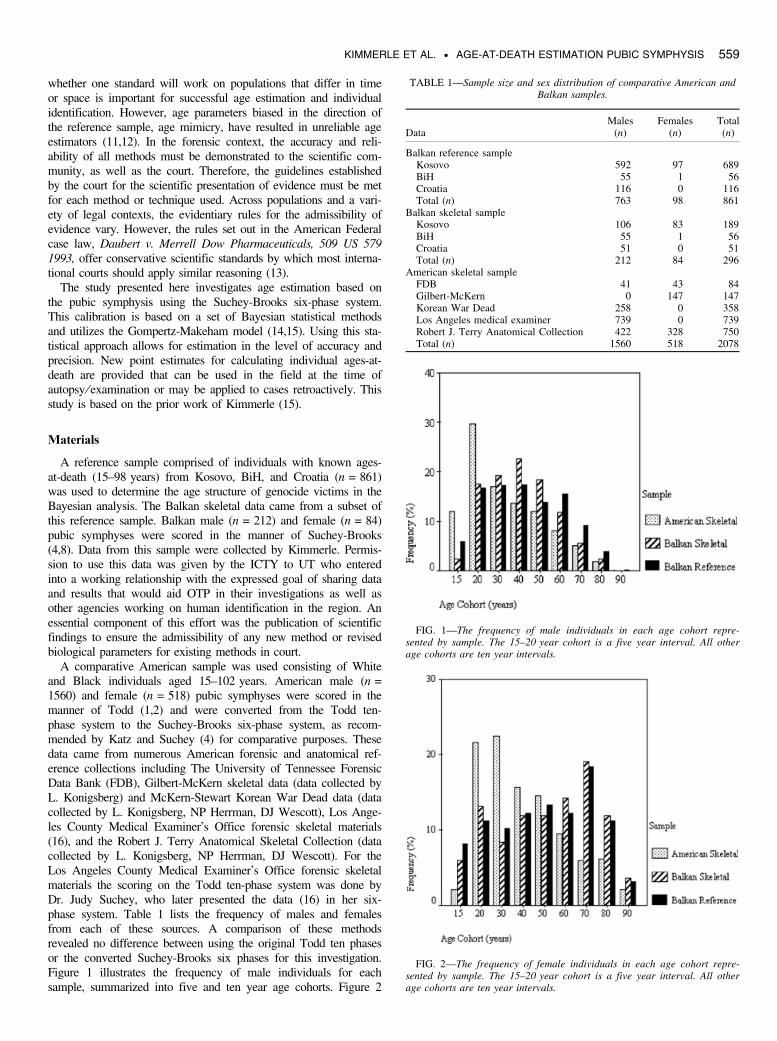
A comparative American sample was used consisting of Whiteand Black individuals aged 15–102 years. American male (n =1560) and female (n = 518) pubic symphyses were scored in themanner of Todd (1,2) and were converted from the Todd ten-phase system to the Suchey-Brooks six-phase system, as recom-mended by Katz and Suchey (4) for comparative purposes. Thesedata came from numerous American forensic and anatomical ref-erence collections including The University of Tennessee ForensicData Bank (FDB), Gilbert-McKern skeletal data (data collected byL. Konigsberg) and McKern-Stewart Korean War Dead data (datacollected by L. Konigsberg, NP Herrman, DJ Wescott), Los Ange-les County Medical Examiner’s Office forensic skeletal materials(16), and the Robert J. Terry Anatomical Skeletal Collection (datacollected by L. Konigsberg, NP Herrman, DJ Wescott). For theLos Angeles County Medical Examiner’s Office forensic skeletalmaterials the scoring on the Todd ten-phase system was done byDr. Judy Suchey, who later presented the data (16) in her six-phase system. Table 1 lists the frequency of males and femalesfrom each of these sources. A comparison of these methodsrevealed no difference between using the original Todd ten phasesor the converted Suchey-Brooks six phases for this investigation.Figure 1 illustrates the frequency of male individuals for eachsample, summarized into five and ten year age cohorts. Figure 2
TABLE 1—Sample size and sex distribution of comparative American andBalkan samples.
DataMales
(n)Females
(n)Total(n)
Balkan reference sampleKosovo 592 97 689BiH 55 1 56Croatia 116 0 116Total (n) 763 98 861
Balkan skeletal sampleKosovo 106 83 189BiH 55 1 56Croatia 51 0 51Total (n) 212 84 296
American skeletal sampleFDB 41 43 84Gilbert-McKern 0 147 147Korean War Dead 258 0 358Los Angeles medical examiner 739 0 739Robert J. Terry Anatomical Collection 422 328 750Total (n) 1560 518 2078
FIG. 1—The frequency of male individuals in each age cohort repre-sented by sample. The 15–20 year cohort is a five year interval. All otherage cohorts are ten year intervals.
FIG. 2—The frequency of female individuals in each age cohort repre-sented by sample. The 15–20 year cohort is a five year interval. All otherage cohorts are ten year intervals.
KIMMERLE ET AL. • AGE-AT-DEATH ESTIMATION PUBIC SYMPHYSIS 559
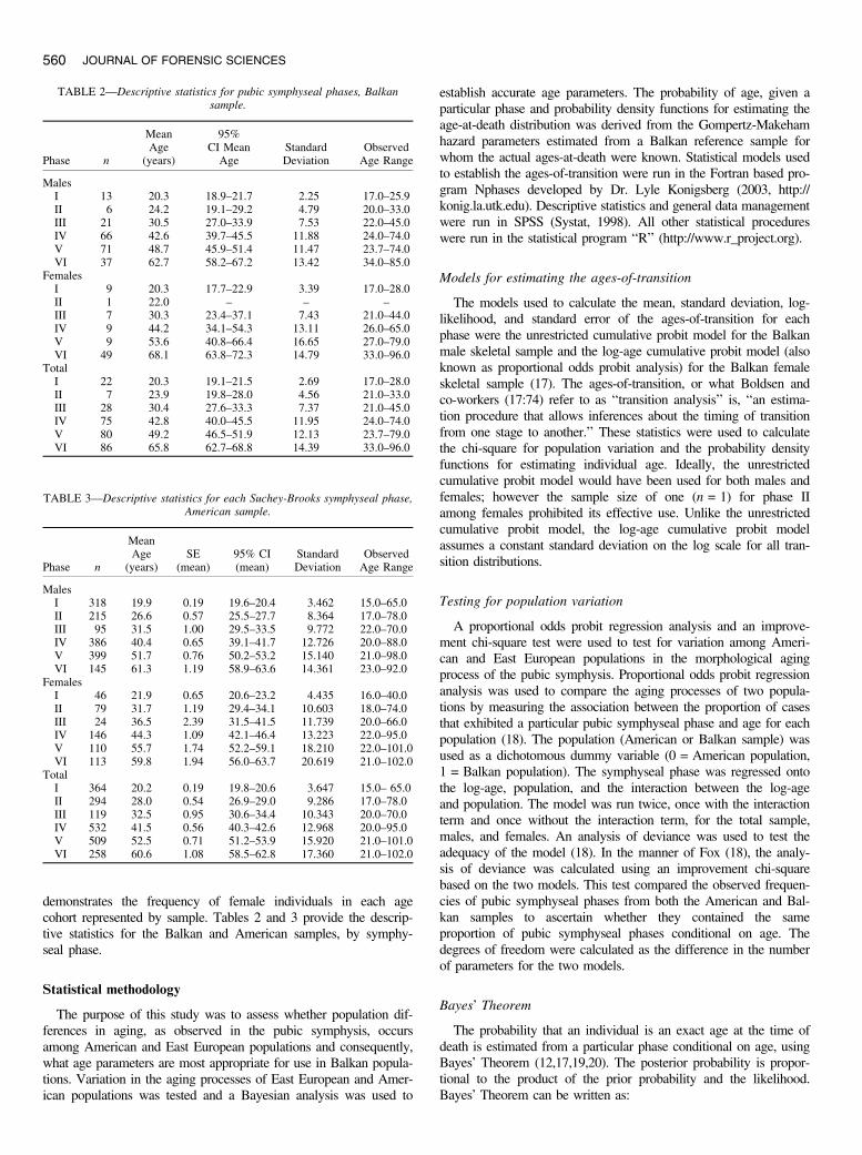
demonstrates the frequency of female individuals in each agecohort represented by sample. Tables 2 and 3 provide the descrip-tive statistics for the Balkan and American samples, by symphy-seal phase.
Statistical methodology
The purpose of this study was to assess whether population dif-ferences in aging, as observed in the pubic symphysis, occursamong American and East European populations and consequently,what age parameters are most appropriate for use in Balkan popula-tions. Variation in the aging processes of East European and Amer-ican populations was tested and a Bayesian analysis was used to
establish accurate age parameters. The probability of age, given aparticular phase and probability density functions for estimating theage-at-death distribution was derived from the Gompertz-Makehamhazard parameters estimated from a Balkan reference sample forwhom the actual ages-at-death were known. Statistical models usedto establish the ages-of-transition were run in the Fortran based pro-gram Nphases developed by Dr. Lyle Konigsberg (2003, http://konig.la.utk.edu). Descriptive statistics and general data managementwere run in SPSS (Systat, 1998). All other statistical procedureswere run in the statistical program ‘‘R’’ (http://www.r_project.org).
Models for estimating the ages-of-transition
The models used to calculate the mean, standard deviation, log-likelihood, and standard error of the ages-of-transition for eachphase were the unrestricted cumulative probit model for the Balkanmale skeletal sample and the log-age cumulative probit model (alsoknown as proportional odds probit analysis) for the Balkan femaleskeletal sample (17). The ages-of-transition, or what Boldsen andco-workers (17:74) refer to as ‘‘transition analysis’’ is, ‘‘an estima-tion procedure that allows inferences about the timing of transitionfrom one stage to another.’’ These statistics were used to calculatethe chi-square for population variation and the probability densityfunctions for estimating individual age. Ideally, the unrestrictedcumulative probit model would have been used for both males andfemales; however the sample size of one (n = 1) for phase IIamong females prohibited its effective use. Unlike the unrestrictedcumulative probit model, the log-age cumulative probit modelassumes a constant standard deviation on the log scale for all tran-sition distributions.
Testing for population variation
A proportional odds probit regression analysis and an improve-ment chi-square test were used to test for variation among Ameri-can and East European populations in the morphological agingprocess of the pubic symphysis. Proportional odds probit regressionanalysis was used to compare the aging processes of two popula-tions by measuring the association between the proportion of casesthat exhibited a particular pubic symphyseal phase and age for eachpopulation (18). The population (American or Balkan sample) wasused as a dichotomous dummy variable (0 = American population,1 = Balkan population). The symphyseal phase was regressed ontothe log-age, population, and the interaction between the log-ageand population. The model was run twice, once with the interactionterm and once without the interaction term, for the total sample,males, and females. An analysis of deviance was used to test theadequacy of the model (18). In the manner of Fox (18), the analy-sis of deviance was calculated using an improvement chi-squarebased on the two models. This test compared the observed frequen-cies of pubic symphyseal phases from both the American and Bal-kan samples to ascertain whether they contained the sameproportion of pubic symphyseal phases conditional on age. Thedegrees of freedom were calculated as the difference in the numberof parameters for the two models.
Bayes’ Theorem
The probability that an individual is an exact age at the time ofdeath is estimated from a particular phase conditional on age, usingBayes’ Theorem (12,17,19,20). The posterior probability is propor-tional to the product of the prior probability and the likelihood.Bayes’ Theorem can be written as:
TABLE 2—Descriptive statistics for pubic symphyseal phases, Balkansample.
Phase n
MeanAge
(years)
95%CI Mean
AgeStandardDeviation
ObservedAge Range
MalesI 13 20.3 18.9–21.7 2.25 17.0–25.9II 6 24.2 19.1–29.2 4.79 20.0–33.0III 21 30.5 27.0–33.9 7.53 22.0–45.0IV 66 42.6 39.7–45.5 11.88 24.0–74.0V 71 48.7 45.9–51.4 11.47 23.7–74.0VI 37 62.7 58.2–67.2 13.42 34.0–85.0
FemalesI 9 20.3 17.7–22.9 3.39 17.0–28.0II 1 22.0 – – –III 7 30.3 23.4–37.1 7.43 21.0–44.0IV 9 44.2 34.1–54.3 13.11 26.0–65.0V 9 53.6 40.8–66.4 16.65 27.0–79.0VI 49 68.1 63.8–72.3 14.79 33.0–96.0
TotalI 22 20.3 19.1–21.5 2.69 17.0–28.0II 7 23.9 19.8–28.0 4.56 21.0–33.0III 28 30.4 27.6–33.3 7.37 21.0–45.0IV 75 42.8 40.0–45.5 11.95 24.0–74.0V 80 49.2 46.5–51.9 12.13 23.7–79.0VI 86 65.8 62.7–68.8 14.39 33.0–96.0
TABLE 3—Descriptive statistics for each Suchey-Brooks symphyseal phase,American sample.
Phase n
MeanAge
(years)SE
(mean)95% CI(mean)
StandardDeviation
ObservedAge Range
MalesI 318 19.9 0.19 19.6–20.4 3.462 15.0–65.0II 215 26.6 0.57 25.5–27.7 8.364 17.0–78.0III 95 31.5 1.00 29.5–33.5 9.772 22.0–70.0IV 386 40.4 0.65 39.1–41.7 12.726 20.0–88.0V 399 51.7 0.76 50.2–53.2 15.140 21.0–98.0VI 145 61.3 1.19 58.9–63.6 14.361 23.0–92.0
FemalesI 46 21.9 0.65 20.6–23.2 4.435 16.0–40.0II 79 31.7 1.19 29.4–34.1 10.603 18.0–74.0III 24 36.5 2.39 31.5–41.5 11.739 20.0–66.0IV 146 44.3 1.09 42.1–46.4 13.223 22.0–95.0V 110 55.7 1.74 52.2–59.1 18.210 22.0–101.0VI 113 59.8 1.94 56.0–63.7 20.619 21.0–102.0
TotalI 364 20.2 0.19 19.8–20.6 3.647 15.0– 65.0II 294 28.0 0.54 26.9–29.0 9.286 17.0–78.0III 119 32.5 0.95 30.6–34.4 10.343 20.0–70.0IV 532 41.5 0.56 40.3–42.6 12.968 20.0–95.0V 509 52.5 0.71 51.2–53.9 15.920 21.0–101.0VI 258 60.6 1.08 58.5–62.8 17.360 21.0–102.0
560 JOURNAL OF FORENSIC SCIENCES
f(AjS) ¼ Pr(SjA)*f(A)R
Pr(SjA)* f(A)ð1:1Þ
In equation (1.1) Pr(S|A) is the probability of obtaining theobserved symphyseal stage from someone who is exactly A yearsold. This probability is found from the probit model. f(A) is a prob-ability density function (PDF) for age, starting at age 15 years (theminimum age we consider) and running to x, the maximum possi-ble age (for which we take 100 years), which is estimated by fittinga Gompertz hazard model to the known ages.
The Bayesian approach to estimate age is based on a ‘‘classicalregression’’ method and thereby avoids the problem of ‘‘regressionto the mean’’ (20) where the mean refers to the reference sample.Instead, the method takes the Gompertz-Makeham model as aninformative prior.
Hazard analysis
Kaplan-Meier (KM) survivorship analysis is a nonparametricmethod of calculating life tables that estimates the survival functionfrom ages-at-death. A parametric model, the Gompertz-Makeham(GM) (15,20), was used to estimate the age-at-death distributionsand was compared to the Kaplan-Meier survivorship curve to deter-mine whether the hazard model could adequately fit the nonpara-metric survivorship. The Gompertz-Makeham hazard model hasthree parameters (a2, a3, b3) and is expressed as:
hðtÞ ¼ a2þ a3expðb3tÞ
s(t) ¼ expð�a2t þ a3=b3ð1� expðb3 � tÞÞÞ ð1:2Þ
where h = the hazard rate, t = age shifted by 15 years, ands = survivorship (20). The 95% confidence intervals wereplaced around the KM survivorship curves of known ages foreach sample. These parameters were used to calculate the distri-bution of age, f(age).
Estimating individual ages-at-death
A Bayesian approach requires that the distribution of age, f(age),be estimated. This distribution is the PDF. Therefore, the resultsfrom a Bayesian analysis are not ‘‘point estimates,’’ but rather theposterior distribution. As more cases become available, the poster-ior distribution changes. However, individual estimates can beobtained from the distribution, and are known as the highest poster-ior density regions. To estimate the age-at-death for an individual,the highest posterior density regions for each symphyseal phasewere calculated (20). These age estimates were based on the GMhazard parameters and the ages-of-transition between one pubicsymphyseal phase to the next. The PDF was calculated by multi-plying the survivorship to an age with the hazard rate at that age:f(age) ¼ h(age)*s(age); where f(age) ¼ PDF, s(age) ¼ survivor-ship, and h(age) ¼ hazard rate.
Results
Modeling pubic symphyseal ages-of-transition
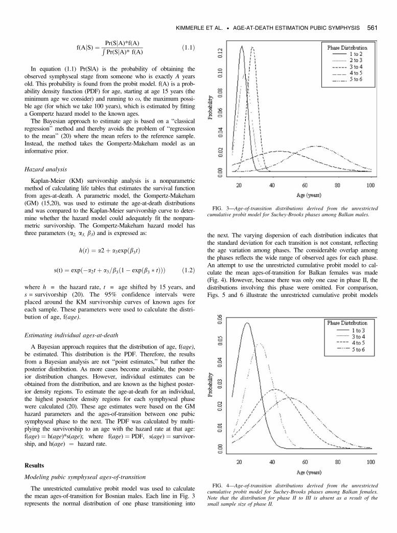
The unrestricted cumulative probit model was used to calculatethe mean ages-of-transition for Bosnian males. Each line in Fig. 3represents the normal distribution of one phase transitioning into
the next. The varying dispersion of each distribution indicates thatthe standard deviation for each transition is not constant, reflectingthe age variation among phases. The considerable overlap amongthe phases reflects the wide range of observed ages for each phase.An attempt to use the unrestricted cumulative probit model to cal-culate the mean ages-of-transition for Balkan females was made(Fig. 4). However, because there was only one case in phase II, thedistributions involving this phase were omitted. For comparison,Figs. 5 and 6 illustrate the unrestricted cumulative probit models
FIG. 3—Age-of-transition distributions derived from the unrestrictedcumulative probit model for Suchey-Brooks phases among Balkan males.
FIG. 4—Age-of-transition distributions derived from the unrestrictedcumulative probit model for Suchey-Brooks phases among Balkan females.Note that the distribution for phase II to III is absent as a result of thesmall sample size of phase II.
KIMMERLE ET AL. • AGE-AT-DEATH ESTIMATION PUBIC SYMPHYSIS 561
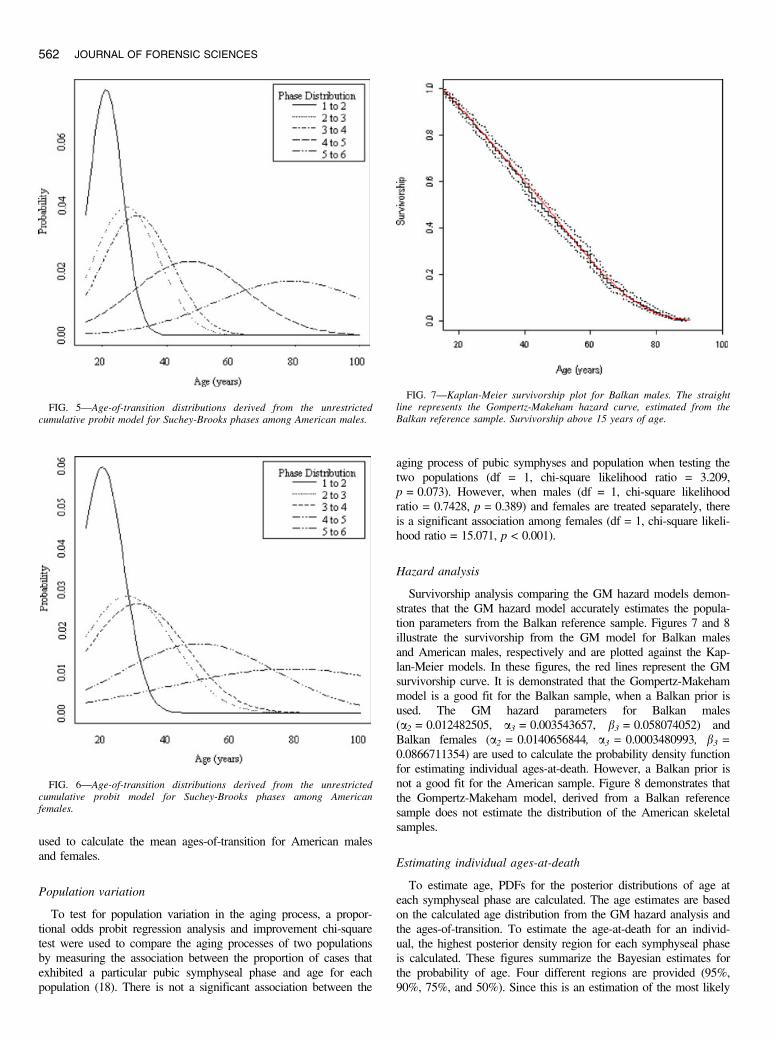
used to calculate the mean ages-of-transition for American malesand females.
Population variation
To test for population variation in the aging process, a propor-tional odds probit regression analysis and improvement chi-squaretest were used to compare the aging processes of two populationsby measuring the association between the proportion of cases thatexhibited a particular pubic symphyseal phase and age for eachpopulation (18). There is not a significant association between the
aging process of pubic symphyses and population when testing thetwo populations (df = 1, chi-square likelihood ratio = 3.209,p = 0.073). However, when males (df = 1, chi-square likelihoodratio = 0.7428, p = 0.389) and females are treated separately, thereis a significant association among females (df = 1, chi-square likeli-hood ratio = 15.071, p < 0.001).
Hazard analysis
Survivorship analysis comparing the GM hazard models demon-strates that the GM hazard model accurately estimates the popula-tion parameters from the Balkan reference sample. Figures 7 and 8illustrate the survivorship from the GM model for Balkan malesand American males, respectively and are plotted against the Kap-lan-Meier models. In these figures, the red lines represent the GMsurvivorship curve. It is demonstrated that the Gompertz-Makehammodel is a good fit for the Balkan sample, when a Balkan prior isused. The GM hazard parameters for Balkan males(a2 = 0.012482505, a3 = 0.003543657, b3 = 0.058074052) andBalkan females (a2 = 0.0140656844, a3 = 0.0003480993, b3 =0.0866711354) are used to calculate the probability density functionfor estimating individual ages-at-death. However, a Balkan prior isnot a good fit for the American sample. Figure 8 demonstrates thatthe Gompertz-Makeham model, derived from a Balkan referencesample does not estimate the distribution of the American skeletalsamples.
Estimating individual ages-at-death
To estimate age, PDFs for the posterior distributions of age ateach symphyseal phase are calculated. The age estimates are basedon the calculated age distribution from the GM hazard analysis andthe ages-of-transition. To estimate the age-at-death for an individ-ual, the highest posterior density region for each symphyseal phaseis calculated. These figures summarize the Bayesian estimates forthe probability of age. Four different regions are provided (95%,90%, 75%, and 50%). Since this is an estimation of the most likely
FIG. 5—Age-of-transition distributions derived from the unrestrictedcumulative probit model for Suchey-Brooks phases among American males.
FIG. 6—Age-of-transition distributions derived from the unrestrictedcumulative probit model for Suchey-Brooks phases among Americanfemales.
FIG. 7—Kaplan-Meier survivorship plot for Balkan males. The straightline represents the Gompertz-Makeham hazard curve, estimated from theBalkan reference sample. Survivorship above 15 years of age.
562 JOURNAL OF FORENSIC SCIENCES
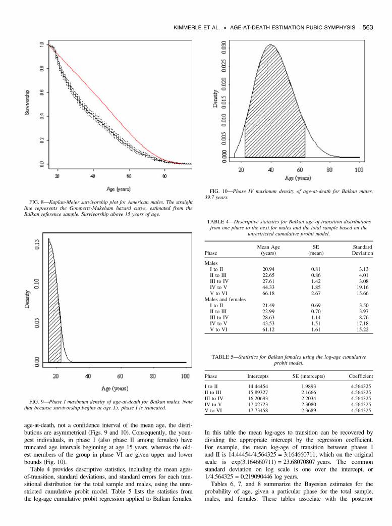
age-at-death, not a confidence interval of the mean age, the distri-butions are asymmetrical (Figs. 9 and 10). Consequently, the youn-gest individuals, in phase I (also phase II among females) havetruncated age intervals beginning at age 15 years, whereas the old-est members of the group in phase VI are given upper and lowerbounds (Fig. 10).
Table 4 provides descriptive statistics, including the mean ages-of-transition, standard deviations, and standard errors for each tran-sitional distribution for the total sample and males, using the unre-stricted cumulative probit model. Table 5 lists the statistics fromthe log-age cumulative probit regression applied to Balkan females.
In this table the mean log-ages to transition can be recovered bydividing the appropriate intercept by the regression coefficient.For example, the mean log-age of transition between phases Iand II is 14.44454 ⁄4.564325 = 3.164660711, which on the originalscale is exp(3.164660711) = 23.68070807 years. The commonstandard deviation on log scale is one over the intercept, or1 ⁄ 4.564325 = 0.219090446 log years.
Tables 6, 7, and 8 summarize the Bayesian estimates for theprobability of age, given a particular phase for the total sample,males, and females. These tables associate with the posterior
FIG. 9—Phase I maximum density of age-at-death for Balkan males. Notethat because survivorship begins at age 15, phase I is truncated.
TABLE 4—Descriptive statistics for Balkan age-of-transition distributionsfrom one phase to the next for males and the total sample based on the
unrestricted cumulative probit model.
PhaseMean Age
(years)SE
(mean)StandardDeviation
MalesI to II 20.94 0.81 3.13II to III 22.65 0.86 4.01III to IV 27.61 1.42 3.08IV to V 44.33 1.85 19.16V to VI 66.18 2.67 15.66
Males and femalesI to II 21.49 0.69 3.50II to III 22.99 0.70 3.97III to IV 28.63 1.14 8.76IV to V 43.53 1.51 17.18V to VI 61.12 1.61 15.22
TABLE 5—Statistics for Balkan females using the log-age cumulativeprobit model.
Phase Intercepts SE (intercepts) Coefficient
I to II 14.44454 1.9893 4.564325II to III 15.89327 2.1666 4.564325III to IV 16.20693 2.2034 4.564325IV to V 17.02723 2.3080 4.564325V to VI 17.73458 2.3689 4.564325
FIG. 10—Phase IV maximum density of age-at-death for Balkan males,39.7 years.
FIG. 8—Kaplan-Meier survivorship plot for American males. The straightline represents the Gompertz-Makeham hazard curve, estimated from theBalkan reference sample. Survivorship above 15 years of age.
KIMMERLE ET AL. • AGE-AT-DEATH ESTIMATION PUBIC SYMPHYSIS 563
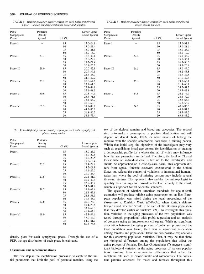
density plots for each symphyseal phase. Through the use of aPDF, the age distribution of each phase is estimated.
Discussion and recommendations
The first step in the identification process is to establish the ini-tial parameters that limit the pool of potential matches, using the
sex of the skeletal remains and broad age categories. The secondstep is to make a presumptive or positive identification and willdepend on dental charts, DNA, or other means of linking theremains with the specific antemortem data from a missing person.Within that initial step, the objectives of the investigator may varysuch as establishing broad age cohorts for identification or creatinga demographic profile for a whole site, all of which may influencehow the age parameters are defined. Therefore, the level of CI usedto estimate an individual case is left up to the investigator andshould be approached on a case-by-case basis. This approach dif-fers from typical forensic casework encountered in the UnitedStates but reflects the context of violations to international humani-tarian law where the pool of missing persons may include severalthousand victims. This approach also enables the anthropologist toquantify their findings and provide a level of certainty to the court,which is important for all scientific standards.
The question of whether American standards for age-at-deathestimation will produce reliable aging parameters on an East Euro-pean population was raised during the legal proceedings of theProsecutor v. Radislav Krsti_c (IT-98-33), when Krsti_c’s defenselawyer asked whether ‘‘could it be said of the Bosnian populationthat they develop earlier or quicker?’’ (21). To investigate this ques-tion, variation in the aging processes of the two populations wastested through proportional odds probit regression and an analysisof deviance using an improvement chi-square. While no significantassociation between the aging process of pubic symphyses and thetotal population was found, there was a significant associationamong females and population. There are two possible explanationsfor this observed population variation. First, it may be that thereare biological differences among the populations that affect theaging process of females. Kemkes-Grottenthaler (7) suggests signif-icant biological variation in the aging processes of various popula-tions resulting from environmental differences that affect themetabolic rate such as caloric intake and osteoporosis. The consis-tent patterns observed for males and females throughout this
TABLE 7—Highest posterior density region for each pubic symphysealphase among males.
PubicSymphysealPhase
PosteriorDensity(years) CI (%)
Lower–upperBound (years)
Phase I – 95 15.0–23.990 15.0–22.675 15.0–20.550 15.0–18.4
Phase II 23.2 95 17.4–29.990 18.2–28.975 19.6–27.050 21.0–25.4
Phase III 27.8 95 20.1–41.990 20.9–39.475 22.5–35.450 24.5–31.9
Phase IV 39.7 95 19.9–67.490 22.4–63.175 26.8–55.950 31.7–48.9
Phase V 52.8 95 20.6–76.390 25.4–73.675 33.7–68.350 41.9–62.4
Phase VI 69.0 95 42.3–89.690 47.0–86.775 54.0–81.950 60.5–76.8
TABLE 6—Highest posterior density region for each pubic symphysealphase � unisex standard combining males and females.
PubicSymphysealPhase
PosteriorDensity(years) CI (%)
Lower–upperBound (years)
Phase I – 95 15.0–24.890 15.0–23.475 15.0–21.150 15.0–18.7
Phase II 23.3 95 16.6–30.190 17.6–29.275 19.2–27.450 20.9–25.7
Phase III 28.0 95 20.0–42.990 20.9–39.575 22.6–35.750 24.6–32.2
Phase IV 39.7 95 20.6–64.690 23.1–61.575 27.4–54.850 32.1–48.3
Phase V 50.9 95 20.8–74.590 25.3–71.675 33.1–66.250 40.6–60.3
Phase VI 67.3 95 39.8–88.790 44.5–85.775 51.8–80.750 58.4–75.4
TABLE 8—Highest posterior density region for each pubic symphysealphase among females.
PubicSymphysealPhase
PosteriorDensity(years) CI (%)
Lower–upperBound (years)
Phase I – 95 15.0–31.990 15.0–28.675 15.0–23.950 15.0–19.9
Phase II 22.4 95 15.0–38.990 15.0–35.175 16.3–30.650 18.5–26.9
Phase III 26.5 95 15.0–47.090 16.2–43.875 18.7–37.850 21.6–32.6
Phase IV 35.3 95 19.7–66.190 21.4–60.175 24.7–51.250 28.5–43.8
Phase V 44.9 95 26.2–77.990 28.4–72.975 32.2–64.350 36.7–55.7
Phase VI 74.9 95 40.6–93.390 45.5–91.275 54.5–87.550 63.6–83.2
564 JOURNAL OF FORENSIC SCIENCES
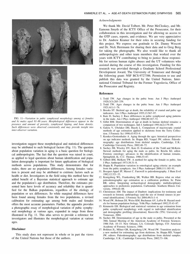
investigation suggest these morphological and statistical differencesmay be attributed to such biological factors (Fig. 11). The questionabout population variation in aging is a basic question for biologi-cal anthropologists. The fact that the question was raised in court,as applied to legal questions about human identification and popu-lation demography is important for future applications of biologicalmethods across populations. This study demonstrates that formales, there are no population differences. Among females varia-tion is present and may be attributed to extrinsic factors such ashealth or diet. Investigators in the field using this method have theadded benefit of a Bayesian statistical approach to estimate ageand the population’s age distribution. Therefore, the estimates pre-sented here have levels of accuracy and reliability that is quanti-fied for the Balkan populations, regardless of the etiology ofpopulation or sex differences. So while marginal differences havebeen found among females from the two populations, a revisedcalibration for estimating age among both males and femalesoffers the most accurate parameters. Further, the appendix providesa photographic essay of morphological variation of pubic symphy-seal morphology at various ages, expanded in range from thoseillustrated in Fig. 11. This atlas serves to provide a reference forinvestigators and illustrates the morphological variation at variousages.
Disclaimer
This study does not represent in whole or in part the viewsof the United Nations but those of the authors.
Acknowledgments
We thank Mr. David Tolbert, Mr. Peter McCloskey, and Mr.Eamonn Smyth of the ICTY Office of the Prosecutor, for theircollaboration in this investigation and for allowing us access tothe OTP cases, reports, and evidence. We are very appreciativeto Dr. Andrew Kramer for their roles in securing funding forthis project. We express our gratitude to Dr. Danny Wescottand Dr. Nick Herrmann for sharing their data and to Greg Bergfor taking the photographs. We also would like to thank allanthropologists and other team members that worked over theyears with ICTY contributing to bring to justice those responsi-ble for serious human rights abuses and the UT volunteers whoassisted during the course of this investigation. Funding for thisresearch was provided through a Graduate School ProfessionalDevelopment Award, The University of Tennessee and throughthe following grant: NSF BCS-9727386. Permission to use andpublish this data was granted by the United Nations, Inter-national Criminal Tribunal for the Former Yugoslavia, Office ofthe Prosecutor and Registry.
References
1. Todd TW. Age changes in the pubic bone. Am J Phys Anthropol1920;3(3):285–339.
2. Todd TW. Ages changes in the pubic bone. Am J Phys Anthropol1921;4(1):1–76.
3. Brooks ST. Skeletal age at death, the reliability of cranial and pubic ageindicators. Am J Phys Anthropol 1955;13:567–97.
4. Katz D, Suchey J. Race differences in pubic symphyseal aging patternsin the male. Am J Phys Anthropol 1986;80:167–72.
5. Gillet RM. Determination of age at death in human skeletal remains: acomparison of two techniques. Intl J Anthropol 1991;6(2):179–89.
6. Galera V, Ubelaker DH, Hayek L. Comparison of macroscopic cranialmethods of age estimation applied to skeletons from the Terry Collec-tion. J Forensic Sci 1998;43:933–9.
7. Kemkes-Grottenthaler A. Aging through the ages: historical perspectiveson age indicator methods. In: Hoppa RD, Vaupel JW, editors. Paleode-mography: age distributions from skeletal samples. Cambridge, U.K.:Cambridge University Press, 2002;48–72.
8. Suchey JM, Wiseley DV, Katz D. Evaluation of the Todd and McKern-Stewart methods for aging the male os pubis. In: Reichs KJ, editor.Forensic osteology: advances in the identification of human remains.Springfield, IL: C.C. Thomas, 1986;33–67.
9. Gilbert BM, McKern TW. A method for aging the female os pubis. AmJ Phys Anthropol 1973;38:31–8.
10. Hoppa R. Population variation in osteological aging criteria: an examplefrom the pubic symphysis. Am J Phys Anthropol 2000;111:185–91.
11. Bocquet-Appel JP, Masset C. Farewell to paleodemography. J Hum Evol1982;12:321–33.
12. Konigsberg WL, Frankenberg SR, Walker RB. Regress what on what:paleodemographic age estimation as a calibration problem. In: PaineRR, editor. Integrating archaeological demography: multidisciplinaryapproaches to prehistoric population. Carbondale: Southern Illinois Uni-versity, 1994;64–88.
13. Christensen AM. The impact of Daubert: implications for testimony andresearch in forensic anthropology (and the use of frontal sinuses in per-sonal identification). J Forensic Sci 2004;49(3):1–4.
14. Wood JW, Holman DJ, Weiss KM, Buchanan AV, LeFor B. Hazard mod-els for human population biology. Yrbk Phys Anthropol 1992;35:43–87.
15. Kimmerle EH. Biological and statistical variation in age estimation frompubic symphyseal morphology with regard to individual identificationand demographic profiling [dissertation]. Knoxville (TN): University ofTennessee, 2004.
16. Suchey JM. Determination of age in the male os pubis. Presented at the38th Annual Meeting of the American Academy of Forensic Sciences;1986 Feb. 10–15. New Orleans, LA. Colorado Springs, CO: AmericanAcademy of Forensic Sciences, 1986.
17. Boldsen JL, Milner GR, Konigsberg LW, Wood JW. Transition analysis:a new method for estimating age from skeletons. In: Hoppa RD, VaupelJW, editors. Paleodemography: age distributions from skeletal samples.Cambridge, U.K.: Cambridge University Press, 2002;73–106.
FIG. 11—Variation in pubic symphyseal morphology among a) femalesand b) males aged 81–86 years. Morphological differences appear in thepresence and amount of porosity, symphyseal lipping, and bone density.Such differences were observed consistently and may provide insight intointer-observer variation.
KIMMERLE ET AL. • AGE-AT-DEATH ESTIMATION PUBIC SYMPHYSIS 565
18. Fox J. An R and S-plus companion to applied regression. ThousandOaks: Sage Publications, 2002, http://www.r-project.org/.
19. Lucy D, Aykroyd RG, Pollard A, Solheim M. A Bayesian approach toadult human age estimation from dental observations by Johanson’s agechanges. J Forensic Sci 1996;41(2):189–94.
20. Konigsberg LW, Frankenberg SR. Deconstructing death in paleodemgra-phy. Am J Phys Anthropol 2002;117:297–309.
21. Prosecutor v. Radislav Krstic, Case No. IT-98-33. Trial transcript(May 30, 2000, T. 3806) http://www.un.org/icty/transe33/000530it.htm.Accessed March 18, 2008.
Additional information and reprint requests:Erin H. Kimmerle, Ph.D.
Department of AnthropologyUniversity of South Florida
4202 E. Fowler Avenue SOC 107Tampa, FL 33620
E-mail: [email protected]
Appendix
Atlas of morphological variation of the pubic symphyseal faceas a function of age
One of the goals set forth in the collaboration between theICTY, the Office of the Prosecutor (OTP), and the Forensic
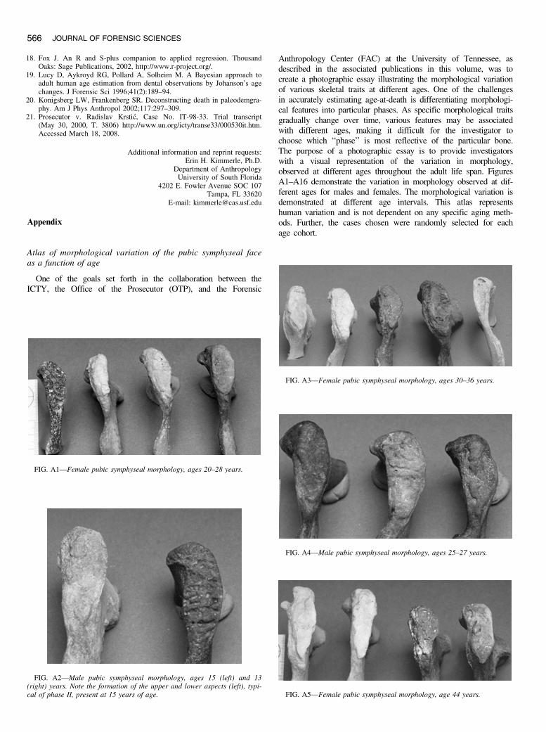
Anthropology Center (FAC) at the University of Tennessee, asdescribed in the associated publications in this volume, was tocreate a photographic essay illustrating the morphological variationof various skeletal traits at different ages. One of the challengesin accurately estimating age-at-death is differentiating morphologi-cal features into particular phases. As specific morphological traitsgradually change over time, various features may be associatedwith different ages, making it difficult for the investigator tochoose which ‘‘phase’’ is most reflective of the particular bone.The purpose of a photographic essay is to provide investigatorswith a visual representation of the variation in morphology,observed at different ages throughout the adult life span. FiguresA1–A16 demonstrate the variation in morphology observed at dif-ferent ages for males and females. The morphological variation isdemonstrated at different age intervals. This atlas representshuman variation and is not dependent on any specific aging meth-ods. Further, the cases chosen were randomly selected for eachage cohort.
FIG. A1—Female pubic symphyseal morphology, ages 20–28 years.
FIG. A3—Female pubic symphyseal morphology, ages 30–36 years.
FIG. A2—Male pubic symphyseal morphology, ages 15 (left) and 13(right) years. Note the formation of the upper and lower aspects (left), typi-cal of phase II, present at 15 years of age.
FIG. A4—Male pubic symphyseal morphology, ages 25–27 years.
FIG. A5—Female pubic symphyseal morphology, age 44 years.
566 JOURNAL OF FORENSIC SCIENCES
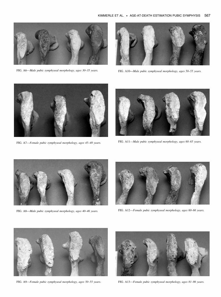
FIG. A10—Male pubic symphyseal morphology, ages 50–55 years.FIG. A6—Male pubic symphyseal morphology, ages 30–35 years.
FIG. A7—Female pubic symphyseal morphology, ages 45–48 years.
FIG. A8—Male pubic symphyseal morphology, ages 40–46 years.
FIG. A9—Female pubic symphyseal morphology, ages 50–55 years.
FIG. A11—Male pubic symphyseal morphology, ages 60–65 years.
FIG. A12—Female pubic symphyseal morphology, ages 60–66 years.
FIG. A13—Female pubic symphyseal morphology, ages 81–86 years.
KIMMERLE ET AL. • AGE-AT-DEATH ESTIMATION PUBIC SYMPHYSIS 567
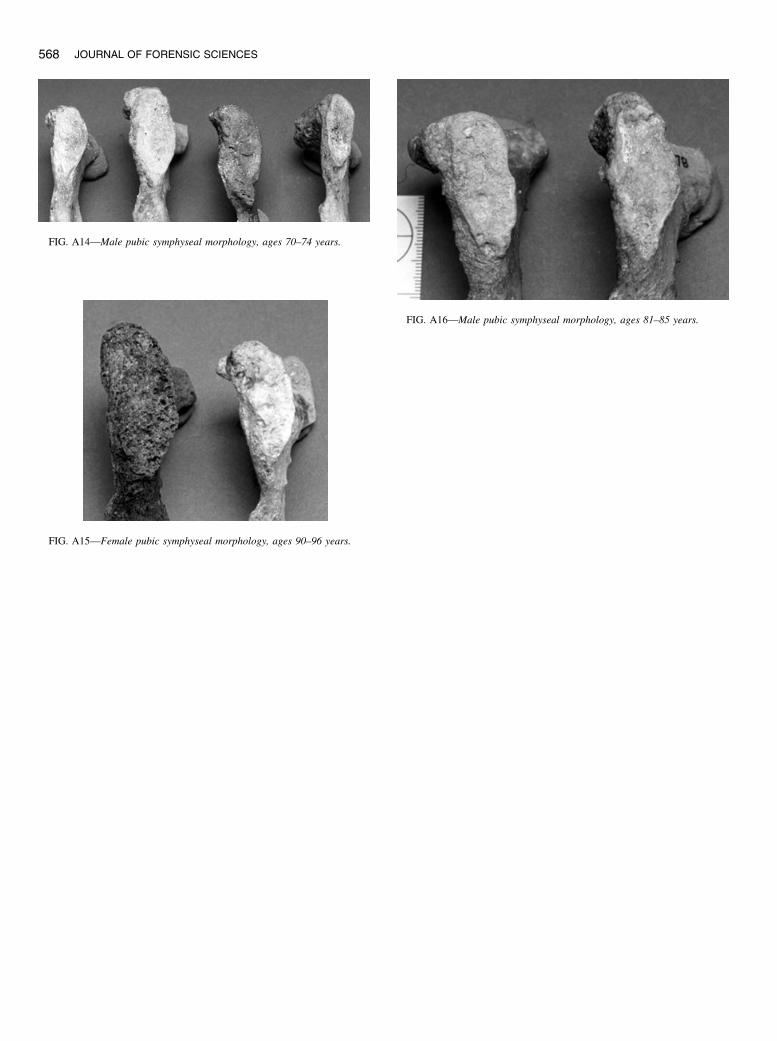
FIG. A16—Male pubic symphyseal morphology, ages 81–85 years.
FIG. A14—Male pubic symphyseal morphology, ages 70–74 years.
FIG. A15—Female pubic symphyseal morphology, ages 90–96 years.
568 JOURNAL OF FORENSIC SCIENCES