Embed Size (px)
Citation preview
ORIGINAL INVESTIGATIONS
INTERNATIONAL JOURNAL OF SPORT BIOMECHANICS, 1988, 4, 315-325
Analysis of Lower Limb Muscle Function in Ergometer Rowing
1.-M. John Wilson, D. Gordon E. Robertson, and J. Peter Stothart
In an effort to seek further understanding of lower limb muscle function in the rowing movement, an electromyographic analysis was undertaken of row- ers rowing on a Gjessing ergometer. A strain gauged transducer was insert- ed in the ergometer linkage between handle and flywheel to detect pulling force. Electrodes were placed on the following lower limb muscles: gluteus maximus, biceps femoris, rectus femoris, vastus lateralis, gastrocnemius, and tibialis anterior. Linear envelope electromyograms from each muscle and the force signals were sampled synchronously at 50 Hz. The results indicated that all six muscles were active from catch to finish of the drive phase. Bi- ceps femoris, gluteus maximus, gastrocnemius, and vastus lateralis all be- gan their activity at or just prior to catch and reached maximal excitation near peak force of the stroke. Rectus femoris and tibialis anterior activity began prior to the catch and reached maximal excitation subsequent to peak force. The coactivation of the five leg muscles, of which four were biarticu- lar, included potentially antagonistic actions that would cancel each other's effects. Clearly, however, other explanations must be considered. Both gas- trocnemius and biceps femoris have been shown to act as knee extensors and may do so in the case of the rowing action. Furthermore, rectus femoris may act with unchanging length as a knee extensor by functioning as a rigid link between the pelvis and tibia. In this manner, energy created by the hip ex- tensors is transferred across the knee joint via the isometrically contracting rectus femoris muscle.
The increasing popularity of rowing and advances in technology in this de- cade have prompted many kinesiological investigations of simulated rowing. Despite numerous physiologically oriented investigations-maximal oxygen con- sumption abilities, blood lactate levels, muscle biopsies (Hagerman, 1984), and ECG analyses (Larsson, 1980)-biomechanical information about rowing move- ments is scarce. Moreover, the majority of investigators have focused on quan-
The authors are with the University of Ottawa. Address correspondence to Gordon Robertson, School of Human Kinetics, University of Ottawa, Ottawa, Canada KIN 6N5.
316 WILSON, ROBERTSON, AND STOTHART
tifying the external forces exerted by the athlete, yet very little is known about where and how these forces were produced within the individual.
Although there are extensive EMG studies in sports such as cycling (Houtz & Fisher, 1959; Jorge & Hull, 1983), sprint running (Simonsen, Thomsen, & Klausen, 1985), and walking (Pedotti, 1977), rowing has remained relatively un- touched in this aspect. As a means of sidestepping research problems created by the sport's aquatic environment, most investigations have been carried out on specialized rowing ergometers. Even though rowing technique is only approxi- mated with such devices, Nelson and Widule (1983) have shown kinematic similar- ities between ergometer rowing and sculling on the water. When segment energies were examined, however, Martindale and Robertson (1984) reported significant differences between the two skills, calling into question the use of results from ergometer research to the on-water situation.
EMG analyses of novice and competitively classed oarsmen were first reported by Daireaux and Pottier (1983). These investigators studied raw and integrated electromyographical signals of several muscles, namely the biceps brachii, wrist flexors, wrist extensors, rhomboids, trapezius, erector spinae, and vastus lateralis. Rowing simulation was performed on a hydraulic resistance er- gometer (Vogatore Skiff). Even though intensity and rowing technique were not specified, their findings indicated that more experienced oarsmen generated larger vastus lateralis and lower biceps femoris integrated EMGs throughout the entire cycle when compared to novices. The latter were also observed to have greater variance in raw EMGs of all six muscles as well as elevated vastus lateralis ac- tivity during the recovery phase. This was attributed to eccentric contractions required in braking a fast gliding movement during recovery.
Marr and Stafford (1983) also utilized electromyographical techniques as a means of differentiating hamstring, latissimus dorsi, and erector spinae muscle activity between a novice and an experienced junior oarsman. Novice technique was reported as being more segmented (no overlap between hamstring and latis- simus dorsi bursts of activity) than that of the junior. These results have limited applicability due to the small sample size and deficiencies in the reporting of the data collection techniques.
In an effort to determine the most efficient technique of rowing, Robertson (1985) performed an electromyographic analysis of rowing on a Gjessing ergo- meter. The idea was to examine whether rowers used reci~rocal inhibition of antagonistic muscles to minimize energy expenditure during the rowing stroke (Standard Slide technique according to Koerner, 1980). In a pilot study of two female rowers, it was discovered that there was no reciprocal inhibition of leg muscles. In contrast, several antagonists acted in a synergistic fashion. For ex- ample, biceps femoris and gastrocnemius, both knee flexors, were recruited max- imally with vastus lateralis and vastus medialis, which are knee extensors. Furthermore, gluteus maximus and rectus femoris, two antagonistic hip muscles, were active synchronously during the pull (drive) although rectus femoris was not recruited fully. As a means of substantiating the conclusions derived from that study, the present study was conducted using a larger sample of male rowers.
ERGOMETER ROWING 31 7
Purpose
It was the purpose of this study to examine the recruitment pattern of the major leg muscles of male rowers using a Gjessing ergometer. The following muscles were monitored: gluteus maximus, rectus femoris, vastus lateralis, biceps femoris, gastrocnemius, and tibialis anterior. These muscles were selected as being the most important leg muscles in rowing.
Subjects and Methods
Nine lightweight university classed male rowers served as subjects for this in- vestigation. Mean height and age of the group was 182.9 cm and 23.5 years, respectively. All subjects with the exception of one, a Canadian Junior National, had a minimum of 3 years rowing experience using the Standard Slide technique (in Canada the Modem Orthodox technique).
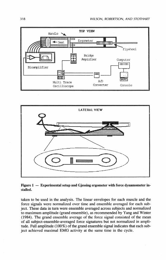
To measure tensile force applied by the rower, the Gjessing ergometer was instrumented with a strain gauge transducer between the oar handle and draw cable, as shown in Figure 1. The transducer consisted of a flat 10.3-cm stainless steel double O-ring link on which were mounted four strain gauges. The config- uration of gauges on the link was such that tension/compression sensitivity of the Wheatstone bridge circuit was 2.6 times that of a single 20-rnm gauge. Out- put from this circuit was amplified through a bridge amplifier (Honeywell, Ac- cudata 218). The linearity and hysteresis of the transducer was 1.5 and 2.8% FSO, respectively. Dynamic calibration of the transducer with a force platform (Kistler) showed less than 5 % attenuation and no phase lag at frequencies below 6 Hz.
Prior to testing, each subject was permitted a 5- to 10-minute warm-up period on the ergometer in which the necessary ergometer adjustments were made. Pairs of Beclanan silverlsilver chloride electrodes (2.5 cm apart) were then placed directly over the motor points of the tibialis anterior (TA), gastrocnemius (GC), vastus lateralis (VL), biceps femoris (BF), rectus femoris (RF), and gluteus max- imus (GM) muscles according to Delagi, Perotto, Iazzetti, and Morrison (1975). EMG signals were amplified and processed by high gain (to 1000), high input impedance (10 megohms), differential amplifiers with a 10-700 Hz band pass filter to remove artifacts and high frequency noise. These amplifiers were capa- ble of generating raw, full wave rectified, linear envelope, and integrated elec- tromyographical signals. The linear envelope signals were monitored by means of a four channel digital oscilloscope (Tektronix).
After the warm-up and familiarization period, the subjects started rowing the ergometer and used about six to eight strokes to attain their designated stroke rate (approx. 30 strokeslminute against a resistance of 29.4 N.m 13 kp]). Once the athlete signaled that he was ready, linear envelope EMGs (2nd order, 5 Hz low pass analog filter) of all six muscles and the force transducer signal were sampled, synchronously, at a frequency of 50 Hz by means of a minicomputer (Data General rnicroEclipse). The experimental setup is shown in Figure 1. Fifteen second samples were recorded for each subject, from which five strokes were
318 WILSON, ROBERTSON, AND STOTHART
Figure 1 - Experimental setup and Gjessing ergometer with force dynamometer in- stalled.
taken to be used in the analysis. The linear envelopes for each muscle and the force signals were normalized over time and ensemble averaged for each sub- ject. These data in turn were ensemble averaged across subjects and normalized to maximum amplitude (grand ensemble), as recommended by Yang and Winter (1984). The grand ensemble average of the force signal consisted of the mean of all subject-ensemble-averaged force signatures but not normalized in ampli- tude. Full amplitude (100%) of the grand ensemble signal indicates that each sub- ject achieved maximal EMG activity at the same time in the cycle.
ERGOMETER ROWING 319
0 20 40 60 80 100
X o f Cycle
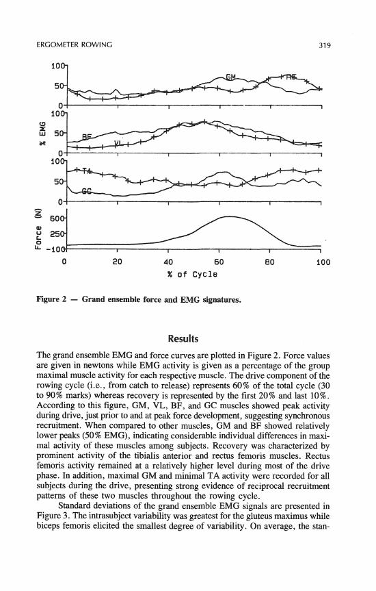
Figure 2 - Grand ensemble force and EMG signatures.
Results
The grand ensemble EMG and force curves are plotted in Figure 2. Force values are given in newtons while EMG activity is given as a percentage of the group maximal muscle activity for each respective muscle. The drive component of the rowing cycle (i.e., from catch to release) represents 60% of the total cycle (30 to 90% marks) whereas recovery is represented by the first 20% and last 10%. According to this figure, GM, VL, BF, and GC muscles showed peak activity during drive, just prior to and at peak force development, suggesting synchronous recruitment. When compared to other muscles, GM and BF showed relatively lower peaks (50% EMG), indicating considerable individual differences in maxi- mal activity of these muscles among subjects. Recovery was characterized by prominent activity of the tibialis anterior and rectus femoris muscles. Rectus femoris activity remained at a relatively higher level during most of the drive phase. In addition, maximal GM and minimal TA activity were recorded for all subjects during the drive, presenting strong evidence of reciprocal recruitment patterns of these two muscles throughout the rowing cycle.
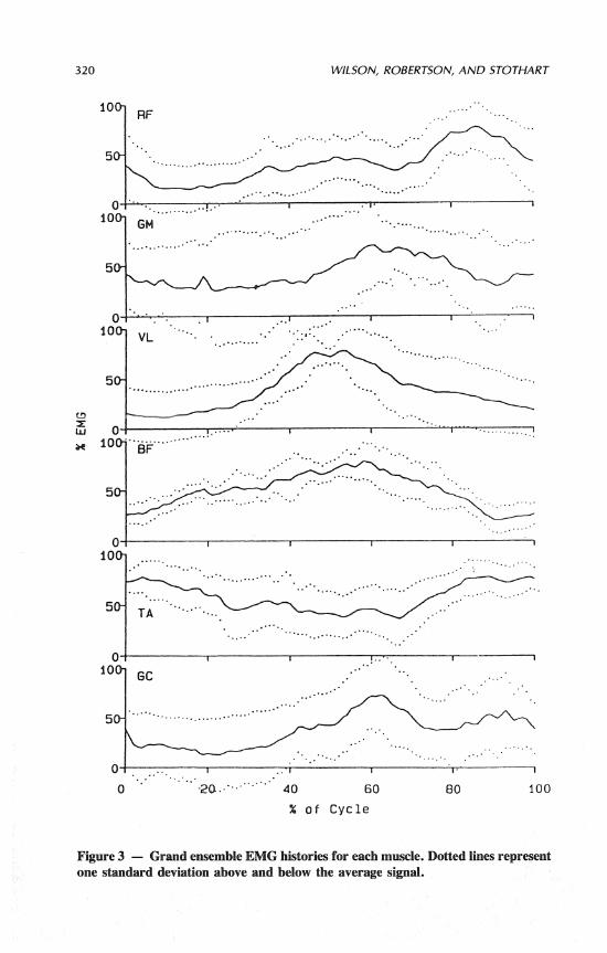
Standard deviations of the grand ensemble EMG signals are presented in Figure 3. The intrasubject variability was greatest for the gluteus maximus while biceps femoris elicited the smallest degree of variability. On average, the stan-
320 WILSON, ROBERTSON, AND STOTHART
% o f Cycle
Figure 3 - Grand ensemble EMG histories for each muscle. Dotted l i i represent one standard deviation above and below the average signal.
ERGOMETER ROWING
Table 1
Lower Limb Muscle Co-contraction Truth Table
Each fraction represents the number of subjects who solicited co-contraction of the respec- tive muscles out of the total number of subjects from which the required EMG information was available.
dard deviation represented 10 to 20% of the mean signal. Table 1 shows the number of subjects who displayed co-contraction of the various muscles as a frac- tion of the number of subjects where EMG recordings were made. Co-contraction was operationally defined as when the ensemble EMG levels of both muscles si- multaneously surpassed 50% of their cyclic maximum. It should be noted that EMG data for each muscle were not available from all subjects.
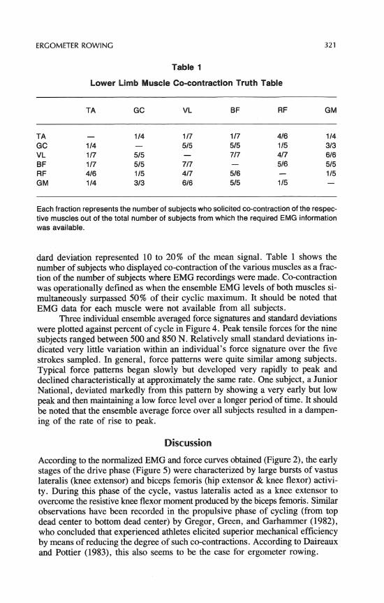
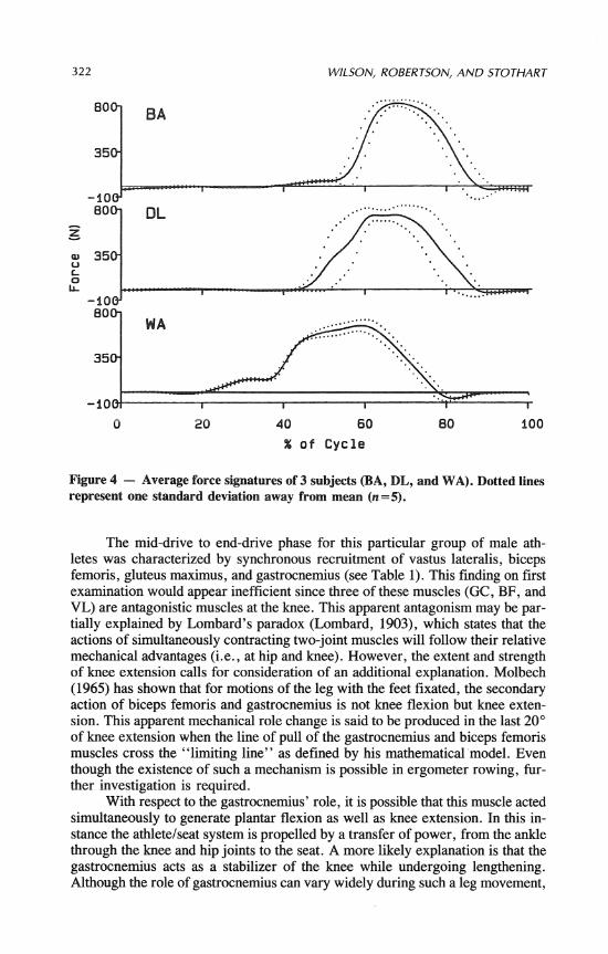
Three individual ensemble averaged force signatures and standard deviations were plotted against percent of cycle in Figure 4. Peak tensile forces for the nine subjects ranged between 500 and 850 N. Relatively small standard deviations in- dicated very little variation within an individual's force signature over the five strokes sampled. In general, force patterns were quite similar among subjects. Typical force patterns began slowly but developed very rapidly to peak and declined characteristically at approximately the same rate. One subject, a Junior National, deviated markedly from this pattern by showing a very early but low peak and then maintaining a low force level over a longer period of time. It should be noted that the ensemble average force over all subjects resulted in a dampen- ing of the rate of rise to peak.
Discussion
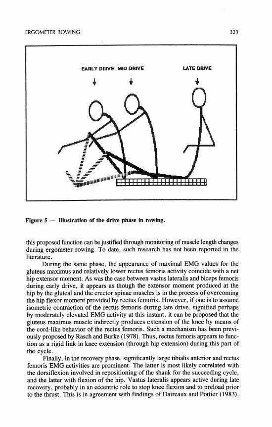
According to the normalized EMG and force curves obtained (Figure 2), the early stages of the drive phase (Figure 5) were characterized by large bursts of vastus lateralis (knee extensor) and biceps femoris (hip extensor & knee flexor) activi- ty. During this phase of the cycle, vastus lateralis acted as a knee extensor to overcome the resistive knee flexor moment produced by the biceps femoris. Similar observations have been recorded in the propulsive phase of cycling (from top dead center to bottom dead center) by Gregor, Green, and Garhamrner (1982), who concluded that experienced athletes elicited superior mechanical efficiency by means of reducing the degree of such co-contractions. According to Daireaux and Pottier (1983), this also seems to be the case for ergometer rowing.
322 WILSON, ROBERTSON, AND STOTHART
-.Luu1 I I I I
0 20 40 60 60 100
X o f Cycle
Figure 4 - Average force signatures of 3 subjects (BA, DL, and WA). Dotted lines represent one standard deviation away from mean (n=5).
The mid-drive to end-drive phase for this particular group of male ath- letes was characterized by synchronous recruitment of vastus lateralis, biceps femoris, gluteus maxirnus, and gastrocnemius (see Table 1). This finding on first examination would appear inefficient since three of these muscles (GC, BF, and VL) are antagonistic muscles at the knee. This apparent antagonism may be par- tially explained by Lombard's paradox (Lombard, 1903), which states that the actions of simultaneously contracting two-joint muscles will follow their relative mechanical advantages (i.e., at hip and knee). However, the extent and strength of knee extension calls for consideration of an additional explanation. Molbech (1965) has shown that for motions of the leg with the feet fixated, the secondary action of biceps femoris and gastrocnemius is not knee flexion but knee exten- sion. This apparent mechanical role change is said to be produced in the last 20' of knee extension when the line of pull of the gastrocnemius and biceps femoris muscles cross the "limiting line" as defined by his mathematical'model. Even though the existence of such a mechanism is possible in ergometer rowing, fur- ther investigation is required.
With respect to the gastrocnemius' role, it is possible that this muscle acted simultaneously to generate plantar flexion as well as knee extension. In this in- stance the athletelseat system is propelled by a transfer of power, from the ankle through the knee and hip joints to the seat. A more likely explanation is that the gastrocnemius acts as a stabilizer of the knee while undergoing lengthening. Although the role of gastrocnemius can vary widely during such a leg movement,
ERGOMETER ROWING 323
EARLY DRIVE MID DRIVE LATE DRIVE
Figure 5 - Illustration of the drive phase in rowing.
this proposed function can be justified through monitoring of muscle length changes during ergometer rowing. To date, such research has not been reported in the literature.
During the same phase, the appearance of maximal EMG values for the gluteus maximus and relatively lower rectus femoris activity coincide with a net hip extensor moment. As was the case between vastus lateralis and biceps femoris during early drive, it appears as though the extensor moment produced at the hip by the gluteal and the erector spinae muscles is in the process of overcoming the hip flexor moment provided by rectus femoris. However, if one is to assume isometric contraction of the rectus femoris during late drive, signified perhaps by moderately elevated EMG activity at this instant, it can be proposed that the gluteus maximus muscle indirectly produces extension of the knee by means of the cord-like behavior of the rectus femoris. Such a mechanism has been previ- ously proposed by Rasch and Burke (1978). Thus, rectus femoris appears to func- tion as a rigid link in knee extension (through hip extension) during this part of the cycle.
Finally, in the recovery phase, significantly large tibialis anterior and rectus femoris EMG activities are prominent. The latter is most likely correlated with the dorsiflexion involved in repositioning of the shank for the succeeding cycle, and the latter with flexion of the hip. Vastus lateralis appears active during late recovery, probably in an eccentric role to stop knee flexion and to preload prior to the thrust. This is in agreement with findings of Daireaux and Pottier (1983).
324 WILSON, ROBERTSON, AND STOTHART
Conclusion
According to the electromyographical data collected, the drive phase of rowing is characterized by coactivation of the vastus lateralis, rectus femoris, gluteus maximus, biceps femoris, and gastrocnemius. The apparently antagonistic ac- tivity of biceps femoris and gastrocnemius against the knee extensors-vastus later- alis and rectus femoris-may be explained by Lombard's paradox or Molbech's principle that all these muscles can act as knee extensors. Gastrocnemius in this case is revealed as having a double role: knee extension and plantar flexion. Fur- thermore, it is believed that knee extension is induced by the action of the gluteus maximus about the hip via the cord-like behavior of the rectus femoris muscle. On the other hand, movements involved in recovery, dorsiflexion, and hip flex- ion are produced in part by tibialis anterior and rectus femoris, respectively. In the latter stages of recovery, vastus lateralis is involved eccentrically in the inhi- bition of knee flexion and muscle preloading. Since these conclusions are based on electromyographical observations alone, further research is needed for con- firmation. Such knowledge provides insight as to the roles of biarticular muscles in skilled movement and contributes to rehabilitation, coaching development, and athlete training.
References
Daireaux, A., & Pottier, M. (1983). Etude electromyographique (E.M.G.) de muscles representatifs du mouvement de l'aviron [Electromyographic study of muscles in- volved in rowing]. Medecine du Sport, 57(2), 21-27.
Delagi, E.F., Perotto, A., Iazzetti, I., & Momson, D. (1975). Anatomic guide for the electromyographer-The limbs. Springfield, IL: Thomas.
Gregor, R.J., Green, D., & Garhammer, J.J. (1982). An electromyographic analysis of selected muscular activity in elite competitive cyclists. Biomechanics VII @p. 537-541). Baltimore: University Park Press.
Hagerman, F.C. (1984). Applied physiology of rowing. Sports Medicine, 1, 303-326. Houtz, S.A., & Fisher, F.J. (1959). An analysis of the muscle action and ioint excursion
during exercise on a stationary bicycle. Journal of Bone and Joint Surgery, 41(a), 123-131.
Jorge, R.R., & Hull, M.L. (1983). An analysis of EMG measurements during bicycle pedalling. Dept. of Mechanical Engineering Report, University of California, Davis.
Koerner, D.E. (1980). Analysis of two major international rowing styles. Paper pre- sented at 7th FISA International Rowing Coaches Colloquium in Werder, German Democratic Republic.
Larsson, L. (1980). Morphological muscle characteristics in rowers. Canadian Journal of Applied Sports Sciences, 5, 239-244.
Lombard, W. (1903). The action of two-joint muscles. American Physical Education Review, 8, 141-145.
Marr, A., & Stafford, P.A. (1983, February). A kinematic and electromyographical comparison of a junior and a novice rower. Pelops, pp. 1-6.
Martindale, W.O., & Robertson, D.G.E. (1984). Mechanical energy in sculling and in rowing an ergometer. Canadian Journal of Applied Sports Sciences, 9(3), 153-163.
Molbech, S. (1965). On the paradoxical effect of some two-joint muscles. Acta Morpho- logical Neerlando-Scandinavica, 6 , 17 1-177.
ERGOMETER ROWING 325
Nelson, W.N., & Widule, C.J. (1983). Kinematic analysis and efficiency estimate of inter- collegiate female rowers. Medicine and Science in Sports and Exercise, 15(6), 535-541.
Pedotti, A. (1977). A study of motor coordination and neuromuscular activities in human locomotion. Biological Cybernetics, 26, 53-62.
Rasch, P.J., & Burke, R.K. (1978). Kinesiology and applied anatomy. Philadelphia: Lea & Febiger.
Robertson, D.G.E. (1985). Impulse and electromyographic analysis of ergometer rowing. Canadian Journal of Applied Sport Sciences, 10, 12.
Simonsen, E.B., Thomsen, L., & Klausen, K. (1985). Activity of mono- and biarticular leg muscles during sprint running. European Journal of Applied Physiology, 54, 524-532.
Yang, J.F., & Winter, D. A. (1984). Electromyographic amplitude normalization methods: Improving their sensitivity as diagnostic tools in gait analysis. Archives of Physical Medicine and Rehabilitation, 65, 517-521.
Acknowledgments
Financial assistance for this research was provided by Sport Canada in affiliation with the Canadian Amateur Rowing Association. Technical support was provided by Ralph Fournier.