Embed Size (px)
Citation preview

r e v b r a s o r t o p . 2 0 1 5;5 0(6):673–679
O
Ac
CA
FB
a
A
R
A
A
K
E
J
C
A
P
A
C
C
A
(
h2
www.rbo.org .br
riginal Article
natomical study on the innervation of the elbowapsule�
ristina Schmitt Cavalheiro ∗, Mauro Razuk Filho, João Rozas, João Wey,ntonio Marcos de Andrade, Edie Benedito Caetano
aculdade de Ciências Médicas e da Saúde de Sorocaba (FCMS), Pontifícia Universidade Católica de São Paulo (PUC-SP), Sorocaba, SP,razil
r t i c l e i n f o
rticle history:
eceived 22 September 2014
ccepted 10 November 2014
vailable online 19 October 2015
eywords:
lbow joint
oint capsule
adaver
natomy
a b s t r a c t
Objectives: To put forward an anatomical description of the innervation of the elbow capsule,
illustrated through morphological analysis on dissections.
Methods: Thirty elbows from fresh fixed adult cadavers aged 32–74 years, of both sexes, were
dissected.
Results: Among the dissected arms, we observed that the median nerve did not have any
branches in two arms, while it had one branch in five arms, two branches in two arms,
three branches in ten arms, four branches in nine arms and five branches in two arms. The
radial nerve did not have any branches in two arms, while it had one branch in two arms,
two branches in nine arms, three branches in ten arms, four branches in five arms and five
branches in two arms. The ulnar nerve did not have any branches in three arms, while it
had one branch in six arms, two branches in four arms, three branches in five arms, four
branches in seven arms, five branches in four arms and six branches in one arm.
Conclusions: We observed branches of the radial, ulnar and medial nerves in the elbow joint,
and a close relationship between their capsular and motor branches.
© 2015 Sociedade Brasileira de Ortopedia e Traumatologia. Published by Elsevier Editora
Ltda. All rights reserved.
Estudo anatômico da inervacão da cápsula do cotovelo
r e s u m o
alavras-chave: Objetivos: Promover a descricão anatômica da inervacão da cápsula do cotovelo com
a morfologia das dissecacões.
rticulacão do cotovelo ilustracão por meio d ápsula articularadáver
natomia
Métodos: Foram dissecados 30 cotovelos de cadáveres adultos frescos e fixados, com idade
entre 32 e 74 anos, de ambos os sexos.
� Work performed at the Faculdade de Ciências Médicas e da Saúde de Sorocaba (FCMS), Pontifícia Universidade Católica de São PauloPUC-SP), Sorocaba, SP, Brazil.∗ Corresponding author.
E-mails: [email protected], [email protected] (C.S. Cavalheiro).ttp://dx.doi.org/10.1016/j.rboe.2015.10.001255-4971/© 2015 Sociedade Brasileira de Ortopedia e Traumatologia. Published by Elsevier Editora Ltda. All rights reserved.
674 r e v b r a s o r t o p . 2 0 1 5;5 0(6):673–679
Resultados: Observamos, dentre os bracos dissecados, dois com nenhum ramo do nervo
mediano, cinco com um ramo, dois com dois ramos, 10 com três ramos, nove com quatro
ramos e dois com cinco ramos. Quando se trata do nervo radial, dois bracos não apresen-
taram ramos, dois mostraram dois ramos, nove continham dois ramos, 10 contaram com três
ramos, cinco tinham quatro ramos e dois tinham cinco ramos. Em relacão ao nervo ulnar,
tivemos três bracos sem ramos articulares, seis com um ramo, quatro com dois ramos, cinco
com três ramos, sete com quatro ramos, quatro com cinco ramos e um com seis ramos.
Conclusões: Constatamos ramos do nervo radial, ulnar e medial na articulacão do cotovelo,
assim como a relacão próxima entre os seus ramos capsulares e motores.
© 2015 Sociedade Brasileira de Ortopedia e Traumatologia. Publicado por Elsevier
Editora Ltda. Todos os direitos reservados.
muscle, the median nerve branches out into small sectionsthat go to the capsular region of the anterior medial epicondyle
Introduction
The first mentions of the nerve branches of the elbow capsuledate from 1844, in descriptions of a branch of the cutaneousnerve perforating the brachial muscle and reaching the cap-sule; branches of the median nerve penetrating the elbowjoint; and branches of the ulnar nerve branching out betweenmedial epicondyle and the olecranon. A branch of the radialnerve extending to the long head of the triceps and head-ing toward the olecranon and posterior capsule was alsodescribed.
In 1857, small branches of the musculocutaneous andmedian nerve extending to the anterior part of the capsule andvariable branches of the anterior interosseous nerve appear-ing between the radius and ulna and innervating the capsulearound the radial head were described. With regard to the pos-terior part of the capsule, a branch derived from the radialnerve that originated from the muscle branch of the lateraland medial head of the triceps brachii muscle was described.
A study conducted in 1877 reported the presence of smallfilaments from the median nerve going to the anteromedialregion of the capsule and branches of ulnar origin going to theposteromedial capsule.
In subsequent years, studies began to describe this subjectwith greater precision through dissections. From dissectionson seven adult elbows and five fetal elbows, the contributionsof the four main nerves innervating the elbow capsule (ulnar,median, musculocutaneous and radial) were demonstrated. Astudy in 1949 only mentioned ramifications going to the ole-cranon process, and did not describe capsular branches of theradial nerve.
The present-day main anatomy textbooks, such as Gray,Hollinshead, Latarjet and Liard, do not cite the radial nerve.1–8
The elbow capsule is extensive and coats the distal extrem-ity of the humerus and proximal extremity of the ulna andradius. Anteriorly and proximally, it is inserted above the fossaof the coronoid process and capitellum. Distally, it adheresmedially to the coronoid process of the ulna and laterally tothe annular ligament of the radius (Fig. 1A).
Posteriorly and proximally, the capsule adheres above theolecranon fossa, goes around the margin and continues acrossthe entire medial and lateral column, where it covers all of the
sigmoid fossa (Fig. 1B).The anterior joint capsule is usually thinner and moretransparent. It remains under tension when the elbow is
extended and relaxes when the elbow is flexed. The greatestcapacity of the joint capsule is 30–35 ml at 80◦ of flexion, whenit is fully distended.
In relation to the musculocutaneous nerve, it is known thatthe area that it innervates is the anterior capsule. This nerveissues a small branch from its main trunk, which penetratesthe middle third of the brachial muscle and goes in deeper toreach the anterior part of the humerus and supply the perios-teum. It then reaches the elbow capsule, where it divides intoa variable number of branches (Fig. 2A). This nerve is the mostconstant supplier of the capsule, both macroscopically andmicroscopically. In some cases, this capsule branch may formanastomoses with branches of the median nerve and thencontinue to the capsule (Fig. 2B). The region of the muscu-locutaneous nerve may be juxtaposed both to median and tolateral areas.9–12
Before passing between the heads of the pronator teres
Fig. 1 – Anterior limits of the elbow capsule (A). Posteriorlimits of the elbow capsule (B).
r e v b r a s o r t o p . 2 0 1 5;5 0(6):673–679 675
Fig. 2 – Ramification of the musculocutaneous nerve onreaching the elbow capsule (A). The capsule branch mayform anastomoses with branches of the median nerve andt
(mtni
Ftcmc
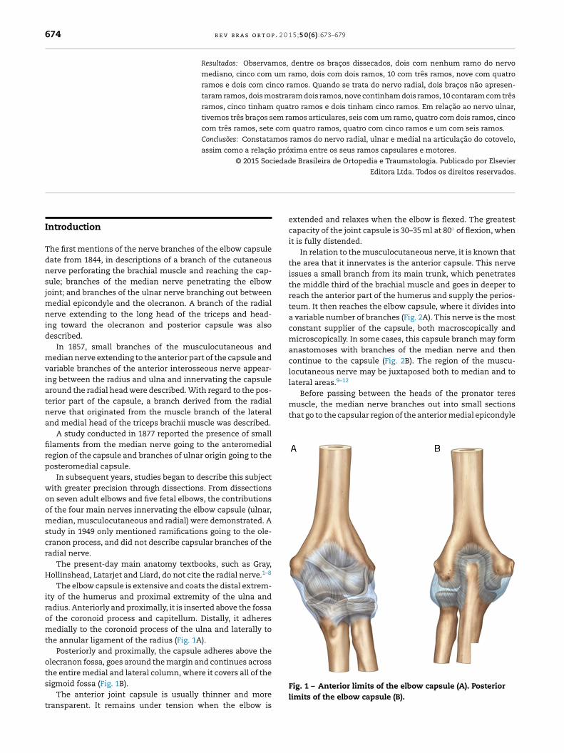
Fig. 4 – Branches of the ulnar nerve beginning in the groovebetween the medial epicondyle and the olecranon (A). Joint
hen continue to the capsule (B).
Fig. 3A). A joint branch may also occur, which developsore proximally to the elbow, posteriorly to the bifurca-
ion of the brachial artery, and joins the musculocutaneouserve to innervate the anterior capsule (Fig. 3B). The anterior
nterosseous nerve gives rise to a small filament that supplies
ig. 3 – Ramification of the median nerve in small sectionshat go from the anterior medial epicondyle toward theapsule region (A). Union of the median nerve with theusculocutaneous muscle for innervating the anterior
apsule (B).
branches originating above the cubital tunnel (B).
the posteroinferior part of the capsule, adjacent to the ulna.Thus, the median nerve usually innervates the anteromedialpart of the joint capsule and this area may be overlain by themusculocutaneous nerve.13–15
The ulnar nerve usually appears as three branches, whichbegin at the sulcus between the medial epicondyle and theolecranon (Fig. 4A). Joint branches that arise several centime-ters above the cubital tunnel have been described (Fig. 4B).These supply the posteromedial part of the capsules and theneighborhood of the medial epicondyle and olecranon, bothin the cubital tunnel. This area may be overlain by the radialnerve.16–18
A descending branch is issued from the main trunk of theradial nerve, and this follows the lateral head of the tricepsmuscle. When it reaches the olecranon, it bifurcates to thecapsule in the region of the olecranon fossa (Fig. 5A). There isalso a small filament that arises from the branch going to theanconeus muscle and innervates the posterolateral region ofthe capsule (Fig. 5B). In some cases, the posterior and proximalcapsules, which involve the extremity of the olecranon, areinnervated by thin branches from the ulnar collateral nerve,which is a branch of the main trunk of the radial nerve (Fig. 5C).This region may be overlain by ulnar innervation. Regardingthe anterior capsule, after this passes through the intramus-cular septum of the supinator muscle, it generally divides intosmall branches that may form anastomoses with structuresof the musculocutaneous nerve (Fig. 5D).18–25
Thus, it is clear that the anterior part of the capsule is
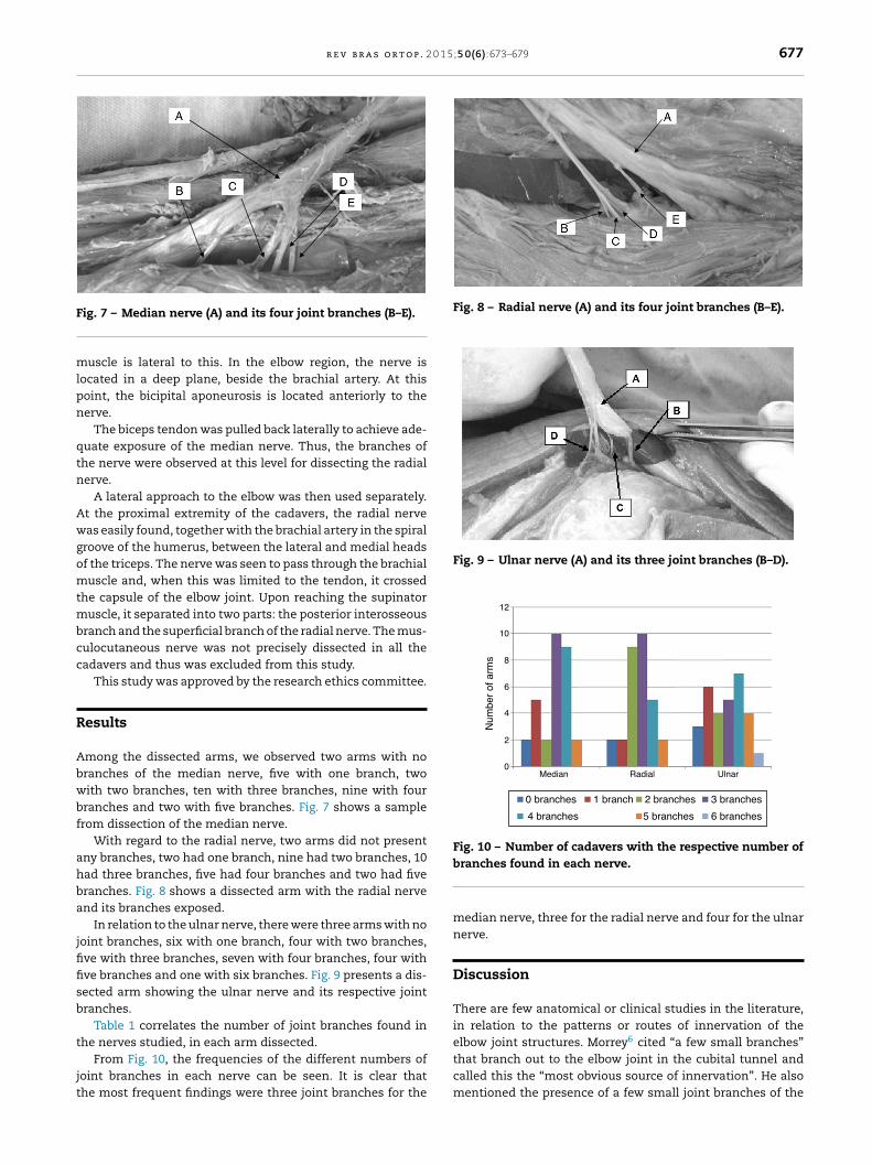
usually innervated by the musculocutaneous nerve. Thismay be overlain laterally by branches of the radial nerveand medially by branches of the median nerve (Fig. 6A). Theposterior capsule is innervated medially by the ulnar nerve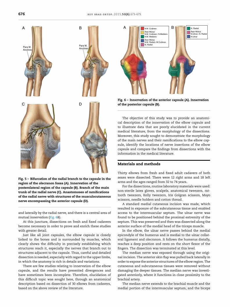
676 r e v b r a s o r t o p . 2 0 1 5;5 0(6):673–679
Para M,Ancôneo
Para M,Ancôneo
A B
DC
Fig. 5 – Bifurcation of the radial branch to the capsule in theregion of the olecranon fossa (A). Innervation of theposterolateral region of the capsule (B). Branch of the maintrunk of the radial nerve (C). Anastomoses of ramifications
N.M. Cutâneo
N.M. Mediano
N. Radial
N. Radial
Áear MútuaN.M. Cutâneo+ N.Mediano
Áear MútuaN.Ulnar+ N. Radial
Áear MútuaN.M. Radial+ M.Cutâneo
N.Ulnar
BA
Fig. 6 – Innervation of the anterior capsule (A). Innervation
of the radial nerve with structures of the musculocutaneousnerve encompassing the anterior capsule (D).
and laterally by the radial nerve, and there is a central area ofmutual innervation (Fig. 6B).
At this juncture, dissections on fresh and fixed cadaversbecome necessary in order to prove and enrich these studieswith greater detail.
Just like all joint capsules, the elbow capsule is closelylinked to the bones and is surrounded by muscles, whichclearly shows the difficulty in precisely establishing whichstructures reach it, especially the nerves that branch out tostructures adjacent to the capsule. Thus, careful and detaileddissection is needed, especially with regard to the upper limbs,in which the anatomy is rich in details and variations.
There are few studies relating to innervation of the elbowcapsule, and the results have presented divergences and
have sometimes been incomplete. Therefore, elucidation ofthis difficult topic was sought here, through an anatomicaldescription based on dissection of 30 elbows from cadavers,based on the above review of the literature.of the posterior capsule (B).
The objective of this study was to provide an anatomi-cal description of the innervation of the elbow capsule andto illustrate data that are poorly elucidated in the currentmedical literature, from the morphology of the dissections.Moreover, this study sought to demonstrate the morphologyof the main nerves and their ramifications to the elbow cap-sule, identify the locations of nerve insertions of the elbowcapsule and compare the findings from dissections with theinformation in the medical literature.
Materials and methods
Thirty elbows from fresh and fixed adult cadavers of bothsexes were dissected. There were 12 right arms and 18 leftarms and the ages ranged from 32 to 74 years.
For the dissections, routine laboratory materials were used:non-sterile latex gloves, scalpels, anatomical tweezers, rat-tooth tweezers, Kelly tweezers, Iris Golgran scissors, Mayoscissors, needle holders and cotton thread.
A standard medial cutaneous incision was made, whichresulted in exposure of the subcutaneous tissue and enabledaccess to the intermuscular septum. The ulnar nerve wasfound to be positioned behind the proximal extremity of theseptum. This was preserved and then was dissected along theanterior surface of the medial head of the triceps muscle.
In the elbow, the ulnar nerve passes behind the medialepicondyle of the humerus and is medial to the ulnar collat-eral ligament and olecranon. It follows the humerus distally,reaches a deep position and rests on the short flexor of thefingers. The dissection was terminated at this level.
The median nerve was exposed through using the origi-nal incision. The anterior skin flap was pulled back laterally inorder to expose the anterior structures of the elbow region. Thecutaneous and subcutaneous tissues were removed withoutdamaging the deeper tissues. The median nerve was investi-
gated anteriorly, where it functions in close proximity to thebrachial artery.The median nerve extends to the brachial muscle and themedial portion of the intermuscular septum, and the biceps
r e v b r a s o r t o p . 2 0 1 5;5 0(6):673–679 677
F
mlpn
qtn
Awgomtmbcc
R
Abwbf
ahba
jfifisb
t
jt
Fig. 8 – Radial nerve (A) and its four joint branches (B–E).
Fig. 9 – Ulnar nerve (A) and its three joint branches (B–D).
12
10
8
6
4
2
0Median UlnarRadial
Num
ber
of a
rms
0 branches
4 branches
1 branch
5 branches
2 branches
6 branches
3 branches
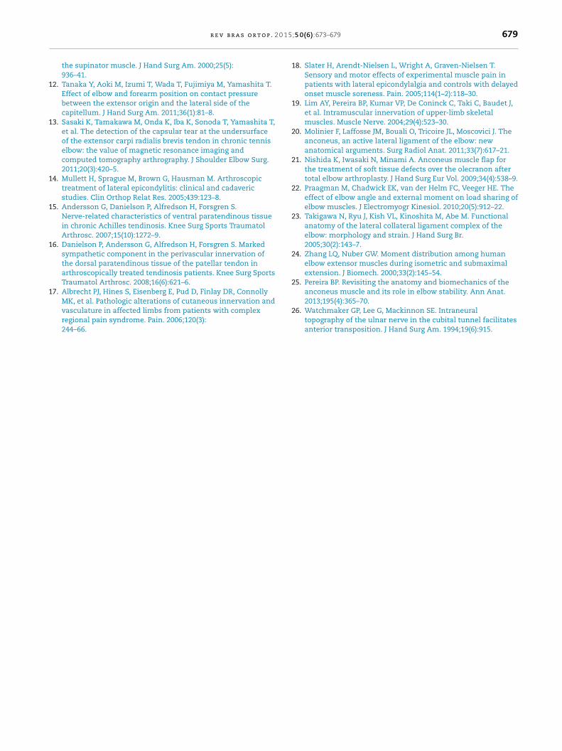
Fig. 10 – Number of cadavers with the respective number of
ig. 7 – Median nerve (A) and its four joint branches (B–E).
uscle is lateral to this. In the elbow region, the nerve isocated in a deep plane, beside the brachial artery. At thisoint, the bicipital aponeurosis is located anteriorly to theerve.
The biceps tendon was pulled back laterally to achieve ade-uate exposure of the median nerve. Thus, the branches ofhe nerve were observed at this level for dissecting the radialerve.
A lateral approach to the elbow was then used separately.t the proximal extremity of the cadavers, the radial nerveas easily found, together with the brachial artery in the spiral
roove of the humerus, between the lateral and medial headsf the triceps. The nerve was seen to pass through the brachialuscle and, when this was limited to the tendon, it crossed
he capsule of the elbow joint. Upon reaching the supinatoruscle, it separated into two parts: the posterior interosseous
ranch and the superficial branch of the radial nerve. The mus-ulocutaneous nerve was not precisely dissected in all theadavers and thus was excluded from this study.
This study was approved by the research ethics committee.
esults
mong the dissected arms, we observed two arms with noranches of the median nerve, five with one branch, twoith two branches, ten with three branches, nine with fourranches and two with five branches. Fig. 7 shows a samplerom dissection of the median nerve.
With regard to the radial nerve, two arms did not presentny branches, two had one branch, nine had two branches, 10ad three branches, five had four branches and two had fiveranches. Fig. 8 shows a dissected arm with the radial nervend its branches exposed.
In relation to the ulnar nerve, there were three arms with nooint branches, six with one branch, four with two branches,ve with three branches, seven with four branches, four withve branches and one with six branches. Fig. 9 presents a dis-ected arm showing the ulnar nerve and its respective jointranches.
Table 1 correlates the number of joint branches found in
he nerves studied, in each arm dissected.From Fig. 10, the frequencies of the different numbers ofoint branches in each nerve can be seen. It is clear thathe most frequent findings were three joint branches for the
branches found in each nerve.
median nerve, three for the radial nerve and four for the ulnarnerve.
Discussion
There are few anatomical or clinical studies in the literature,in relation to the patterns or routes of innervation of theelbow joint structures. Morrey6 cited “a few small branches”
that branch out to the elbow joint in the cubital tunnel andcalled this the “most obvious source of innervation”. He alsomentioned the presence of a few small joint branches of the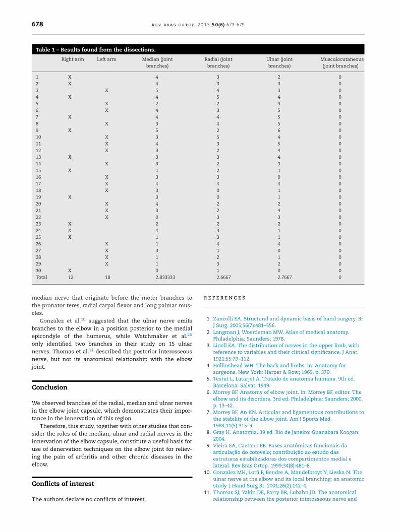
678 r e v b r a s o r t o p . 2 0 1 5;5 0(6):673–679
Table 1 – Results found from the dissections.
Right arm Left arm Median (jointbranches)
Radial (jointbranches)
Ulnar (jointbranches)
Musculocutaneous(joint branches)
1 X 4 3 2 02 X 4 3 3 03 X 5 4 3 04 X 4 5 4 05 X 2 2 3 06 X 4 3 5 07 X 4 4 5 08 X 3 4 5 09 X 5 2 6 010 X 3 5 4 011 X 4 3 5 012 X 3 2 4 013 X 3 3 4 014 X 3 2 3 015 X 1 2 1 016 X 3 3 0 017 X 4 4 4 018 X 3 0 1 019 X 3 0 1 020 X 4 2 2 021 X 3 2 4 022 X 0 3 3 023 X 2 2 2 024 X 4 3 1 025 X 1 3 1 026 X 1 4 4 027 X 3 1 0 028 X 1 2 1 029 X 1 3 2 0
r
1
30 X 0
Total 12 18 2.833333
median nerve that originate before the motor branches tothe pronator teres, radial carpal flexor and long palmar mus-cles.
Gonzalez et al.10 suggested that the ulnar nerve emitsbranches to the elbow in a position posterior to the medialepicondyle of the humerus, while Watchmaker et al.26
only identified two branches in their study on 15 ulnarnerves. Thomas et al.11 described the posterior interosseousnerve, but not its anatomical relationship with the elbowjoint.
Conclusion
We observed branches of the radial, median and ulnar nervesin the elbow joint capsule, which demonstrates their impor-tance in the innervation of this region.
Therefore, this study, together with other studies that con-sider the roles of the median, ulnar and radial nerves in theinnervation of the elbow capsule, constitute a useful basis foruse of denervation techniques on the elbow joint for reliev-ing the pain of arthritis and other chronic diseases in theelbow.
Conflicts of interest
The authors declare no conflicts of interest.1
1 0 02.6667 2.7667 0
e f e r e n c e s
1. Zancolli EA. Structural and dynamic basis of hand surgery. BrJ Surg. 2005;56(7):481–556.
2. Langman J, Woerdeman MW. Atlas of medical anatomy.Philadelphia: Saunders; 1978.
3. Linell EA. The distribution of nerves in the upper limb, withreference to variables and their clinical significance. J Anat.1921;55:79–112.
4. Hollinshead WH. The back and limbs. In: Anatomy forsurgeons. New York: Harper & Row; 1969. p. 379.
5. Testut L, Latarjet A. Tratado de anatomia humana. 9th ed.Barcelona: Salvat; 1949.
6. Morrey BF. Anatomy of elbow joint. In: Morrey BF, editor. Theelbow and its disorders. 3rd ed. Philadelphia: Saunders; 2000.p. 13–42.
7. Morrey BF, An KN. Articular and ligamentous contributions tothe stability of the elbow joint. Am J Sports Med.1983;11(5):315–9.
8. Gray H. Anatomia. 39 ed. Rio de Janeiro: Guanabara Koogan;2004.
9. Vieira EA, Caetano EB. Bases anatômicas funcionais daarticulacão do cotovelo; contribuicão ao estudo dasestruturas estabilizadoras dos compartimentos medial elateral. Rev Bras Ortop. 1999;34(8):481–8.
0. Gonzalez MH, Lotfi P, Bendre A, Mandelbroyt Y, Lieska N. The
ulnar nerve at the elbow and its local branching: an anatomicstudy. J Hand Surg Br. 2001;26(2):142–4.1. Thomas SJ, Yakin DE, Parry BR, Lubahn JD. The anatomicalrelationship between the posterior interosseous nerve and
0 1 5
1
1
1
1
1
1
1
1
2
2
2
2
2
2
r e v b r a s o r t o p . 2
the supinator muscle. J Hand Surg Am. 2000;25(5):936–41.
2. Tanaka Y, Aoki M, Izumi T, Wada T, Fujimiya M, Yamashita T.Effect of elbow and forearm position on contact pressurebetween the extensor origin and the lateral side of thecapitellum. J Hand Surg Am. 2011;36(1):81–8.
3. Sasaki K, Tamakawa M, Onda K, Iba K, Sonoda T, Yamashita T,et al. The detection of the capsular tear at the undersurfaceof the extensor carpi radialis brevis tendon in chronic tenniselbow: the value of magnetic resonance imaging andcomputed tomography arthrography. J Shoulder Elbow Surg.2011;20(3):420–5.
4. Mullett H, Sprague M, Brown G, Hausman M. Arthroscopictreatment of lateral epicondylitis: clinical and cadavericstudies. Clin Orthop Relat Res. 2005;439:123–8.
5. Andersson G, Danielson P, Alfredson H, Forsgren S.Nerve-related characteristics of ventral paratendinous tissuein chronic Achilles tendinosis. Knee Surg Sports TraumatolArthrosc. 2007;15(10):1272–9.
6. Danielson P, Andersson G, Alfredson H, Forsgren S. Markedsympathetic component in the perivascular innervation ofthe dorsal paratendinous tissue of the patellar tendon inarthroscopically treated tendinosis patients. Knee Surg SportsTraumatol Arthrosc. 2008;16(6):621–6.
7. Albrecht PJ, Hines S, Eisenberg E, Pud D, Finlay DR, Connolly
MK, et al. Pathologic alterations of cutaneous innervation andvasculature in affected limbs from patients with complexregional pain syndrome. Pain. 2006;120(3):244–66.2
;5 0(6):673–679 679
8. Slater H, Arendt-Nielsen L, Wright A, Graven-Nielsen T.Sensory and motor effects of experimental muscle pain inpatients with lateral epicondylalgia and controls with delayedonset muscle soreness. Pain. 2005;114(1–2):118–30.
9. Lim AY, Pereira BP, Kumar VP, De Coninck C, Taki C, Baudet J,et al. Intramuscular innervation of upper-limb skeletalmuscles. Muscle Nerve. 2004;29(4):523–30.
0. Molinier F, Laffosse JM, Bouali O, Tricoire JL, Moscovici J. Theanconeus, an active lateral ligament of the elbow: newanatomical arguments. Surg Radiol Anat. 2011;33(7):617–21.
1. Nishida K, Iwasaki N, Minami A. Anconeus muscle flap forthe treatment of soft tissue defects over the olecranon aftertotal elbow arthroplasty. J Hand Surg Eur Vol. 2009;34(4):538–9.
2. Praagman M, Chadwick EK, van der Helm FC, Veeger HE. Theeffect of elbow angle and external moment on load sharing ofelbow muscles. J Electromyogr Kinesiol. 2010;20(5):912–22.
3. Takigawa N, Ryu J, Kish VL, Kinoshita M, Abe M. Functionalanatomy of the lateral collateral ligament complex of theelbow: morphology and strain. J Hand Surg Br.2005;30(2):143–7.
4. Zhang LQ, Nuber GW. Moment distribution among humanelbow extensor muscles during isometric and submaximalextension. J Biomech. 2000;33(2):145–54.
5. Pereira BP. Revisiting the anatomy and biomechanics of theanconeus muscle and its role in elbow stability. Ann Anat.
2013;195(4):365–70.6. Watchmaker GP, Lee G, Mackinnon SE. Intraneuraltopography of the ulnar nerve in the cubital tunnel facilitatesanterior transposition. J Hand Surg Am. 1994;19(6):915.