Embed Size (px)
Citation preview
Radiotherapy and Oncology 84 (2007) 335–347www.thegreenjournal.com
Educational review
Anatomical bases for the radiological delineationof lymph node areas. Upper limbs, chest and abdomen
Benoit Lengelea, Catherine Nyssen-behetsa, Pierre Scallietb,*
aDepartment of Experimental Morphology, and bDepartment of Radiation Oncology, Cliniques Universitaires Saint Luc,Universite Catholique de Louvain, Bruxelles, Belgium
Abstract
Cancer spreads locally through direct infiltration into soft tissues, or at distance by invading vascular structures, thenmigrating through the lymphatic or blood flow. Although cancer cells carried in the blood can end in virtually any cornerof the body, lymphatic migration is usually stepwise, through successive nodal stops, which can temporarily delay furtherprogression. In radiotherapy, irradiation of lymphatic paths relevant to the localisation of the primary has been commonpractice for decades. Similarly, excision of cancer is often completed by lymphatic dissection.Both in radiotherapy and in surgery, advanced knowledge of the lymphatic pathways relevant to any tumour location is
an important information for treatment preparation and execution. This second part describes the lymphatics of theupper limb, of the thorax and of the upper abdomen. Providing anatomical bases for the radiological delineation of lymphnodes areas in the axilla, in the chest and in the abdomen, it also offers a simplified classification for labeling themediastinal and intra-abdominal nodal levels, grouped in each location inside three major functional areas (called I, IIand III) which are all divided into three sublevels (named a, b or c).
�c 2007 Elsevier Ireland Ltd. All rights reserved. Radiotherapy and Oncology 84 (2007) 335–347.
Keywords: Radiotherapy; Thoracic cancer; Abdominal cancer; Lymphatics; CTV
The first part of this series described the major lymphaticcollecting trunks and the lymphatic drainage of the headand neck region (to be published in Radiotherapy and Oncol-ogy [9]). The second part, ‘‘Anatomical bases for the radio-logical delineation of lymph node areas’’, deals with thelymphatic of the upper limbs, thorax and abdomen. It isan anatomical description of major and minor lymphatics,more detailed than previously published material [18]. Asin the head and neck region, there is still some inconsis-tency in the way various observers actually delineate lymphnode stations relevant to the radiotherapy of various chesttumours [16]. It points to the need of an unambiguous ana-tomical description of the localisation of lymphatics.
The topography of the lymphatic system in the upperlimbs is presented for the first time, of interest for thetreatment of melanoma and other skin cancers, as well assome particular types of soft tissue sarcoma with a lympha-tic spread (angiosarcoma, synoviosarcoma, etc.).
Also of interest are the anatomical landmarks used todelineate levels I, II and III in the axilla. It should beacknowledged that this distinction is not anatomical, sinceit does not reflect distinct afferent pathways as it does inthe head and neck region (and elsewhere). Rather, it is aguide for a systematic approach to nodal dissection in thecourse of breast cancer surgery. Two relevant stations are
0167-8140/$ - see front matter �c 2007 Elsevier Ireland Ltd. All rights re
added, the interpectoral (iP) and the parasternal (pS) sta-tions (see Fig. 1).
Further, the thoracic lymphatics are presented accord-ing to a classification in functional areas, following the log-ics of afferent pathways, rather than using the less clearclassification in levels of the American Thoracic Society[10].
A distal extension of the ATS classification has also beenproposed by Korst et al. [6], to account for the frequentinvasion of upper abdominal nodes by oesophageal tumours.It adds levels 15–20, to the levels 1–14 of the former.However, this nomenclature is not consistent with the onecommonly proposed for gastric cancer surgery by the Japa-nese Research Society for the Study of Gastric Cancer [4].This creates confusion and inconsistency in the reportingof nodal dissection of tumours located at the cardia.
The present way of labeling the chest and abdominallymph node stations is thus relevant to both thoracic cancer(lung cancer, oesophageal cancer, and to a lesser extentlymphomas) and upper abdominal cancer (gastric cancer,bile duct cancer, etc.). It offers also clear vascular and ver-tebral landmarks for delineation of surgical and conformalradiotherapy target areas, which is not the case in theATS or JRSGC maps, unless the more recent classificationof Korst is used [6].
served. doi:10.1016/j.radonc.2007.07.016
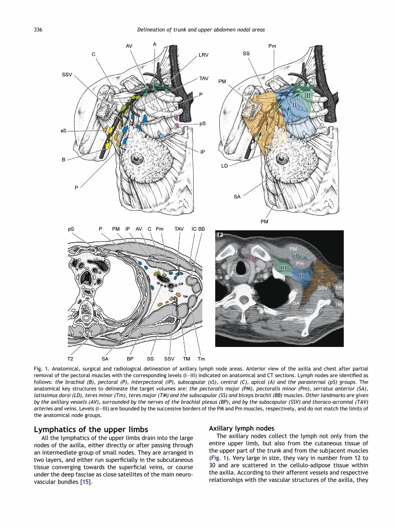
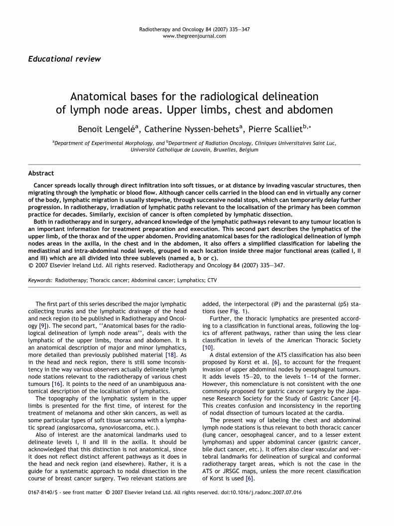
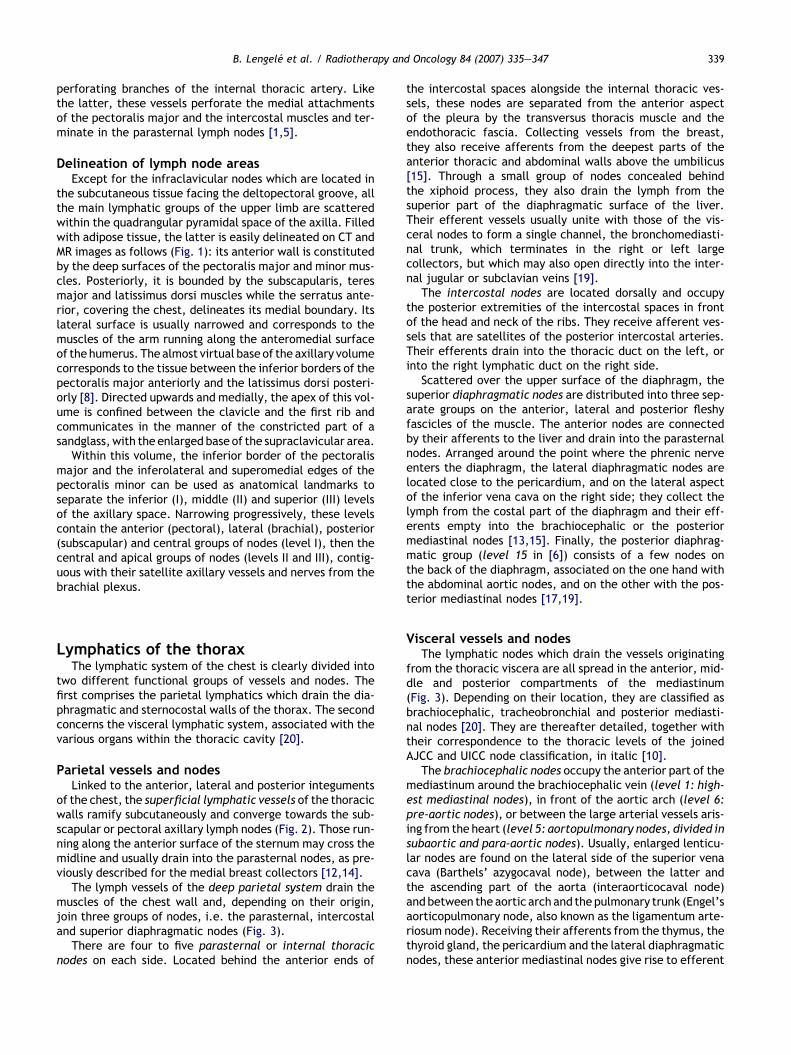
Fig. 1. Anatomical, surgical and radiological delineation of axillary lymph node areas. Anterior view of the axilla and chest after partialremoval of the pectoral muscles with the corresponding levels (I–III) indicated on anatomical and CT sections. Lymph nodes are identified asfollows: the brachial (B), pectoral (P), interpectoral (iP), subscapular (sS), central (C), apical (A) and the parasternal (pS) groups. Theanatomical key structures to delineate the target volumes are: the pectoralis major (PM), pectoralis minor (Pm), serratus anterior (SA),latissimus dorsi (LD), teres minor (Tm), teres major (TM) and the subscapular (SS) and biceps brachii (BB) muscles. Other landmarks are givenby the axillary vessels (AV), surrounded by the nerves of the brachial plexus (BP), and by the subscapular (SSV) and thoraco-acromial (TAV)arteries and veins. Levels (I–III) are bounded by the successive borders of the PM and Pm muscles, respectively, and do not match the limits ofthe anatomical node groups.
336 Delineation of trunk and upper abdomen nodal areas
Lymphatics of the upper limbsAll the lymphatics of the upper limbs drain into the large
nodes of the axilla, either directly or after passing throughan intermediate group of small nodes. They are arranged intwo layers, and either run superficially in the subcutaneoustissue converging towards the superficial veins, or courseunder the deep fasciae as close satellites of the main neuro-vascular bundles [15].
Axillary lymph nodesThe axillary nodes collect the lymph not only from the
entire upper limb, but also from the cutaneous tissue ofthe upper part of the trunk and from the subjacent muscles(Fig. 1). Very large in size, they vary in number from 12 to30 and are scattered in the cellulo-adipose tissue withinthe axilla. According to their afferent vessels and respectiverelationships with the vascular structures of the axilla, they
B. Lengele et al. / Radiotherapy and Oncology 84 (2007) 335–347 337
are divided into five groups which, however, are not clearlydelineated [12,14]:
The lateral or brachial group includes four to six nodessituated on the infero-medial side of the axillary vein.Their afferent vessels drain the lymph from the superfi-cial and deep compartments of upper limb, except forthe superficial vessels of the arm that run alongside thecephalic vein. Their efferents have a threefold termina-tion: most of them terminate in the central or apicalgroups, while others pass into the supraclavicular nodes[12].The anterior or pectoral group is composed of four tofive nodes located behind the pectoralis major muscleand along the lower border of the pectoralis minor.Forming a chain along and behind the lateral thoracicvessels, these nodes receive afferent vessels from theskin and muscles of the anterior and lateral walls ofthe trunk above the umbilicus. They also drain the lat-eral parts of the breast, and their efferent vesselsextend to the central and apical groups of axillarynodes [19].The posterior or subscapular group comprises six toseven nodes arranged above one another in a chain thatfollows the subscapular vessels, in the groove which sep-arates, on the posterior wall of the axilla, the teresmajor and subscapularis muscles. The afferent vesselsof this group collect the lymph arising from the musclesand skin of the back and from the scapular area down-wards to the iliac crest. Their efferent vessels drain intothe central and apical lymph nodes [14].The central group of axillary nodes usually containsthree to five extremely large nodes, located in the cen-tral part of the adipose tissue of the axilla between thepreceding chains which progressively converge towardsthem. Their efferent vessels then extend to the apicalgroup [20].The apical group contains six to 12 large lenticular nodeswhich occupy the apex of the axilla, behind the upperportion of the pectoralis minor and partly above thismuscle. The majority of these nodes rest on the infero-medial side of the proximal part of the axillary vein, inclose contact with the upper digitations of the serratusanterior. Receiving afferent vessels from all the otheraxillary nodes, they also drain some superficial vesselsrunning along the cephalic vein. The efferent vessels ofthis group unite to form the subclavian trunk whichfinally opens into the right lymphatic duct on the rightside, or into the thoracic duct on the left side [19].
ig. 2. Superficial lymphatic pathways of the upper limbs. Anteriornd posterior anatomical views showing the distribution of vesselsnd nodes of the upper limbs. Nodes are identified as follows: theupratrochlear (sT), infraclavicular (iC) and supraclavicular (sC)odes. Collecting vessels form three ascending drainage pathwayslassified as the medial brachial (B), central brachial (cB) andateral brachial (LB) pathways. Parietal vessels of the anterior andosterior chest walls run into the pectoral (P), parasternal (pS) andubscapular (sS) groups of nodes, respectively.
Superficial lymph nodesLocated on the surface of the deep fasciae, the superfi-
cial lymph nodes of the upper limbs are few in numberand are invariably located in the subcutaneous tissue[12,15]. Interposed on the superficial lymphatic pathways,they are known as the supratrochlear and infraclaviculargroups (Fig. 2).
The supratrochlear node is usually isolated and deeplyembedded in the subcutaneous fat, just over the deepfascia about 4–5 cm above the medial epicondyle of the
Faasnclps
humerus. Draining the superficial lymphatic pathwaysascending from the ulnar side of the forearm, it sends effer-
338 Delineation of trunk and upper abdomen nodal areas
ent vessels, which accompany the basilic vein to join thedeep subfascial vessels.
The infraclavicular nodes are one to two in number, andconsist of small interrupting nodes located near the cephalicvein in the deltopectoral groove. They are traversed by thesuperficial lymphatic vessels, which drain the lateral skin ofthe arm and shoulder. Their efferent vessels pierce the clav-ipectoral fascia immediately below the clavicle and termi-nate in the apical group of axillary nodes. Nevertheless,some other vessels cross the anterior border of the clavicleand finally reach the deep cervical nodes of the supraclavic-ular group [19].
Functional drainage pathwaysOn a functional level, the superficial and deep lympha-
tic pathways of the upper limbs are almost completely seg-regated by the deep fasciae. Nevertheless, they finallyconverge towards the axilla and communicate with eachother at both locations where the superficial vessels, run-ning alongside the basilic and cephalic veins, accompanythem through Morestin’s and Cruveilhier’s fascial foraminaand then join the deep perivascular lymphatic channels[12].
The superficial lymphatic pathways (Fig. 2) issue from allparts of the cutaneous layers of the upper limb and origi-nate in the hand from an extremely dense network withmaximal development on the palmar surface of the fingers.These digital plexuses are drained by small collecting ves-sels which first follow the corresponding collateral arterybut then incline backwards and pass into the dorsal aspectof the hand. The remainder of the palm is drained by vesselswhich course in front of the wrist and divide into medial,lateral and central small trunklets which then ascend to-wards the forearm along its ulnar, radial and palmar aspects[14]. On the posterior surface of the forearm, the dorsalantebrachial channels pass progressively around the medialand lateral borders of the limb to join the vessels thatcourse in front of the elbow. As they run upwards, thesechannels gradually decrease in number, and finally separateinto three distinct superficial brachial pathways [8]:
The medial brachial collecting vessels follow the basilicvein, and some of them pass through the supratrochlearnodes above the elbow. Thereafter they perforate thefascia with the vein, join the deep collectors, and endin the brachial group of axillary nodes.The central brachial collecting vessels run longitudinallyover the fascial sheath of the biceps brachii, pierce theaxillary fascia along the anterior axillary fold, and termi-nate in the brachial nodes.The lateral brachial collecting vessels are associatedwith the cephalic vein and continue their course on thelateral side of the biceps brachii until they reach the del-topectoral groove. At this point most of them empty intothe brachial group of axillary nodes. A few transit by theinfraclavicular nodes which also receive lateral afferentsfrom the deltoid area. Efferents from these nodes end aspreviously stated in the apical axillary nodes and some-times in the cervical supraclavicular node [17], adjacentto region IV of the head and neck region [3].
During their course, the longitudinal superficial vesselsundergo many divisions which sometimes diverge and some-times converge, thereby creating several connections be-tween the dorsal, central, lateral and medial pathwaysorganised around the arm and forearm. Because of the largenumber of randomly distributed anastomosing channels, thepattern of lymphatic spread of a cutaneous tumour such as amelanoma is difficult to predict: for instance, the dissemi-nation of a dorsomedial melanoma of the hand can first in-volve the palmar supratrochlear node, then the brachialgroup of the axilla [8]. The potential however exists thatif the dorsal collecting trunklets incline more likely aroundthe lateral border of the forearm, there is a possibility ofprimary metastatic nodes being present in the infraclavicu-lar group and then immediately in the apex of the axilla.
The deep lymphatic pathways of the upper limb compriselarge collectors which are few in number and relatively lessanastomosing than the superficial channels. Running aroundthe axial vessels, they form radial, ulnar, interosseous andbrachial ascending pathways which drain into the lateralbrachial nodes of the axilla. Along their course small nodescan be found. Within the axilla, the main lymphatic chan-nels arising from the lateral, anterior and posterior nodessuccessively pass through the central and apical groups ofnodes. During their course along the axillary vein, theysequentially cross three topographical segments [1,7] lo-cated, respectively, behind the lower part of the pectoralismajor (level I), behind the pectoralis minor (level II), and fi-nally above the upper border of the pectoralis minor in thesubclavicular triangle (level III), adjacent to level IV of thehead and neck region.
Closely linked to the lymphatic pathways of the upperlimb, the lymphatic vessels of the breast are mainly directedtoward the axilla (Fig. 1). Originating from a dense plexus inthe interlobular connective tissue of the breast they commu-nicate with the overlying subcutaneous network, especiallyaround the nipple, giving rise to a subareolar circular plexus[15]. The latter is drained by two or three main collectorswhich turn around the inferior border of the pectoralis majorand which become satellites of the lateral thoracic vessels[2,7,17]. Behind the muscle the principal lymphatic pathwayof the breast thus reaches the anterior pectoral group of ax-illary nodes. Nevertheless, three alternative drainage path-ways also exist, explaining the other primary locations ofmetastatic lymph nodes observed in breast cancer.
The first accessory route is constituted by direct lympha-tic vessels of the inferolateral part of the breast whichadopt a more dorsal route and join the posterior subscapularnodes of the axilla [7].
The second route involves lymphatic channels which arisefrom the upper parts of the gland and tend to follow the cuta-neous branches of the thoraco-acromial artery. Most of thesevessels pass through the fascicles of the pectoralis major anddrain immediately into the apical axillary nodes. Betweenthe pectoralis major and minor muscles, some of these ves-sels are interrupted by a large inconstant interpectoral node,usually known as Rotter’s node [5,7]. Others encounter theinfraclavicular and supraclavicular nodes.
The third alternative pathway is directed medially andcomprises several channels running alongside the cutaneous
B. Lengele et al. / Radiotherapy and Oncology 84 (2007) 335–347 339
perforating branches of the internal thoracic artery. Likethe latter, these vessels perforate the medial attachmentsof the pectoralis major and the intercostal muscles and ter-minate in the parasternal lymph nodes [1,5].
Delineation of lymph node areasExcept for the infraclavicular nodes which are located in
the subcutaneous tissue facing the deltopectoral groove, allthe main lymphatic groups of the upper limb are scatteredwithin the quadrangular pyramidal space of the axilla. Filledwith adipose tissue, the latter is easily delineated on CT andMR images as follows (Fig. 1): its anterior wall is constitutedby the deep surfaces of the pectoralis major and minor mus-cles. Posteriorly, it is bounded by the subscapularis, teresmajor and latissimus dorsi muscles while the serratus ante-rior, covering the chest, delineates its medial boundary. Itslateral surface is usually narrowed and corresponds to themuscles of the arm running along the anteromedial surfaceof the humerus. The almost virtual base of the axillary volumecorresponds to the tissue between the inferior borders of thepectoralis major anteriorly and the latissimus dorsi posteri-orly [8]. Directed upwards and medially, the apex of this vol-ume is confined between the clavicle and the first rib andcommunicates in the manner of the constricted part of asandglass, with the enlarged base of the supraclavicular area.
Within this volume, the inferior border of the pectoralismajor and the inferolateral and superomedial edges of thepectoralis minor can be used as anatomical landmarks toseparate the inferior (I), middle (II) and superior (III) levelsof the axillary space. Narrowing progressively, these levelscontain the anterior (pectoral), lateral (brachial), posterior(subscapular) and central groups of nodes (level I), then thecentral and apical groups of nodes (levels II and III), contig-uous with their satellite axillary vessels and nerves from thebrachial plexus.
Lymphatics of the thoraxThe lymphatic system of the chest is clearly divided into
two different functional groups of vessels and nodes. Thefirst comprises the parietal lymphatics which drain the dia-phragmatic and sternocostal walls of the thorax. The secondconcerns the visceral lymphatic system, associated with thevarious organs within the thoracic cavity [20].
Parietal vessels and nodesLinked to the anterior, lateral and posterior integuments
of the chest, the superficial lymphatic vessels of the thoracicwalls ramify subcutaneously and converge towards the sub-scapular or pectoral axillary lymph nodes (Fig. 2). Those run-ning along the anterior surface of the sternum may cross themidline and usually drain into the parasternal nodes, as pre-viously described for the medial breast collectors [12,14].
The lymph vessels of the deep parietal system drain themuscles of the chest wall and, depending on their origin,join three groups of nodes, i.e. the parasternal, intercostaland superior diaphragmatic nodes (Fig. 3).
There are four to five parasternal or internal thoracicnodes on each side. Located behind the anterior ends of
the intercostal spaces alongside the internal thoracic ves-sels, these nodes are separated from the anterior aspectof the pleura by the transversus thoracis muscle and theendothoracic fascia. Collecting vessels from the breast,they also receive afferents from the deepest parts of theanterior thoracic and abdominal walls above the umbilicus[15]. Through a small group of nodes concealed behindthe xiphoid process, they also drain the lymph from thesuperior part of the diaphragmatic surface of the liver.Their efferent vessels usually unite with those of the vis-ceral nodes to form a single channel, the bronchomediasti-nal trunk, which terminates in the right or left largecollectors, but which may also open directly into the inter-nal jugular or subclavian veins [19].
The intercostal nodes are located dorsally and occupythe posterior extremities of the intercostal spaces in frontof the head and neck of the ribs. They receive afferent ves-sels that are satellites of the posterior intercostal arteries.Their efferents drain into the thoracic duct on the left, orinto the right lymphatic duct on the right side.
Scattered over the upper surface of the diaphragm, thesuperior diaphragmatic nodes are distributed into three sep-arate groups on the anterior, lateral and posterior fleshyfascicles of the muscle. The anterior nodes are connectedby their afferents to the liver and drain into the parasternalnodes. Arranged around the point where the phrenic nerveenters the diaphragm, the lateral diaphragmatic nodes arelocated close to the pericardium, and on the lateral aspectof the inferior vena cava on the right side; they collect thelymph from the costal part of the diaphragm and their eff-erents empty into the brachiocephalic or the posteriormediastinal nodes [13,15]. Finally, the posterior diaphrag-matic group (level 15 in [6]) consists of a few nodes onthe back of the diaphragm, associated on the one hand withthe abdominal aortic nodes, and on the other with the pos-terior mediastinal nodes [17,19].
Visceral vessels and nodesThe lymphatic nodes which drain the vessels originating
from the thoracic viscera are all spread in the anterior, mid-dle and posterior compartments of the mediastinum(Fig. 3). Depending on their location, they are classified asbrachiocephalic, tracheobronchial and posterior mediasti-nal nodes [20]. They are thereafter detailed, together withtheir correspondence to the thoracic levels of the joinedAJCC and UICC node classification, in italic [10].
The brachiocephalic nodes occupy the anterior part of themediastinum around the brachiocephalic vein (level 1: high-est mediastinal nodes), in front of the aortic arch (level 6:pre-aortic nodes), or between the large arterial vessels aris-ing from the heart (level 5: aortopulmonary nodes, divided insubaortic and para-aortic nodes). Usually, enlarged lenticu-lar nodes are found on the lateral side of the superior venacava (Barthels’ azygocaval node), between the latter andthe ascending part of the aorta (interaorticocaval node)and between the aortic arch and thepulmonary trunk (Engel’saorticopulmonary node, also known as the ligamentum arte-riosum node). Receiving their afferents from the thymus, thethyroid gland, the pericardium and the lateral diaphragmaticnodes, these anterior mediastinal nodes give rise to efferent
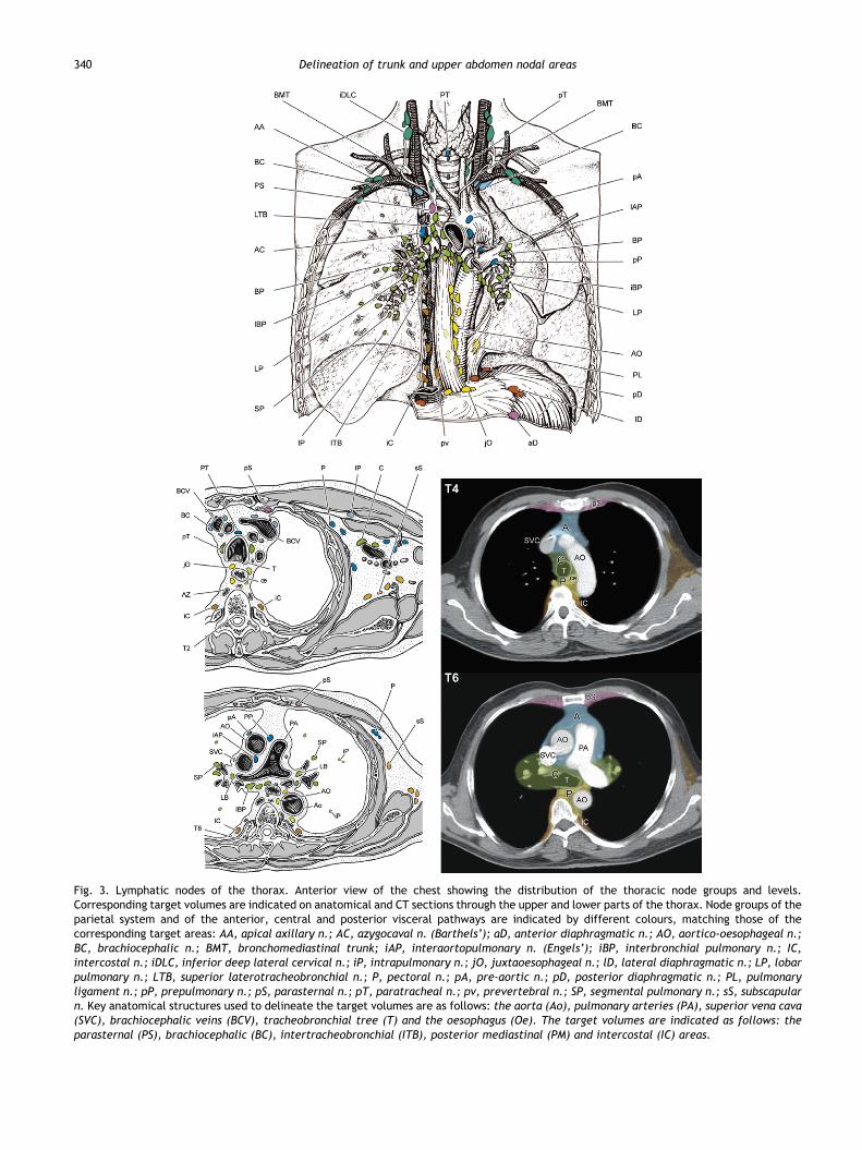
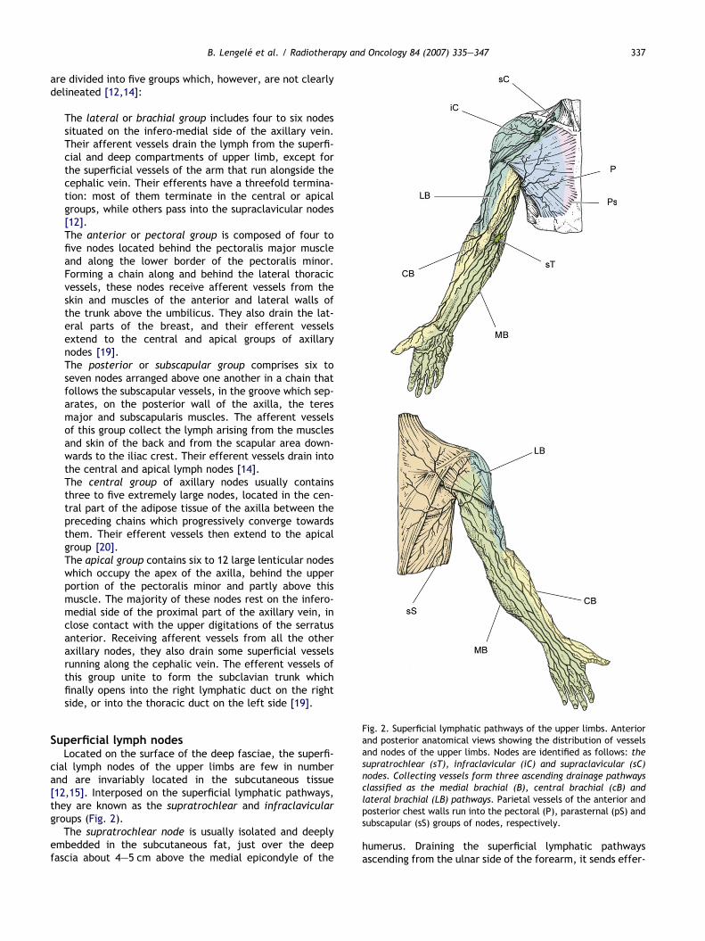
Fig. 3. Lymphatic nodes of the thorax. Anterior view of the chest showing the distribution of the thoracic node groups and levels.Corresponding target volumes are indicated on anatomical and CT sections through the upper and lower parts of the thorax. Node groups of theparietal system and of the anterior, central and posterior visceral pathways are indicated by different colours, matching those of thecorresponding target areas: AA, apical axillary n.; AC, azygocaval n. (Barthels’); aD, anterior diaphragmatic n.; AO, aortico-oesophageal n.;BC, brachiocephalic n.; BMT, bronchomediastinal trunk; iAP, interaortopulmonary n. (Engels’); iBP, interbronchial pulmonary n.; IC,intercostal n.; iDLC, inferior deep lateral cervical n.; iP, intrapulmonary n.; jO, juxtaoesophageal n.; lD, lateral diaphragmatic n.; LP, lobarpulmonary n.; LTB, superior laterotracheobronchial n.; P, pectoral n.; pA, pre-aortic n.; pD, posterior diaphragmatic n.; PL, pulmonaryligament n.; pP, prepulmonary n.; pS, parasternal n.; pT, paratracheal n.; pv, prevertebral n.; SP, segmental pulmonary n.; sS, subscapularn. Key anatomical structures used to delineate the target volumes are as follows: the aorta (Ao), pulmonary arteries (PA), superior vena cava(SVC), brachiocephalic veins (BCV), tracheobronchial tree (T) and the oesophagus (Oe). The target volumes are indicated as follows: theparasternal (PS), brachiocephalic (BC), intertracheobronchial (ITB), posterior mediastinal (PM) and intercostal (IC) areas.
340 Delineation of trunk and upper abdomen nodal areas
B. Lengele et al. / Radiotherapy and Oncology 84 (2007) 335–347 341
ducts which unite with those from the tracheobronchialnodes to form the bronchomediastinal trunk [2,19].
The tracheobronchial nodes are concentrated around thetracheal bifurcation and include five different groups whichfrequently contain the largest nodes of the body: the para-tracheal nodes (level 2: upper paratracheal nodes) on thelateral sides of the trachea; the superior (latero) tracheo-bronchial nodes (level 4: lower paratracheal, includingazygocaval nodes), situated in the angles between the tra-chea and the right and left main bronchi; the inferior(inter)tracheobronchial nodes (level 7: subcarinal nodes),located below the carina between the two main bronchialstems, the bronchopulmonary nodes (level 10: hilar nodes)in the hilum of each lung, and the intrapulmonary nodes(levels 11–14: interlobar, lobar, segmental, and subseg-mental nodes), scattered within the central lung substanceinside the divisions of the lobar, segmental and subsegmen-tal branches of the bronchial tree [9,19]. The afferent ves-sels of the tracheobronchial nodes drain the lymph from thesuperficial (subpleural) and deep (peribronchial) networksof the lung, and also from the thoracic part of the tracheaand the heart [2,14]. Together with those of the brachioce-phalic group their efferents constitute the right and leftbronchomediastinal trunks [19].
The posterior mediastinal nodes are spread within theposterior mediastinal fat, behind the trachea (level 3: ret-rotracheal nodes) then behind the pericardium, along thepulmonary ligament (level 9: pulmonary ligament nodes),and around the aorta and the oesophagus (level 8: perio-esophageal nodes, located below carina and often dividedas middle – level 8M – and lower – level 8L – paraoesoph-ageal nodes). Receiving afferent vessels from the posteriorpart of the pericardium, the oesophagus and the posteriordiaphragmatic nodes, they send efferents which mostly ter-minate in the thoracic duct, although some may end in thetracheobronchial chain [2,13,17].
Functional drainage pathwaysOf immediate interest in understanding the way in which
intrathoracic tumours are disseminated, the lymph nodes ofthe chest and their connecting vessels are arranged in threemain ascending streams [8]:
The anterior stream is located in the anterior mediasti-num. Including the xiphoid, parasternal and brachioce-phalic nodes, it may become invaded as a result of thedissemination of breast, thyroid, or thymic tumours.The central stream occupies the middle part of themediastinum. Linked inferiorly to the lateral diaphrag-matic nodes, it is formed by the successive subgroupsof tracheobronchial nodes. Immediately adjacent tothe respiratory tree, this main drainage pathway of thethoracic viscera represents the usual route of dissemina-tion for lung cancers, but also for malignant tumours ofthe oesophagus.The posterior stream runs behind the heart, in the nar-row fatty space of the posterior mediastinum. Groupingthe posterior diaphragmatic nodes and the posteriormediastinal nodes arranged in a continuous chain aroundthe aorta and the oesophagus, it mostly ends in the tho-
racic duct. Its metastatic involvement is usually observedin cases of oesophageal cancer.
Although each represents an individual preferential path-way of lymphatic dispersion, the anterior, central and pos-terior mediastinal chains described herein are notcompletely separated from one another [2,8]. In the supe-rior mediastinum, the terminal ducts of the anterior andcentral chains unite to give rise to an ascending commonbronchomediastinal trunk (joining the low end of level IVof the head and neck region), while the posterior chain re-mains isolated, mainly connected to the thoracic duct[14,17]. On the contrary, the posterior and central path-ways may encounter one another inferiorly since the lowerperioesophageal nodes send divergent efferent vessels thatend either in the inferior tracheobronchial nodes or in thethoracic duct [15,19]. Finally, it should be noted that bothanterior and posterior pathways are connected by transdia-phragmatic channels with the parietal lymphatics of theperitoneal cavity: they may thus become involved in thespread of intra-abdominal tumours [17]. Along the loweroesophagus, in the direction of the cardia, lymph nodes (le-vel 8L: lower paraoesophageal) may drain either craniallyalong the posterior mediastinal stream following the tho-racic part of the oesophagus, or caudally, in the directionof the abdomen, through the paracardial nodes (level 16)and left gastric nodes (level 17), readily reaching the coe-liac nodes (level 20) and the thoracic duct [6]. The lympha-tic vessels draining the lower oesophagus give thus rise to ahighly anastomotic network, linked cranially to the multiplenode stations of the posterior mediastinal chain but alsoshowing an alternative route with less resistivity in thedirection of the thoracic duct through a shorter descendingabdominal pathway. This fact presumably explains why in-tra-abdominal nodes along the lesser curvature are so easilyinvaded by oesophageal cancer.
Delineation of lymph node areasOn transverse sections of the chest, the lymphatic target
volumes can be delineated according to the following ana-tomical landmarks (Fig. 3).
The parasternal lymphatic area is a lenticular laminarplane bounded anteriorly by the deep surface of the ster-num and anterior intercostal spaces and dorsally by thetransversus thoracis muscle. It extends from the xiphoidprocess upwards to the sternoclavicular joints with theinternal thoracic artery as a central anatomical landmark.
The brachiocephalic lymphatic area (anterior area I)occupies the anterior mediastinal fat, in front of the largesupracardiac vessels. Anteriorly facing the posterior aspectof the sternum, this volume is bounded laterally by theanterior parts of the right and left mediastinal pleurae. Cau-dally it disappears nearly below the level of the sixth tho-racic vertebra and cranially it communicates through thethoracic inlet, alongside the carotid arteries and internaljugular veins, with the lower part of the cervical jugulocar-otid areas. Conventionally, the inferior edge of clavicle canbe used as landmark to trace the limit between these intra-thoracic and deep cervical areas. Taking the brachiocephalicvein, the aortic arch and the pulmonary trunk as
342 Delineation of trunk and upper abdomen nodal areas
successive anatomical landmarks, this volume can be di-vided into supra-aortic (area Ia), pre-aortic (area Ib) andsubaortic (area Ic) stages which, respectively, contain theupper (level 1: upper mediastinal), middle (level 6: pre-aortic) and lower (level 5: aortopulmonary) anterior medi-astinal lymph node groups [8]. The level 1 for thoracic stag-ing is adjacent to the level VI of the head and neck region.However, these levels are not connected, as they both draindirectly into the central venous system (see part I of thepresent article).
The peritracheobronchial area (central area II) is cen-tered around the thoracic trachea and the main bronchi. Itsanterior and posterior limits can be defined as running,respectively, along the posterior aspect of the superior venacava and aortic arch anteriorly and along the ventral aspectof the oesophagus posteriorly. Delineated by the middleparts of the mediastinal pleurae, its lateral boundaries in-clude the right and left pulmonary hila which enter into bothlungs [8]. Inferiorly, this area does not extend below the levelof the sixth thoracic vertebra, but includes all the nodegroups associated with the trachea (level 2: upper paratrac-heal, level 4: lower paratracheal and level 7: subcarinall)and with the origin of the bronchial tree (level 10: tracheo-bronchial or hilar). Using the aortic arch, the tracheal bifur-cation as successive boundary landmarks, it may be divedinto three successive stages containing the upper (area IIa),middle (area IIb) and lower (area IIc) peritracheobronchiallymph node stations. Outside the mediastinal fatty spaceand inside the parenchyma of each lung, this area spreadsinto the lateral pulmonary volumes which include two addi-tional right and left pulmonary areas (intrapulmonary areasIVR and IVL), containing the multiple successive lymph nodestations scattered inside the lungs (levels 11: interlobar, 12:lobar, 13: segmental, and 14: subsegmental).
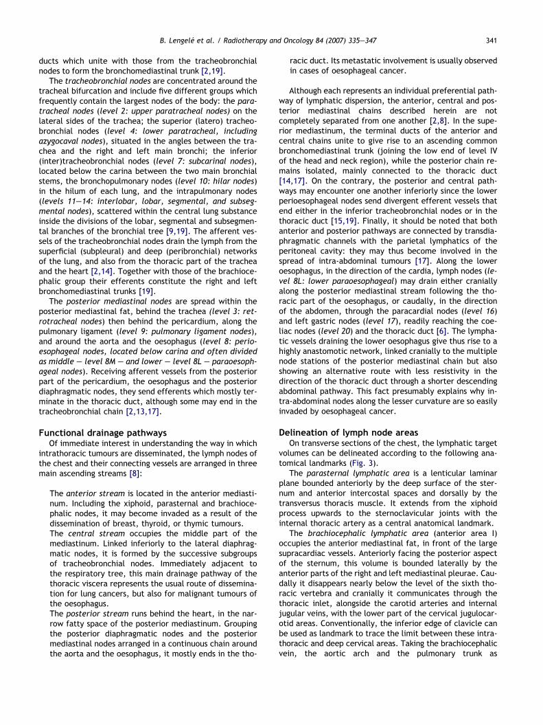
Table 1Visceral node groups of the chest, with corresponding surgical levels an
Node groups AJCC levels Area
Anterior medistinal area (brachiocephalic, I)Brachiocephalic (supra-aortic) Level 1 IaPre-aortic Level 5 IbAortopulmonary (sub-para aortic) Level 6 Ic
Central mediastinal area (peritracheobronchic, II)Upper paratracheal Level 2 IIaLower paratracheal (laterotracheobronchic) Level 4 IIb
Subcarinal (intertracheobronchic) Level 7 IIcPulmonary hilar (bronchopulmonary) Level 10 IIc
Posterior mediastinal area (perioesophageal, III)Retrotracheal (upper post. Mediastinum) Level 3 IIIaMiddle paraoesophageal Level 8M IIIbLower paraoesophageal Level 8L IIIcPulmonary ligament Level 9 IIIcDiaphragmatic Level 15 IIIc
Lateral pulmonary areas (right and left intrapulmonary)Interlobar Level 11 IVaLobar Level 12 IVbSegmental Level 13 IVcSubsegmental Level 14 IVd
The posterior mediastinal area is a narrow fatty channellocated behind a plane running along the posterior aspect ofthe heart caudally and along the anterior surface of theoesophagus cranially. It is bounded by the ventral aspectof the vertebral column posteriorly, and laterally by thedorsal part of the mediastinal pleurae. This volume containsthe thoracic oesophagus, the descending aorta, both azygosand hemi-azygos veins and between these landmarks, thethoracic duct and the posterior mediastinal nodes (level 3:retrotracheal, levels 8M and 8L: middle and lower parao-esophageal, levels 9 of the pulmonary ligaments and level15: diaphragmatic). Using the tracheal bifurcation and thepulmonary hila as successive boundary landmarks, it maybe divided into three successive stages containing the upper(area IIIa), middle (area IIIb) and lower (area IIIc) perio-esophageal lymph node stations. Posterolateral extensionson both sides of the vertebrae up to the costal angles allowit to also include the posterior intercostal node groups [8],located along the proximal course of the posterior intercos-tals vessels and on the medial aspect of the thoracic sympa-thetic nerve chains.
Table 1 summarizes the here described simplified func-tional delineation of the three major chest visceral lymphnode areas (I–II–III), emphasizing their critical anatomicallandmarks and correlating their successive stages (a, b andc) with the corresponding levels listed in the AJCC and UICCnode classifications.
Lymphatics of the abdomenFollowing the same arrangement as that in the thorax,
the lymphatics of the abdomen are divided into parietaland visceral vessels and nodes, and, respectively, drain
d target areas for conformal radiotherapy (nodal CTV)
Location Anatomical landmarks
T1–T2 Along brachiocephalic veinsT3 In front of aortic archT4 Around ascending aorta and
pulmonary arteries
T1–T2 Along trachea, above aortic archT3 Along trachea, behind aortic arch (L)
and below azygos arch (R)T4 Around tracheal bifurcationT5–T6 Along main bronchus at hilum
T1–T4 Behind tracheaT5–T6 Below carina, facing pulmonary hilumT7–T11 Below pulmonary hilumT7–T9 Below hilum, facing left atriumT10–T11 Behind vertical part of diaphragm
– Long main bronchus, in lung– Central position in lung, along lobar bronchi– Medial position in lung, along segmental bronchi– Peripheral position in lung, along distal airways
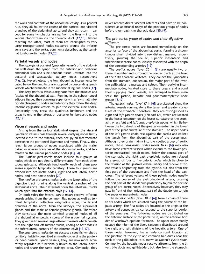
B. Lengele et al. / Radiotherapy and Oncology 84 (2007) 335–347 343
the walls and contents of the abdominal cavity. As a generalrule, they all follow the course of the parietal and visceralbranches of the abdominal aorta and they all return – ex-cept for some lymphatics arising from the liver – into thevenous bloodstream via the thoracic duct [12,15]. Beforereaching the latter, most of them are interrupted by verylarge retroperitoneal nodes scattered around the inferiorvena cava and the aorta, commonly described as the termi-nal lumbo-aortic nodes [19,20].
Parietal vessels and nodesThe superficial parietal lymphatic vessels of the abdomi-
nal wall drain the lymph from the anterior and posteriorabdominal skin and subcutaneous tissue upwards into thepectoral and subscapular axillary nodes, respectively(Fig. 2). Nevertheless, the low abdominal integuments lo-cated below the umbilicus are supplied by descending lymphvessels which terminate in the superficial inguinal nodes [17].
The deep parietal vessels originate from the muscles andfasciae of the abdominal wall. Running in the subperitonealadipose tissue, they converge superiorly in a few small infe-rior diaphragmatic nodes and inferiorly they follow the deepinferior epigastric vessels to join the external iliac nodes.Posteriorly, they cross the quadratus lumborum and thepsoas to end in the lateral or posterior lumbo-aortic nodes[12,19].
Visceral vessels and nodesArising from the various abdominal organs, the visceral
lymphatic vessels pass through several outlying nodes firstlylocated close to the viscera, then occupy an intermediateposition in the peritoneal ligaments and mesos. Finally, theyreach larger groups of nodes associated with the majorpaired or uneven branches of the abdominal aorta, and ter-minate in the lumbar peri-aortic nodes (Fig. 4).
The lumbar peri-aortic nodes include four groups ofnodes which are not clearly differentiated from each othertopographically, although functionally each of them pos-sesses a specific lymphatic territory. These four groups aredivided into pre-aortic nodes, right and left lateral aorticnodes, and post-aortic nodes [20].
The median pre-aortic nodes drain the lymphatics of thedigestive tract running along the ventral branches of theabdominal aorta. Their efferents form the intestinal trunkswhich open into the cisterna chyli [12,14].
On both sides the lateral aortic nodes receive efferentvessels arising from the common iliac nodes as well as ter-minal lymphatic collectors originating along the lateralbranches of the aorta, from the kidneys, the suprarenalglands and the male or female gonadic glands. Thereforethey constitute the main terminal group of nodes of allthe abdominal or pelvic viscera of the urogenital system.They give rise to several large efferent vessels which consti-tute the right and left lumbar trunks, ending on both sides inthe inferolateral corners of the cisterna chyli [12,17].
The post-aortic nodes do not possess a specific lymphaticterritory. Initially described as mostly collecting the poster-ior deep parietal lymph vessels, they are now more accu-rately regarded as functionally linked to the lateral aorticnodes and share the same drainage area. Obviously, they
never receive direct visceral afferents and have to be con-sidered as additional relays of the previous groups of nodesbefore they reach the thoracic duct [15,19].
The pre-aortic group of nodes and their digestiveaffluents
The pre-aortic nodes are located immediately on theanterior surface of the abdominal aorta, forming a discon-tinuous chain divided into three distinct masses, respec-tively, grouping the coeliac, superior mesenteric andinferior mesenteric nodes, closely associated with the originof the corresponding arteries [19].
The coeliac nodes (level 20 in [6]) are usually two orthree in number and surround the coeliac trunk at the levelof the 12th thoracic vertebra. They collect the lymphaticsfrom the stomach, duodenum, the major part of the liver,the gallbladder, pancreas and spleen. Their outlying inter-mediate nodes, located close to these organs and aroundtheir supplying blood vessels, are arranged in three mainsets: the gastric, hepatic and pancreaticosplenic nodegroups [8,17].
The gastric nodes (level 17 in [6]) are situated along thearterial vessels running along the lesser and greater curva-tures of the stomach. Therefore, they are distinguished asright and left gastric nodes (17R and 17L) which are locatedin the lesser omentum on the lesser curvature of the stom-ach, or as right and left gastro-epiploic nodes, which lie be-tween the two sheaths of the greater omentum in the lowerpart of the great curvature of the stomach. The upper nodesof the left gastric chain rest against the cardia and collectthe lymph from the abdominal part of the oesophagus.Although they drain mainly into the coeliac group of aorticnodes, these paracardial nodes (level 16 in [6]) may alsohave some efferent vessels which extend to the lower pos-terior mediastinal lymph nodes. Below the central part ofthe stomach, the right gastro-epiploic nodes are relayedby a group of four to five pyloric nodes which lie close tothe division of the gastroduodenal artery and receive affer-ent vessels originating from the pylorus but also from thefirst part of the duodenum and from the head of the pan-creas. The efferent vessels of these pyloric nodes usuallyfollow the course of the gastroduodenal artery, crossingthe first part of the duodenum posteriorly to join the coeliacgroup of pre-aortic nodes. Alternatively however, they maypass in front of the horizontal part of the duodenum to jointhe superior mesenteric nodes.
The hepatic nodes (level 18 in [6]) form a chain of threeto six nodes which are situated along the course of the he-patic artery. The first nodes are located at the origin of theartery and consequently correspond to the superior borderof the pancreas. The following nodes are distributed onthe anterior surface of the portal vein, on the anterior bor-der of Winslow’s epiploic foramen. The upper nodes finallyoccupy the hilum of the liver, randomly distributed aroundthe right and left divisions of the hepatic artery. One ofthese nodes, however, has a fairly constant location atthe junction of the cystic and common bile ducts near theneck of the gallbladder: it is known as Quenu’s cystic node.Commonly, the hepatic nodes receive afferents from the li-ver, bile ducts and gallbladder, but also from the stomach,
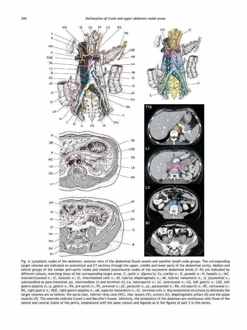
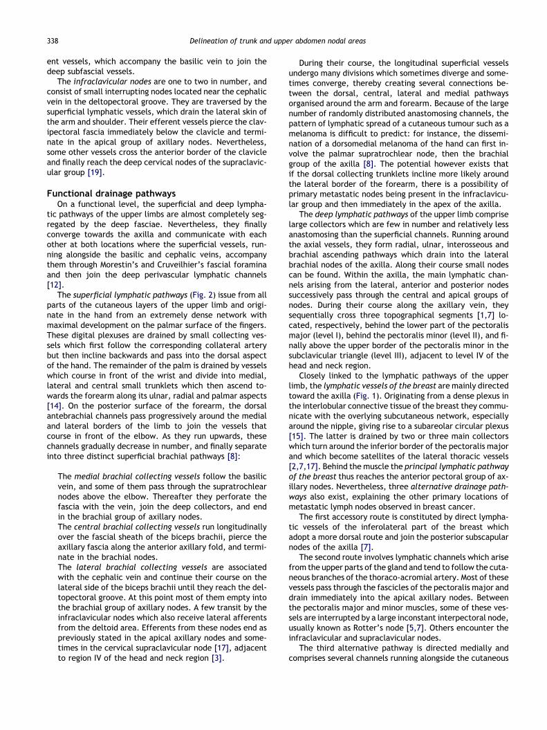
Fig. 4. Lymphatic nodes of the abdomen. Anterior view of the abdominal blood vessels and satellite lymph node groups. The correspondingtarget volumes are indicated on anatomical and CT sections through the upper, middle and lower parts of the abdominal cavity. Median andlateral groups of the lumbar peri-aortic nodes and related juxtavisceral nodes of the successive abdominal levels (I–III) are indicated bydifferent colours, matching those of the corresponding target areas. C, cystic n. (Quenu’s); Co, coeliac n.; G, gonadic n.; H, hepatic n.; IAC,interaorticocaval n.; IC, ileocolic n.; iC, intermediate colic n.; iD, inferior diaphragmatic n.; iM, inferior mesenteric n.; Ji, jejunoileal n.;subclassified as para-intestinal (p), intermediate (i) and terminal (t); LA, lateroaortic n.; LC, laterocaval n.; LG, left gastric n.; LGE, leftgastro-epiploic n.; p, pyloric n.; PA, pre-aortic n.; PC, precaval n.; pC, paracolic n.; pc, paracardial n.; RA, retroaortic n.; RC, retrocaval n.;RG, right gastric n.; RGE, right gastro-epiploic n.; sM, superior mesenteric n.; tC, terminal colic n. Key anatomical structures to delineate thetarget volumes are as follows: the aorta (Ao), inferior vena cava (IVC), iliac vessels (IV), ureters (U), diaphragmatic pillars (D) and the psoasmuscles (P). The asterisks indicate Cuneo’s and Marcille’s fossae. Inferiorly, the lymphatics of the abdomen are continuous with those of thelateral and central chains of the pelvis, emphasized with the same colours and legends as in the figures of part 3 in this series.
344 Delineation of trunk and upper abdomen nodal areas
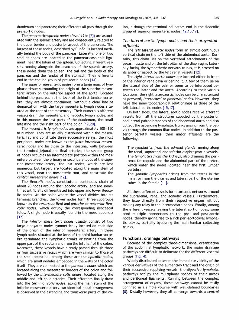
B. Lengele et al. / Radiotherapy and Oncology 84 (2007) 335–347 345
duodenum and pancreas; their efferents all pass through thepre-aortic nodes.
The pancreaticosplenic nodes (level 19 in [6]) are associ-ated with the splenic artery and are consequently related tothe upper border and posterior aspect of the pancreas. Thelargest of these nodes, described by Cuneo, is located medi-ally behind the body of the pancreas. Laterally, one or twosmaller nodes are located in the pancreaticosplenic liga-ment, near the hilum of the spleen. Collecting afferent ves-sels running alongside the branches of the splenic artery,these nodes drain the spleen, the tail and the body of thepancreas and the fundus of the stomach. Their efferentsend in the coeliac group of pre-aortic nodes [14].
The superior mesenteric nodes form a large mass of lym-phatic tissue surrounding the origin of the superior mesen-teric artery on the anterior aspect of the aorta. Locatedbehind the pancreas at the level of the first lumbar verte-bra, they are almost continuous, without a clear line ofdemarcation, with the large mesenteric lymph nodes situ-ated at the root of the mesentery. Their numerous afferentvessels drain the mesenteric and ileocolic lymph nodes, andin this manner the last parts of the duodenum, the smallintestine and the right part of the colon [17,20].
The mesenteric lymph nodes are approximately 100–150in number. They are usually distributed within the mesen-teric fat and constitute three successive relays: the mostperipheral nodes are known as the juxta-intestinal mesen-teric nodes and lie close to the intestinal walls betweenthe terminal jejunal and ileal arteries; the second groupof nodes occupies an intermediate position within the mes-entery between the primary or secondary loops of the supe-rior mesenteric artery; the last nodes, which are lessnumerous but larger, are located along the main stem ofthis vessel, near the mesenteric root, and constitute thecentral mesenteric nodes [12].
The ileocolic nodes constitute a continuous chain ofabout 20 nodes around the ileocolic artery, and are some-times artificially differentiated into upper and lower ileoco-lic nodes. At the point where the vessel divides into itsterminal branches, the lower nodes form three subgroupsknown as the recurrent ileal and anterior or posterior ileo-colic nodes, which occupy the corresponding ileocaecalfolds. A single node is usually found in the meso-appendix[12].
The inferior mesenteric nodes usually consist of twolarge elongated nodes symmetrically located on each sideof the origin of the inferior mesenteric artery. In theselymph nodes situated at the level of the third lumbar verte-bra terminate the lymphatic trunks originating from theupper part of the rectum and from the left half of the colon.Moreover, these vessels have already passed through threeor four successive relays which are very similar to those ofthe small intestine: among these are the epicolic nodes,which are small nodules embedded in the walls of the colonitself. They are connected to the paracolic nodes which arelocated along the mesenteric borders of the colon and fol-lowed by the intermediate colic nodes, located along themiddle and left colic arteries. Their efferents finally draininto the terminal colic nodes, along the main stem of theinferior mesenteric artery. An identical nodal arrangementis observed in the ascending and transverse parts of the co-
lon, although the terminal collectors end in the ileocolicgroup of superior mesenteric nodes [12,15,17].
The lateral aortic lymph nodes and their urogenitalaffluents
The left lateral aortic nodes form an almost continuousvertical chain on the left side of the abdominal aorta. Dor-sally, this chain lies on the vertebral attachments of thepsoas muscle and on the left pillar of the diaphragm. Later-ally facing the sympathetic nervous trunks, it is crossed onits anterior aspect by the left renal vessels [12].
The right lateral aortic nodes are located either in frontof the inferior vena cava or behind it. A few of them lie onthe lateral side of the vein or seem to be interposed be-tween the latter and the aorta. According to their variouslocations, the right lateroaortic nodes can be characterizedas precaval, laterocaval or postcaval nodes. However, theyhave the same topographical relationships as those of theleft lateral aortic nodes [15,17].
On both sides, the lateral aortic nodes receive afferentvessels from all the structures supplied by the posteriorand lateral paired branches of the abdominal aorta and alsocollect the ascending lymphatic trunks arising from the pel-vis through the common iliac nodes. In addition to the pos-terior parietal vessels, their major affluents are thefollowing:
The lymphatics from the adrenal glands running alongthe renal, suprarenal and inferior diaphragmatic vessels.The lymphatics from the kidneys, also draining the peri-renal fat capsule and the abdominal part of the ureter,which enter the nodes located near the first lumbarvertebra.The gonadic lymphatics arising from the testes in themale, or from the ovaries and lateral part of the uterinetubes in the female [11].
All these afferent vessels form tortuous networks aroundthe suprarenal, renal and gonadic vessels. Furthermore,they issue directly from their respective organs withoutmaking any relay in the intermediate nodes. Finally, amongthe efferent vessels leaving the lateral aortic nodes, somesend multiple connections to the pre- and post-aorticnodes, thereby giving rise to a rich peri-aortocaval lympha-tic plexus, partially bypassing the main lumbar collectingtrunks.
Functional drainage pathwaysBecause of the complex three-dimensional organisation
of the abdominal lymphatic network, the major drainagepathways are difficult to delineate for the different visceralgroups (Fig. 4).
Widely distributed between the immediate vicinity of thevarious derivatives of the alimentary tract and the origin oftheir successive supplying vessels, the digestive lymphaticpathways occupy the multiplanar spaces of their mesosand peritoneal ligaments. Running between the complexarrangement of organs, these pathways cannot be easilyconfined in a simple volume with well-defined boundaries[8]. Distally however, they all converge towards a central

Table 2Drainage of the intra-abdominal organs in the various anatomical levels of Fig. 4
Levels I II III
Sublevels Ia Ib IIa IIb IIIa IIIb
Organs Liver Adrenals Small bowel Kidneys Left colon OvariesStomach Kidneys (upper pole) Right colon Ovaries Sigmoid TestisSpleen Pancreas and
duodenumTestis Rectum
Bile duct
Location In front of T12 In front of L1 In front of L3
Anatomical landmarks Around big vesselsaorta, IVC (median)
Anterior tophren. pilar andpsoas m. (lateral)
Around bigvessels aorta,IVC (median)
Anterior topsoas muscle(lateral)
Around bigvessels aorta,IVC (median)
Anterior topsoas muscle(lateral)
All levels labeled ‘‘a’’ are median and drain the intra-peritoneal organs. All levels labeled ‘‘b’’ are lateral and drain retroperitoneal organsand structures.
346 Delineation of trunk and upper abdomen nodal areas
ascending axis, giving rise to a median pre-aortic pathway.The latter longitudinal chain can be divided into three suc-cessive functional levels:
The coeliac level (Ia, corresponding to level 20 in [6]) islocated in front of the T12 vertebra. It receives the ter-minal lymphatic pathways of all the viscera located inthe supra-mesocolic part of the peritoneal cavity. Conse-quently, its metastatic involvement is thus usuallyobserved in tumours of the liver, bile ducts, stomach,abdominal oesophagus or pancreas.The height of the superior mesenteric level (IIa) corre-sponds to that of the L1 vertebra. Receiving the lympha-tic pathways from the small intestine and righthemicolon, it may also be invaded in cases of pancreati-coduodenal cancer.The height of the inferior mesenteric level (IIIa) is situ-ated in front of the L3 vertebra and may show enlargedmetastatic nodes as a result of the lymphatic spread ofmalignant tumours of the left hemicolon or upper partof the rectum.
More precisely located in the retroperitoneal space, thegenito-urinary lymphatic pathways on both sides of theabdominal aorta form two lateral ascending chains facingthe psoas muscles posteriorly and the right and left dia-phragmatic pillars. As in the case of the central digestiveaxis, the anatomical landmarks of the T12, L1 and L3 ver-tebrae can be used to define the three functional levels(Ib, IIb and IIIb) where primary metastatic nodes of supra-renal, renal and gonadic tumours may be, respectively,found [8].
Table 2 summarizes the drainage of the intra-abdominalorgans in the various anatomical levels of Fig. 4.
Delineation of lymph node areasAs an extension of the previous considerations, it is diffi-
cult to include all the juxtavisceral or intermediate groupsof abdominal nodes and interposed lymph vessels withinsimple geometric compartments with well-defined anatom-ical boundaries. However, the possibility exists of moreaccurately delineating the three-dimensional space that
contains the terminal lumbar peri-aortic nodes, their effer-ent trunks, the cisterna chyli and the abdominal part of thethoracic duct (Fig. 4).
On radiological CT or MR sections, the so-obtained lumbarperi-aortic lymphatic area is boundeddorsally by the anterioraspect of the vertebral column and extends laterally towardsthe lateral borders of the psoas muscles and, more superiorlyto the edges of both pillars of the diaphragm. Ventrally, itsanterior limit corresponds to the posterior peritoneal liningof theomental bursa, then to the posterior surface of the pan-creas, and finally to the root of the mesentery [8]. This longi-tudinal volume extends from the 12th thoracic vertebradownwards to the fourth lumbar vertebra, and in additionto the various lumbar groups of nodes belonging to levels I,II and III, includes the abdominal aorta, the inferior venacava, the ureters, the gonadic, renal and suprarenal vesselsand the sympathetic nerves of the coeliac plexus, all embed-ded within the retroperitoneal fat.
* Corresponding author. Pierre Scalliet, Department of Radia-tion Oncology, Cliniques Universitaires Saint Luc, Universite Cath-olique de Louvain, 10, Avenue Hippocrate, B-1200 Bruxelles,Belgium. E-mail address: [email protected]
Received 15 March 2007; received in revised form 9 July 2007;accepted 14 July 2007; Available online 24 August 2007
References[1] Berg JW. The significance of axillary node levels in the study of
breast cancer. Cancer 1955;63:776–8.[2] Caplan I. Anatomical review of the lymph nodes of the
mediastinum. Surg Radiol Anat 1990;12:9–18.[3] Gregoire V, Coche E, Cosnard G, et al. Selection and delinea-
tion of lymph node target volumes in head and neck conformalradiotherapy. Proposal for standardizing terminology andprocedure based on the surgical experience. Radiother Oncol2000;56:135–50.
[4] Kajitani T. Japanese research society for the study of gastriccancer. The general rules for gastric cancer study in surgeryand pathology. Jpn J Surg 1981;11:127–45.
[5] Kirikuta IC. Target volume selection and delineation in breastcancer conformal radiotherapy. In: Gregoire V, Scalliet P, AngKK, editors. Clinical target volume in conformal and intensitymodulated radiotherapy. A clinical guide to cancer treat-
B. Lengele et al. / Radiotherapy and Oncology 84 (2007) 335–347 347
ment. Berlin, Heidelberg, New York: Springer Verlag; 1998. p.121–43.
[6] Korst RJ, Rush VW, Venkatraman E, et al. Proposed revision ofthe staging classification for esophageal cancer. J ThoracicCardiovasc Surg 1998;3:660–70.
[7] Kubik S. Efferente LymphgefaBe und die regionale Lymphnoten derBrustdruse. In: Foldi, M, Kubik, S, editor. Lehrbuch der Lymphologie.Ath ed. Stuttgart, Jean, Lubeck, Ulm: Fischer; 1999. p. 137–52.
[8] Lengele BG. The lymphatic system. In: Gregoire V, Scalliet P, AngKK, editors. Clinical target volume in conformal and intensitymodulatedradiotherapy.Aclinical guidetocancertreatment. Ber-lin, Heidelberg, New York: Springer Verlag; 2004. p. 1–36.
[9] Lengele BG, Hamoir M, Scalliet P, Gregoire V. Anatomicalbases for the radiological delineation of lymph node areas.Major collecting trunks, head and neck. Radiother Oncol 2007;doi:10.1016/j.radonc.2007.02.009.
[10] Moutain CF, Dressler CM. Regional lymph node classificationfor lung cancer staging. Chest 1997;111:1718–23.
[11] Plentl AA, Friedman EA. Lymphatic system of the femalegenitalia. The morphologic basis of oncologic diagnosis andtherapy. Philadelphia: Saunders; 1971.
[12] Poirier P, Cuneo B, Delamere G. The lymphatics. Westmin-ster: Archibald Constable & Co. Ltd.; 1903.
[13] Riquet M, Pimpec-barthes LE, Hidden G. Lymphatic drainage ofthe pericardium to the mediastinal lymph nodes. Surg RadiolAnat 2001;23:317–9.
[14] Rouviere H, Tobias MJ. Anatomy of the human lymphaticsystem. Ann Arbor: Edwards; 1938.
[15] Sappey PC. Des vaisseaux lymphatiques. In: Sappey PC, editor.Traite d’anatomie descriptive, vol. 2. Paris: Delahaye A,Lecrosnier E; 1888. p. 731–842.
[16] SenanS,VanSornsenDeKoste j, SamsonM,et al. Evaluationof atarget contouring protocol for 3D conformal radiotherapy innon-small-cell lung cancer. Radiother Oncol 1999;53:247–55.
[17] Testut L. Des lymphatiques. In: Testut L, editor. Traited’anatomie humaine, vol. 2. Paris: Doin; 1893. p. 267–308.
[18] Valentini V, Dinapoli N, Nori S, et al. An application of visiblehuman database in radiotherapy: tutorial for image guidedexternal radiotherapy (TIGER). Radiother Oncol2004;70:165–9.
[19] Warwick R, Williams P. Topography of the lymph nodes andvessels. In: Gray’s anatomy. Edinburgh: Longman; 1973. p.727–44.
[20] Whitmore I. Lymphoid system. In: Federative Committee onAnatomical Terminology – terminologica anatomica. Stutt-gart: Thieme Verlag; 1998. p. 100–3.