Embed Size (px)
Citation preview
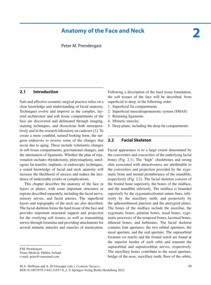
29M.A. Shiffman and A. Di Giuseppe (eds.), Cosmetic Surgery, DOI 10.1007/978-3-642-21837-8_2, © Springer-Verlag Berlin Heidelberg 2012
Anatomy of the Face and Neck
Peter M. Prendergast
2
2.1 Introduction
Safe and effective cosmetic surgical practice relies on a clear knowledge and understanding of facial anatomy. Techniques evolve and improve as the complex, lay-ered architecture and soft tissue compartments of the face are discovered and delineated through imaging, staining techniques, and dissections both intraopera-tively and in the research laboratory on cadavers [ 1 ] . To create a more youthful, natural-looking form, the sur-geon endeavors to reverse some of the changes that occur due to aging. These include volumetric changes in soft tissue compartments, gravitational changes, and the attenuation of ligaments. Whether the plan of reju-venation includes rhytidectomy, platysmaplasty, autol-ogous fat transfer, implants, or endoscopic techniques, a sound knowledge of facial and neck anatomy will increase the likelihood of success and reduce the inci-dence of undesirable results or complications.
This chapter describes the anatomy of the face in layers or planes, with some important structures or regions described separately, including the facial nerve, sensory nerves, and facial arteries. The superfi cial layers and topography of the neck are also described. The facial skeleton forms the hard tissue of the face and provides important structural support and projection for the overlying soft tissues, as well as transmitting nerves through foramina and providing attachments for several mimetic muscles and muscles of mastication.
Following a description of the hard tissue foundation, the soft tissues of the face will be described, from superfi cial to deep, in the following order: 1. Superfi cial fat compartments 2. Superfi cial musculoaponeurotic system (SMAS) 3. Retaining ligaments 4. Mimetic muscles 5. Deep plane, including the deep fat compartments
2.2 Facial Skeleton
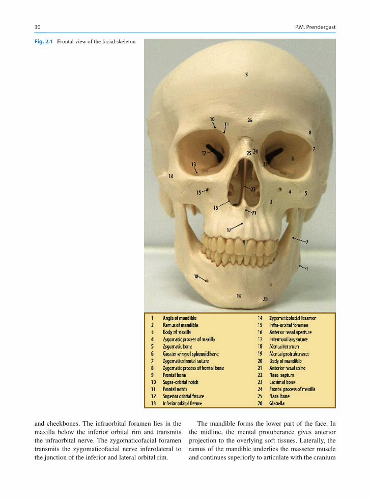
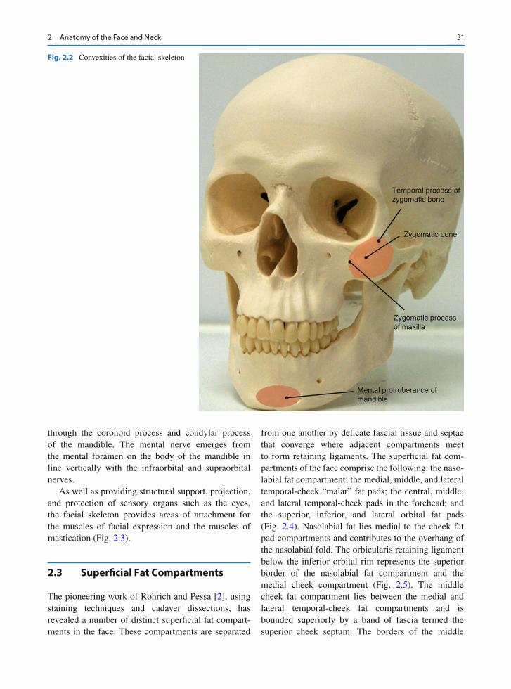
Facial appearance is to a large extent determined by the convexities and concavities of the underlying facial bones (Fig. 2.1 ). The “high” cheekbones and strong chin associated with attractiveness are attributable to the convexities and projection provided by the zygo-matic bone and mental protuberance of the mandible, respectively (Fig. 2.2 ). The facial skeleton consists of the frontal bone superiorly, the bones of the midface, and the mandible inferiorly. The midface is bounded superiorly by the zygomaticofrontal suture lines, infe-riorly by the maxillary teeth, and posteriorly by the sphenoethmoid junction and the pterygoid plates. The bones of the midface include the maxillae, the zygomatic bones, palatine bones, nasal bones, zygo-matic processes of the temporal bones, lacrimal bones, ethmoid bones, and turbinates. The facial skeleton contains four apertures: the two orbital apertures, the nasal aperture, and the oral aperture. The supraorbital foramen (or notch) and the frontal notch are found at the superior border of each orbit and transmit the supraorbital and supratrochlear nerves, respectively. The maxillary bones contribute to the nasal aperture, bridge of the nose, maxillary teeth, fl oor of the orbits,
P. M. Prendergast Venus Medical , Dublin , Ireland e-mail: [email protected]
30 P.M. Prendergast
and cheekbones. The infraorbital foramen lies in the maxilla below the inferior orbital rim and transmits the infraorbital nerve. The zygomaticofacial foramen transmits the zygomaticofacial nerve inferolateral to the junction of the inferior and lateral orbital rim.
The mandible forms the lower part of the face. In the midline, the mental protuberance gives anterior projection to the overlying soft tissues. Laterally, the ramus of the mandible underlies the masseter muscle and continues superiorly to articulate with the cranium
Fig. 2.1 Frontal view of the facial skeleton
312 Anatomy of the Face and Neck
through the coronoid process and condylar process of the mandible. The mental nerve emerges from the mental foramen on the body of the mandible in line vertically with the infraorbital and supraorbital nerves.
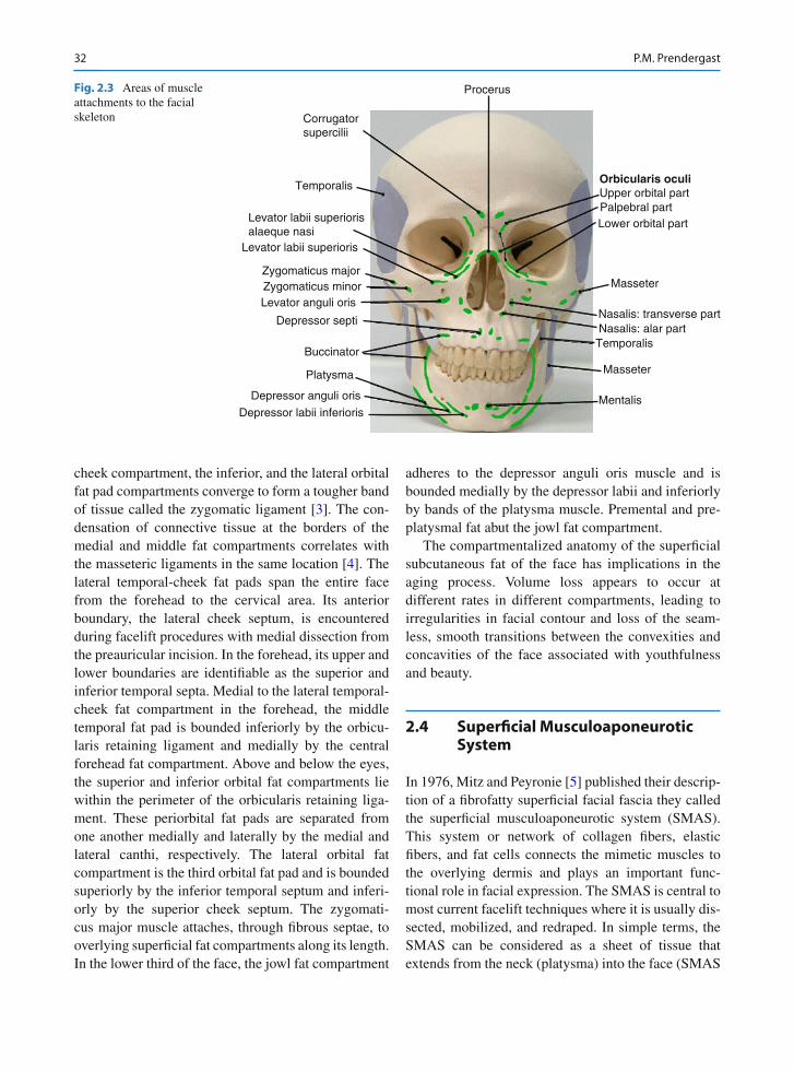
As well as providing structural support, projection, and protection of sensory organs such as the eyes, the facial skeleton provides areas of attachment for the muscles of facial expression and the muscles of mastication (Fig. 2.3 ).
2.3 Superfi cial Fat Compartments
The pioneering work of Rohrich and Pessa [ 2 ] , using staining techniques and cadaver dissections, has revealed a number of distinct superfi cial fat compart-ments in the face. These compartments are separated
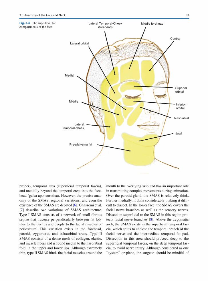
from one another by delicate fascial tissue and septae that converge where adjacent compartments meet to form retaining ligaments. The superfi cial fat com-partments of the face comprise the following: the naso-labial fat compartment; the medial, middle, and lateral temporal-cheek “malar” fat pads; the central, middle, and lateral temporal-cheek pads in the forehead; and the superior, inferior, and lateral orbital fat pads (Fig. 2.4 ). Nasolabial fat lies medial to the cheek fat pad compartments and contributes to the overhang of the nasolabial fold. The orbicularis retaining ligament below the inferior orbital rim represents the superior border of the nasolabial fat compartment and the medial cheek compartment (Fig. 2.5 ). The middle cheek fat compartment lies between the medial and lateral temporal-cheek fat compartments and is bounded superiorly by a band of fascia termed the superior cheek septum. The borders of the middle
Temporal process ofzygomatic bone
Zygomatic bone
Zygomatic processof maxilla
Mental protruberance ofmandible
Fig. 2.2 Convexities of the facial skeleton
32 P.M. Prendergast
cheek compartment, the inferior, and the lateral orbital fat pad compartments converge to form a tougher band of tissue called the zygomatic ligament [ 3 ] . The con-densation of connective tissue at the borders of the medial and middle fat compartments correlates with the masseteric ligaments in the same location [ 4 ] . The lateral temporal-cheek fat pads span the entire face from the forehead to the cervical area. Its anterior boundary, the lateral cheek septum, is encountered during facelift procedures with medial dissection from the preauricular incision. In the forehead, its upper and lower boundaries are identifi able as the superior and inferior temporal septa. Medial to the lateral temporal-cheek fat compartment in the forehead, the middle temporal fat pad is bounded inferiorly by the orbicu-laris retaining ligament and medially by the central forehead fat compartment. Above and below the eyes, the superior and inferior orbital fat compartments lie within the perimeter of the orbicularis retaining liga-ment. These periorbital fat pads are separated from one another medially and laterally by the medial and lateral canthi, respectively. The lateral orbital fat compartment is the third orbital fat pad and is bounded superiorly by the inferior temporal septum and inferi-orly by the superior cheek septum. The zygomati-cus major muscle attaches, through fi brous septae, to overlying superfi cial fat compartments along its length. In the lower third of the face, the jowl fat compartment
adheres to the depressor anguli oris muscle and is bounded medially by the depressor labii and inferiorly by bands of the platysma muscle. Premental and pre-platysmal fat abut the jowl fat compartment.
The compartmentalized anatomy of the superfi cial subcutaneous fat of the face has implications in the aging process. Volume loss appears to occur at different rates in different compartments, leading to irregularities in facial contour and loss of the seam-less, smooth transitions between the convexities and concavities of the face associated with youthfulness and beauty.
2.4 Superfi cial Musculoaponeurotic System
In 1976, Mitz and Peyronie [ 5 ] published their descrip-tion of a fi brofatty superfi cial facial fascia they called the superfi cial musculoaponeurotic system (SMAS). This system or network of collagen fi bers, elastic fi bers, and fat cells connects the mimetic muscles to the overlying dermis and plays an important func-tional role in facial expression. The SMAS is central to most current facelift techniques where it is usually dis-sected, mobilized, and redraped. In simple terms, the SMAS can be considered as a sheet of tissue that extends from the neck (platysma) into the face (SMAS
Procerus
Corrugator supercilii
Temporalis
Levator labii superiorisalaeque nasi
Levator labii superioris
Zygomaticus majorZygomaticus minorLevator anguli oris
Depressor septi
Buccinator
Platysma
Depressor anguli oris
Depressor labii inferioris
Lower orbital part
Orbicularis oculiUpper orbital partPalpebral part
Masseter
Nasalis: transverse partNasalis: alar partTemporalis
Masseter
Mentalis
Fig. 2.3 Areas of muscle attachments to the facial skeleton
332 Anatomy of the Face and Neck
proper), temporal area (superfi cial temporal fascia), and medially beyond the temporal crest into the fore-head (galea aponeurotica). However, the precise anat-omy of the SMAS, regional variations, and even the existence of the SMAS are debated [ 6 ] . Ghassemi et al. [ 7 ] describe two variations of SMAS architecture. Type I SMAS consists of a network of small fi brous septae that traverse perpendicularly between fat lob-ules to the dermis and deeply to the facial muscles or periosteum. This variation exists in the forehead, parotid, zygomatic, and infra orbital areas. Type II SMAS consists of a dense mesh of collagen, elastic, and muscle fi bers and is found medial to the nasolabial fold, in the upper and lower lips. Although extremely thin, type II SMAS binds the facial muscles around the
mouth to the overlying skin and has an important role in transmitting complex movements during animation. Over the parotid gland, the SMAS is relatively thick. Further medially, it thins considerably making it diffi -cult to dissect. In the lower face, the SMAS covers the facial nerve branches as well as the sensory nerves. Dissection superfi cial to the SMAS in this region pro-tects facial nerve branches [ 8 ] . Above the zygomatic arch, the SMAS exists as the superfi cial temporal fas-cia, which splits to enclose the temporal branch of the facial nerve and the intermediate temporal fat pad. Dissection in this area should proceed deep to the superfi cial temporal fascia, on the deep temporal fas-cia, to avoid nerve injury. Although considered as one “system” or plane, the surgeon should be mindful of
Lateral Temporal-Cheek(forehead)
Middle forehead
Central
Lateral orbital
Medial
Superiororbital
Inferiororbital
Nasolabial
Jowl
Middle
Lateraltemporal-cheek
Pre-platysma fat
Fig. 2.4 The superfi cial fat compartments of the face
34 P.M. Prendergast
the regional differences in SMAS anatomy from supe-rior to inferior and lateral to medial.
2.5 Retaining Ligaments
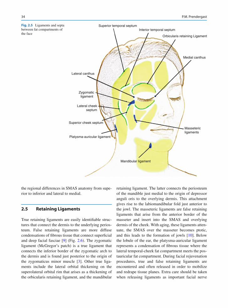
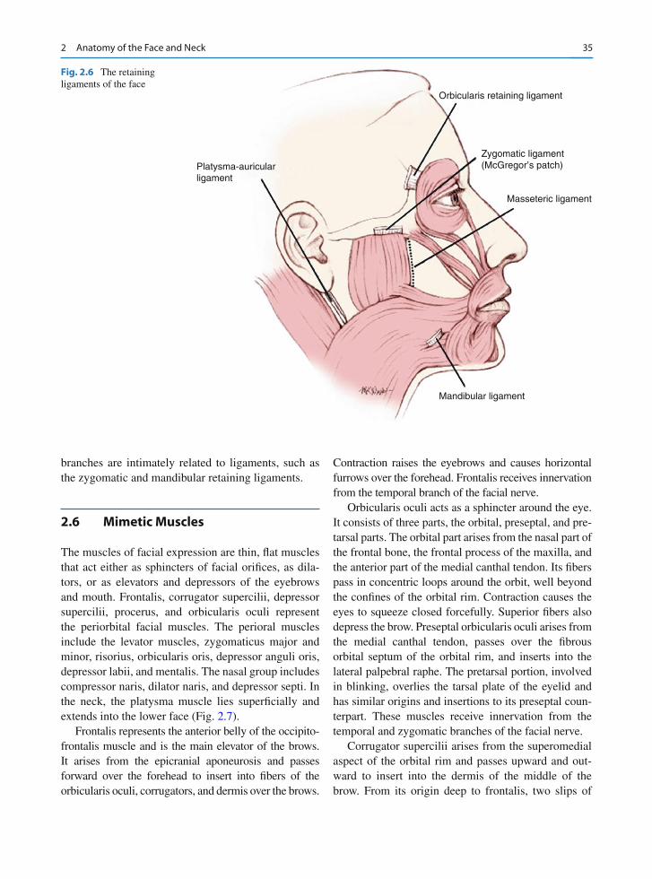
True retaining ligaments are easily identifi able struc-tures that connect the dermis to the underlying perios-teum. False retaining ligaments are more diffuse condensations of fi brous tissue that connect superfi cial and deep facial fasciae [ 9 ] (Fig. 2.6 ). The zygomatic ligament (McGregor’s patch) is a true ligament that connects the inferior border of the zygomatic arch to the dermis and is found just posterior to the origin of the zygomaticus minor muscle [ 3 ] . Other true liga-ments include the lateral orbital thickening on the superolateral orbital rim that arises as a thickening of the orbicularis retaining ligament, and the mandibular
retaining ligament. The latter connects the periosteum of the mandible just medial to the origin of depressor anguli oris to the overlying dermis. This attachment gives rise to the labiomandibular fold just anterior to the jowl. The masseteric ligaments are false retaining ligaments that arise from the anterior border of the masseter and insert into the SMAS and overlying dermis of the cheek. With aging, these ligaments atten-uate, the SMAS over the masseter becomes ptotic, and this leads to the formation of jowls [ 10 ] . Below the lobule of the ear, the platysma-auricular ligament represents a condensation of fi brous tissue where the lateral temporal-cheek fat compartment meets the pos-tauricular fat compartment. During facial rejuvenation procedures, true and false retaining ligaments are encountered and often released in order to mobilize and redrape tissue planes. Extra care should be taken when releasing ligaments as important facial nerve
Massetericligaments
Superior temporal septumInterior temporal septum
Orbicularis retaining Ligament
Medial canthus
Mandibular ligament
Platysma-auricular ligament
Superior cheek septum
Lateral cheekseptum
Zygomaticligament
Lateral canthus
Fig. 2.5 Ligaments and septa between fat compartments of the face
352 Anatomy of the Face and Neck
branches are intimately related to ligaments, such as the zygomatic and mandibular retaining ligaments.
2.6 Mimetic Muscles
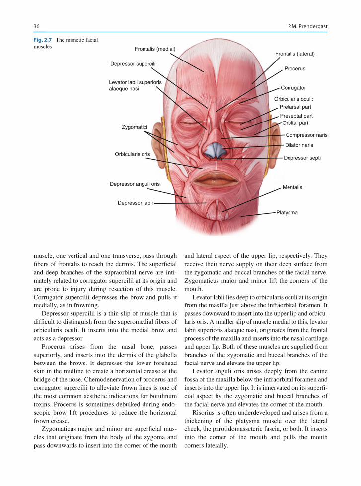
The muscles of facial expression are thin, fl at muscles that act either as sphincters of facial orifi ces, as dila-tors, or as elevators and depressors of the eyebrows and mouth. Frontalis, corrugator supercilii, depressor supercilii, procerus, and orbicularis oculi represent the periorbital facial muscles. The perioral muscles include the levator muscles, zygomaticus major and minor, risorius, orbicularis oris, depressor anguli oris, depressor labii, and mentalis. The nasal group includes compressor naris, dilator naris, and depressor septi. In the neck, the platysma muscle lies superfi cially and extends into the lower face (Fig. 2.7 ).
Frontalis represents the anterior belly of the occipito-frontalis muscle and is the main elevator of the brows. It arises from the epicranial aponeurosis and passes forward over the forehead to insert into fi bers of the orbicularis oculi, corrugators, and dermis over the brows.
Contraction raises the eyebrows and causes horizontal furrows over the forehead. Frontalis receives innervation from the temporal branch of the facial nerve.
Orbicularis oculi acts as a sphincter around the eye. It consists of three parts, the orbital, preseptal, and pre-tarsal parts. The orbital part arises from the nasal part of the frontal bone, the frontal process of the maxilla, and the anterior part of the medial canthal tendon. Its fi bers pass in concentric loops around the orbit, well beyond the confi nes of the orbital rim. Contraction causes the eyes to squeeze closed forcefully. Superior fi bers also depress the brow. Preseptal orbicularis oculi arises from the medial canthal tendon, passes over the fi brous orbital septum of the orbital rim, and inserts into the lateral palpebral raphe. The pretarsal portion, involved in blinking, overlies the tarsal plate of the eyelid and has similar origins and insertions to its preseptal coun-terpart. These muscles receive innervation from the temporal and zygomatic branches of the facial nerve.
Corrugator supercilii arises from the superomedial aspect of the orbital rim and passes upward and out-ward to insert into the dermis of the middle of the brow. From its origin deep to frontalis, two slips of
Orbicularis retaining ligament
Zygomatic ligament(McGregor’s patch)
Masseteric ligament
Mandibular ligament
Platysma-auricularligament
Fig. 2.6 The retaining ligaments of the face
36 P.M. Prendergast
muscle, one vertical and one transverse, pass through fi bers of frontalis to reach the dermis. The superfi cial and deep branches of the supraorbital nerve are inti-mately related to corrugator supercilii at its origin and are prone to injury during resection of this muscle. Corrugator supercilii depresses the brow and pulls it medially, as in frowning.
Depressor supercilii is a thin slip of muscle that is diffi cult to distinguish from the superomedial fi bers of orbicularis oculi. It inserts into the medial brow and acts as a depressor.
Procerus arises from the nasal bone, passes superiorly, and inserts into the dermis of the glabella between the brows. It depresses the lower forehead skin in the midline to create a horizontal crease at the bridge of the nose. Chemodenervation of procerus and corrugator supercilii to alleviate frown lines is one of the most common aesthetic indications for botulinum toxins. Procerus is sometimes debulked during endo-scopic brow lift procedures to reduce the horizontal frown crease.
Zygomaticus major and minor are superfi cial mus-cles that originate from the body of the zygoma and pass downwards to insert into the corner of the mouth
and lateral aspect of the upper lip, respectively. They receive their nerve supply on their deep surface from the zygomatic and buccal branches of the facial nerve. Zygomaticus major and minor lift the corners of the mouth.
Levator labii lies deep to orbicularis oculi at its origin from the maxilla just above the infraorbital foramen. It passes downward to insert into the upper lip and orbicu-laris oris. A smaller slip of muscle medial to this, levator labii superioris alaeque nasi, originates from the frontal process of the maxilla and inserts into the nasal cartilage and upper lip. Both of these muscles are supplied from branches of the zygomatic and buccal branches of the facial nerve and elevate the upper lip.
Levator anguli oris arises deeply from the canine fossa of the maxilla below the infraorbital foramen and inserts into the upper lip. It is innervated on its superfi -cial aspect by the zygomatic and buccal branches of the facial nerve and elevates the corner of the mouth.
Risorius is often underdeveloped and arises from a thickening of the platysma muscle over the lateral cheek, the parotidomasseteric fascia, or both. It inserts into the corner of the mouth and pulls the mouth corners laterally.
Frontalis (lateral)
Procerus
Corrugator
Orbicularis oculi:
Pretarsal part
Preseptal partOrbital part
Compressor naris
Dilator naris
Depressor septi
Mentalis
Platysma
Depressor labii
Depressor anguli oris
Orbicularis oris
Zygomatici
Levator labii superiorisalaeque nasi
Depressor supercilii
Frontalis (medial)
Fig. 2.7 The mimetic facial muscles
372 Anatomy of the Face and Neck
Orbicularis oris acts as a sphincter around the mouth and its fi bers interlace with all of the other facial mus-cles that act on the mouth. The buccal and marginal mandibular branches of the facial nerve provide motor supply to orbicularis oris, which has various actions, including pursing, dilation, and closure of the lips.
Depressor anguli oris arises from the periosteum of the mandible along the oblique line lateral to depressor labii inferioris. Its fi bers converge on the modiolus with fi bers of orbicularis oris, risorius, and sometimes levator anguli oris. It is supplied by the marginal man-dibular branch of the facial nerve and depresses the mouth corners on contraction. Depressor labii inferio-ris arises from the oblique line of the mandible in front of the mental foramen, where fi bers of depressor anguli oris cover it. It passes upward and medially to insert into the skin and mucosa of the lower lip and into fi bers of orbicularis oris.
Mentalis arises from the incisive fossa of the man-dible and descends to insert into the dermis of the chin. Contraction elevates and protrudes the lower lip and creates the characteristic “peach-pit” dimpling of the skin over the chin. Motor supply arises from the mar-ginal mandibular nerve.
Nasalis consists of two parts, the transverse part (compressor naris) and alar part (dilator naris). Compressor naris arises from the maxilla over the canine tooth and passes over the dorsum of the nose to interlace with fi bers from the contralateral side. It compresses the nasal aperture. Dilator naris originates from the maxilla just below and medial to compressor naris and inserts into the alar cartilage of the nose. It dilates the nostrils during respiration. Depressor septi is a slip of muscle arising from the maxilla above the central incisor, deep to the mucous membrane of the upper lip. It inserts into the cartilaginous nasal septum and pulls the nose tip inferiorly. Nasalis and depressor septi receive innervation from the superior buccal branches of the facial nerve.
2.7 Deep Plane Including the Deep Fat Compartments
The superfi cial fat compartments described above lie above the muscles of facial expression in the subcuta-neous plane. In the midface, the suborbicularis oculi fat and deep cheek fat represent deeper fat compartments that provide volume and shape to the face and act as gliding planes within which the muscles of facial
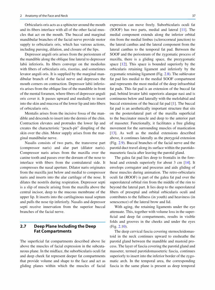
expression can move freely. Suborbicularis oculi fat (SOOF) has two parts, medial and lateral [ 11 ] . The medial component extends along the inferior orbital rim from the medial limbus (sclerocorneal junction) to the lateral canthus and the lateral component from the lateral canthus to the temporal fat pad. Between the SOOF and the periosteum of the zygomatic process of maxilla, there is a gliding space, the prezygomatic space [ 12 ] . This space is bounded superiorly by the orbicularis retaining ligament and inferiorly by the zygomatic retaining ligament (Fig. 2.8 ). The sublevator fat pad lies medial to the medial SOOF compartment and represents the most medial of the deep infraorbital fat pads. This fat pad is an extension of the buccal fat pad, behind levator labii superioris alaeque nasi and is continuous below and laterally with the melolabial and buccal extensions of the buccal fat pad [ 1 ] . The buccal fat pad is an aesthetically important structure that sits on the posterolateral part of the maxilla superfi cial to the buccinator muscle and deep to the anterior part of masseter. Functionally, it facilitates a free gliding movement for the surrounding muscles of mastication [ 13 ] . As well as the medial extensions described above, it continues laterally as the pterygoid extension (Fig. 2.9 ). Buccal branches of the facial nerve and the parotid duct travel along its surface within the parotido-masseteric fascia after leaving the parotid gland.
The galea fat pad lies deep to frontalis in the fore-head and extends superiorly for about 3 cm [ 14 ] . It envelops corrugator and procerus and aids gliding of these muscles during animation. The retro-orbicularis oculi fat (ROOF) is part of the galea fat pad over the superolateral orbital rim from the middle of the rim to beyond the lateral part. It lies deep to the superolateral fi bers of preseptal and orbital orbicularis oculi and contributes to the fullness (in youth) and heaviness (in senescence) of the lateral brow and lid.
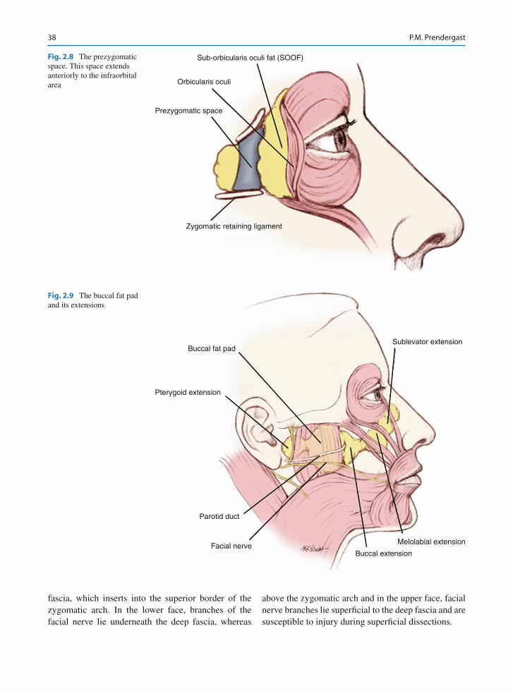
With aging, the retaining ligaments under the eye attenuate. This, together with volume loss in the super-fi cial and deep fat compartments, results in visible folds and grooves in the cheeks and under the eyes (Fig. 2.10 ).
The deep cervical fascia covering sternocleidomas-toid in the neck continues upward to ensheathe the parotid gland between the mandible and mastoid pro-cess. The layer of fascia covering the parotid gland and masseter, termed parotidomasseteric fascia, continues superiorly to insert into the inferior border of the zygo-matic arch. In the temporal area, the corresponding fascia in the same plane is present as deep temporal
38 P.M. Prendergast
fascia, which inserts into the superior border of the zygomatic arch. In the lower face, branches of the facial nerve lie underneath the deep fascia, whereas
above the zygomatic arch and in the upper face, facial nerve branches lie superfi cial to the deep fascia and are susceptible to injury during superfi cial dissections.
Sublevator extension
Melolabial extension
Buccal extensionFacial nerve
Parotid duct
Pterygoid extension
Buccal fat pad
Fig. 2.9 The buccal fat pad and its extensions
Sub-orbicularis oculi fat (SOOF)
Orbicularis oculi
Prezygomatic space
Zygomatic retaining ligament
Fig. 2.8 The prezygomatic space. This space extends anteriorly to the infraorbital area
392 Anatomy of the Face and Neck
2.8 Neck
Surgical rejuvenation of the neck is frequently included in an overall plan of facial rejuvenation to maintain harmony and enhance results. Cosmetic sur-gical procedures in the neck typically address the superfi cial structures: skin, subcutaneous fat, and plat-ysma. Occasionally, subplatysmal fat and even the digastric muscles are partially resected to improve neck contour [ 15, 16 ] . The aim of surgery is to improve or restore the defi nition of the topographical landmarks of the neck. These include a sharp mento-cervical angle, defi ned mandibular border, and promi-nent anterior border of sternocleidomastoid.
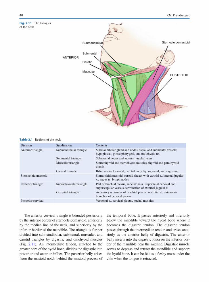
The neck can be divided into anterior, posterior, posterior cervical, and sternocleidomastoid regions (Fig. 2.11 ). Most cosmetic surgical intervention takes place in the anterior region or triangle. The contents of each region are described in Table 2.1 .
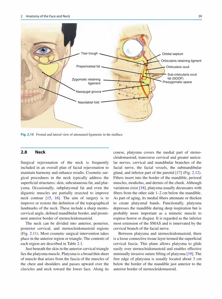
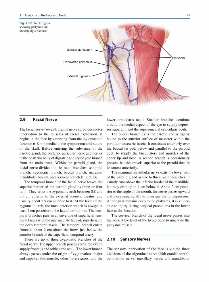
Just beneath the skin in the anterior cervical triangle lies the platysma muscle. Platysma is a broad thin sheet of muscle that arises from the fascia of the muscles of the chest and shoulders and passes upward over the clavicles and neck toward the lower face. Along its
course, platysma covers the medial part of sterno-cleidomastoid, transverse cervical and greater auricu-lar nerves, cervical and mandibular branches of the facial nerve, the facial vessels, the submandibular gland, and inferior part of the parotid [ 17 ] (Fig. 2.12 ). Fibers insert into the border of the mandible, perioral muscles, modiolus, and dermis of the cheek. Although variations exist [ 18 ] , platysma usually decussates with fi bers from the other side 1–2 cm below the mandible. As part of aging, its medial fi bers attenuate or thicken to create platysmal bands. Functionally, platysma depresses the mandible during deep inspiration but is probably more important as a mimetic muscle to express horror or disgust. It is regarded as the inferior most extension of the SMAS and is innervated by the cervical branch of the facial nerve.
Between platysma and sternocleidomastoid, there is a loose connective tissue layer termed the superfi cial cervical fascia. This plane allows platysma to glide easily over sternocleidomastoid and enables effective minimally invasive suture lifting of platysma [ 19 ] . The free edge of platysma is usually located about 3 cm below the border of the mandible just anterior to the anterior border of sternocleidomastoid.
Tear trough
Preperiosteal fat
Orbital septum
Orbicularis retaining ligment
Orbicularis oculi
Prezygomatic space
Sub-orbicularis oculifat (SOOF)
Nasojugal groove
Nasolabial fold
Zygomatic retainingligament
Fig. 2.10 Frontal and lateral view of attenuated ligaments in the midface
40 P.M. Prendergast
The anterior cervical triangle is bounded posteriorly by the anterior border of sternocleidomastoid, anteriorly by the median line of the neck, and superiorly by the inferior border of the mandible. The triangle is further divided into submandibular, submental, muscular, and carotid triangles by digastric and omohyoid muscles (Fig. 2.11 ). An intermediate tendon, attached to the greater horn of the hyoid bone, divides the digastric into posterior and anterior bellies. The posterior belly arises from the mastoid notch behind the mastoid process of
the temporal bone. It passes anteriorly and inferiorly below the mandible toward the hyoid bone where it becomes the digastric tendon. The digastric tendon passes through the intermediate tendon and arises ante-riorly as the anterior belly of digastric. The anterior belly inserts into the digastric fossa on the inferior bor-der of the mandible near the midline. Digastric muscle serves to depress and retract the mandible and support the hyoid bone. It can be felt as a fl eshy mass under the chin when the tongue is retracted.
Submandibular
Submental
Carotid
Muscular
ANTERIOR
POSTERIOR
Sternocleidomastoid
Fig. 2.11 The triangles of the neck
Table 2.1 Regions of the neck
Division Subdivision Contents
Anterior triangle Submandibular triangle Submandibular gland and nodes; facial and submental vessels; hypoglossal, glossopharygeal, and mylohyoid nn.
Submental triangle Submental nodes and anterior jugular veins Muscular triangle Sternothyroid and sternohyoid muscles, thyroid and parathyroid
glands Carotid triangle Bifurcation of carotid, carotid body, hypoglossal, and vagus nn.
Sternocleidomastoid Sternocleidomastoid, carotid sheath with carotid a., internal jugular v., vagus n., lymph nodes
Posterior triangle Supraclavicular triangle Part of brachial plexus, subclavian a., superfi cial cervical and suprascapular vessels, termination of external jugular v.
Occipital triangle Accessory n., trunks of brachial plexus, occipital a., cutaneous branches of cervical plexus
Posterior cervical Vertebral a., cervical plexus, nuchal muscles
412 Anatomy of the Face and Neck
2.9 Facial Nerve
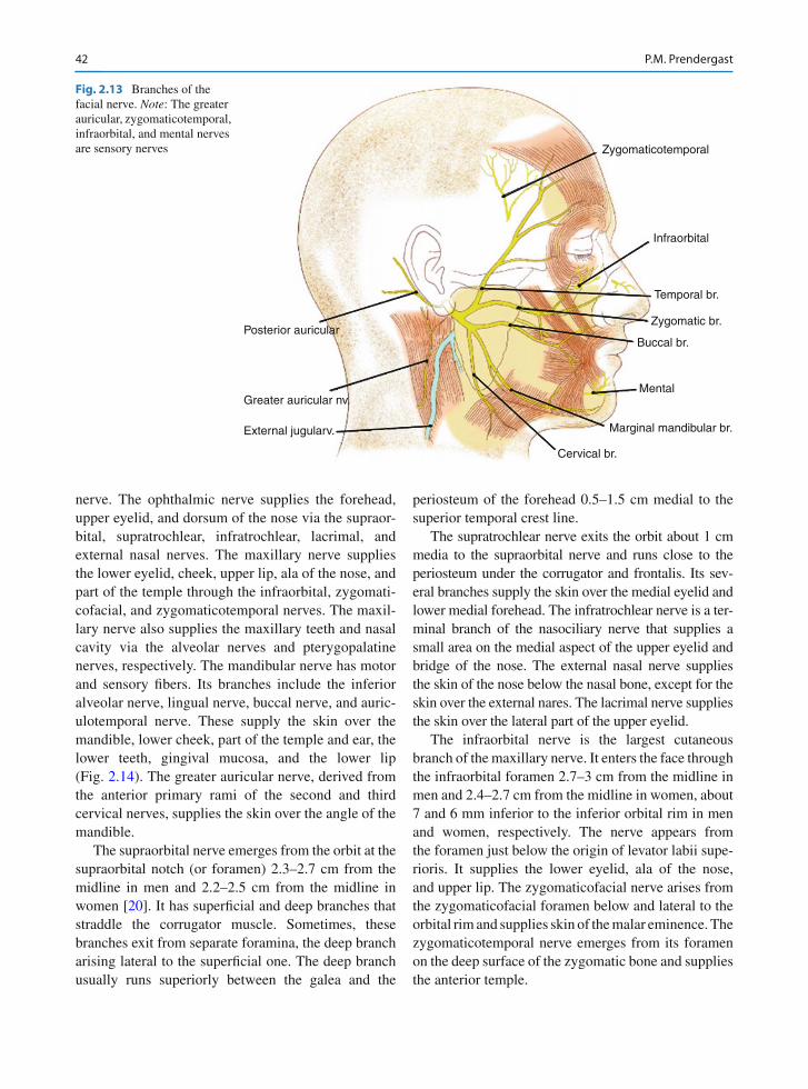
The facial nerve (seventh cranial nerve) provides motor innervation to the muscles of facial expression. It begins in the face by emerging from the stylomastoid foramen 6–8 mm medial to the tympanomastoid suture of the skull. Before entering the substance of the parotid gland, the posterior auricular nerve and nerves to the posterior belly of digastric and stylohyoid branch from the main trunk. Within the parotid gland, the facial nerve divides into its main branches: temporal branch, zygomatic branch, buccal branch, marginal mandibular branch, and cervical branch (Fig. 2.13 ).
The temporal branch of the facial nerve leaves the superior border of the parotid gland as three or four rami. They cross the zygomatic arch between 0.8 and 3.5 cm anterior to the external acoustic meatus, and usually about 2.5 cm anterior to it. At the level of the zygomatic arch, the most anterior branch is always at least 2 cm posterior to the lateral orbital rim. The tem-poral branches pass in an envelope of superfi cial tem-poral fascia with the intermediate fat pad, superfi cial to the deep temporal fascia. The temporal branch enters frontalis about 2 cm above the brow, just below the anterior branch of the superfi cial temporal artery.
There are up to three zygomatic branches of the facial nerve. The upper branch passes above the eye to supply frontalis and orbicularis oculi. The lower branch always passes under the origin of zygomaticus major and supplies this muscle, other lip elevators, and the
lower orbicularis oculi. Smaller branches continue around the medial aspect of the eye to supply depres-sor supercilii and the superomedial orbicularis oculi.
The buccal branch exits the parotid and is tightly bound to the anterior surface of masseter within the parotidomasseteric fascia. It continues anteriorly over the buccal fat pad, below and parallel to the parotid duct, to supply the buccinators and muscles of the upper lip and nose. A second branch is occasionally present, but this travels superior to the parotid duct in its course anteriorly.
The marginal mandibular nerve exits the lower part of the parotid gland as one to three major branches. It usually runs above the inferior border of the mandible, but may drop up to 4 cm below it. About 2 cm poste-rior to the angle of the mouth, the nerve passes upward and more superfi cially to innervate the lip depressors. Although it remains deep to the platysma, it is vulner-able to injury during surgical procedures in the lower face at this location.
The cervical branch of the facial nerve passes into the neck at the level of the hyoid bone to innervate the platysma muscle.
2.10 Sensory Nerves
The sensory innervation of the face is via the three divisions of the trigeminal nerve (fi fth cranial nerve): ophthalmic nerve, maxillary nerve, and mandibular
Platysma
Greater auricular n.
Transverse cervical n.
External jugular v.
Fig. 2.12 Neck region showing platysma and underlying structures
42 P.M. Prendergast
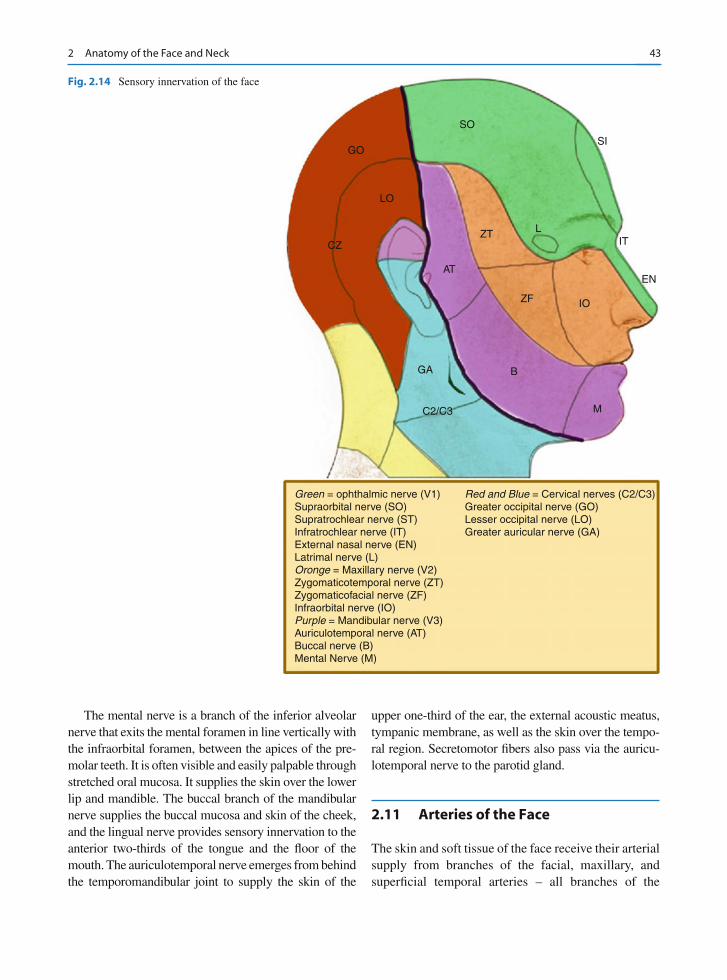
nerve. The ophthalmic nerve supplies the forehead, upper eyelid, and dorsum of the nose via the supraor-bital, supratrochlear, infratrochlear, lacrimal, and external nasal nerves. The maxillary nerve supplies the lower eyelid, cheek, upper lip, ala of the nose, and part of the temple through the infraorbital, zygomati-cofacial, and zygomaticotemporal nerves. The maxil-lary nerve also supplies the maxillary teeth and nasal cavity via the alveolar nerves and pterygopalatine nerves, respectively. The mandibular nerve has motor and sensory fi bers. Its branches include the inferior alveolar nerve, lingual nerve, buccal nerve, and auric-ulotemporal nerve. These supply the skin over the mandible, lower cheek, part of the temple and ear, the lower teeth, gingival mucosa, and the lower lip (Fig. 2.14 ). The greater auricular nerve, derived from the anterior primary rami of the second and third cervical nerves, supplies the skin over the angle of the mandible.
The supraorbital nerve emerges from the orbit at the supraorbital notch (or foramen) 2.3–2.7 cm from the midline in men and 2.2–2.5 cm from the midline in women [ 20 ] . It has superfi cial and deep branches that straddle the corrugator muscle. Sometimes, these branches exit from separate foramina, the deep branch arising lateral to the superfi cial one. The deep branch usually runs superiorly between the galea and the
periosteum of the forehead 0.5–1.5 cm medial to the superior temporal crest line.
The supratrochlear nerve exits the orbit about 1 cm media to the supraorbital nerve and runs close to the periosteum under the corrugator and frontalis. Its sev-eral branches supply the skin over the medial eyelid and lower medial forehead. The infratrochlear nerve is a ter-minal branch of the nasociliary nerve that supplies a small area on the medial aspect of the upper eyelid and bridge of the nose. The external nasal nerve supplies the skin of the nose below the nasal bone, except for the skin over the external nares. The lacrimal nerve supplies the skin over the lateral part of the upper eyelid.
The infraorbital nerve is the largest cutaneous branch of the maxillary nerve. It enters the face through the infraorbital foramen 2.7–3 cm from the midline in men and 2.4–2.7 cm from the midline in women, about 7 and 6 mm inferior to the inferior orbital rim in men and women, respectively. The nerve appears from the foramen just below the origin of levator labii supe-rioris. It supplies the lower eyelid, ala of the nose, and upper lip. The zygomaticofacial nerve arises from the zygomaticofacial foramen below and lateral to the orbital rim and supplies skin of the malar eminence. The zygomaticotemporal nerve emerges from its foramen on the deep surface of the zygomatic bone and supplies the anterior temple.
Zygomaticotemporal
Infraorbital
Zygomatic br.
Buccal br.
Marginal mandibular br.
Cervical br.
Posterior auricular
Greater auricular nv.
External jugularv.
Temporal br.
Mental
Fig. 2.13 Branches of the facial nerve. Note : The greater auricular, zygomaticotemporal, infraorbital, and mental nerves are sensory nerves
432 Anatomy of the Face and Neck
The mental nerve is a branch of the inferior alveolar nerve that exits the mental foramen in line vertically with the infraorbital foramen, between the apices of the pre-molar teeth. It is often visible and easily palpable through stretched oral mucosa. It supplies the skin over the lower lip and mandible. The buccal branch of the mandibular nerve supplies the buccal mucosa and skin of the cheek, and the lingual nerve provides sensory innervation to the anterior two-thirds of the tongue and the fl oor of the mouth. The auriculotemporal nerve emerges from behind the temporomandibular joint to supply the skin of the
upper one-third of the ear, the external acoustic meatus, tympanic membrane, as well as the skin over the tempo-ral region. Secretomotor fi bers also pass via the auricu-lotemporal nerve to the parotid gland.
2.11 Arteries of the Face
The skin and soft tissue of the face receive their arterial supply from branches of the facial, maxillary, and superfi cial temporal arteries – all branches of the
GO
LO
CZ
AT
ZT
SO
SI
L
EN
IT
ZF
B
M
GA
C2/C3
IO
Green = ophthalmic nerve (V1)Supraorbital nerve (SO)Supratrochlear nerve (ST)Infratrochlear nerve (IT)External nasal nerve (EN)Latrimal nerve (L)Oronge = Maxillary nerve (V2)Zygomaticotemporal nerve (ZT)Zygomaticofacial nerve (ZF)Infraorbital nerve (IO)Purple = Mandibular nerve (V3)Auriculotemporal nerve (AT)Buccal nerve (B)Mental Nerve (M)
Red and Blue = Cervical nerves (C2/C3)Greater occipital nerve (GO)Lesser occipital nerve (LO)Greater auricular nerve (GA)
Fig. 2.14 Sensory innervation of the face
44 P.M. Prendergast
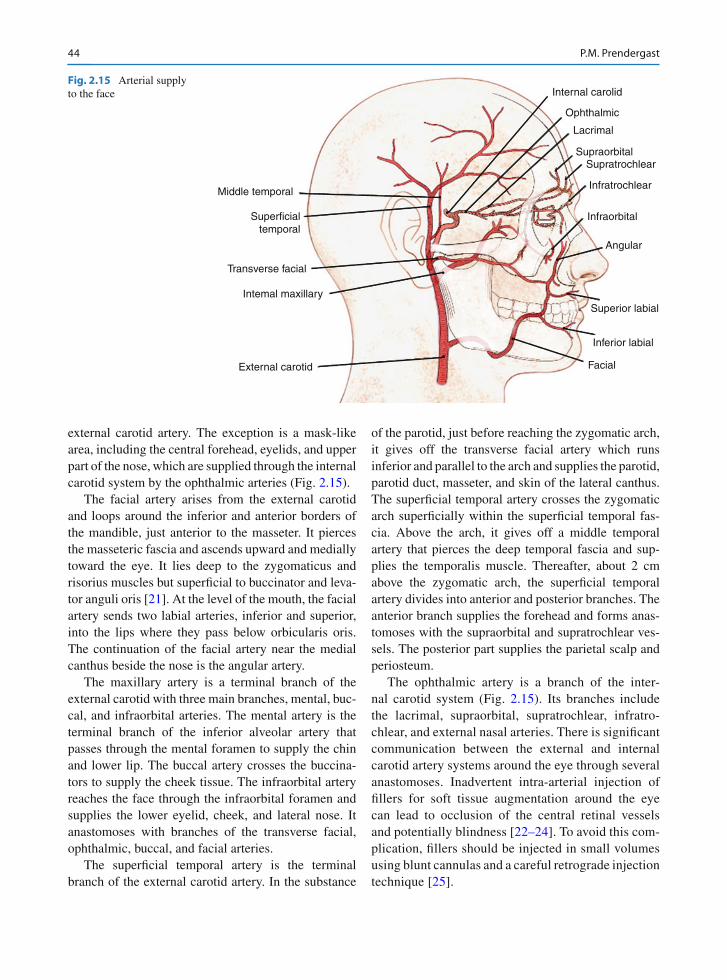
external carotid artery. The exception is a mask-like area, including the central forehead, eyelids, and upper part of the nose, which are supplied through the internal carotid system by the ophthalmic arteries (Fig. 2.15 ).
The facial artery arises from the external carotid and loops around the inferior and anterior borders of the mandible, just anterior to the masseter. It pierces the masseteric fascia and ascends upward and medially toward the eye. It lies deep to the zygomaticus and risorius muscles but superfi cial to buccinator and leva-tor anguli oris [ 21 ] . At the level of the mouth, the facial artery sends two labial arteries, inferior and superior, into the lips where they pass below orbicularis oris. The continuation of the facial artery near the medial canthus beside the nose is the angular artery.
The maxillary artery is a terminal branch of the external carotid with three main branches, mental, buc-cal, and infraorbital arteries. The mental artery is the terminal branch of the inferior alveolar artery that passes through the mental foramen to supply the chin and lower lip. The buccal artery crosses the buccina-tors to supply the cheek tissue. The infraorbital artery reaches the face through the infraorbital foramen and supplies the lower eyelid, cheek, and lateral nose. It anastomoses with branches of the transverse facial, ophthalmic, buccal, and facial arteries.
The superfi cial temporal artery is the terminal branch of the external carotid artery. In the substance
of the parotid, just before reaching the zygomatic arch, it gives off the transverse facial artery which runs inferior and parallel to the arch and supplies the parotid, parotid duct, masseter, and skin of the lateral canthus. The superfi cial temporal artery crosses the zygomatic arch superfi cially within the superfi cial temporal fas-cia. Above the arch, it gives off a middle temporal artery that pierces the deep temporal fascia and sup-plies the temporalis muscle. Thereafter, about 2 cm above the zygomatic arch, the superfi cial temporal artery divides into anterior and posterior branches. The anterior branch supplies the forehead and forms anas-tomoses with the supraorbital and supratrochlear ves-sels. The posterior part supplies the parietal scalp and periosteum.
The ophthalmic artery is a branch of the inter-nal carotid system (Fig. 2.15 ). Its branches include the lacrimal, supraorbital, supratrochlear, infratro-chlear, and external nasal arteries. There is signifi cant communication between the external and internal carotid artery systems around the eye through several anastomoses. Inadvertent intra-arterial injection of fi llers for soft tissue augmentation around the eye can lead to occlusion of the central retinal vessels and potentially blindness [ 22– 24 ] . To avoid this com-plication, fi llers should be injected in small volumes using blunt cannulas and a careful retrograde injection technique [ 25 ] .
Internal carolid
Ophthalmic
Lacrimal
SupraorbitalSupratrochlear
Infratrochlear
Infraorbital
Angular
Superior labial
Inferior labial
Facial
Middle temporal
Superficialtemporal
Transverse facial
Intemal maxillary
External carotid
Fig. 2.15 Arterial supply to the face
452 Anatomy of the Face and Neck
References
1. Gassner HG, Rafi i A, Young A, Murakami C, Moe K, Larrabee WF (2008) Surgical anatomy of the face. Implications for modern face-lift techniques. Arch Facial Plast Surg 10(1):9–19
2. Rohrich RJ, Pessa JE (2007) The fat compartments of the face: anatomy and clinical implications for cosmetic sur-gery. Plast Reconstr Surg 119(7):2219–2227
3. Furnas DW (1989) The retaining ligaments of the cheek. Plast Reconstr Surg 83(1):11–16
4. Stuzin JM, Baker TJ, Gordon HL (1992) The relationship of the superfi cial and deep facial fascias: relevance to rhytidec-tomy and aging. Plast Reconstr Surg 89(3):441–449
5. Mitz V, Peyronie M (1976) The superfi cial musculo- aponeurotic system in the parotid and cheek area. Plast Reconstr Surg 58(1):80–88
6. Gardetto A, Dabernig J, Rainer C, Piegger J, Piza-Katzer H, Fritsch H (2003) Does a superfi cial musculoaponeurotic system exist in the face and neck? An anatomical study by the tissue plastination technique. Plast Reconstr Surg 111(2):664–672
7. Ghassemi A, Prescher A, Riediger D, Axer H (2003) Anatomy of the SMAS revisited. Aesthetic Plast Surg 27(4):258–264
8. Wobig JL, Dailey RA (2004) Facial anatomy. In: Wobig JL, Dailey RA (eds) Oculofacial plastic surgery. Thieme, New York, p 5
9. Jones BM, Grover R (2008) Anatomical considerations. In: Jones BM, Grover R (eds) Facial rejuvenation surgery. Mosby Press, London, pp 18–22
10. Mendelson BC, Freeman ME, Wu W, Huggins RJ (2008) Surgical anatomy of the lower face: the premasseter space, the jowl, and the labiomandibular fold. Aesthetic Plast Surg 32(2):185–195
11. Rohrich R, Arbique GM, Wong C, Brown S, Pessa JE (2009) The anatomy of suborbicularis fat: implications for perior-bital rejuvenation. Plast Reconstr Surg 124(3):946–951
12. Mendelson BC, Muzaffar AR, Adams WP (2002) Surgical anatomy of the midcheek and malar mounds. Plast Reconstr Surg 110(3):885–896
13. Larrabee WF, Makielski KH, Henderson JL (2004) Cheeks and neck. In: Larrabee WF, Makielski KH, Henderson JL (eds) Surgical anatomy of the face. Lippincott Williams & Wilkins, Philadelphia, p 178
14. Zide BM (2006) ROOF and beyond (superolateral zone). In: Zide BM, Jelks GW (eds) Surgical anatomy around the orbit. The system of zones. Lippincott Williams & Wilkins, Philadelphia, p 57
15. Rohrich RJ, Pessa JE (2010) The subplatysmal supramy-lohyoid fat. Plast Reconstr Surg 126(2):589–595
16. Connell BF, Shamoun JM (1997) The signifi cance of digas-tric muscle contouring for rejuvenation of the submental area of the face. Plast Reconstr Surg 99(6):1586–1590
17. De Castro CC (2000) The changing role of platysma in face lifting. Plast Reconstr Surg 105(2):764–775
18. De Castro CC (1980) The anatomy of the platysma muscle. Plast Reconstr Surg 66(5):680–683
19. Labbe D, Franco RG, Nicolas J (2006) Platysma suspen-sion and platysmaplasty during neck lift. Anatomical study and analysis of 30 cases. Plast Reconstr Surg 117(6):2001–2007
20. Zide BM (2006) Supraorbital nerve. Nuances/dissections from above. In: Zide BM, Jelks GW (eds) Surgical anatomy around the orbit. The system of zones. Lippincott Williams & Wilkins, Philadelphia, p 77
21. Berkovitz BKB, Moxham BJ (2002). Head and neck anat-omy. a clinical reference, Martin Dunitz, London, p 118
22. Silva MT, Curi AL (2004) Blindness and total ophthal-moplegia after aesthetic polymethylmethacrylate injection: case report. Arg Neuropsiquiatr 62(3B):873–874
23. McCleve D, Goldstein JC (1995) Blindness secondary to injections in the nose, mouth, and face: cause and preven-tion. Ear Nose Throat J 74:182–188
24. Dreizen NG, Framm L (1989) Sudden unilateral visual loss after autologous fat injection into the glabellar area. Am J Ophthalmol 107:85–87
25. Coleman SR (2002) Avoidance of arterial occlusion from injection of soft tissue fi llers. Aesthet Surg J 22(6):555–557

http://www.springer.com/978-3-642-21836-1