Embed Size (px)
DESCRIPTION
A n a t o m y o f th e P e r i c a rdi a l S p a c e and Mediastinum: Relevance to E p i c a rd i a l M a p p i n g and AblationSabine Ernst, MD, FESCa,b, Damian Sanchez-Quintana, MD, PhDc, Siew Yen Ho, PhD, FRCPathd,e,*KEYWORDS Cardiac anatomy Epicardial space Catheter ablation Puncture technique Ventricular tachycardia Atrial fibrillationCatheter ablation for most arrhythmia substrates is performed frequently from within the cardiac chambers using an exclusively endocardial app
Citation preview
Anatomy of thePericardial Spaceand Mediastinum:Relevance toEpicardial Mappingand Ablation
Sabine Ernst, MD, FESCa,b,Damian Sanchez-Quintana, MD, PhDc,Siew Yen Ho, PhD, FRCPathd,e,*KEYWORDS
� Cardiac anatomy � Epicardial space� Catheter ablation � Puncture technique� Ventricular tachycardia � Atrial fibrillation
Catheter ablation for most arrhythmia substrates isperformed frequently from within the cardiacchambers using an exclusively endocardialapproach. Although epicardial ablations havebeen performed for many years; they have beenfrom within the cardiac veins (mainly the coronarysinus) or from above the semilunar valves.1,2 Inmost of these procedures, a fairly small area (eg,an accessory pathway or a focal origin of ventric-ular ectopy) was targeted, requiring only focalenergy application. However, epicardial arrhyth-mias may require deployment of more complexlesions, such as linear lesions.
Because of the clinical need to manage ventric-ular tachycardia in patients with Chagas disease,the earliest experience of epicardial mappingand ablation using a subxiphoidal approachcame from Brazil.3 Although intraoperative
Prof Ho’s unit receives funding support from the Royal Ba Cardiology Department, Royal Brompton and Harefielb National Heart and Lung Institute, Imperial College Loc Departamento de Anatomıa Humana, Facultad de MBadajoz, Spaind Cardiac Morphology Unit, Imperial College London, Loe Cardiac Morphology Unit, Royal Brompton and Harefi* Corresponding author. Cardiac Morphology Unit, RoyLondon SW3 6NP, UK.E-mail address: [email protected] (S.Y. Ho).
Card Electrophysiol Clin 2 (2010) 1–8doi:10.1016/j.ccep.2009.11.0031877-9182/10/$ – see front matter ª 2010 Published by E
mapping and subsequent arrhythmia surgerywas commonly performed in the 1980s and90s,4 nowadays, arrhythmia surgery mostlyconcentrates on atrial fibrillation.5,6 Arrhythmiasurgery is mainly undertaken as an add-on orconcomitant surgery, seldom as a stand-aloneprocedure. Because new tools have becomeavailable for an epicardial approach withoutthe need to open the left atrium (LA), this proce-dure can be performed off-pump and in a purelythoracoscopic fashion.
Recently, reports on ‘‘sandwich’’ mapping andablation for ischemic ventricular tachycardia haveencouraged clinicians to widen their diagnosticand therapeutic window to this space (Fig. 1).7–9
This article describes the anatomic backgroundto the technical steps for the pericardial approach,which is an emerging clinical necessity and
rompton and Harefield Hospital Charitable Fund.d Hospital, Sydney Street, London SW3 6NP, UKndon, London SW3 6LY, UKedicina, University of Extremadura, UEX, E-06071
ndon SW3 6LY, UKeld Hospital, Sydney Street, London SW3 6NP, UKal Brompton and Harefield Hospital, Sydney Street,
lsevier Inc. card
iacE
P.th
ecli
nics
.com
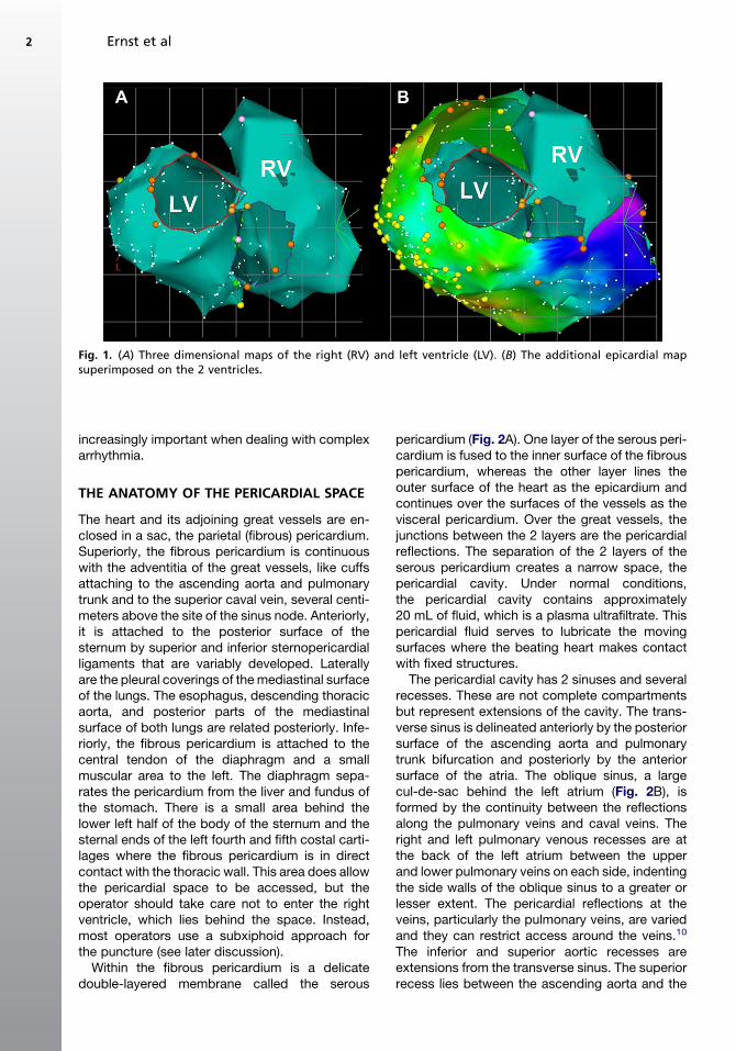
Fig. 1. (A) Three dimensional maps of the right (RV) and left ventricle (LV). (B) The additional epicardial mapsuperimposed on the 2 ventricles.
Ernst et al2
increasingly important when dealing with complexarrhythmia.
THE ANATOMY OF THE PERICARDIAL SPACE
The heart and its adjoining great vessels are en-closed in a sac, the parietal (fibrous) pericardium.Superiorly, the fibrous pericardium is continuouswith the adventitia of the great vessels, like cuffsattaching to the ascending aorta and pulmonarytrunk and to the superior caval vein, several centi-meters above the site of the sinus node. Anteriorly,it is attached to the posterior surface of thesternum by superior and inferior sternopericardialligaments that are variably developed. Laterallyare the pleural coverings of the mediastinal surfaceof the lungs. The esophagus, descending thoracicaorta, and posterior parts of the mediastinalsurface of both lungs are related posteriorly. Infe-riorly, the fibrous pericardium is attached to thecentral tendon of the diaphragm and a smallmuscular area to the left. The diaphragm sepa-rates the pericardium from the liver and fundus ofthe stomach. There is a small area behind thelower left half of the body of the sternum and thesternal ends of the left fourth and fifth costal carti-lages where the fibrous pericardium is in directcontact with the thoracic wall. This area does allowthe pericardial space to be accessed, but theoperator should take care not to enter the rightventricle, which lies behind the space. Instead,most operators use a subxiphoid approach forthe puncture (see later discussion).
Within the fibrous pericardium is a delicatedouble-layered membrane called the serous
pericardium (Fig. 2A). One layer of the serous peri-cardium is fused to the inner surface of the fibrouspericardium, whereas the other layer lines theouter surface of the heart as the epicardium andcontinues over the surfaces of the vessels as thevisceral pericardium. Over the great vessels, thejunctions between the 2 layers are the pericardialreflections. The separation of the 2 layers of theserous pericardium creates a narrow space, thepericardial cavity. Under normal conditions,the pericardial cavity contains approximately20 mL of fluid, which is a plasma ultrafiltrate. Thispericardial fluid serves to lubricate the movingsurfaces where the beating heart makes contactwith fixed structures.
The pericardial cavity has 2 sinuses and severalrecesses. These are not complete compartmentsbut represent extensions of the cavity. The trans-verse sinus is delineated anteriorly by the posteriorsurface of the ascending aorta and pulmonarytrunk bifurcation and posteriorly by the anteriorsurface of the atria. The oblique sinus, a largecul-de-sac behind the left atrium (Fig. 2B), isformed by the continuity between the reflectionsalong the pulmonary veins and caval veins. Theright and left pulmonary venous recesses are atthe back of the left atrium between the upperand lower pulmonary veins on each side, indentingthe side walls of the oblique sinus to a greater orlesser extent. The pericardial reflections at theveins, particularly the pulmonary veins, are variedand they can restrict access around the veins.10
The inferior and superior aortic recesses areextensions from the transverse sinus. The superiorrecess lies between the ascending aorta and the
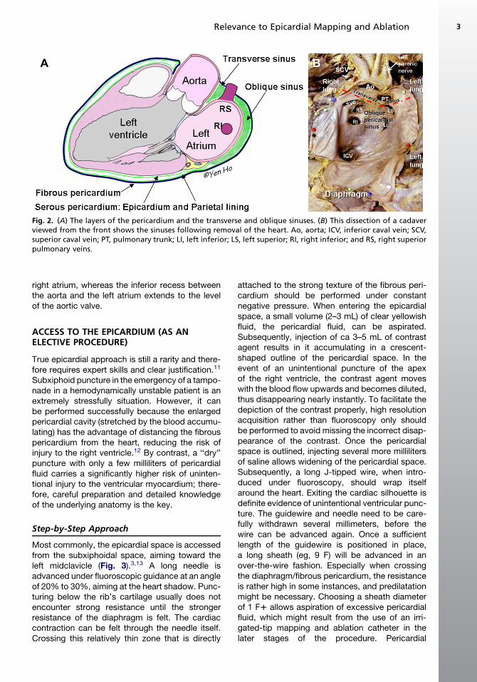
Fig. 2. (A) The layers of the pericardium and the transverse and oblique sinuses. (B) This dissection of a cadaverviewed from the front shows the sinuses following removal of the heart. Ao, aorta; ICV, inferior caval vein; SCV,superior caval vein; PT, pulmonary trunk; LI, left inferior; LS, left superior; RI, right inferior; and RS, right superiorpulmonary veins.
Relevance to Epicardial Mapping and Ablation 3
right atrium, whereas the inferior recess betweenthe aorta and the left atrium extends to the levelof the aortic valve.
ACCESS TO THE EPICARDIUM (AS ANELECTIVE PROCEDURE)
True epicardial approach is still a rarity and there-fore requires expert skills and clear justification.11
Subxiphoid puncture in the emergency of a tampo-nade in a hemodynamically unstable patient is anextremely stressfully situation. However, it canbe performed successfully because the enlargedpericardial cavity (stretched by the blood accumu-lating) has the advantage of distancing the fibrouspericardium from the heart, reducing the risk ofinjury to the right ventricle.12 By contrast, a ‘‘dry’’puncture with only a few milliliters of pericardialfluid carries a significantly higher risk of uninten-tional injury to the ventricular myocardium; there-fore, careful preparation and detailed knowledgeof the underlying anatomy is the key.
Step-by-Step Approach
Most commonly, the epicardial space is accessedfrom the subxiphoidal space, aiming toward theleft midclavicle (Fig. 3).3,13 A long needle isadvanced under fluoroscopic guidance at an angleof 20% to 30%, aiming at the heart shadow. Punc-turing below the rib’s cartilage usually does notencounter strong resistance until the strongerresistance of the diaphragm is felt. The cardiaccontraction can be felt through the needle itself.Crossing this relatively thin zone that is directly
attached to the strong texture of the fibrous peri-cardium should be performed under constantnegative pressure. When entering the epicardialspace, a small volume (2–3 mL) of clear yellowishfluid, the pericardial fluid, can be aspirated.Subsequently, injection of ca 3–5 mL of contrastagent results in it accumulating in a crescent-shaped outline of the pericardial space. In theevent of an unintentional puncture of the apexof the right ventricle, the contrast agent moveswith the blood flow upwards and becomes diluted,thus disappearing nearly instantly. To facilitate thedepiction of the contrast properly, high resolutionacquisition rather than fluoroscopy only shouldbe performed to avoid missing the incorrect disap-pearance of the contrast. Once the pericardialspace is outlined, injecting several more millilitersof saline allows widening of the pericardial space.Subsequently, a long J-tipped wire, when intro-duced under fluoroscopy, should wrap itselfaround the heart. Exiting the cardiac silhouette isdefinite evidence of unintentional ventricular punc-ture. The guidewire and needle need to be care-fully withdrawn several millimeters, before thewire can be advanced again. Once a sufficientlength of the guidewire is positioned in place,a long sheath (eg, 9 F) will be advanced in anover-the-wire fashion. Especially when crossingthe diaphragm/fibrous pericardium, the resistanceis rather high in some instances, and predilatationmight be necessary. Choosing a sheath diameterof 1 F1 allows aspiration of excessive pericardialfluid, which might result from the use of an irri-gated-tip mapping and ablation catheter in thelater stages of the procedure. Pericardial
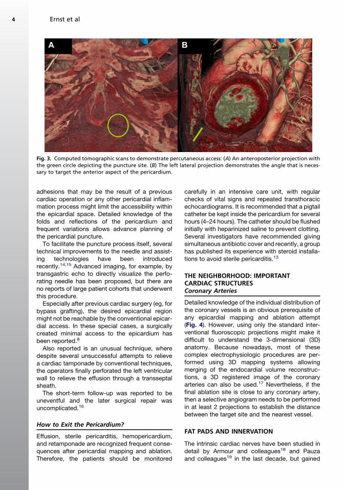
Fig. 3. Computed tomographic scans to demonstrate percutaneous access: (A) An anteroposterior projection withthe green circle depicting the puncture site. (B) The left lateral projection demonstrates the angle that is neces-sary to target the anterior aspect of the pericardium.
Ernst et al4
adhesions that may be the result of a previouscardiac operation or any other pericardial inflam-mation process might limit the accessibility withinthe epicardial space. Detailed knowledge of thefolds and reflections of the pericardium andfrequent variations allows advance planning ofthe pericardial puncture.
To facilitate the puncture process itself, severaltechnical improvements to the needle and assist-ing technologies have been introducedrecently.14,15 Advanced imaging, for example, bytransgastric echo to directly visualize the perfo-rating needle has been proposed, but there areno reports of large patient cohorts that underwentthis procedure.
Especially after previous cardiac surgery (eg, forbypass grafting), the desired epicardial regionmight not be reachable by the conventional epicar-dial access. In these special cases, a surgicallycreated minimal access to the epicardium hasbeen reported.8
Also reported is an unusual technique, wheredespite several unsuccessful attempts to relievea cardiac tamponade by conventional techniques,the operators finally perforated the left ventricularwall to relieve the effusion through a transseptalsheath.
The short-term follow-up was reported to beuneventful and the later surgical repair wasuncomplicated.16
How to Exit the Pericardium?
Effusion, sterile pericarditis, hemopericardium,and retamponade are recognized frequent conse-quences after pericardial mapping and ablation.Therefore, the patients should be monitored
carefully in an intensive care unit, with regularchecks of vital signs and repeated transthoracicechocardiograms. It is recommended that a pigtailcatheter be kept inside the pericardium for severalhours (4–24 hours). The catheter should be flushedinitially with heparinized saline to prevent clotting.Several investigators have recommended givingsimultaneous antibiotic cover and recently, a grouphas published its experience with steroid installa-tions to avoid sterile pericarditis.13
THE NEIGHBORHOOD: IMPORTANTCARDIAC STRUCTURESCoronary Arteries
Detailed knowledge of the individual distribution ofthe coronary vessels is an obvious prerequisite ofany epicardial mapping and ablation attempt(Fig. 4). However, using only the standard inter-ventional fluoroscopic projections might make itdifficult to understand the 3-dimensional (3D)anatomy. Because nowadays, most of thesecomplex electrophysiologic procedures are per-formed using 3D mapping systems allowingmerging of the endocardial volume reconstruc-tions, a 3D registered image of the coronaryarteries can also be used.17 Nevertheless, if thefinal ablation site is close to any coronary artery,then a selective angiogram needs to be performedin at least 2 projections to establish the distancebetween the target site and the nearest vessel.
FAT PADS AND INNERVATION
The intrinsic cardiac nerves have been studied indetail by Armour and colleagues18 and Pauzaand colleagues19 in the last decade, but gained
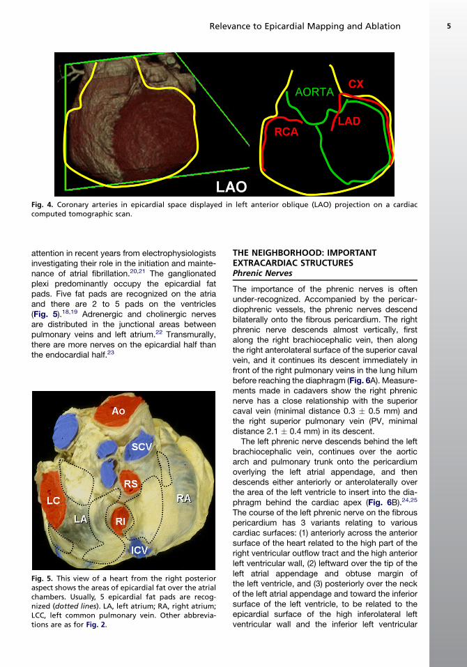
Fig. 4. Coronary arteries in epicardial space displayed in left anterior oblique (LAO) projection on a cardiaccomputed tomographic scan.
Relevance to Epicardial Mapping and Ablation 5
attention in recent years from electrophysiologistsinvestigating their role in the initiation and mainte-nance of atrial fibrillation.20,21 The ganglionatedplexi predominantly occupy the epicardial fatpads. Five fat pads are recognized on the atriaand there are 2 to 5 pads on the ventricles(Fig. 5).18,19 Adrenergic and cholinergic nervesare distributed in the junctional areas betweenpulmonary veins and left atrium.22 Transmurally,there are more nerves on the epicardial half thanthe endocardial half.23
Fig. 5. This view of a heart from the right posterioraspect shows the areas of epicardial fat over the atrialchambers. Usually, 5 epicardial fat pads are recog-nized (dotted lines). LA, left atrium; RA, right atrium;LCC, left common pulmonary vein. Other abbrevia-tions are as for Fig. 2.
THE NEIGHBORHOOD: IMPORTANTEXTRACARDIAC STRUCTURESPhrenic Nerves
The importance of the phrenic nerves is oftenunder-recognized. Accompanied by the pericar-diophrenic vessels, the phrenic nerves descendbilaterally onto the fibrous pericardium. The rightphrenic nerve descends almost vertically, firstalong the right brachiocephalic vein, then alongthe right anterolateral surface of the superior cavalvein, and it continues its descent immediately infront of the right pulmonary veins in the lung hilumbefore reaching the diaphragm (Fig. 6A). Measure-ments made in cadavers show the right phrenicnerve has a close relationship with the superiorcaval vein (minimal distance 0.3 � 0.5 mm) andthe right superior pulmonary vein (PV, minimaldistance 2.1 � 0.4 mm) in its descent.
The left phrenic nerve descends behind the leftbrachiocephalic vein, continues over the aorticarch and pulmonary trunk onto the pericardiumoverlying the left atrial appendage, and thendescends either anteriorly or anterolaterally overthe area of the left ventricle to insert into the dia-phragm behind the cardiac apex (Fig. 6B).24,25
The course of the left phrenic nerve on the fibrouspericardium has 3 variants relating to variouscardiac surfaces: (1) anteriorly across the anteriorsurface of the heart related to the high part of theright ventricular outflow tract and the high anteriorleft ventricular wall, (2) leftward over the tip of theleft atrial appendage and obtuse margin ofthe left ventricle, and (3) posteriorly over the neckof the left atrial appendage and toward the inferiorsurface of the left ventricle, to be related to theepicardial surface of the high inferolateral leftventricular wall and the inferior left ventricular
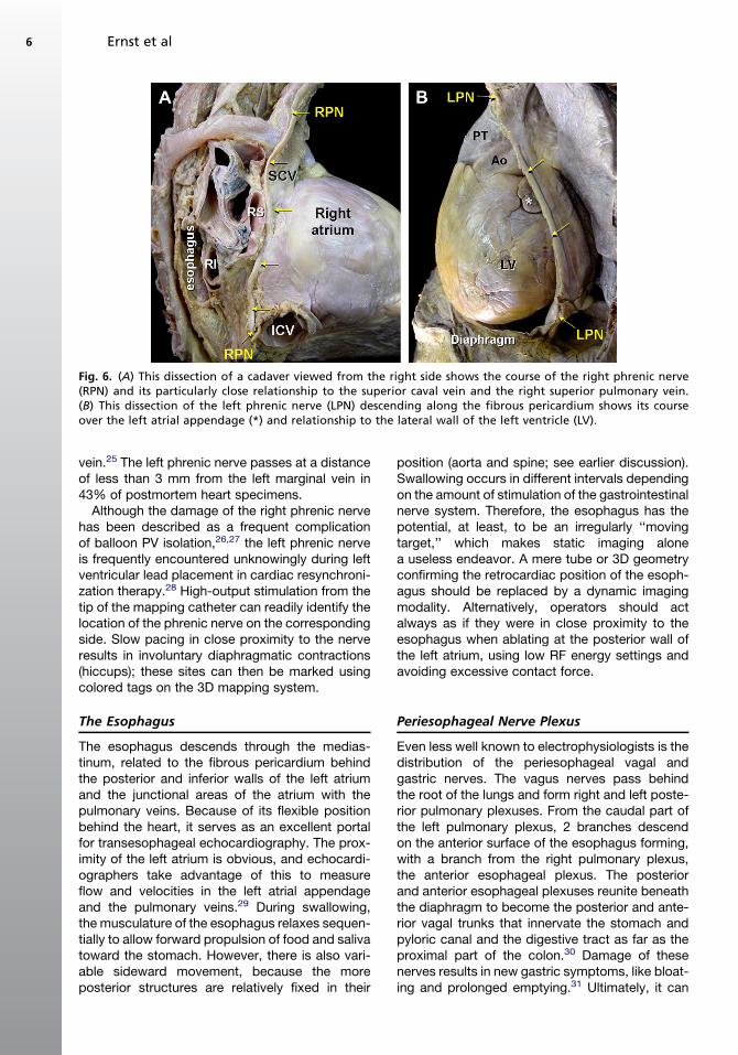
Fig. 6. (A) This dissection of a cadaver viewed from the right side shows the course of the right phrenic nerve(RPN) and its particularly close relationship to the superior caval vein and the right superior pulmonary vein.(B) This dissection of the left phrenic nerve (LPN) descending along the fibrous pericardium shows its courseover the left atrial appendage (*) and relationship to the lateral wall of the left ventricle (LV).
Ernst et al6
vein.25 The left phrenic nerve passes at a distanceof less than 3 mm from the left marginal vein in43% of postmortem heart specimens.
Although the damage of the right phrenic nervehas been described as a frequent complicationof balloon PV isolation,26,27 the left phrenic nerveis frequently encountered unknowingly during leftventricular lead placement in cardiac resynchroni-zation therapy.28 High-output stimulation from thetip of the mapping catheter can readily identify thelocation of the phrenic nerve on the correspondingside. Slow pacing in close proximity to the nerveresults in involuntary diaphragmatic contractions(hiccups); these sites can then be marked usingcolored tags on the 3D mapping system.
The Esophagus
The esophagus descends through the medias-tinum, related to the fibrous pericardium behindthe posterior and inferior walls of the left atriumand the junctional areas of the atrium with thepulmonary veins. Because of its flexible positionbehind the heart, it serves as an excellent portalfor transesophageal echocardiography. The prox-imity of the left atrium is obvious, and echocardi-ographers take advantage of this to measureflow and velocities in the left atrial appendageand the pulmonary veins.29 During swallowing,the musculature of the esophagus relaxes sequen-tially to allow forward propulsion of food and salivatoward the stomach. However, there is also vari-able sideward movement, because the moreposterior structures are relatively fixed in their
position (aorta and spine; see earlier discussion).Swallowing occurs in different intervals dependingon the amount of stimulation of the gastrointestinalnerve system. Therefore, the esophagus has thepotential, at least, to be an irregularly ‘‘movingtarget,’’ which makes static imaging alonea useless endeavor. A mere tube or 3D geometryconfirming the retrocardiac position of the esoph-agus should be replaced by a dynamic imagingmodality. Alternatively, operators should actalways as if they were in close proximity to theesophagus when ablating at the posterior wall ofthe left atrium, using low RF energy settings andavoiding excessive contact force.
Periesophageal Nerve Plexus
Even less well known to electrophysiologists is thedistribution of the periesophageal vagal andgastric nerves. The vagus nerves pass behindthe root of the lungs and form right and left poste-rior pulmonary plexuses. From the caudal part ofthe left pulmonary plexus, 2 branches descendon the anterior surface of the esophagus forming,with a branch from the right pulmonary plexus,the anterior esophageal plexus. The posteriorand anterior esophageal plexuses reunite beneaththe diaphragm to become the posterior and ante-rior vagal trunks that innervate the stomach andpyloric canal and the digestive tract as far as theproximal part of the colon.30 Damage of thesenerves results in new gastric symptoms, like bloat-ing and prolonged emptying.31 Ultimately, it can
Relevance to Epicardial Mapping and Ablation 7
result in pyloric spasms, which may necessitatedilatation or botulinum injections.32
The Diaphragm
The pericardium rests on an almost flat area of thediaphragm called the cardiac plateau, whichextends more to the left than the right. The profileof the diaphragm rises on either side of the cardiacplateau to a smooth convex dome that is higherand slightly broader on the right than on the left.Most of the inferior surface of the diaphragmis covered by the peritoneum. The right side ismolded over the convex surface of the right lobeof the liver, the right kidney, and the right supra-renal gland. The left side conforms to the left lobeof the liver, the fundus of the stomach, the spleen,the left kidney, and the left suprarenal gland.
SUMMARY
Detailed anatomic knowledge of the pericardialspace and the neighboring structures inside themediastinum is the key to entering this novelspace. Accurate imaging and understanding ofthe 3D relationship should avoid causing uninten-tional ‘‘collateral damage’’ by catheter ablation.
REFERENCES
1. Ouyang F, Fotuhi P, Ho SY, et al. Repetitive mono-
morphic ventricular tachycardia originating from
the aortic sinus cusp: electrocardiographic charac-
terization for guiding catheter ablation. J Am Coll
Cardiol 2002;39(3):500–8.
2. Sun Y, Arruda M, Otomo K, et al. Coronary sinus-
ventricular accessory connections producing post-
eroseptal and left posterior accessory pathways:
incidence and electrophysiological identification.
Circulation 2002;106(11):1362–7.
3. Sosa E, Scanavacca M, d’Avila A, et al. A new tech-
nique to perform epicardial mapping in the electro-
physiology laboratory. J Cardiovasc Electrophysiol
1996;7(6):531–6.
4. Waxman HL, Buxton AE, Marchlinski FE, et al.
Medical versus surgical treatment of tachydysrhyth-
mias. Eur Heart J 1984;5(Suppl B):103–8.
5. Cui YQ, Sun LB, Li Y, et al. Intraoperative modified
Cox mini-maze procedure for long-standing persis-
tent atrial fibrillation. Ann Thorac Surg 2008;85(4):
1283–9.
6. McClelland JH, Duke D, Reddy R. Preliminary
results of a limited thoracotomy: new approach to
treat atrial fibrillation. J Cardiovasc Electrophysiol
2007;18(12):1289–95.
7. Schweikert RA, Saliba WI, Tomassoni G, et al. Percuta-
neous pericardial instrumentation for endo-epicardial
mapping of previously failed ablations. Circulation
2003;108(11):1329–35.
8. Soejima K, Stevenson WG, Sapp JL, et al. Endocar-
dial and epicardial radiofrequency ablation of
ventricular tachycardia associated with dilated
cardiomyopathy: the importance of low-voltage
scars. J Am Coll Cardiol 2004;43(10):1834–42.
9. Garcia FC, Bazan V, Zado ES, et al. Epicardial
substrate and outcome with epicardial ablation of
ventricular tachycardia in arrhythmogenic right
ventricular cardiomyopathy/dysplasia. Circulation
2009;120(5):366–75.
10. D’Avila A, Scanavacca M, Sosa E, et al. Pericardial
anatomy for the interventional electrophysiologist.
J Cardiovasc Electrophysiol 2003;14(4):422–30.
11. Cappato R. NICE guidance on catheter ablation of atrial
fibrillation using an epicardial (non-thorascoscopic)
approach. Heart 2009;95:1956–7.
12. Tsang TS, Freeman WK, Barnes ME, et al. Rescue
echocardiographically guided pericardiocentesis
for cardiac perforation complicating catheter-based
procedures. The Mayo Clinic experience. J Am Coll
Cardiol 1998;32(5):1345–50.
13. Grimard C, Lacotte J, Hidden-Lucet F, et al. Percuta-
neous epicardial radiofrequency ablation of ventric-
ular arrhythmias after failure of endocardial
approach: a 9-year experience. J Cardiovasc Elec-
trophysiol 2009. [Epub ahead of print].
14. d’Avila A, Scanavacca M, Sosa E. Transthoracic
epicardial catheter ablation of ventricular tachy-
cardia. Heart Rhythm 2006;3(9):1110–1.
15. Hou D, March KL. A novel percutaneous technique
for accessing the normal pericardium: a single-
center successful experience of 53 porcine proce-
dures. J Invasive Cardiol 2003;15(1):13–7.
16. Hsu LF, Scavee C, Jais P, et al. Transcardiac pericar-
diocentesis: an emergency life-saving technique for
cardiac tamponade. J Cardiovasc Electrophysiol
2003;14(9):1001–3.
17. Zeppenfeld K, Tops LF, Bax JJ, et al. Images in
cardiovascular medicine. Epicardial radiofrequency
catheter ablation of ventricular tachycardia in the
vicinity of coronary arteries is facilitated by fusion
of 3-dimensional electroanatomical mapping with
multislice computed tomography. Circulation 2006;
114(3):e51–2.
18. Armour JA, Murphy DA, Yuan BX, et al. Gross and
microscopic anatomy of the human intrinsic cardiac
nervous system. Anat Rec 1997;247(2):289–98.
19. Pauza DH, Skripka V, Pauziene N, et al. Morphology,
distribution, and variability of the epicardiac neural
ganglionated subplexuses in the human heart.
Anat Rec 2000;259(4):353–82.
20. Hou Y, Scherlag BJ, Lin J, et al. Interactive atrial
neural network: determining the connections
between ganglionated plexi. Heart Rhythm 2007;
4(1):56–63.
Ernst et al8
21. Scanavacca M, Pisani CF, Hachul D, et al. Selective
atrial vagal denervation guided by evoked vagal
reflex to treat patients with paroxysmal atrial fibrilla-
tion. Circulation 2006;114(9):876–85.
22. Tan AY, Li H, Wachsmann-Hogiu S, et al. Autonomic
innervation and segmental muscular disconnections
at the human pulmonary vein-atrial junction: implica-
tions for catheter ablation of atrial-pulmonary vein
junction. J Am Coll Cardiol 2006;48(1):132–43.
23. Tan AY, Chen PS, Chen LS, et al. Autonomic nerves
in pulmonary veins. Heart Rhythm 2007;4(Suppl 3):
S57–60.
24. Sanchez-Quintana D, Cabrera JA, Climent V, et al.
How close are the phrenic nerves to cardiac struc-
tures? Implications for cardiac interventionalists.
J Cardiovasc Electrophysiol 2005;16(3):309–13.
25. Sanchez-Quintana D, Ho SY, Climent V, et al.
Anatomic evaluation of the left phrenic nerve rele-
vant to epicardial and endocardial catheter ablation:
implications for phrenic nerve injury. Heart Rhythm
2009;6(6):764–8.
26. Chun KR, Schmidt B, Metzner A, et al. The ‘single
big cryoballoon’ technique for acute pulmonary
vein isolation in patients with paroxysmal atrial fibril-
lation: a prospective observational single centre
study. Eur Heart J 2009;30(6):699–709.
27. Neumann T, Vogt J, Schumacher B, et al. Circumfer-
ential pulmonary vein isolation with the cryoballoon
technique results from a prospective 3-center study.
J Am Coll Cardiol 2008;52(4):273–8.
28. Leclercq C, Gadler F, Kranig W, et al. A randomized
comparison of triple-site versus dual-site ventricular
stimulation in patients with congestive heart failure.
J Am Coll Cardiol 2008;51(15):1455–62.
29. Schneider C, Ernst S, Malisius R, et al. Transeso-
phageal echocardiography: a follow-up tool after
catheter ablation of atrial fibrillation and interven-
tional therapy of pulmonary vein stenosis and
occlusion. J Interv Card Electrophysiol 2007;18(2):
195–205.
30. Ho SY, Cabrera JA, Sanchez-Quintana D. Vagaries
of the vagus nerve: relevance to ablationists.
J Cardiovasc Electrophysiol 2006;17(3):330–1.
31. Shah D, Dumonceau JM, Burri H, et al. Acute pyloric
spasm and gastric hypomotility: an extracardiac
adverse effect of percutaneous radiofrequency
ablation for atrial fibrillation. J Am Coll Cardiol
2005;46(2):327–30.
32. Pisani CF, Hachul D, Sosa E, et al. Gastric hypomo-
tility following epicardial vagal denervation ablation
to treat atrial fibrillation. J Cardiovasc Electrophysiol
2008;19(2):211–3.