Embed Size (px)
Citation preview
Anatomy & Physiology of the Excretory System
Anatomy & Physiology 13-14
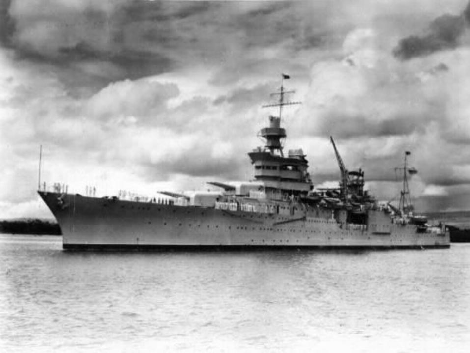
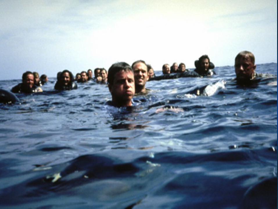
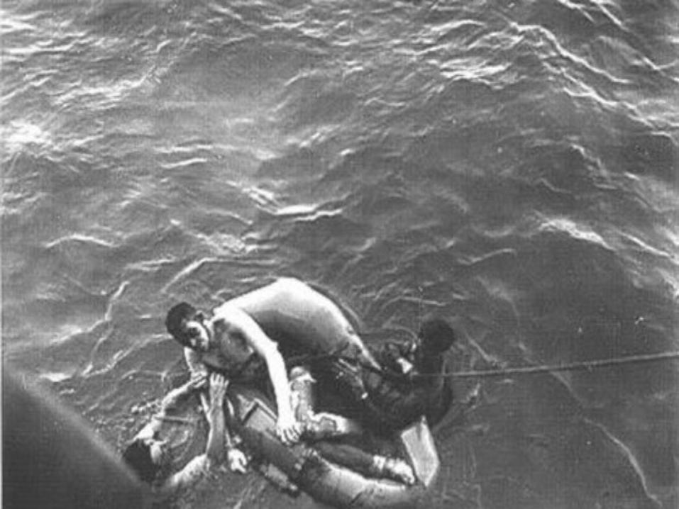

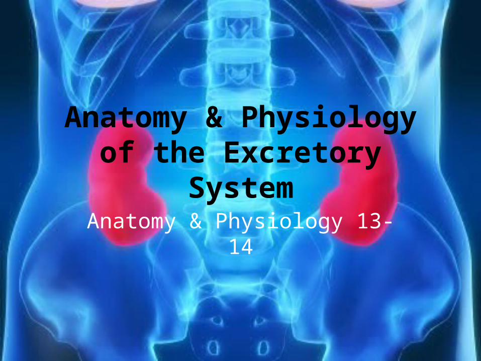
Why Are We Not Advised To Drink Seawater?
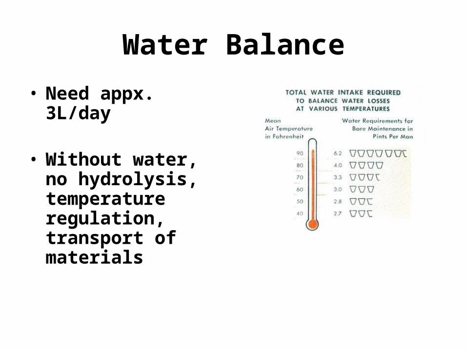
Water Balance
• Need appx. 3L/day
• Without water, no hydrolysis, temperature regulation, transport of materials
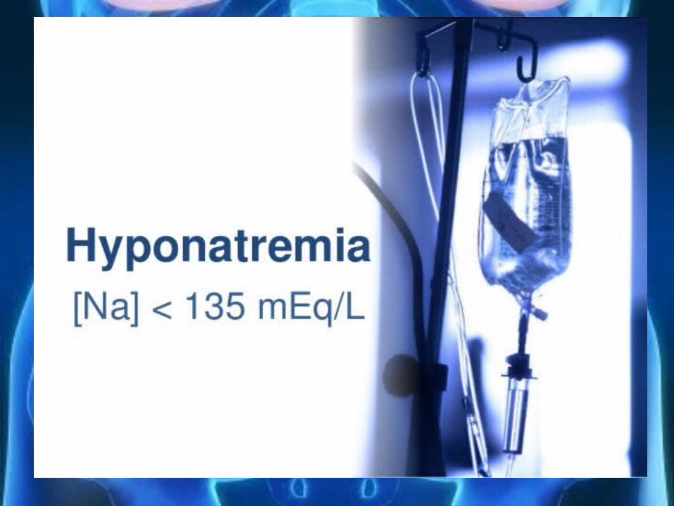
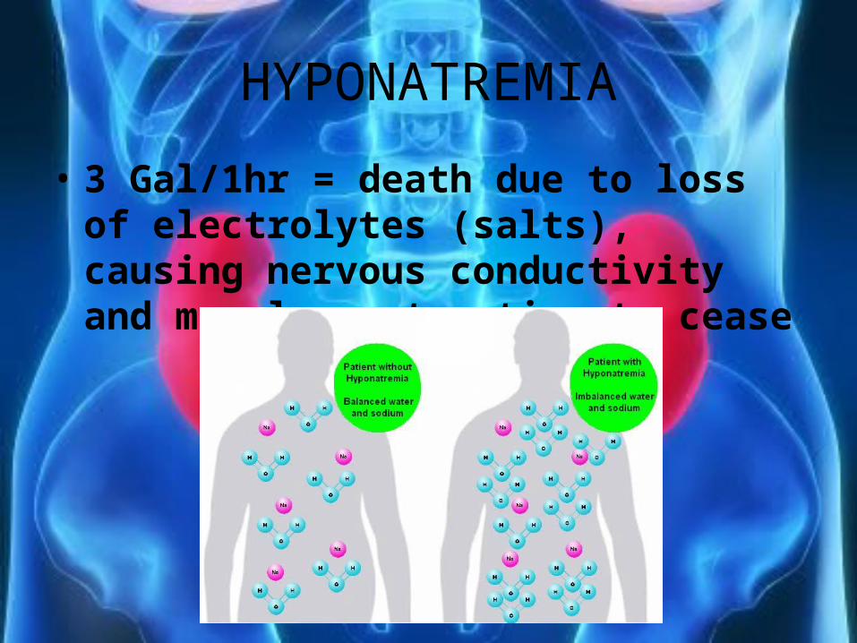
HYPONATREMIA
• 3 Gal/1hr = death due to loss of electrolytes (salts), causing nervous conductivity and muscle contraction to cease
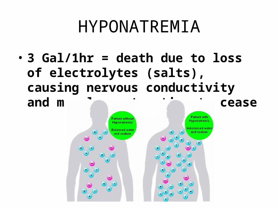
HYPONATREMIA
• 3 Gal/1hr = death due to loss of electrolytes (salts), causing nervous conductivity and muscle contraction to cease
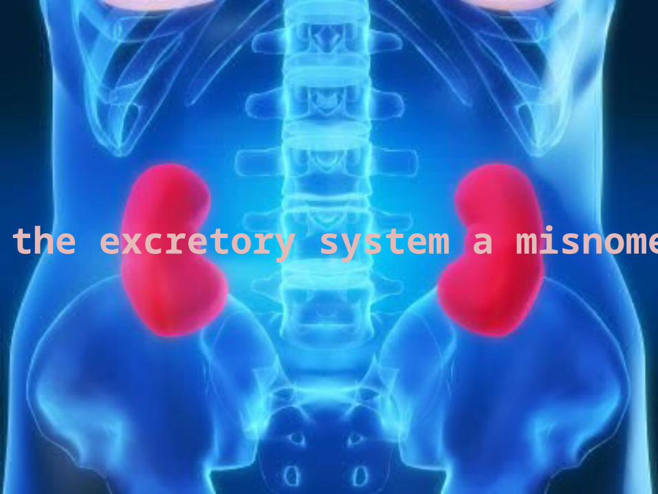
Is the excretory system a misnomer?
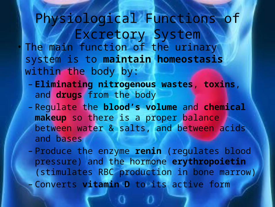
Physiological Functions of Excretory System• The main function of the urinary system is to
maintain homeostasis within the body by:– Eliminating nitrogenous wastes, toxins, and drugs
from the body– Regulate the blood’s volume and chemical
makeup so there is a proper balance between water & salts, and between acids and bases
– Produce the enzyme renin (regulates blood pressure) and the hormone erythropoietin (stimulates RBC production in bone marrow)
– Converts vitamin D to its active form
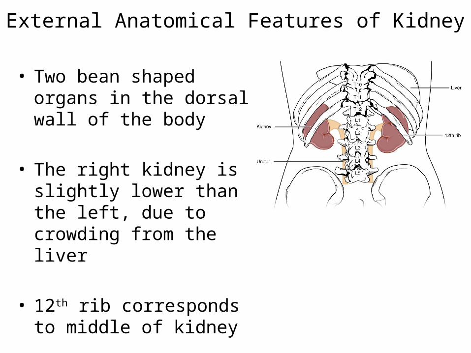
External Anatomical Features of Kidney
• Two bean shaped organs in the dorsal wall of the body
• The right kidney is slightly lower than the left, due to crowding from the liver
• 12th rib corresponds to middle of kidney
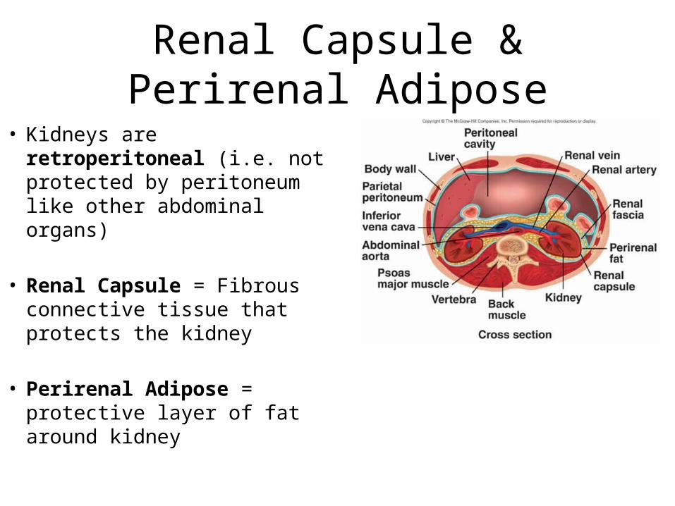
Renal Capsule & Perirenal Adipose
• Kidneys are retroperitoneal (i.e. not protected by peritoneum like other abdominal organs)
• Renal Capsule = Fibrous connective tissue that protects the kidney
• Perirenal Adipose = protective layer of fat around kidney
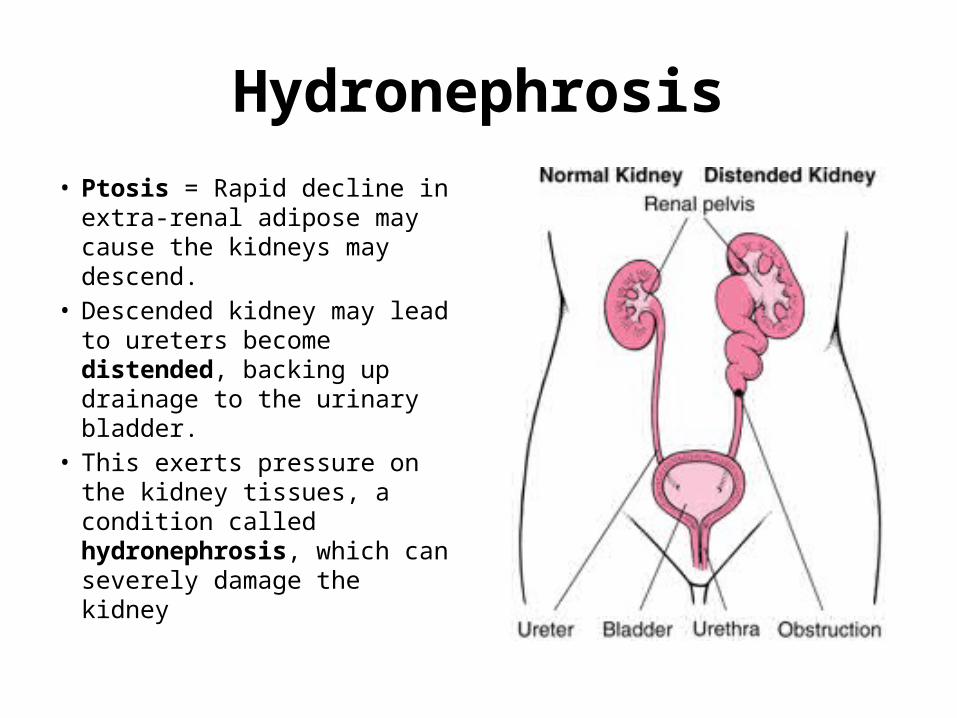
Hydronephrosis• Ptosis = Rapid decline in
extra-renal adipose may cause the kidneys may descend.
• Descended kidney may lead to ureters become distended, backing up drainage to the urinary bladder.
• This exerts pressure on the kidney tissues, a condition called hydronephrosis, which can severely damage the kidney
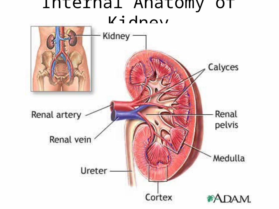
Internal Anatomy of Kidney
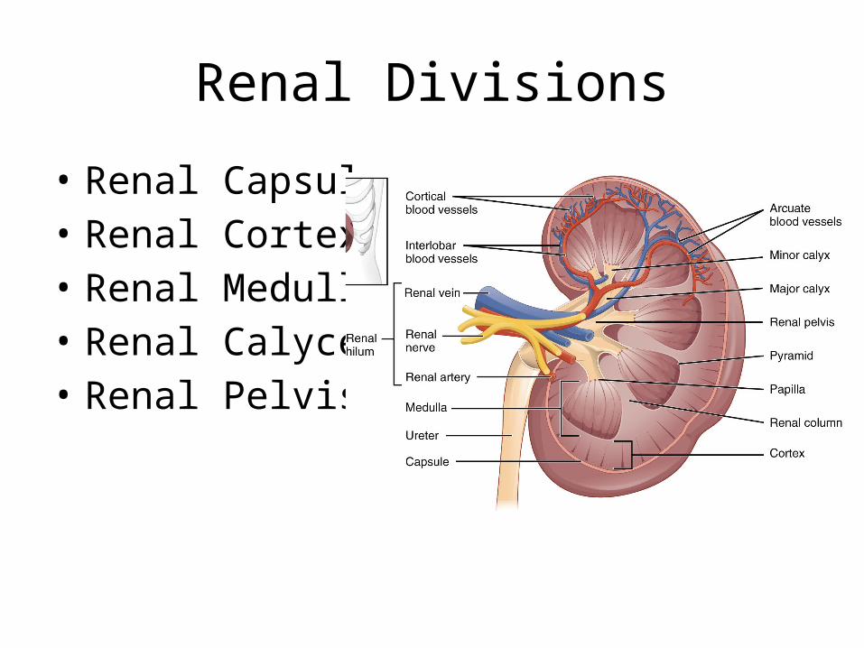
Renal Divisions
• Renal Capsule• Renal Cortex• Renal Medulla• Renal Calyces• Renal Pelvis
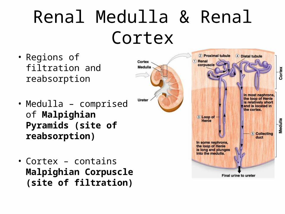
Renal Medulla & Renal Cortex
• Regions of filtration and reabsorption
• Medulla – comprised of Malpighian Pyramids (site of reabsorption)
• Cortex – contains Malpighian Corpuscle (site of filtration)
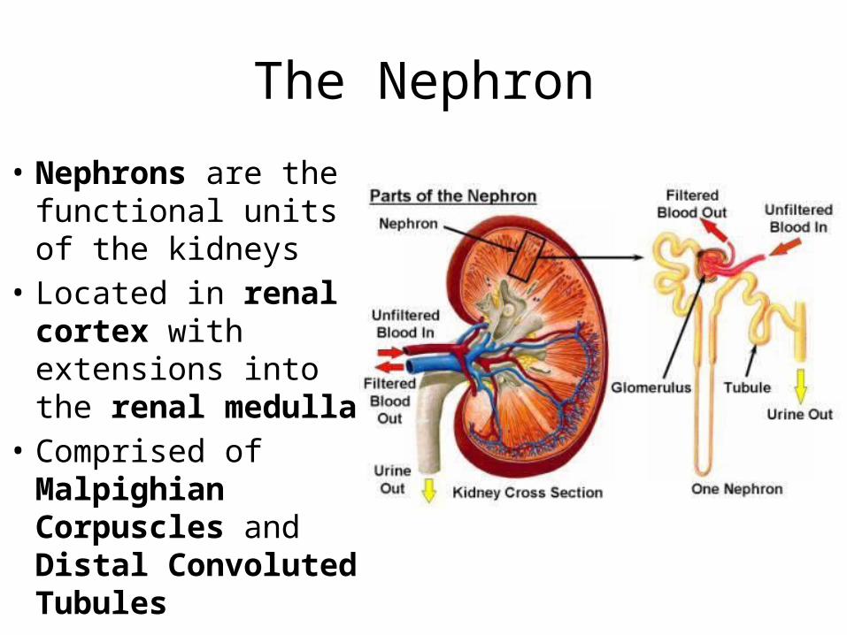
The Nephron
• Nephrons are the functional units of the kidneys
• Located in renal cortex with extensions into the renal medulla
• Comprised of Malpighian Corpuscles and Distal Convoluted Tubules
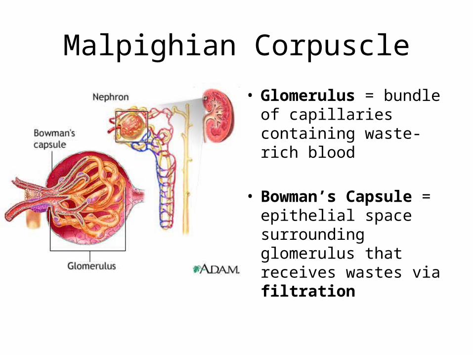
Malpighian Corpuscle
• Glomerulus = bundle of capillaries containing waste-rich blood
• Bowman’s Capsule = epithelial space surrounding glomerulus that receives wastes via filtration
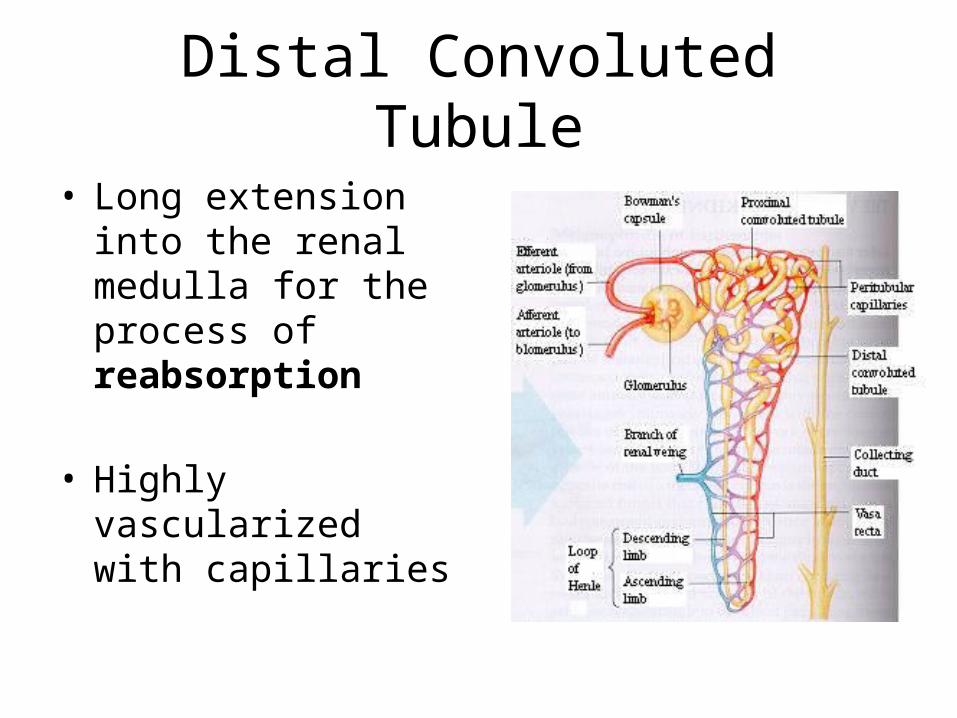
Distal Convoluted Tubule
• Long extension into the renal medulla for the process of reabsorption
• Highly vascularized with capillaries
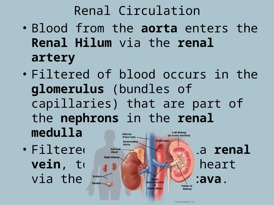
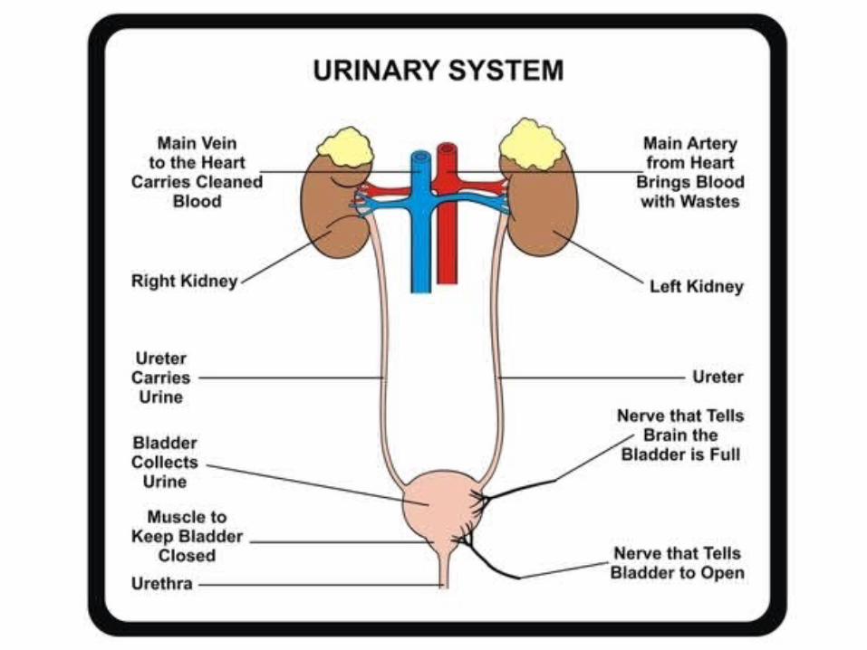
Renal Circulation• Blood from the aorta enters the Renal Hilum
via the renal artery• Filtered of blood occurs in the glomerulus
(bundles of capillaries) that are part of the nephrons in the renal medulla
• Filtered blood exits via renal vein, to return to the heart via the inferior vena cava.
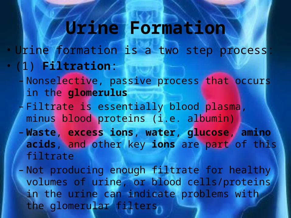
Urine Formation• Urine formation is a two step process:• (1) Filtration: – Nonselective, passive process that occurs in the
glomerulus– Filtrate is essentially blood plasma, minus blood
proteins (i.e. albumin)– Waste, excess ions, water, glucose, amino acids, and
other key ions are part of this filtrate– Not producing enough filtrate for healthy volumes of
urine, or blood cells/proteins in the urine can indicate problems with the glomerular filters
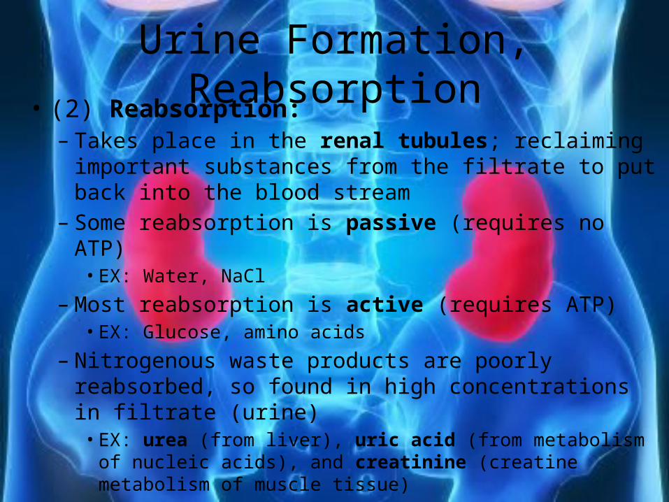
Urine Formation, Reabsorption• (2) Reabsorption:– Takes place in the renal tubules; reclaiming important
substances from the filtrate to put back into the blood stream
– Some reabsorption is passive (requires no ATP)• EX: Water, NaCl
– Most reabsorption is active (requires ATP)• EX: Glucose, amino acids
– Nitrogenous waste products are poorly reabsorbed, so found in high concentrations in filtrate (urine)• EX: urea (from liver), uric acid (from metabolism of nucleic
acids), and creatinine (creatine metabolism of muscle tissue)
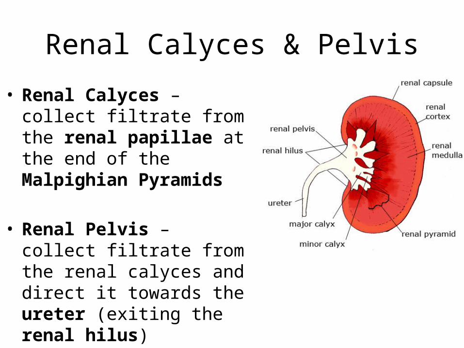
Renal Calyces & Pelvis
• Renal Calyces – collect filtrate from the renal papillae at the end of the Malpighian Pyramids
• Renal Pelvis – collect filtrate from the renal calyces and direct it towards the ureter (exiting the renal hilus)
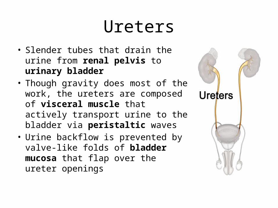
Ureters• Slender tubes that drain the urine
from renal pelvis to urinary bladder• Though gravity does most of the
work, the ureters are composed of visceral muscle that actively transport urine to the bladder via peristaltic waves
• Urine backflow is prevented by valve-like folds of bladder mucosa that flap over the ureter openings
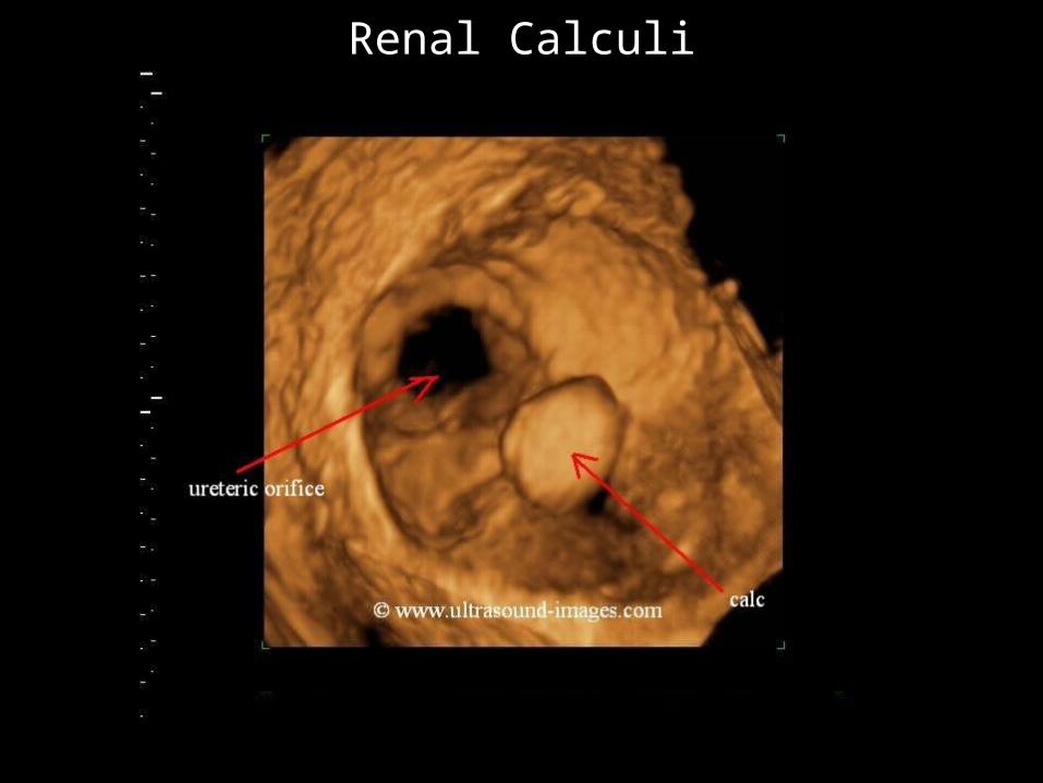
Renal Calculi
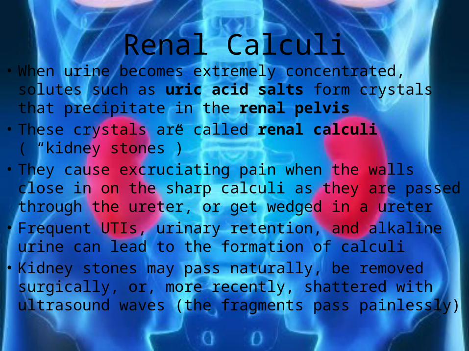
Renal Calculi• When urine becomes extremely concentrated, solutes
such as uric acid salts form crystals that precipitate in the renal pelvis
• These crystals are called renal calculi ( “kidney stones”)• They cause excruciating pain when the walls close in on
the sharp calculi as they are passed through the ureter, or get wedged in a ureter
• Frequent UTIs, urinary retention, and alkaline urine can lead to the formation of calculi
• Kidney stones may pass naturally, be removed surgically, or, more recently, shattered with ultrasound waves (the fragments pass painlessly)
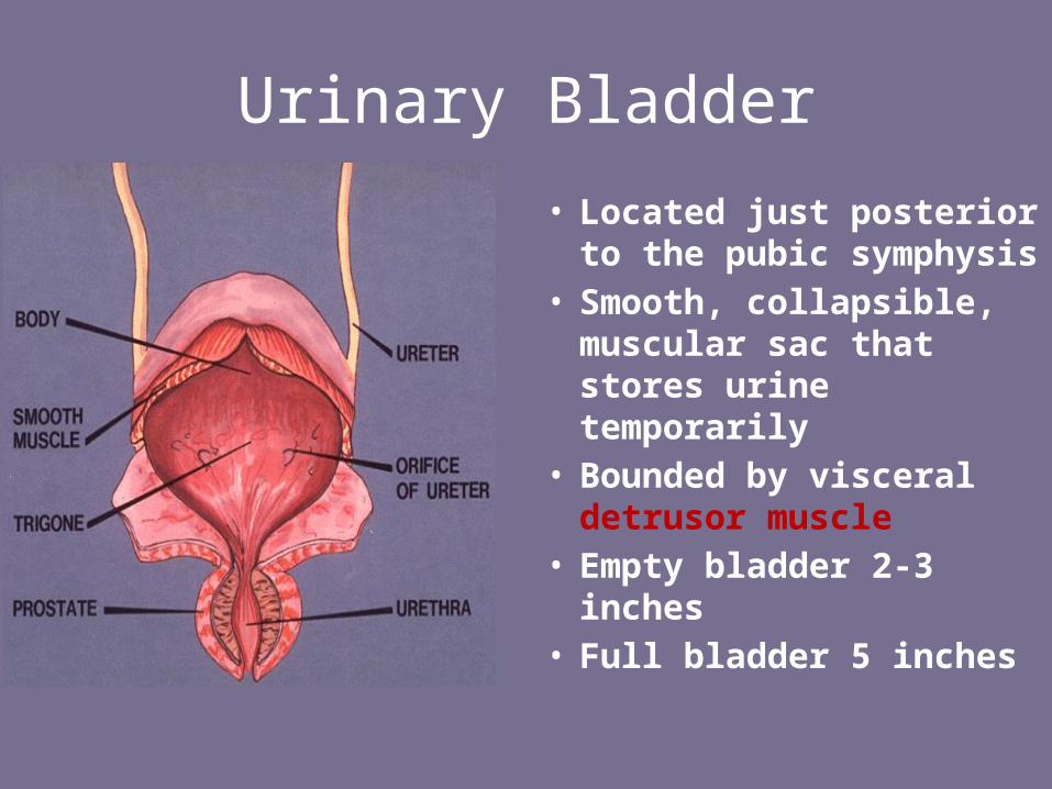
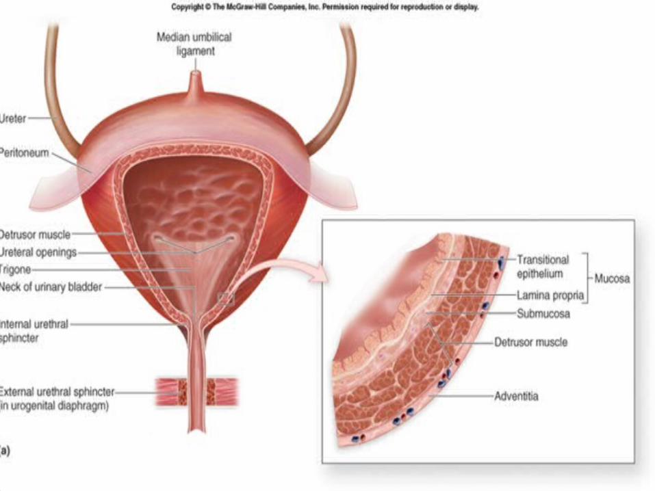
Urinary Bladder
• Located just posterior to the pubic symphysis
• Smooth, collapsible, muscular sac that stores urine temporarily
• Bounded by visceral detrusor muscle
• Empty bladder 2-3 inches• Full bladder 5 inches
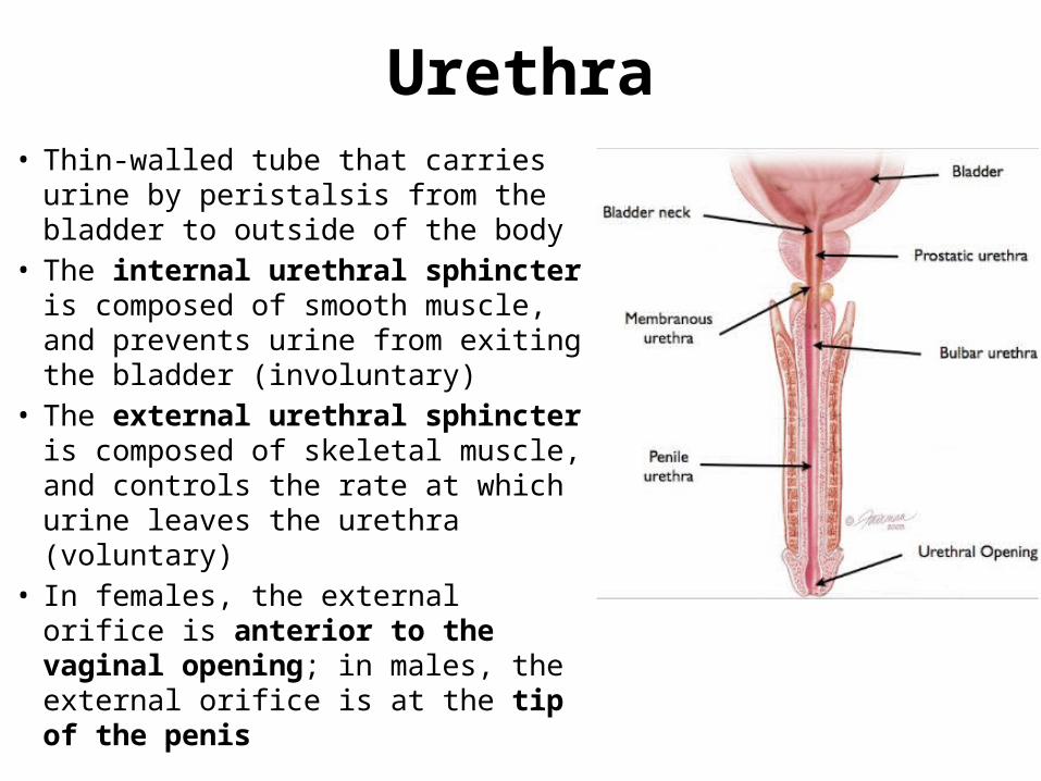
Urethra• Thin-walled tube that carries urine by
peristalsis from the bladder to outside of the body
• The internal urethral sphincter is composed of smooth muscle, and prevents urine from exiting the bladder (involuntary)
• The external urethral sphincter is composed of skeletal muscle, and controls the rate at which urine leaves the urethra (voluntary)
• In females, the external orifice is anterior to the vaginal opening; in males, the external orifice is at the tip of the penis
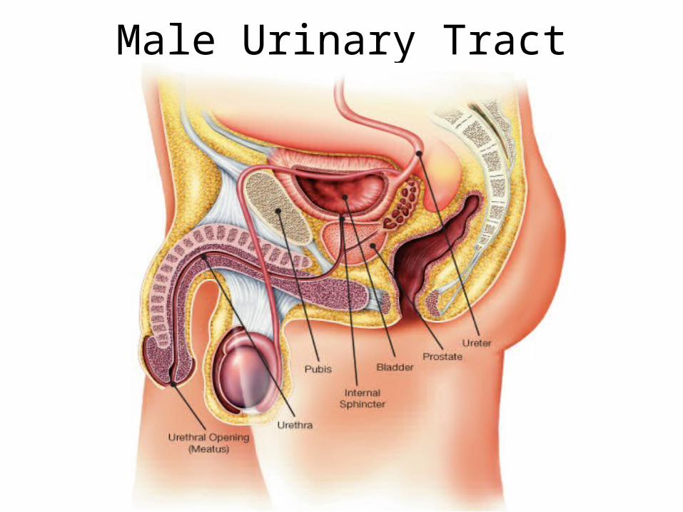
Male Urinary Tract
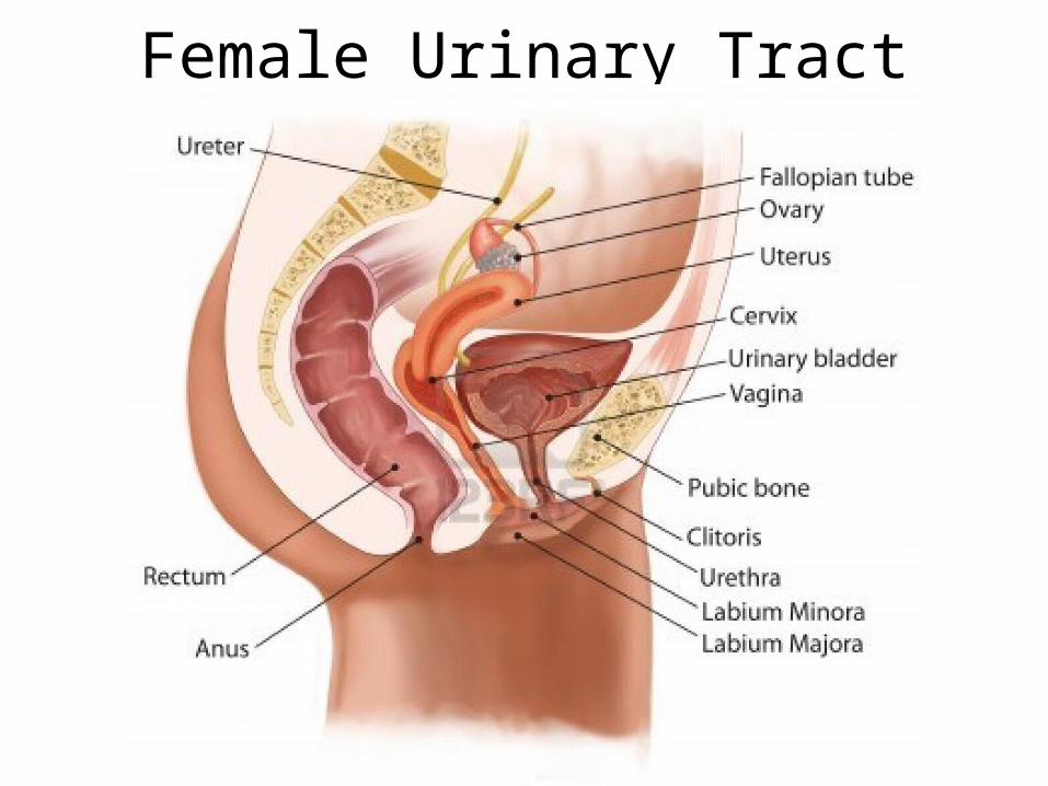
Female Urinary Tract
Micturition• “Voiding” (emptying bladder)• When ~200mL of urine have collected in the
bladder, this stretching activates receptors• The pelvic splanchnic nerves trigger the
internal urethral sphincter to open, allowing urine into the upper part of the urethra
• This is when you feel the urge to go to the bathroom; however, you control the external urethral sphincter (to a point)
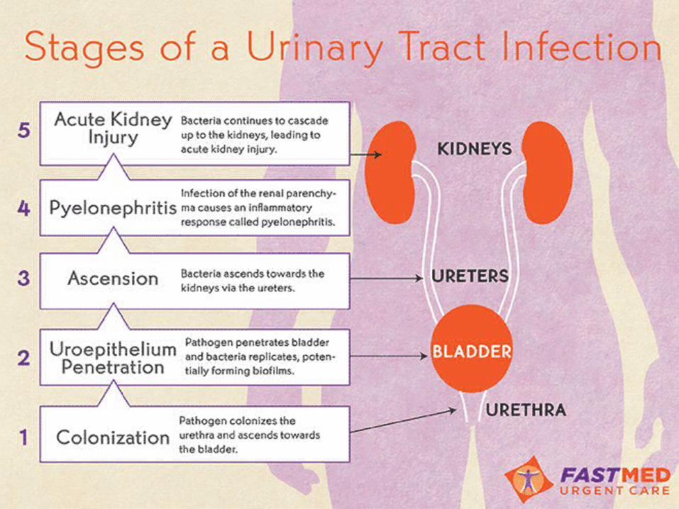
Urinary Tract Infections (UTIs)• Bacterial infection of one part of the urinary tract• Cystitis is a bladder infection; typically
accompanied by frequent urges, burning sensation during urination, cloudy urine
• Urethritis is inflammation of the ureters; marked by frequent urges to urinate & painful urination
• Women are 30 times more likely to develop a UTI than men, due to anatomical differences in the opening of the urethra
• Cranberries contain a substance that helps prevent bacteria from adhering to the bladder walls