Embed Size (px)
Citation preview
Proc. Natl. Acad. Sci. USAVol. 86, pp. 1939-1943, March 1989Genetics
Ancient DNA: Extraction, characterization, molecular cloning,and enzymatic amplification
(oxidative damage/electron microscopy/Alu sequences/mitochondrial DNA/polymerase chain reaction)
SVANTE PAABODepartment of Biochemistry, University of California, Berkeley, CA 94720*; and Institut fur Molekularbiologie II der Universitat Zurich,Honggerberg, CH-8093 Zirich, Switzerland
Communicated by George Klein, December 15, 1988
ABSTRACT Several chemical and enzymatic propertieswere examined in the DNA extracted from dry remains of softtissues that vary in age from 4 to 13,000 years and representfour species, including two extinct animals (the marsupial wolfand giant ground sloth). The DNA obtained was invariably ofa low average molecular size and damaged by oxidative pro-cesses, which primarily manifest themselves as modifications ofpyrimidines and sugar residues as well as baseless sites andintermolecular cross-links. This renders molecular cloningdifficult. However, the polymerase chain reaction can be usedto amplify and study short mitochondrial DNA sequences thatare of anthropological and evolutionary significance. Thisopens up the prospect of performing diachronical studies ofmolecular evolutionary genetics.
DNA from ancient organic remains has been extracted in anumber of cases (e.g., see refs. 1-5). On two occasions,molecular cloning made it possible to obtain DNA sequencesfrom such remains (6, 7). However, a serious concernpertinent to the study of ancient DNA is the occurrence ofpostmortem damage in DNA extracted from archaeologicalspecimens, since such damage may make impossible theapplication of many molecular biological techniques and/orcause erroneous sequence information to be obtained (8, 9).To make general statements about the state ofpreservation
of DNA in ancient dry remains of soft tissues, I haveextracted nucleic acids from 12 specimens representing awide variety of geographical regions and different timeperiods. Here I report the nature of the chemical modifica-tions present in these DNA samples and show that, whereasthese modifications render molecular cloning techniquesdifficult and possibly error prone, the recently developedpolymerase chain reaction (PCR) is likely to produce reliablesequence information from many ancient tissue remains. Thishas wide implications in that it opens up the possibility ofusing museum specimens as well as archaeological finds toaddress questions of historic, evolutionary, and taxonomicsignificance.
MATERIALS AND METHODSSamples. Twelve specimens (A-L) were used for DNA
extraction: A, a 4-year-old piece of dried pork from Loten,Norway; B, a piece of skin from a stuffed marsupial wolf(Thylacinus cynocephalus), kept since 1869 in the ZoologicalMuseum of the University, Zurich; C, a 13,000-year-old skinfrom a ground sloth (Mylodon) originating from the UltimaEsperanza Cave, Chile, now in the British Museum (NaturalHistory), London; D, skin from a natural mummy fromSayala, Egyptian Nubia, 6th-12th century AD (body K9/7);E, same as D (body K13/2); F, skin from a 5000-year-old
natural mummy from Gebelein, Egypt (British Museum, no.32754) (10); G, same as F (no. 32753); H, same as F (no.32755); 1, colon tissue from Egyptian mummy ROM I, ca.1200 BC, in the Royal Ontario Museum, Toronto (11); J, largeorgan package, found to the left in the chest cavity ofEgyptian mummy AS73b, 3rd century BC, belonging to theInstitute for Anthropology and Human Genetics, Universityof Munich (12); K, a mummified human head from Peru, 4th-5th century AD (Las Trancas 83, new number 606), in theInstitute for Anthropology and Human Genetics, Universityof Munich (13); L, liver tissue from canopic jar of Egyptianmummy 21470, 20th-19th century BC, in the ManchesterMuseum, Manchester, U.K. (14).DNA Extraction and Miscellaneous Methods. DNA was
extracted by a modification of the Blin and Stafford proce-dure (15) that has been described by Paabo et al. (16). Afterethanol precipitation, a brown contaminant, probably repre-senting Maillard products of reducing sugars (17), was re-moved from the nucleic acids by centrifugation through a 10-40% sucrose gradient in 10 mM Tris HCl, pH 8.0/1 M NaCi(18) at 30,000 rpm (100,000 x g) (15'C for 24 hr). The elutedfractions were ethanol precipitated and resuspended in 50,ulof Tris salt buffer. Fractions containing nucleic acids werepooled from the middle third of each gradient. The browncontaminants remained on top of the gradients. For theextracts used for enzymatic amplification, ethanol precipita-tion and sucrose gradients were replaced by centrifugation onCentricon 30 filters (Amicon) to minimize losses of DNA. Inaddition, control extractions were performed in an identicalmanner except that no tissue was added to the extractionmixture.Agarose gels, ethidium bromide staining, nick-translation
of the probe containing an Alu repeat (19), end-labeling(without prior phosphatase treatment), precipitation withtrichloroacetic acid (TCA), hybridization, and autoradiogra-phy were by standard techniques (18). Hydrolysis of DNAand high-performance liquid chromatography (HPLC) of thebases were performed as described (20).
Quantitation of the ancient DNA was performed by anethidium bromide dot assay (18) as well as estimations fromethidium bromide-stained gels. Determinations of concentra-tions by absorbance at 260 nm proved impossible to performin most extracts because of an unknown component thatexhibited peak absorption at -215 nm. All determinations ofancient DNA concentrations should be regarded as tentativesince it is not known how the lesions present in the DNAaffect its ability to intercalate fluorescent dyes.
Alkali sensitivity was assayed by adding 0.5 .ug of salmonsperm DNA to the end-labeled sample followed by NaOH toa final concentration of 0.3 M. Incubation was at room
Abbreviations: PCR, polymerase chain reaction; TCA, trichloroace-tic acid; endo III and IV, endonucleases III and IV; AP, apur-inic/apyrimidinic.*Present address.
1939
The publication costs of this article were defrayed in part by page chargepayment. This article must therefore be hereby marked "advertisement"in accordance with 18 U.S.C. §1734 solely to indicate this fact.

Proc. Natl. Acad. Scd. USA 86 (1989)
temperature overnight. Endonuclease IV (endo IV) digestionwas performed in 10 mM NaCl/50 mM Hepes, pH 7.8/5 mMdithiothreitol; endonuclease III (endo III) digestion was in100 mM KCI/50 mM Hepes, pH 7.8/1 mM EDTA/1 mMdithiothreitol; and uracil-DNA glycosylase digestion was in70 mM Hepes, pH 7.8/1 mM EDTA/1 mM dithiothreitol. Allenzymatic digestions were for 1 hr at 370C. In the case ofuracil-DNA glycosylase, NaCi was then added to a finalconcentration of 100 mM and endo IV was added for anadditional incubation of 1 hr. Before TCA precipitation, 1 ,gof salmon sperm DNA was added to the digests. Controldigestions were performed as described above except that noenzyme was added to the incubation mixtures. The purifica-tion of endo IV (21), endo III (22), and uracil-DNA glyco-sylase (23) has been described.
Electron microscopy was performed essentially as de-scribed (24).Molecular Cloning and Enzymatic Amplification. Mung
bean nuclease treatment was performed according to themanufacturer's directions. Cloning in pUC19 (25) was doneby standard techniques; isolated supercoiled plasmid DNAwas sequenced by the dideoxynucleotide chain-terminationmethod (26).Enzymatic amplification by the PCR (27) was performed as
described (16) using heat-resistant Thermus aquaticus (Taq)DNA polymerase (28). The primers used to amplify andsequence human mitochondrial DNA were as follows: D3E,5'-GCGAATTCCTAGTGGGTGAGGGGTGGC-3' 16255;D18X, 5'-GCTCTAGACCATGCTTACAAGCAAGT-3'16209; cytb2, 5'-AAACTGCAGCCCCTCAGAATGATATT-TGTCCTCA-3' 15149; M14725(H), 5'-CGAAGCITGATAT-GAAAAACCATCGTTG-3' 14724. Numbers at 3' ends referto Anderson et al. (29). Underlined sequences were added tothe 5' ends to create restriction sites. Primers A and B,specific for region V of the mitochondrial genome, have beendescribed (30). Single-stranded template for sequencing wasgenerated by the unbalanced primer method (31).
RESULTSDNA Extraction. DNA was prepared from 0.1-0.5 g of the
samples and 8% of each extract was analyzed by agarose gelelectrophoresis. In all extracts discussed here and in >90%of all extracts prepared from well-preserved desiccatedtissues, DNA could be visualized by ethidium bromidestaining of agarose gels (Fig. 1). The DNA invariably provedto be degraded to an average molecular size of 100-200 basepairs (bp) with substantial amounts ofDNA migrating in therange of 40-500 bp. The yield of DNA as determined byethidium bromide fluorescence varied between 1 and 200 ,g
ABCDEFGHIJKL
-527
- 122
FIG. 1. Agarose gel electrophoresis of DNA extracted from theremains of 12 old dry tissues. The gel contained 2% agarose and DNAwas visualized by ethidium bromide. Letters refer to the samplesanalyzed (see Materials and Methods). Migration positions ofmolecular size markers are indicated in bp.
per g of dry tissue. The majority of the samples seemed to bedouble-stranded, since they stained green with acridineorange (ref. 2; data not shown). However, when the amountof human DNA in the extracts of ancient human tissue wasdetermined by probing aliquots of the extracts, which hadbeen immobilized on nitrocellulose filters, with a nick-translated probe containing a human Alu repeat, the amountsof extracted human DNA varied between 0 and 200 ng per gof tissue-i.e., 3 orders of magnitude lower (data not shown).Since one explanation for this discrepancy could be asubstantial amount of damage occurring in the ancient DNA,the extracted DNA samples were analyzed for base damage.DNA Damage. Approximately 400 ng ofDNA from each of
the extractions was hydrolyzed under acid conditions and thereleased nitrogenous bases were analyzed by reversed-phaseHPLC. As shown in Fig. 2, a sample extracted from 4-year-old dried porcine muscle contained the four unmodifiedbases as well as two additional minor peaks. This chromato-graphic picture was indistinguishable from a DNA sampleextracted from a human cell line. In contrast, the ancientDNA samples showed a drastically different pattern.Whereas adenine and guanine could still be resolved inrelative amounts approximately similar to those present inthe contemporary sample, cytosine and thymine were pres-ent in greatly reduced amounts, the latter at <5% of theexpected amount. In addition, a number of new peaksappeared in the analyses of the ancient samples. Most ofthese peaks eluted early in the chromatogram and presum-ably represent ring-fragmented and/or ring-saturated pyrim-idine derivatives, which are known to elute early on reversed-phase HPLC analysis (e.g., see ref. 32).
Since pyrimidines, in particular thymine, are known to besubstantially more sensitive to oxidative damage than purines(33), it was speculated that a large proportion of the basemodifications observed might be accounted for by oxidativedamage. Therefore, aliquots of the extracted DNA sampleswere end-labeled and analyzed for lesions by treatment with
i010
.tC 1
0%
Elution volume (ml)
FIG. 2. HPLC analysis of hydrolyzed DNA. Samples analyzedwere four-year-old dried pork (A), skin of marsupial wolf (B), skin ofground sloth (C), 4000-year-old mummified liver (L). a and (,injection artifacts.
1940 Genetics: Pddbo
Proc. Natl. Acad. Sci. USA 86 (1989) 1941
alkali or various enzymes. TCA precipitations were thenperformed to determine the extent of sensitivity of the DNAsamples to the various treatments. Representative results aregiven in Table 1. The 4-year-old pork sample was notsensitive to alkali to any greater extent than a contemporarysalmon sperm DNA. However, the ancient DNAs werehighly alkali sensitive. Since oligonucleotides >20 bases longcan be precipitated by TCA (18), it was estimated that morethan one alkali-sensitive site per 20 bp must exist in theancient DNA.
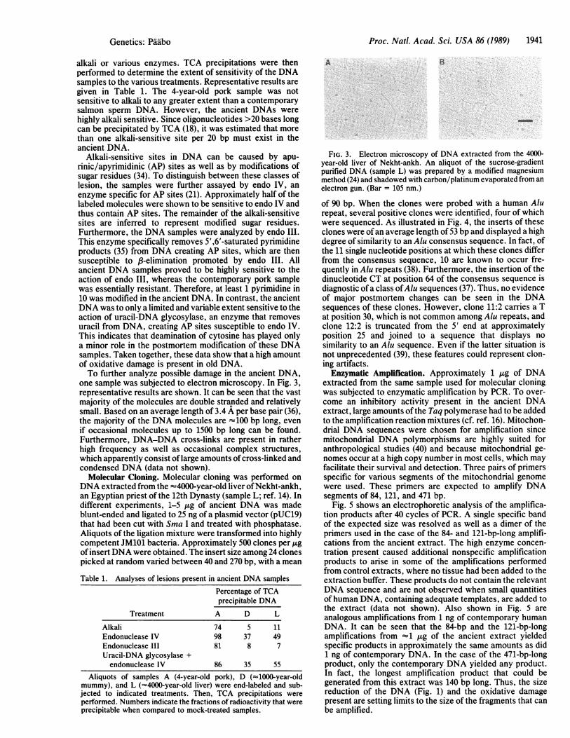
Alkali-sensitive sites in DNA can be caused by apu-rinic/apyrimidinic (AP) sites as well as by modifications ofsugar residues (34). To distinguish between these classes oflesion, the samples were further assayed by endo IV, anenzyme specific for AP sites (21). Approximately half of thelabeled molecules were shown to be sensitive to endo IV andthus contain AP sites. The remainder of the alkali-sensitivesites are inferred to represent modified sugar residues.Furthermore, the DNA samples were analyzed by endo III.This enzyme specifically removes 5',6'-saturated pyrimidineproducts (35) from DNA creating AP sites, which are thensusceptible to p-elimination promoted by endo III. Allancient DNA samples proved to be highly sensitive to theaction of endo III, whereas the contemporary pork samplewas essentially resistant. Therefore, at least 1 pyrimidine in10 was modified in the ancient DNA. In contrast, the ancientDNA was to only a limited and variable extent sensitive to theaction of uracil-DNA glycosylase, an enzyme that removesuracil from DNA, creating AP sites susceptible to endo IV.This indicates that deamination of cytosine has played onlya minor role in the postmortem modification of these DNAsamples. Taken together, these data show that a high amountof oxidative damage is present in old DNA.To further analyze possible damage in the ancient DNA,
one sample was subjected to electron microscopy. In Fig. 3,representative results are shown. It can be seen that the vastmajority of the molecules are double stranded and relativelysmall. Based on an average length of 3.4 A per base pair (36),the majority of the DNA molecules are 4100 bp long, evenif occasional molecules up to 1500 bp long can be found.Furthermore, DNA-DNA cross-links are present in ratherhigh frequency as well as occasional complex structures,which apparently consist oflarge amounts of cross-linked andcondensed DNA (data not shown).
Molecular Cloning. Molecular cloning was performed onDNA extracted from the -4000-year-old liver ofNekht-ankh,an Egyptian priest of the 12th Dynasty (sample L; ref. 14). Indifferent experiments, 1-5 ,ug of ancient DNA was madeblunt-ended and ligated to 25 ng of a plasmid vector (pUC19)that had been cut with Sma I and treated with phosphatase.Aliquots of the ligation mixture were transformed into highlycompetent JM101 bacteria. Approximately 500 clones per ,gof insertDNA were obtained. The insert size among 24 clonespicked at random varied between 40 and 270 bp, with a mean
Table 1. Analyses of lesions present in ancient DNA samples
Percentage of TCAprecipitable DNA
Treatment A D L
Alkali 74 5 11Endonuclease IV 98 37 49Endonuclease III 81 8 7Uracil-DNA glycosylase +endonuclease IV 86 35 55
Aliquots of samples A (4-year-old pork), D ("1000-year-old
A B
FIG. 3. Electron microscopy of DNA extracted from the 4000-year-old liver of Nekht-ankh. An aliquot of the sucrose-gradientpurified DNA (sample L) was prepared by a modified magnesiummethod (24) and shadowed with carbon/platinum evaporated from an
electron gun. (Bar = 105 nm.)
of 90 bp. When the clones were probed with a human Alurepeat, several positive clones were identified, four of whichwere sequenced. As illustrated in Fig. 4, the inserts of theseclones were of an average length of 53 bp and displayed a highdegree of similarity to an Alu consensus sequence. In fact, ofthe 11 single nucleotide positions at which these clones differfrom the consensus sequence, 10 are known to occur fre-quently in Alu repeats (38). Furthermore, the insertion of thedinucleotide CT at position 64 of the consensus sequence isdiagnostic ofa class ofAlu sequences (37). Thus, no evidenceof major postmortem changes can be seen in the DNAsequences of these clones. However, clone 11:2 carries a Tat position 30, which is not common among Alu repeats, andclone 12:2 is truncated from the 5' end at approximatelyposition 25 and joined to a sequence that displays no
similarity to an Alu sequence. Even if the latter situation isnot unprecedented (39), these features could represent clon-ing artifacts.Enzymatic Amplification. Approximately 1 tkg of DNA
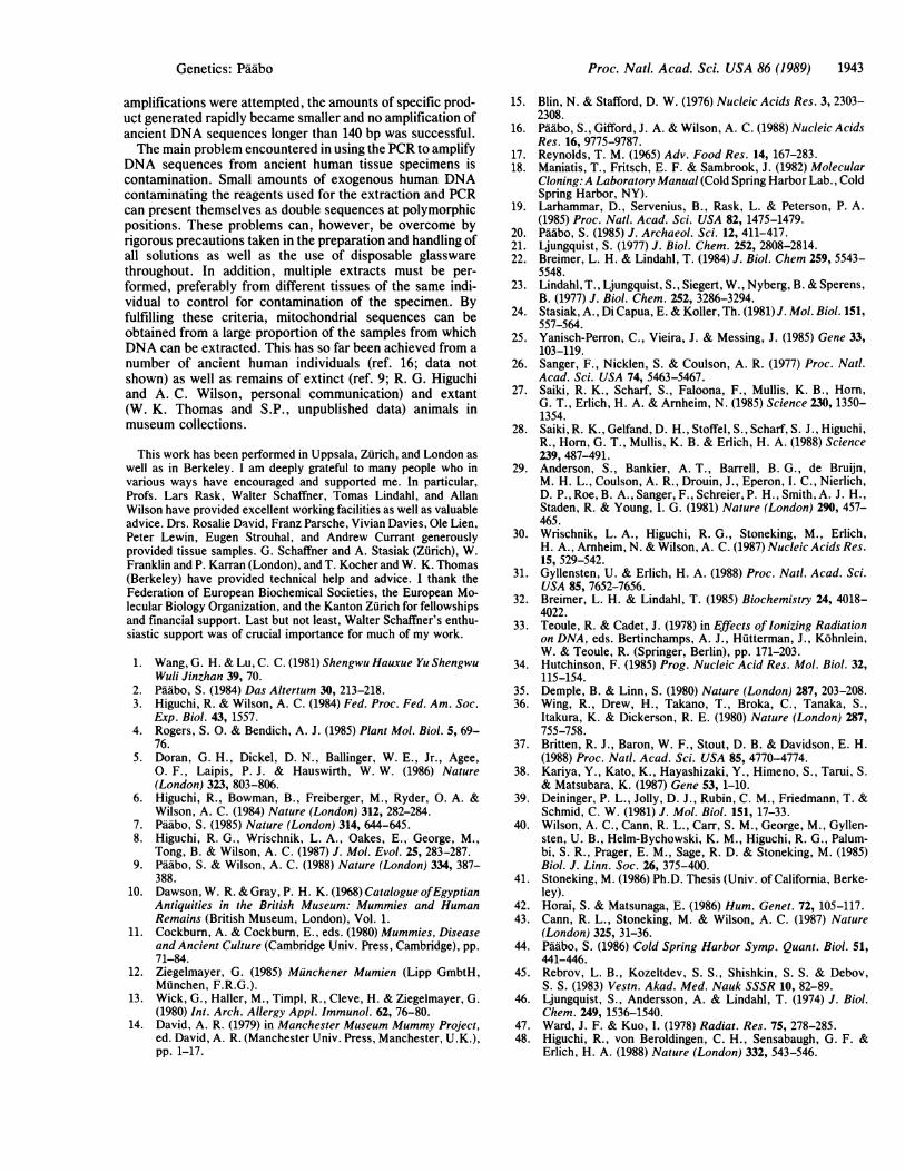
extracted from the same sample used for molecular cloningwas subjected to enzymatic amplification by PCR. To over-come an inhibitory activity present in the ancient DNAextract, large amounts of the Taq polymerase had to be addedto the amplification reaction mixtures (cf. ref. 16). Mitochon-drial DNA sequences were chosen for amplification sincemitochondrial DNA polymorphisms are highly suited foranthropological studies (40) and because mitochondrial ge-nomes occur at a high copy number in most cells, which mayfacilitate their survival and detection. Three pairs of primersspecific for various segments of the mitochondrial genomewere used. These primers are expected to amplify DNAsegments of 84, 121, and 471 bp.
Fig. 5 shows an electrophoretic analysis of the amplifica-tion products after 40 cycles of PCR. A single specific bandof the expected size was resolved as well as a dimer of theprimers used in the case of the 84- and 121-bp-long amplifi-cations from the ancient extract. The high enzyme concen-tration present caused additional nonspecific amplificationproducts to arise in some of the amplifications performedfrom control extracts, where no tissue had been added to theextraction buffer. These products do not contain the relevantDNA sequence and are not observed when small quantitiesof human DNA, containing adequate templates, are added tothe extract (data not shown). Also shown in Fig. 5 areanalogous amplifications from 1 ng of contemporary humanDNA. It can be seen that the 84-bp and the 121-bp-longamplifications from 1 ,ug of the ancient extract yieldedspecific products in approximately the same amounts as did1 ng of contemporary DNA. In the case of the 471-bp-longproduct, only the contemporary DNA yielded any product.In fact, the longest amplification product that could begenerated from this extract was 140 bp long. Thus, the sizereduction of the DNA (Fig. 1) and the oxidative damagepresent are setting limits to the size of the fragments that canbe amplified.
mummy), and L ("4000-year-old liver) were end-labeled and sub-jected to indicated treatments. Then, TCA precipitations wereperformed. Numbers indicate the fractions of radioactivity that wereprecipitable when compared to mock-treated samples.
Genetics: Pddbo
Proc. Natl. Acad. Sci. USA 86 (1989)
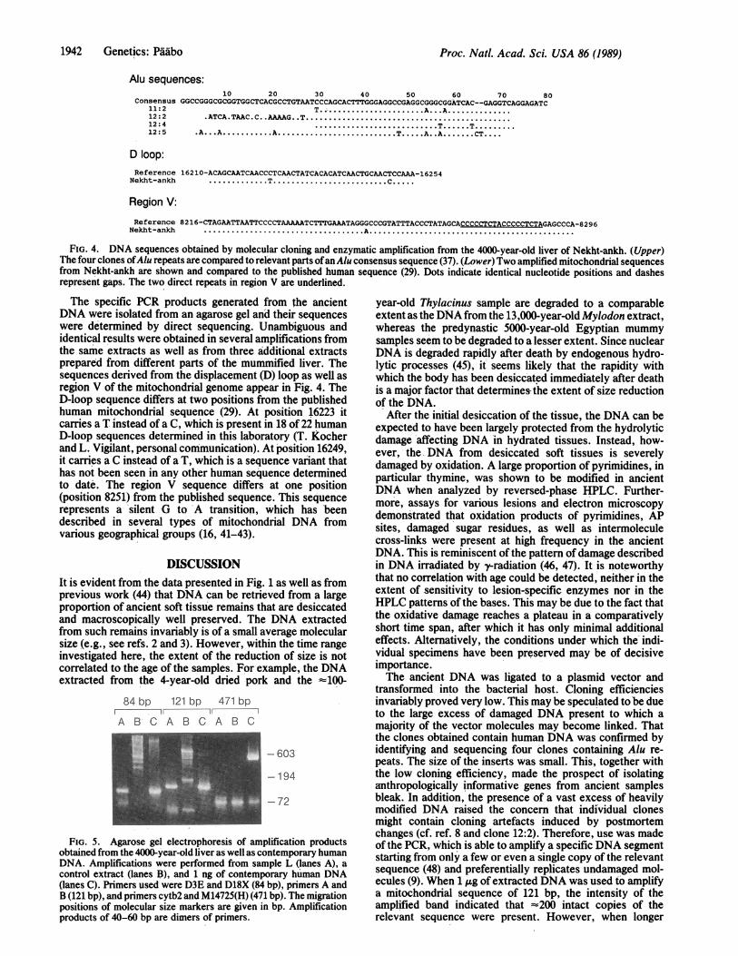
Alu sequences:
Consensus11:212:212:412:5
10 20 30 40 50 60 70 80GGCCGGGCGCGGTGGCTCACGCCTGTAATCCCAGCACTTTGGGAGGCCGAGGCGGGCGGITCAC--GAGGTCAGGAGATC
T.............A.. .A.........ATCA.TAAC.C..AAAAG..T.......................
T. T..A..A . A.T.A..A.CT..
D loop:Reference 16210-ACAGCAATCAACCCTCAACTATCACACATCAACTGCAACTCCAAA-16254
Nekht-ankh .T................... TC.
Region V:
ReferenceNekht-ankh
8216-CTAGAATTAATTCCCCTAAAAATCTTTGAAATAGGGCCCGTATTTACCCTATAGCACCCCCTCTACCCCCTCTAGAGCCCA-8296A..
FIG. 4. DNA sequences obtained by molecular cloning and enzymatic amplification from the 4000-year-old liver of Nekht-ankh. (Upper)The four clones ofAlu repeats are compared to relevant parts ofan Alu consensus sequence (37). (Lower) Two amplified mitochondrial sequencesfrom Nekht-ankh are shown and compared to the published human sequence (29). Dots indicate identical nucleotide positions and dashesrepresent gaps. The two direct repeats in region V are underlined.
The specific PCR products generated from the ancientDNA were isolated from an agarose gel and their sequenceswere determined by direct sequencing. Unambiguous andidentical results were obtained in several amplifications fromthe same extracts as well as from three additional extractsprepared from different parts of the'mummified liver. Thesequences derived from the displacement (D) loop as well asregion V of the mitochondrial genome appear in Fig. 4. TheD-loop sequence differs at two positions from the publishedhuman mitochondrial sequence (29). At position 16223 itcarries a T instead of a C, which is present in 18 of 22 humanD-loop sequences determined in this laboratory (T. Kocherand L. Vigilant, personal communication). At position 16249,it carries a C instead of a T, which is a sequence variant thathas not been seen in any other human sequence determinedto date. The region V sequence differs at one position(position 8251) from the published sequence. This sequencerepresents a silent G to 'A transition, which has 'beendescribed in several types of mitochondrial DNA fromvarious geographical groups (16, 41-43).
DISCUSSIONIt is evident from the data presented in Fig. 1 as well as fromprevious work (44) that DNA can be retrieved from a largeproportion of ancient soft tissue remains that are desiccatedand macroscopically well preserved. The DNA extractedfrom such remains invariably is of a small average molecularsize (e.g., see refs. 2 and 3). However, within the time rangeinvestigated here, the extent of the reduction of size is notcorrelated to the age of the samples. For example, the DNAextracted from the 4-year-old dried pork and the -100-
84 bp 121 bp 471 bp1 1
A B C A 8 C A B C
-603
-194
72
FIG. 5. Agarose gel electrophoresis of amplification productsobtained from the 4000-year-old liver as-well as contemporary humanDNA. Amplifications were performed from sample L (lanes A), acontrol extract (lanes B), and 1 ng of contemporary human DNA(lanes C). Primers used were D3E and D18X (84 bp), primers A andB (121 bp), and primers cytb2 and M14725(H) (471 bp). The migrationpositions of molecular size markers are given in bp. Amplificationproducts of 40-60 bp are dimers of primers.
year-old Thylacinus sample are degraded to a comparableextent as the DNAfrom the 13,000-year-old Mylodon extract,whereas the predynastic 5000-year-old Egyptian mummysamples seem to be degraded to a lesser extent. Since nuclearDNA is degraded rapidly after death by endogenous hydro-lytic processes (45), it! seems likely that the rapidity withwhich the body has been desiccated immediately after deathis a major factor that determines the extent of size reductionof the DNA.
After the initial desiccation of the tissue, the DNA can beexpected to have been largely protected from the hydrolyticdamage'affecting DNA in hydrated tissues. Instead, how-ever, the DNA from desiccated soft tissues is severelydamaged by oxidation. A large proportion of pyrimidines, inparticular thymine, was shown to be modified in ancientDNA when analyzed by reversed-phase HPLC. Further-more, assays for various lesions and electron microscopydemonstrated that oxidation products of pyrimidines, APsites, damaged sugar residues, as well as intermoleculecross-links were present at high frequency in the ancientDNA. This is reminiscent of the pattern of damage describedin DNA irradiated by yrradiation (46, 47). It is noteworthythat no correlation with age could be detected, neither in theextent of sensitivity to lesion-specific enzymes nor in theHPLC patterns of the bases. This may be due to the fact thatthe oxidative damage reaches a plateau in a comparativelyshort time span, after which it has only minimal additionaleffects. Alternatively, the conditions under which the indi-vidual specimens have been preserved may be of decisiveimportance.The ancient DNA was ligated to a plasmid vector and
transformed into the bacterial host. Cloning efficienciesinvariably proved very low. This may be speculated tobe dueto the large excess of damaged DNA present to which amajority of the vector molecules may become linked. Thatthe clones obtained contain human DNA was confirmed byidentifying and sequencing four clones containing Alu re-peats. The size of the inserts was small. This, together withthe low cloning efficiency, made the prospect of isolatinganthropologically informative genes from ancient samplesbleak. In addition, the presence of a vast excess of heavilymodified DNA raised the concern that individual clonesmight contain cloning artefacts induced by postmortemchanges (cf. ref. 8 and clone 12:2). Therefore, use was madeof the PCR, which is able to amplify a specific DNA segmentstarting from only a few or even a single copy of the relevantsequence (48) and preferentially replicates undamaged mol-ecules (9). When 1 ,ug of extracted DNA was used to amplifya mitochondrial sequence of 121 bp, the intensity of theamplified band indicated that =200 intact copies of therelevant sequence were present. However, when longer
1942 Genetics: Pddbo
Proc. Natl. Acad. Sci. USA 86 (1989) 1943
amplifications were attempted, the amounts of specific prod-uct generated rapidly became smaller and no amplification ofancient DNA sequences longer than 140 bp was successful.The main problem encountered in using the PCR to amplify
DNA sequences from ancient human tissue specimens iscontamination. Small amounts of exogenous human DNAcontaminating the reagents used for the extraction and PCRcan present themselves as double sequences at polymorphicpositions. These problems can, however, be overcome byrigorous precautions taken in the preparation and handling ofall solutions as well as the use of disposable glasswarethroughout. In addition, multiple extracts must be per-formed, preferably from different tissues of the same indi-vidual to control for contamination of the specimen. Byfulfilling these criteria, mitochondrial sequences can beobtained from a large proportion of the samples from whichDNA can be extracted. This has so far been achieved from anumber of ancient human individuals (ref. 16; data notshown) as well as remains of extinct (ref. 9; R. G. Higuchiand A. C. Wilson, personal communication) and extant(W. K. Thomas and S.P., unpublished data) animals inmuseum collections.
This work has been performed in Uppsala, Zurich, and London aswell as in Berkeley. I am deeply grateful to many people who invarious ways have encouraged and supported me. In particular,Profs. Lars Rask, Walter Schaffner, Tomas Lindahl, and AllanWilson have provided excellent working facilities as well as valuableadvice. Drs. Rosalie David, Franz Parsche, Vivian Davies, Ole Lien,Peter Lewin, Eugen Strouhal, and Andrew Currant generouslyprovided tissue samples. G. Schaffner and A. Stasiak (Zurich), W.Franklin and P. Karran (London), and T. Kocher and W. K. Thomas(Berkeley) have provided technical help and advice. I thank theFederation of European Biochemical Societies, the European Mo-lecular Biology Organization, and the Kanton Zurich for fellowshipsand financial support. Last but not least, Walter Schaffner's enthu-siastic support was of crucial importance for much of my work.
1. Wang, G. H. & Lu, C. C. (1981) Shengwu Hauxue Yu ShengwuWuli Jinzhan 39, 70.
2. Paabo, S. (1984) Das Altertum 30, 213-218.3. Higuchi, R. & Wilson, A. C. (1984) Fed. Proc. Fed. Am. Soc.
Exp. Biol. 43, 1557.4. Rogers, S. 0. & Bendich, A. J. (1985) Plant Mol. Biol. 5, 69-
76.5. Doran, G. H., Dickel, D. N., Ballinger, W. E., Jr., Agee,
0. F., Laipis, P. J. & Hauswirth, W. W. (1986) Nature(London) 323, 803-806.
6. Higuchi, R., Bowman, B., Freiberger, M., Ryder, 0. A. &Wilson, A. C. (1984) Nature (London) 312, 282-284.
7. Paabo, S. (1985) Nature (London) 314, 644-645.8. Higuchi, R. G., Wrischnik, L. A., Oakes, E., George, M.,
Tong, B. & Wilson, A. C. (1987) J. Mol. Evol. 25, 283-287.9. Paabo, S. & Wilson, A. C. (1988) Nature (London) 334, 387-
388.10. Dawson, W. R. & Gray, P. H. K. (1968) Catalogue ofEgyptian
Antiquities in the British Museum: Mummies and HumanRemains (British Museum, London), Vol. 1.
11. Cockburn, A. & Cockburn, E., eds. (1980) Mummies, Diseaseand Ancient Culture (Cambridge Univ. Press, Cambridge), pp.71-84.
12. Ziegelmayer, G. (1985) Munchener Mumien (Lipp GmbtH,Munchen, F.R.G.).
13. Wick, G., Haller, M., Timpl, R., Cleve, H. & Ziegelmayer, G.(1980) Int. Arch. Allergy Appl. Immunol. 62, 76-80.
14. David, A. R. (1979) in Manchester Museum Mummy Project,ed. David, A. R. (Manchester Univ. Press, Manchester, U.K.),pp. 1-17.
15. Blin, N. & Stafford, D. W. (1976) Nucleic Acids Res. 3, 2303-2308.
16. Paubo, S., Gifford, J. A. & Wilson, A. C. (1988) Nucleic AcidsRes. 16, 9775-9787.
17. Reynolds, T. M. (1965) Adv. Food Res. 14, 167-283.18. Maniatis, T., Fritsch, E. F. & Sambrook, J. (1982) Molecular
Cloning:A Laboratory Manual (Cold Spring Harbor Lab., ColdSpring Harbor, NY).
19. Larhammar, D., Servenius, B., Rask, L. & Peterson, P. A.(1985) Proc. NatI. Acad. Sci. USA 82, 1475-1479.
20. Paubo, S. (1985) J. Archaeol. Sci. 12, 411-417.21. Ljungquist, S. (1977) J. Biol. Chem. 252, 2808-2814.22. Breimer, L. H. & Lindahl, T. (1984) J. Biol. Chem 259, 5543-
5548.23. Lindahl, T., Ljungquist, S., Siegert, W., Nyberg, B. & Sperens,
B. (1977) J. Biol. Chem. 252, 3286-3294.24. Stasiak, A., Di Capua, E. & Koller, Th. (1981) J. Mol. Biol. 151,
557-564.25. Yanisch-Perron, C., Vieira, J. & Messing, J. (1985) Gene 33,
103-119.26. Sanger, F., Nicklen, S. & Coulson, A. R. (1977) Proc. Natl.
Acad. Sci. USA 74, 5463-5467.27. Saiki, R. K., Scharf, S., Faloona, F., Mullis, K. B., Horn,
G. T., Erlich, H. A. & Arnheim, N. (1985) Science 230, 1350-1354.
28. Saiki, R. K., Gelfand, D. H., Stoffel, S., Scharf, S. J., Higuchi,R., Horn, G. T., Mullis, K. B. & Erlich, H. A. (1988) Science239, 487-491.
29. Anderson, S., Bankier, A. T., Barrell, B. G., de Bruijn,M. H. L., Coulson, A. R., Drouin, J., Eperon, I. C., Nierlich,D. P., Roe, B. A., Sanger, F., Schreier, P. H., Smith, A. J. H.,Staden, R. & Young, I. G. (1981) Nature (London) 290, 457-465.
30. Wrischnik, L. A., Higuchi, R. G., Stoneking, M., Erlich,H. A., Arnheim, N. & Wilson, A. C. (1987) Nucleic Acids Res.15, 529-542.
31. Gyllensten, U. & Erlich, H. A. (1988) Proc. Natl. Acad. Sci.USA 85, 7652-7656.
32. Breimer, L. H. & Lindahl, T. (1985) Biochemistry 24, 4018-4022.
33. Teoule, R. & Cadet, J. (1978) in Effects of Ionizing Radiationon DNA, eds. Bertinchamps, A. J., Hutterman, J., Kohnlein,W. & Teoule, R. (Springer, Berlin), pp. 171-203.
34. Hutchinson, F. (1985) Prog. Nucleic Acid Res. Mol. Biol. 32,115-154.
35. Demple, B. & Linn, S. (1980) Nature (London) 287, 203-208.36. Wing, R., Drew, H., Takano, T., Broka, C., Tanaka, S.,
Itakura, K. & Dickerson, R. E. (1980) Nature (London) 287,755-758.
37. Britten, R. J., Baron, W. F., Stout, D. B. & Davidson, E. H.(1988) Proc. Natl. Acad. Sci. USA 85, 4770-4774.
38. Kariya, Y., Kato, K., Hayashizaki, Y., Himeno, S., Tarui, S.& Matsubara, K. (1987) Gene 53, 1-10.
39. Deininger, P. L., Jolly, D. J., Rubin, C. M., Friedmann, T. &Schmid, C. W. (1981) J. Mol. Biol. 151, 17-33.
40. Wilson, A. C., Cann, R. L., Carr, S. M., George, M., Gyllen-sten, U. B., Helm-Bychowski, K. M., Higuchi, R. G., Palum-bi, S. R., Prager, E. M., Sage, R. D. & Stoneking, M. (1985)Biol. J. Linn. Soc. 26, 375-400.
41. Stoneking, M. (1986) Ph.D. Thesis (Univ. of California, Berke-ley).
42. Horai, S. & Matsunaga, E. (1986) Hum. Genet. 72, 105-117.43. Cann, R. L., Stoneking, M. & Wilson, A. C. (1987) Nature
(London) 325, 31-36.44. Paabo, S. (1986) Cold Spring Harbor Symp. Quant. Biol. 51,
441-446.45. Rebrov, L. B., Kozeltdev, S. S., Shishkin, S. S. & Debov,
S. S. (1983) Vestn. Akad. Med. Nauk SSSR 10, 82-89.46. Ljungquist, S., Andersson, A. & Lindahl, T. (1974) J. Biol.
Chem. 249, 1536-1540.47. Ward, J. F. & Kuo, I. (1978) Radiat. Res. 75, 278-285.48. Higuchi, R., von Beroldingen, C. H., Sensabaugh, G. F. &
Erlich, H. A. (1988) Nature (London) 332, 543-546.
Genetics: Pddbo