Embed Size (px)
Citation preview
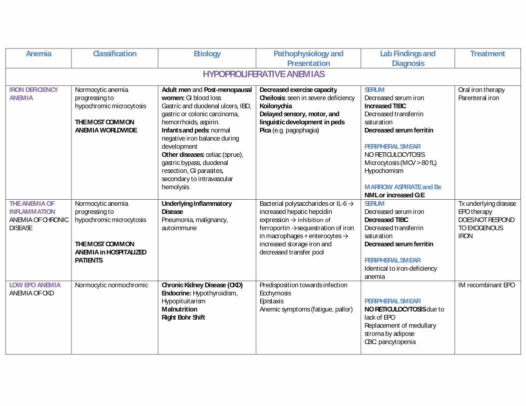
Anemia Classification Etiology Pathophysiology and Presentation
Lab Findings and Diagnosis
Treatment
HYPOPROLIFERATIVE ANEMIAS
IRON DEFICIENCY ANEMIA
Normocytic anemia progressing to hypochromic microcytosis THE MOST COMMON ANEMIA WORLDWIDE
Adult men and Post-menopausal women: GI blood loss Gastric and duodenal ulcers, IBD, gastric or colonic carcinoma, hemorrhoids, aspirin. Infants and peds: normal negative iron balance during development Other diseases: celiac (sprue), gastric bypass, duodenal resection, GI parasites, secondary to intravascular hemolysis
Decreased exercise capacity Cheilosis: seen in severe deficiency Koilonychia Delayed sensory, motor, and linguistic development in peds Pica (e.g. pagophagia)
SERUM Decreased serum iron Increased TIBC Decreased transferrin saturation Decreased serum ferritin PERIPHERAL SMEAR NO RETICULOCYTOSIS Microcytosis (MCV > 80 fL) Hypochomism MARROW ASPIRATE and Bx NML or increased G:E
Oral iron therapy Parenteral iron
THE ANEMIA OF INFLAMMATION ANEMIA OF CHRONIC DISEASE
Normocytic anemia progressing to hypochromic microcytosis THE MOST COMMON ANEMIA in HOSPITALIZED PATIENTS
Underlying Inflammatory Disease Pneumonia, malignancy, autoimmune
Bacterial polysaccharides or IL-6 → increased hepatic hepcidin expression → inhibition of ferroportin →sequestration of iron in macrophages + enterocytes → increased storage iron and decreased transfer pool
SERUM Decreased serum iron Decreased TIBC Decreased transferrin saturation Decreased serum ferritin PERIPHERAL SMEAR Identical to iron-deficiency anemia
Tx underlying disease EPO therapy DOES NOT RESPOND TO EXOGENOUS IRON
LOW EPO ANEMIA ANEMIA OF CKD
Normocytic normochromic Chronic Kidney Disease (CKD) Endocrine: Hypothyroidism, Hypopituitarism Malnutrition Right Bohr Shift
Predisposition towards infection Ecchymosis Epistaxis Anemic symptoms (fatigue, pallor)
PERIPHERAL SMEAR NO RETICULOCYTOSIS due to lack of EPO Replacement of medullary stroma by adipose CBC: pancytopenia
IM recombinant EPO
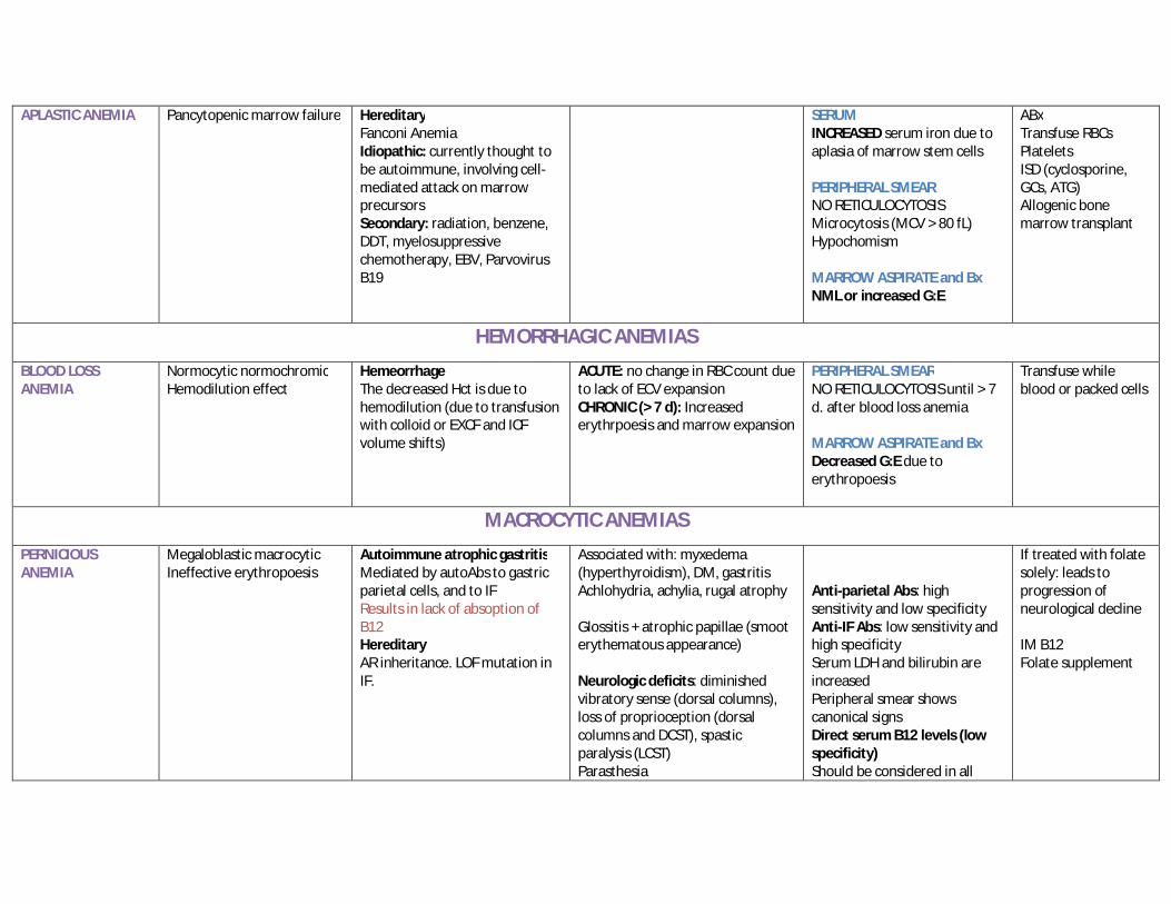
APLASTIC ANEMIA Pancytopenic marrow failure Hereditary Fanconi Anemia Idiopathic: currently thought to be autoimmune, involving cell-mediated attack on marrow precursors Secondary: radiation, benzene, DDT, myelosuppressive chemotherapy, EBV, Parvovirus B19
SERUM INCREASED serum iron due to aplasia of marrow stem cells PERIPHERAL SMEAR NO RETICULOCYTOSIS Microcytosis (MCV > 80 fL) Hypochomism MARROW ASPIRATE and Bx NML or increased G:E
ABx Transfuse RBCs Platelets ISD (cyclosporine, GCs, ATG) Allogenic bone marrow transplant
HEMORRHAGIC ANEMIAS
BLOOD LOSS ANEMIA
Normocytic normochromic Hemodilution effect
Hemeorrhage The decreased Hct is due to hemodilution (due to transfusion with colloid or EXCF and ICF volume shifts)
ACUTE: no change in RBC count due to lack of ECV expansion CHRONIC (> 7 d): Increased erythrpoesis and marrow expansion
PERIPHERAL SMEAR NO RETICULOCYTOSIS until > 7 d. after blood loss anemia MARROW ASPIRATE and Bx Decreased G:E due to erythropoesis
Transfuse while blood or packed cells
MACROCYTIC ANEMIAS
PERNICIOUS ANEMIA
Megaloblastic macrocytic Ineffective erythropoesis
Autoimmune atrophic gastritis Mediated by autoAbs to gastric parietal cells, and to IF Results in lack of absoption of B12 Hereditary AR inheritance. LOF mutation in IF.
Associated with: myxedema (hyperthyroidism), DM, gastritis Achlohydria, achylia, rugal atrophy Glossitis + atrophic papillae (smoot erythematous appearance) Neurologic deficits: diminished vibratory sense (dorsal columns), loss of proprioception (dorsal columns and DCST), spastic paralysis (LCST) Parasthesia
Anti-parietal Abs: high sensitivity and low specificity Anti-IF Abs: low sensitivity and high specificity Serum LDH and bilirubin are increased Peripheral smear shows canonical signs Direct serum B12 levels (low specificity) Should be considered in all
If treated with folate solely: leads to progression of neurological decline IM B12 Folate supplement
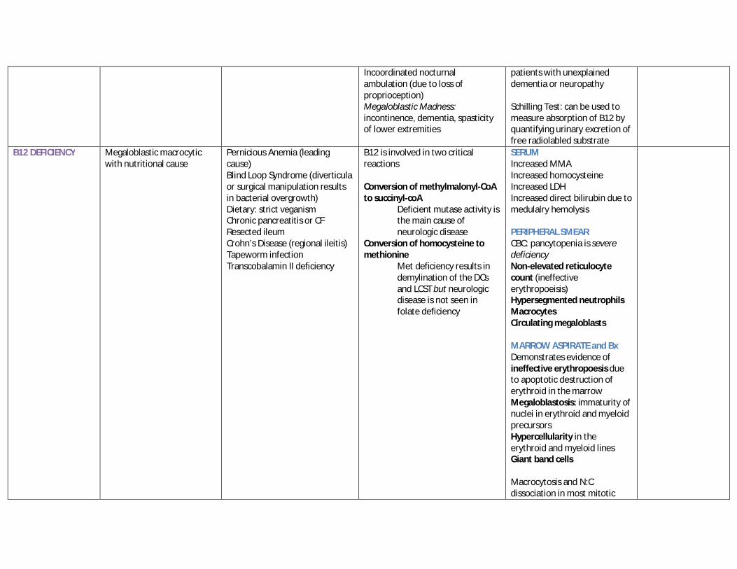
Incoordinated nocturnal ambulation (due to loss of proprioception) Megaloblastic Madness: incontinence, dementia, spasticity of lower extremities
patients with unexplained dementia or neuropathy Schilling Test: can be used to measure absorption of B12 by quantifying urinary excretion of free radiolabled substrate
B12 DEFICIENCY Megaloblastic macrocytic with nutritional cause
Pernicious Anemia (leading cause) Blind Loop Syndrome (diverticula or surgical manipulation results in bacterial overgrowth) Dietary: strict veganism Chronic pancreatitis or CF Resected ileum Crohn’s Disease (regional ileitis) Tapeworm infection Transcobalamin II deficiency
B12 is involved in two critical reactions Conversion of methylmalonyl-CoA to succinyl-coA
Deficient mutase activity is the main cause of neurologic disease
Conversion of homocysteine to methionine
Met deficiency results in demylination of the DCs and LCST but neurologic disease is not seen in folate deficiency
SERUM Increased MMA Increased homocysteine Increased LDH Increased direct bilirubin due to medulalry hemolysis PERIPHERAL SMEAR CBC: pancytopenia is severe deficiency Non-elevated reticulocyte count (ineffective erythropoeisis) Hypersegmented neutrophils Macrocytes Circulating megaloblasts MARROW ASPIRATE and Bx Demonstrates evidence of ineffective erythropoesis due to apoptotic destruction of erythroid in the marrow Megaloblastosis: immaturity of nuclei in erythroid and myeloid precursors Hypercellularity in the erythroid and myeloid lines Giant band cells Macrocytosis and N:C dissociation in most mitotic
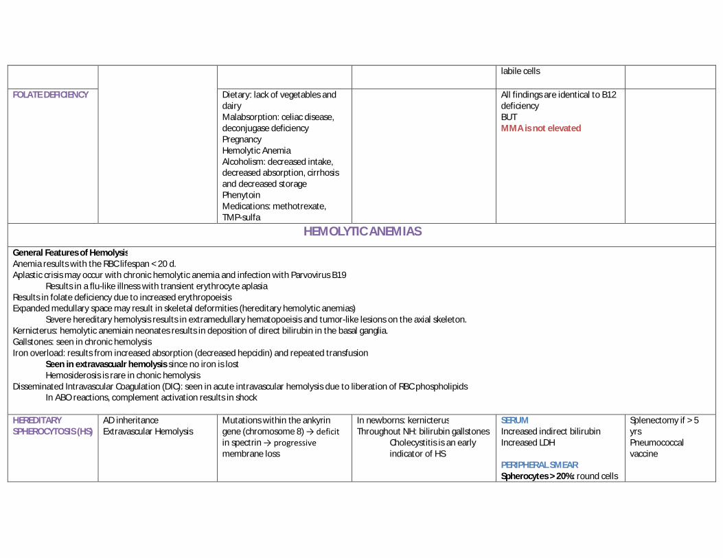
labile cells
FOLATE DEFICIENCY Dietary: lack of vegetables and dairy Malabsorption: celiac disease, deconjugase deficiency Pregnancy Hemolytic Anemia Alcoholism: decreased intake, decreased absorption, cirrhosis and decreased storage Phenytoin Medications: methotrexate, TMP-sulfa
All findings are identical to B12 deficiency BUT MMA is not elevated
HEMOLYTIC ANEMIAS
General Features of Hemolysis Anemia results with the RBC lifespan < 20 d. Aplastic crisis may occur with chronic hemolytic anemia and infection with Parvovirus B19
Results in a flu-like illness with transient erythrocyte aplasia Results in folate deficiency due to increased erythropoeisis Expanded medullary space may result in skeletal deformities (hereditary hemolytic anemias)
Severe hereditary hemolysis results in extramedullary hematopoeisis and tumor-like lesions on the axial skeleton. Kernicterus: hemolytic anemiain neonates results in deposition of direct bilirubin in the basal ganglia. Gallstones: seen in chronic hemolysis Iron overload: results from increased absorption (decreased hepcidin) and repeated transfusion
Seen in extravascualr hemolysis since no iron is lost Hemosiderosis is rare in chonic hemolysis
Disseminated Intravascular Coagulation (DIC): seen in acute intravascular hemolysis due to liberation of RBC phospholipids In ABO reactions, complement activation results in shock
HEREDITARY SPHEROCYTOSIS (HS)
AD inheritance Extravascular Hemolysis
Mutations within the ankyrin gene (chromosome 8) → deficit in spectrin → progressive membrane loss
In newborns: kernicterus Throughout NH: bilirubin gallstones
Cholecystitis is an early indicator of HS
SERUM Increased indirect bilirubin Increased LDH PERIPHERAL SMEAR Spherocytes > 20%: round cells
Splenectomy if > 5 yrs Pneumococcal vaccine
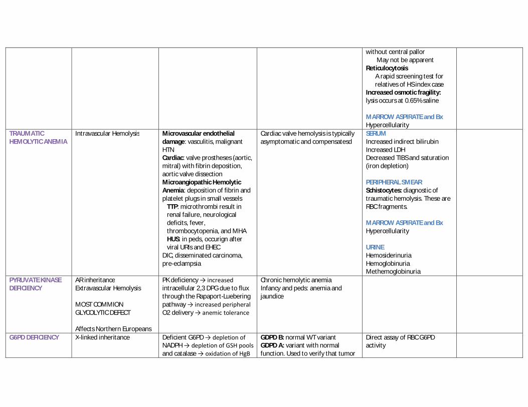
without central pallor May not be apparent Reticulocytosis A rapid screening test for relatives of HS index case Increased osmotic fragility: lysis occurs at 0.65% saline MARROW ASPIRATE and Bx Hypercellularity
TRAUMATIC HEMOLYTIC ANEMIA
Intravascular Hemolysis
Microvascular endothelial damage: vasculitis, malignant HTN Cardiac: valve prostheses (aortic, mitral) with fibrin deposition, aortic valve dissection Microangiopathic Hemolytic Anemia: deposition of fibrin and platelet plugs in small vessels TTP: microthrombi result in renal failure, neurological deficits, fever, thrombocytopenia, and MHA HUS: in peds, occurign after viral URIs and EHEC DIC, disseminated carcinoma, pre-eclampsia
Cardiac valve hemolysis is typically asymptomatic and compensatesd
SERUM Increased indirect bilirubin Increased LDH Decreased TIBS and saturation (iron depletion) PERIPHERAL SMEAR Schistocytes: diagnostic of traumatic hemolysis. These are RBC fragments. MARROW ASPIRATE and Bx Hypercellularity URINE Hemosiderinuria Hemoglobinuria Methemoglobinuria
PYRUVATE KINASE DEFICIENCY
AR inheritance Extravascular Hemolysis MOST COMMION GLYCOLYTIC DEFECT Affects Northern Europeans
PK deficiency → increased intracellular 2,3 DPG due to flux through the Rapaport-Luebering pathway → increased peripheral O2 delivery → anemic tolerance
Chronic hemolytic anemia Infancy and peds: anemia and jaundice
G6PD DEFICIENCY X-linked inheritance
Deficient G6PD → depletion of NADPH → depletion of GSH pools and catalase → oxidation of HgB
GDPD B: normal WT variant GDPD A: variant with normal function. Used to verify that tumor
Direct assay of RBC G6PD activity
Mixed intravascular and extravascular hemolysis MOST COMMON RBC METABOLIC DEFECT Affects Africans, Med basin, Middle East
and membrane SH → formation of disulfide bridges between globin and RBC membrane → Heinz bodies → non-deformability → extravascular hemolysis With severe oxidative stress: oxidation of membrane SH results in perforation and intravascular hemolysis Drugs may trigger hemolysis in the oxidative variants of G6PD deficiency:
Sulfamethoxizole Nitrofurantoin Primaquine Dapsone
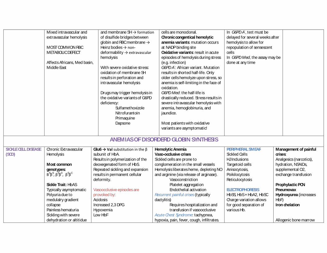
cells are monoclonal. Chronic congentical hemolytic anemia variants: mutation occurs at NADP binding site Oxidative variants: result in acute episodes of hemolysis during stress (e.g. infection) G6PD A-: African variant. Mutation results in shorted half-life. Only older cells hemolyze upon stress, so anemia is self-limiting in the face of oxidation. G6PD Med: the half-life is drastically reduced. Stress results in severe intravascular hemolysis with anemia, hemoglobinuria, and jaundice. Most patients with oxidative variants are asymptomatic!
In G6PD A-, test must be delayed for several weeks after hemolysis to allow for repopulation of sensescent cells In G6PD Med, the assay may be done at any time
ANEMIAS OF DISORDERD GLOBIN SYNTHESIS
SICKLE CELL DISEASE (SCD)
Chronic Extravascular Hemolysis Most common genotypes: ΒSβS, βSβ0, βSβC Sickle Trait: HbAS Typically asymptomatic Polyuria due to medulalry gradient collapse Painless hematuria Sickling with severe dehydration or altitidue
Glu6 → Val substitution in the β subunit of HbA. Results in polymerization of the deoxegenated form of HbS. Repeated sickling and expansion results in permanent cellular deformity. Vasoocclusive episodes are provoked by: Acidosis Increased 2,3 DPG Hypoxemia Low HbF
Hemolytic Anemia Vaso-occlusive crises Sickled cells are prone to conglomeration in the small vessels Hemolysis liberates heme, depleting NO and arginine (via release of arginase).
Vasoconstriction Platelet aggregation Endothelial activation
Recurrent painful crises (typically dactylitis)
Requires hospitalization and transfusion if vasoocclusive
Acute Chest Syndrome: tachypnea, hypoxia, pain, fever, cough, infiltrates.
PERIPHERAL SMEAR Sickled Cells HJ Inclusions Targetoid cells Anisocytosis, Poikilocytosis Reticulocytosis ELECTROPHORESIS HbSS, HbS + HbA2, HbSC Charge variation allows for good separation of various Hb.
Management of painful crises: Analgesics (narcotics), hydration, NSAIDs, supplemental O2, exchange transfusion Prophylactic PCN Pneumovax Hydroxyurea (increases HbF) Iron chelation Allogenic bone marrow
Renal papillary necrosis
βSβ0 results in severe anemia Heterozygosity with α thalessemia trait decreases HbS concentrations
Resembles ARDS. Associated with local or remote infection. Stroke: subclinical infarction Chronic ischemic damage: Aseptic osteonecrosis of the femoral head, Pulmonary HTN CKD, Retinopathy Functional Asplenia Predisposition to infection and sepsis Splenic autoinfarction leads to stabilization of acute anemia (decreased sequestration) The disease typically becomes symptomatic > 6 mo. due to conversion to β chain synthesis.
SERUM Increased indirect bilirubin Increased LDH
transplantation
αTHALASSEMIA SYNDROMES
Hypochromic microcytic Ineffective erythropoeisis Chronic extravascular hemolysis
Typically involve deletion of α globin alleles (chromosome 16).
α thalassemia carrier αα/α- Asymptomatic without anemia Elevated Bart’s hemoglobin (γ4) α thalassemia trait α-/α-: prevalent in Med and Africa
Increased incidence of thalessemia trait and silent carriers
α-/α- and αα/-- : prevalent in East Asia Increased incidence of HbH disease and hydrops fetalis
Hypochromic microcytosis Negligible anemia HbH disease α-/-- Chronic extravascualar hemolytic anemia due to HbH (β4) inclusions
α thalassemia syndromes All hemoglobins are affected May detect Bart’s hemoglobin
HbH disease Transfusion if necessary Folci acid supplement Splenectomy
Hypochromic microcytosis Hemolysis is typically compensated Targetoid cells Oxidant drugs provoke hemolysis Hydrops Fetalis --/-- Most Hb is Bart’s (high O2 affinity and low delivery) Mortality is due to hepatic failure (ischemia and extramedullary hemaptopoeisis)
β THALASSEMIA SYNDROMES
Hypochromic microcytic Ineffective Erythropoesis Chronic extrvascualr hemolysis
Various mutations within the β globin coding region of chromosome 11 → decreased synthesis (Typically not a deletion)
β thalessemia minor (Hb > 9 d/dL) Typically β/β+ Asymptomatic with negligible anemia DESPITE microcytosis (r/o iron deficiency and inflammation) β thalessemia intermedia (Hg 6 -9 g/dL) Typically β+/β+, β+/β0, β/β0 Microcytic anemia with several aberrant RBC morphologies (targetoid, nucleated blasts) β thalessemia major (Hb < 6 g/dL) β0/β0 α4 hemoglobin precipitates, resulting in
Ineffective erythropoeisis Extravascualr hemolysis Severe microcytic anemia
Hepatospenomegaly Marrow expansion and bone deformity Increased GI iron absorption and transfusion hemochromatosis
β thalessemia minor Increased HbA2 (> 3%) Increased HbF Most hemoglobin is HbA β thalessemia intermedia HbF 10 – 90% and HbA2 > 3% Decreased HbA β thalessemia major Absence of HbA Only detect HbF + HbA2
Screen maternal MCV and ferritin Suspect thalessemia with microcytosis and NML ferritin β thalessemia intermedia Transfusion if necessary Folci acid supplement Splenectomy β thalessemia major Transfusion Iron chelation Allogenic stem cell transplant Splenectomy