Embed Size (px)
Citation preview
1
Anemia
• Reduction below normal in oxygen carrying capacity ofthe blood.
• Caused by a variety of disorders.
• FA is one of the inherited anemias that leads to bonemarrow failure leading to a low RBC count.
• FA is diagnosed with the help of blood tests.• FA is treated with bone marrow transplants.
HEMATOPOIESISBlood Cell Formation
• DEFINITION
The process of formation and development of the varioustypes of blood cells and other formed elements (platelets).
• In the adult, all blood cell formation (red bloodcells, white blood cells and platelets) occurs inthe Red Bone Marrow or myeloid tissue.
Hematopoiesis
• In adults, red bone marrow is primarily found inbones of the– Axial skeleton– Pelvic and pectoral girdles– Proximal epiphyses of the humerus and femur.
Active Bone Marrow Locations
Samples can be taken from the bone marrow, smearedin a single cell layer on a slide, and stained
The resulting BONE MARROW SMEAR contains asampling of stem cells and many blood cells in
immature stages of development • Hematopoiesis begins with a stem cell known as thehemocytoblast.
2
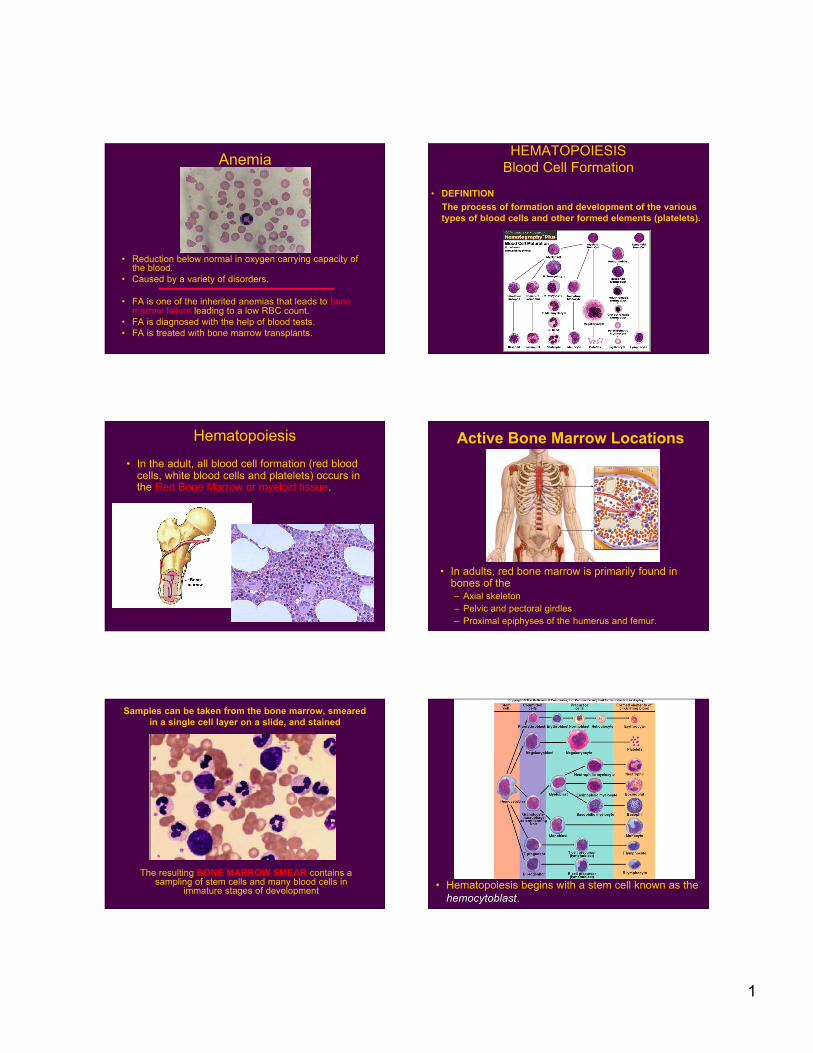
• Red Blood Cell (RBC) synthesis is known asErythropoiesis.
• In erythropoiesis, the hemocytoblast goesthrough a series of morphological changesculminating in the formation of a cell full ofhemoglobin.
ERYTHROPOESISErythropoietin (EPO)
• EPO
– produced and released by the kidneys under low oxygenconditions.
– stimulates increased rate of cell division in erythroblasts and thestem cell that produce erythroblasts.
– Speeds up maturation of RBCs
• Once the erythropoietin stimulates the red bonemarrow to begin manufacturing RBCs, a series ofevents occurs.
• Cell size decreases
• The nucleus becomes condensed (and is eventuallydiscarded)
ERYTHROPOESIS:
• Cytoplasm stains basophilic (purple)• then becomes more acidophilic (pink)• As cells mature, they slowly fill with hemoglobin until
they are bright red reticulocytes• Only mature erythrocytes are released into the
peripheral blood
ERYTHROPOESISthe making of Red Blood Cells (RBCs)
• Note the RBCprecursors
Is this smear from bone marrow or peripheral blood?Why?
ERYTHROPOIESIS Note RBC precursors
3
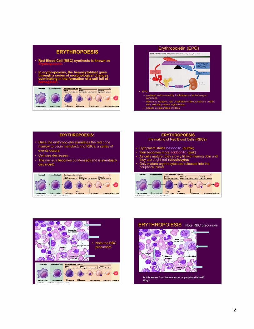
This NORMOBLAST iscaught in the act of expelling its
nucleus
Only mature, anucleate cells are normally released into the blood, so toomany NORMOBLASTS or “BLASTS” found in a blood smear may indicatecancer: usually a leukemia or lymphoma
In fact, many early blood cancers are found in early, asymptomatic stagesduring “routine” blood work!
These cells are termed:“normoblast” while nucleated“erythrocyte” when anucleate
ERYTHROPOIESIS
Appearance and size of normalerythrocytes (RBCs)
The diameter of an RBC (about 7 mL) is often used as a referenceto estimate the size of other cells on a blood smear
The life cycle of a red blood cell.• a – Kidneys respond to low oxygen
concentration in the blood byreleasing erythropoietin.
• b – Erythropoietin travels to the redbone marrow and stimulates anincrease in the production of RBC inthe bone marrow.
• c and d – RBCs mature and thensqueeze through blood vesselmembranes to enter circulation
• e – The heart and lungs work tosupply continuous movement andoxygenation of RBCs.
• f – Damaged or old RBCs aredestroyed primarily by the spleen.
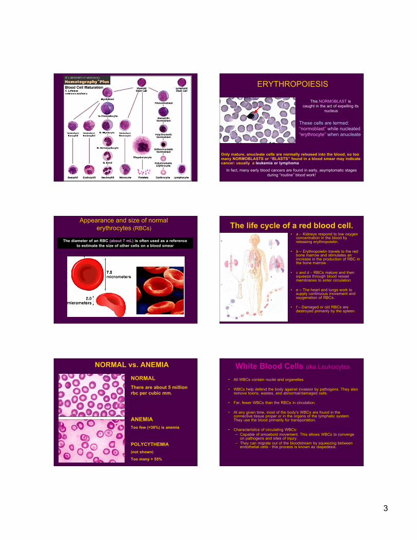
NORMAL vs. ANEMIA
NORMAL
There are about 5 millionrbc per cubic mm.
ANEMIAToo few (<38%) is anemia
POLYCYTHEMIA
(not shown)
Too many > 55%
• All WBCs contain nuclei and organelles.
• WBCs help defend the body against invasion by pathogens. They alsoremove toxins, wastes, and abnormal/damaged cells.
• Far, fewer WBCs than the RBCs in circulation.
• At any given time, most of the body's WBCs are found in theconnective tissue proper or in the organs of the lymphatic system.They use the blood primarily for transportation.
• Characteristics of circulating WBCs:– Capable of amoeboid movement. This allows WBCs to converge
on pathogens and sites of injury.– They can migrate out of the bloodstream by squeezing between
endothelial cells - this process is known as diapedesis.
White Blood Cells aka Leukocytes
4
Types of WBCsClassified as to whether or not they contain
granules that take up Wright's stain and arevisible with the light microscope.
• Granulocytes– Contain visible stained granules– Includes:
• Basophils• Eosinophils• Neutrophils.• Agranulocytes
– Do not contain stained visible granules.» Lymphocytes» Monocytes.
The stem cell is the same or similar to that for erythropoiesis
GRANULOCYTOPOIESIS
GRANULOCYTOPOIESISThe cells in these three lines of development can be readily
identified by the presence of pronounced cytoplasmicgranules, and by the gradual lobulation of the nucleus.
BONEMARROW
GRANULOCYTICPRECURSORS
A. Band or “stab”cell
B. Neutrophil
B
B
A
Granulocytesin peripheral blood
• Named for prominent “granules” in the cytoplasm
• Individually named according to the way the granules stain– Eosinophils- have large red granules
– Basophils- have large blue-purple granules that nearly obscure thenucleus
– Neutrophils- stain neutrally. Cytoplasm is often “purplish” but thegranules are not nearly as prominent as those in Basophils.
Let’s meet them now as they would appear in a peripheral bloodsmear..........
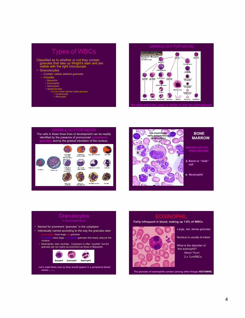
EOSINOPHILFairly infrequent in blood, making up 1-6% of WBCs
Large, red, dense granules
Nucleus is usually bi-lobed
What is the diameter of this eosinophil?
About 14µm
2 x 7µmRBCs
The granules of eosinophils contain (among other things) HISTAMINE.
:

5
When EOSINOPHILS encounter something you are allergic to(pollen, cat dander) they release their granules into the blood andyou have a “histamine reaction”: itchy, runny eyes and nose
A common class of drugs calledANTI-HISTAMINES fight this reaction
BASOPHILSGood luck finding this one in your blood smear today!....
They make up <1% of lymphocytes (WBCs)
Have very large, very dark blue-purple granules
The nucleus is S-shaped, but is usually obscuredby granules
Slightly smaller than as Eosinophils andNeutrophils
Granules contain heparin and vasoactive compounds.
Like eosinophils, BASOPHILS participate in allergic reactions. But unlikeeosinophils, BASOPHILS are involved in the most severe allergic reactions(like bee or wasp stings) that may be deadly if left untreated.
• Migrate to injury sites anddischarge the contents oftheir granules:– Histamine
• Vasodilator
• Increases capillarypermeability.
– Heparin• An anticoagulant.
– These 2 chemicals enhancethe local inflammation initiatedby mast cells and attract otherWBCs.
• Lifespan is not certain.
BASOPHILS NEUTROPHILMOST prevalent granulocyte in the peripheral smear
approx 40-75% of leukocytes
About 12µm in diameter
Distinctive multi-lobed or“sausage link” nucleus
fine granules with aneutral stain•similar in color to RBCs•not “obscuring” nucleus
Neutrophils
NEUTROPHIL
The neutrophil (indicated by black arrow) in the image to theright has phagocytosed bacterial microorganisms (bluerods indicated by thin red arrows) into its cytoplasm.
Monocytopoiesis andLymphocytopoiesis
Development of the agranular leukocytes is difficult tofollow at the light microscopic level.
6
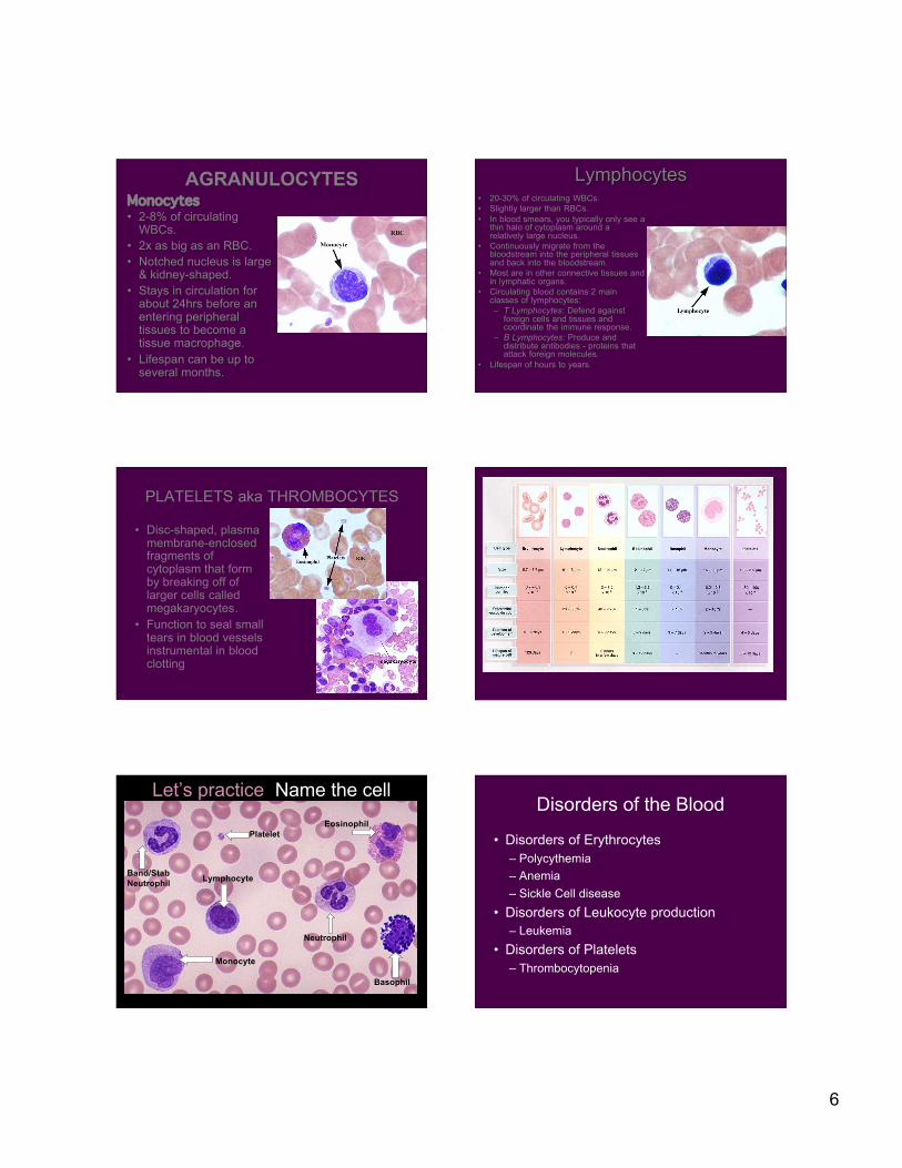
AGRANULOCYTES
• 2-8% of circulatingWBCs.
• 2x as big as an RBC.• Notched nucleus is large
& kidney-shaped.• Stays in circulation for
about 24hrs before anentering peripheraltissues to become atissue macrophage.
• Lifespan can be up toseveral months.
Monocytes
LymphocytesLymphocytes• 20-30% of circulating WBCs.• Slightly larger than RBCs.• In blood smears, you typically only see a
thin halo of cytoplasm around arelatively large nucleus.
• Continuously migrate from thebloodstream into the peripheral tissuesand back into the bloodstream.
• Most are in other connective tissues andin lymphatic organs.
• Circulating blood contains 2 mainclasses of lymphocytes:– T Lymphocytes: Defend against
foreign cells and tissues andcoordinate the immune response.
– B Lymphocytes: Produce anddistribute antibodies - proteins thatattack foreign molecules.
• Lifespan of hours to years.
PLATELETS aka THROMBOCYTES
• Disc-shaped, plasmamembrane-enclosedfragments ofcytoplasm that formby breaking off oflarger cells calledmegakaryocytes.
• Function to seal smalltears in blood vesselsinstrumental in bloodclotting
Let’s practice: Name the cell
Basophil
Eosinophil
Neutrophil
Band/StabNeutrophil
Lymphocyte
Platelet
Monocyte
Disorders of the Blood
• Disorders of Erythrocytes– Polycythemia
– Anemia
– Sickle Cell disease
• Disorders of Leukocyte production– Leukemia
• Disorders of Platelets– Thrombocytopenia
7
Polycythemia
• “Many blood cells” - abnormal excess of erythrocytes in the blood• Can result from a cancer of bone marrow that generates too many
erythrocytes.• Increase in blood viscosity, blocks blood flow through small vessels.• Treated by dilution (remove some blood and replace with sterile
saline or blood transfusion.
Anemia
• Condition in which erythrocyte levels or hemoglobinconcentrations are low
• Results in an abnormally low oxygen carrying capacity.
• Why would an anemic person be intolerant of exercise?
• There are several types of anemia that we're concerned with:
Anemia
Aplastic• Results from destruction of red bone marrow by bacterial toxins, drugs or
radiation. Impacts all blood cells, leading to clotting difficulties andimmune problems.
Iron-Deficiency• What role does iron play in oxygen transport?• Can be secondary to hemorrhagic anemia (blood loss) or due to
inadequate iron intake or absorption.
Pernicious• Can be caused by inadequate intake or absorption of vitamin B12.• The stomach lining produces a chemical called intrinsic factor which is
necessary for the absorption of ingested vitamin B12. It is often a lack ofintrinsic factor that causes pernicious anemia.
Anemia
Sickle-Cell• A mutation in the gene for
the beta chain of Hbresults in an abnormalhemoglobin called HbS.
• Under low-oxygenconditions, the beta chainslink together and becomestiff rods - this gives theRBC a sickle shape.
• Sickled RBCs can thenblock and clog small bloodvessels.
Aplastic Anemia
• Caused by failure of the bone marrow leading toa low RBC count.
Leukocytosis
• White blood cell count above 10,000 (normal range 4000-10,000)
• Usually due to an increase in one of the five types of WBCs• Given the name of the cell that shows the primary increase
– Neutrophilic = neutrophilia– Lymphocytic = lymphocytosis– Eosinophilic = eosinophilia– Monocytic = monocytosis– Basophilic = basophilia
8
Case #2 Neutrophilia Eosinophilia
Pancytopenia
• A shortage of all types of blood cells, includingRBCs and WBCs as well as platelets.
Leukemia
• A form of cancer - uncontrolled proliferation of a leukocyte-forming cell line in the bone marrow.
• The cancer takes over bone marrow and leukocytes flood theinto the blood stream.
• Classified by the cell line involved– Lymphoblastic – from immature lymphocytes
– Myeloblastic – from immature cells of the myeloid line
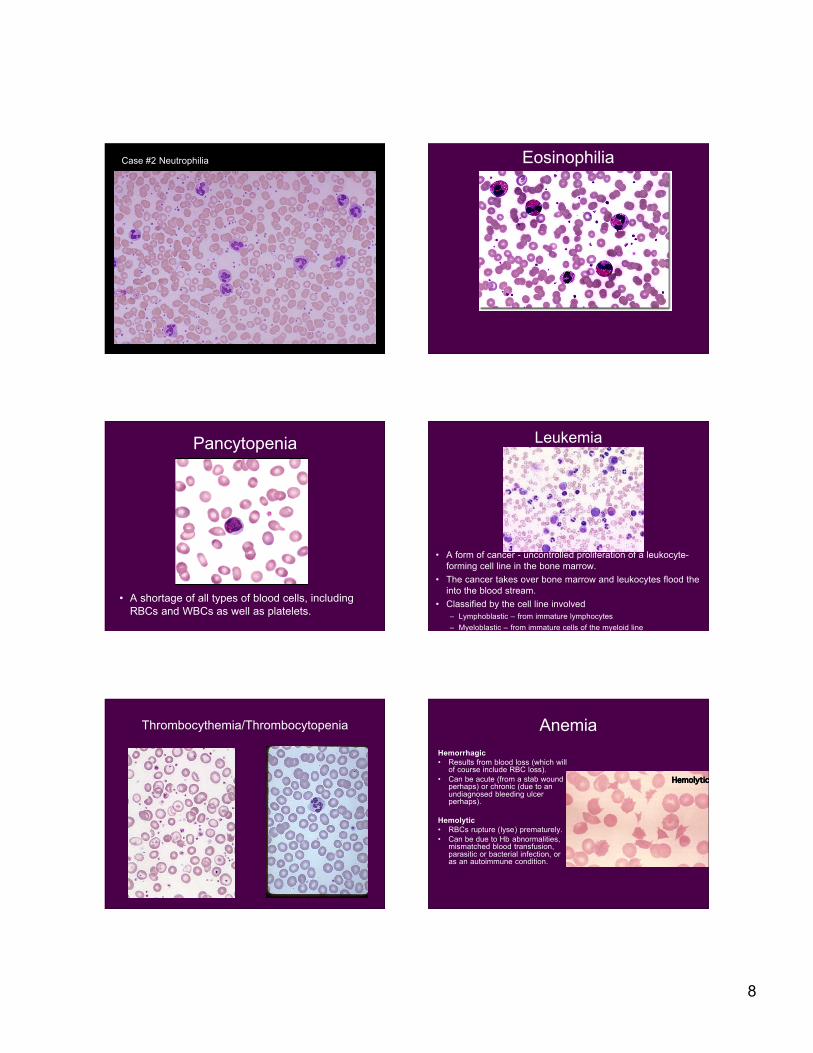
Thrombocythemia/Thrombocytopenia Anemia
Hemorrhagic• Results from blood loss (which will
of course include RBC loss).• Can be acute (from a stab wound
perhaps) or chronic (due to anundiagnosed bleeding ulcerperhaps).
Hemolytic• RBCs rupture (lyse) prematurely.• Can be due to Hb abnormalities,
mismatched blood transfusion,parasitic or bacterial infection, oras an autoimmune condition.
Hemolytic