Embed Size (px)
Citation preview
Chapter 13
Anesthesia for Myocardial Revascularization
Martin J. London, MD • Alexander Mittnacht, MD • Joel A. Kaplan, MD
Epidemiology and Risk Assessment
Pathophysiology of Coronary Artery Disease
AnatomyMyocardial Ischemia and Infarction
Anesthesia for Coronary Artery Bypass Grafting
PremedicationMonitoringInduction and Maintenance
Fast-Track Management for Coronary Artery Bypass Grafting
Coronary Artery Bypass Grafting without Cardiopulmonary Bypass
Monitoring for Off-Pump Coronary Artery Bypass Grafting
Outcomes in Off-Pump Coronary Artery Bypass Grafting
Myocardial Ischemia during Revascularization
Incidence of Myocardial Ischemia in Patients Undergoing Revascularization Surgery
Hemodynamic Changes Related to Myocardial Ischemia
Intraoperative Treatment of Myocardial Ischemia
Coronary Artery and Arterial Conduit Spasm
Summary
References
Providing anesthesia care for patients undergoing coronary artery bypass grafting (CABG) continues to be a challenging yet rewarding endeavor. Surgical, anesthetic, and technologic advances continue to drive changes in clinical routines at a rapid pace, even at a time when the numbers of cases have declined because of the growth of percutaneous coronary interventions (PCIs).
Cardiac anesthesiologists who have been in practice for the past several decades have seen a variety of anesthetic and surgical practices come into vogue and fall out of favor based on new research and economic pressures. Perhaps the most strik-ing example is the rise and fall of high-dose opioid anesthesia, which was initially driven by concern about excessive cardiovascular depression by volatile anesthetics in the 1970s and further accelerated in the mid-1980s by concerns about potential coronary steal with isoflurane. The prolonged postoperative mechanical ventila-tion resulting from the shift to high-dose opioids was also thought important to reduce stress on the recently revascularized myocardium. However, during the fol-lowing decade, this approach was completely reversed by new basic and clinical research, such as lack of evidence for adverse effects of volatile agents, particularly as related to potential effects of coronary vasodilation on coronary steal, and by strong evidence of their benefits via rapid preconditioning; by social and economic factors (i.e., safety and efficacy of fast-tracking for most patients and recognition
293
iv
AN
ESTHESIA
TECH
NIQ
UES FO
R CA
RDIA
C SU
RGIC
AL PRO
CED
URES
that time on the ventilator for many patients is an uncomfortable experience); and by the rapid rise in off-pump coronary artery bypass grafting (OPCAB), which by avoiding adverse physiologic effects of cardiopulmonary bypass (CPB) facili-tates more rapid emergence and recovery in many patients.1,2 Given the increasing emphasis on pain control in all surgical patients and its reported association with enhanced postoperative outcome in a variety of surgical subgroups, there has been a resurgence in the use of neuraxial techniques in cardiac surgery, particularly in European and Asian countries.3 Although not commonly used in the United States because of logistical issues and liability concerns, the rapidly growing literature base mandates that clinicians familiarize themselves with their potential benefits and risks.
EPIDEMIOLOGY AND RISK ASSESSMENT
In 2001, coronary artery disease (CAD) was estimated to occur in 13.2 million individuals in the United States (6.4%), resulting in approximately 500,000 deaths, 2 million hospital discharges, and a societal cost of $133 billion. CABG surgery is clearly the established cornerstone of treatment of advanced degrees of CAD. Although its absolute frequency has recently declined, there is no doubt that it will remain a common procedure and that its complexity will continue to increase for many decades to come. An understanding of the basic epidemiology of CABG surgery and of risk assessment for patients undergoing it is important for the anesthesiologist for a variety of reasons, including interactions with surgeons and cardiologists; enhancing clinical management of patients by recog-nizing high-risk characteristics and situations where preoperative management may not be adequate (such that delay of a planned elective procedure or additional periopera-tive interventions are required); developing a better sense of long-term trends in surgical practice that may impact on future practice volume (e.g., growth or decline of CABG techniques); and changes in complexity of such procedures that may influence reim-bursement or additional training requirements.
Preoperative risk assessment for patients undergoing CABG has evolved dramat-ically over the past 2 decades. The Department of Veterans Affairs in the 1970s established the first large-scale, multicenter surgical outcomes database applying rigorous statistical methodology for comparing outcomes between centers. This group and others have pioneered methodology for adjusting for different severity of illness between patients (i.e., risk adjustment) using multiple preoperative and perioperative variables thought to be of intrinsic value (usually by expert consensus) that could be easily captured and have high consistency of definition.
The Society of Thoracic Surgeons (STS) instituted a voluntary clinical database system with this approach in the early 1990s that has continued to grow rapidly as cardiac surgical groups are increasingly interested in benchmarking their practices against others.4 Many tertiary centers (e.g., Cleveland Clinic) and regional consor-tiums of hospitals (e.g., Northern New England Cardiovascular Disease Study Group) maintain databases, and some publish statistical models. Many states have established and maintain risk-adjusted mandatory reporting systems for hospital and individual surgeon performance (with New York State being an early and influential pioneer). A new scoring system (EuroSCORE) based on outcomes in 128 centers in eight Euro-pean countries has received increasing attention. It appears to compare favorably with the STS model in North American patients.5 It is freely accessible by means of an interactive web-based calculator (www.euroscore.org) and is decidedly simpler and faster to use than the STS’s scoring system, which is now also freely accessible to the public at http://www.sts.org/sections/stsnationaldatabase/riskcalculator/index.html.
294
AN
ESTHESIA
FOR M
YO
CA
RDIA
L REVA
SCU
LARIZA
TION
PATHOPHYSIOLOGY OF CORONARY ARTERY DISEASE
Anatomy
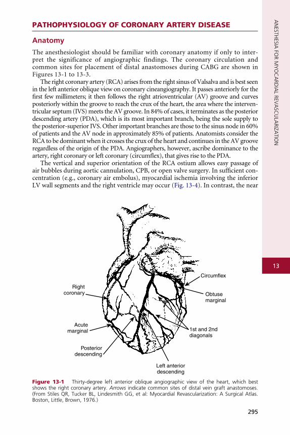
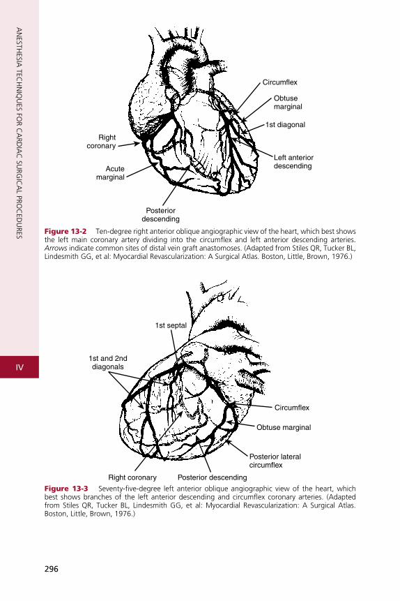
The anesthesiologist should be familiar with coronary anatomy if only to inter-pret the significance of angiographic findings. The coronary circulation and common sites for placement of distal anastomoses during CABG are shown in Figures 13-1 to 13-3.
The right coronary artery (RCA) arises from the right sinus of Valsalva and is best seen in the left anterior oblique view on coronary cineangiography. It passes anteriorly for the first few millimeters; it then follows the right atrioventricular (AV) groove and curves posteriorly within the groove to reach the crux of the heart, the area where the interven-tricular septum (IVS) meets the AV groove. In 84% of cases, it terminates as the posterior descending artery (PDA), which is its most important branch, being the sole supply to the posterior-superior IVS. Other important branches are those to the sinus node in 60% of patients and the AV node in approximately 85% of patients. Anatomists consider the RCA to be dominant when it crosses the crux of the heart and continues in the AV groove regardless of the origin of the PDA. Angiographers, however, ascribe dominance to the artery, right coronary or left coronary (circumflex), that gives rise to the PDA.
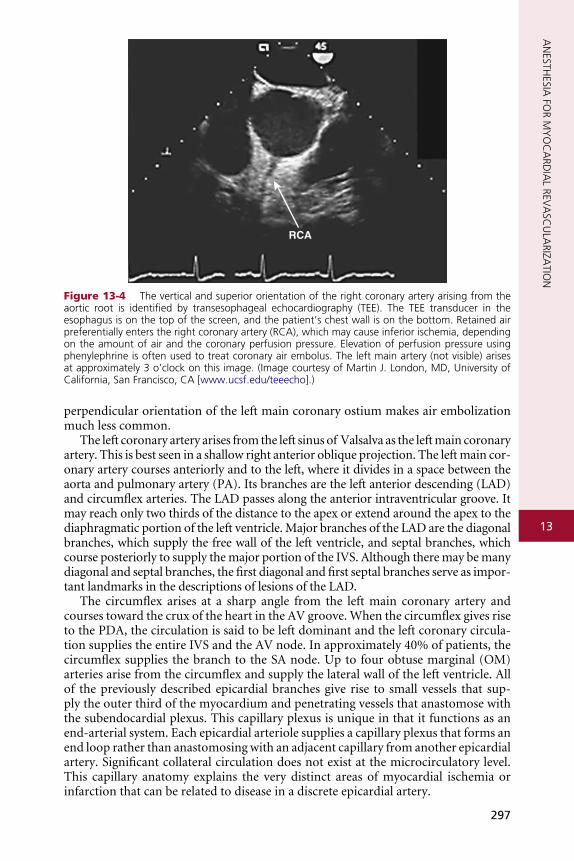
The vertical and superior orientation of the RCA ostium allows easy passage of air bubbles during aortic cannulation, CPB, or open valve surgery. In sufficient con-centration (e.g., coronary air embolus), myocardial ischemia involving the inferior LV wall segments and the right ventricle may occur (Fig. 13-4). In contrast, the near
13
Rightcoronary
Acutemarginal
Posteriordescending
Left anteriordescending
1st and 2nddiagonals
Obtusemarginal
Circumflex
Figure 13-1 Thirty-degree left anterior oblique angiographic view of the heart, which best shows the right coronary artery. Arrows indicate common sites of distal vein graft anastomoses. (From Stiles QR, Tucker BL, Lindesmith GG, et al: Myocardial Revascularization: A Surgical Atlas. Boston, Little, Brown, 1976.)
295
iv
AN
ESTHESIA
TECH
NIQ
UES FO
R CA
RDIA
C SU
RGIC
AL PRO
CED
URES
Rightcoronary
Circumflex
Obtusemarginal
1st diagonal
Left anteriordescendingAcute
marginal
Posteriordescending
Figure 13-2 Ten-degree right anterior oblique angiographic view of the heart, which best shows the left main coronary artery dividing into the circumflex and left anterior descending arteries. Arrows indicate common sites of distal vein graft anastomoses. (Adapted from Stiles QR, Tucker BL, Lindesmith GG, et al: Myocardial Revascularization: A Surgical Atlas. Boston, Little, Brown, 1976.)
1st septal
Circumflex
Obtuse marginal
Posterior lateralcircumflex
1st and 2nddiagonals
Right coronary Posterior descending
Figure 13-3 Seventy-five-degree left anterior oblique angiographic view of the heart, which best shows branches of the left anterior descending and circumflex coronary arteries. (Adapted from Stiles QR, Tucker BL, Lindesmith GG, et al: Myocardial Revascularization: A Surgical Atlas. Boston, Little, Brown, 1976.)
296
AN
ESTHESIA
FOR M
YO
CA
RDIA
L REVA
SCU
LARIZA
TION
RCA
Figure 13-4 The vertical and superior orientation of the right coronary artery arising from the aortic root is identified by transesophageal echocardiography (TEE). The TEE transducer in the esophagus is on the top of the screen, and the patient’s chest wall is on the bottom. Retained air preferentially enters the right coronary artery (RCA), which may cause inferior ischemia, depending on the amount of air and the coronary perfusion pressure. Elevation of perfusion pressure using phenylephrine is often used to treat coronary air embolus. The left main artery (not visible) arises at approximately 3 o’clock on this image. (Image courtesy of Martin J. London, MD, University of California, San Francisco, CA [www.ucsf.edu/teeecho].)
13
perpendicular orientation of the left main coronary ostium makes air embolization much less common.
The left coronary artery arises from the left sinus of Valsalva as the left main coronary artery. This is best seen in a shallow right anterior oblique projection. The left main cor-onary artery courses anteriorly and to the left, where it divides in a space between the aorta and pulmonary artery (PA). Its branches are the left anterior descending (LAD) and circumflex arteries. The LAD passes along the anterior intraventricular groove. It may reach only two thirds of the distance to the apex or extend around the apex to the diaphragmatic portion of the left ventricle. Major branches of the LAD are the diagonal branches, which supply the free wall of the left ventricle, and septal branches, which course posteriorly to supply the major portion of the IVS. Although there may be many diagonal and septal branches, the first diagonal and first septal branches serve as impor-tant landmarks in the descriptions of lesions of the LAD.
The circumflex arises at a sharp angle from the left main coronary artery and courses toward the crux of the heart in the AV groove. When the circumflex gives rise to the PDA, the circulation is said to be left dominant and the left coronary circula-tion supplies the entire IVS and the AV node. In approximately 40% of patients, the circumflex supplies the branch to the SA node. Up to four obtuse marginal (OM) arteries arise from the circumflex and supply the lateral wall of the left ventricle. All of the previously described epicardial branches give rise to small vessels that sup-ply the outer third of the myocardium and penetrating vessels that anastomose with the subendocardial plexus. This capillary plexus is unique in that it functions as an end-arterial system. Each epicardial arteriole supplies a capillary plexus that forms an end loop rather than anastomosing with an adjacent capillary from another epicardial artery. Significant collateral circulation does not exist at the microcirculatory level. This capillary anatomy explains the very distinct areas of myocardial ischemia or infarction that can be related to disease in a discrete epicardial artery.
297
iv
AN
ESTHESIA
TECH
NIQ
UES FO
R CA
RDIA
C SU
RGIC
AL PRO
CED
URES
Myocardial O2 balance
O2 supply
• O2 content of arterial blood• Coronary blood flow
O2 demands:
• Contractile state• Afterload• Preload• Heart rate
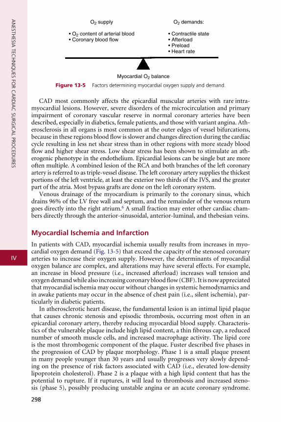
Figure 13-5 Factors determining myocardial oxygen supply and demand.
CAD most commonly affects the epicardial muscular arteries with rare intra-myocardial lesions. However, severe disorders of the microcirculation and primary impairment of coronary vascular reserve in normal coronary arteries have been described, especially in diabetics, female patients, and those with variant angina. Ath-erosclerosis in all organs is most common at the outer edges of vessel bifurcations, because in these regions blood flow is slower and changes direction during the cardiac cycle resulting in less net shear stress than in other regions with more steady blood flow and higher shear stress. Low shear stress has been shown to stimulate an ath-erogenic phenotype in the endothelium. Epicardial lesions can be single but are more often multiple. A combined lesion of the RCA and both branches of the left coronary artery is referred to as triple-vessel disease. The left coronary artery supplies the thickest portions of the left ventricle, at least the exterior two thirds of the IVS, and the greater part of the atria. Most bypass grafts are done on the left coronary system.
Venous drainage of the myocardium is primarily to the coronary sinus, which drains 96% of the LV free wall and septum, and the remainder of the venous return goes directly into the right atrium.6 A small fraction may enter other cardiac cham-bers directly through the anterior-sinusoidal, anterior-luminal, and thebesian veins.
Myocardial Ischemia and Infarction
In patients with CAD, myocardial ischemia usually results from increases in myo-cardial oxygen demand (Fig. 13-5) that exceed the capacity of the stenosed coronary arteries to increase their oxygen supply. However, the determinants of myocardial oxygen balance are complex, and alterations may have several effects. For example, an increase in blood pressure (i.e., increased afterload) increases wall tension and oxygen demand while also increasing coronary blood flow (CBF). It is now appreciated that myocardial ischemia may occur without changes in systemic hemodynamics and in awake patients may occur in the absence of chest pain (i.e., silent ischemia), par-ticularly in diabetic patients.
In atherosclerotic heart disease, the fundamental lesion is an intimal lipid plaque that causes chronic stenosis and episodic thrombosis, occurring most often in an epicardial coronary artery, thereby reducing myocardial blood supply. Characteris-tics of the vulnerable plaque include high lipid content, a thin fibrous cap, a reduced number of smooth muscle cells, and increased macrophage activity. The lipid core is the most thrombogenic component of the plaque. Fuster described five phases in the progression of CAD by plaque morphology. Phase 1 is a small plaque present in many people younger than 30 years and usually progresses very slowly depend-ing on the presence of risk factors associated with CAD (i.e., elevated low-density lipoprotein cholesterol). Phase 2 is a plaque with a high lipid content that has the potential to rupture. If it ruptures, it will lead to thrombosis and increased steno-sis (phase 5), possibly producing unstable angina or an acute coronary syndrome.
298
13
AN
ESTHESIA
FOR M
YO
CA
RDIA
L REVA
SCU
LARIZA
TION
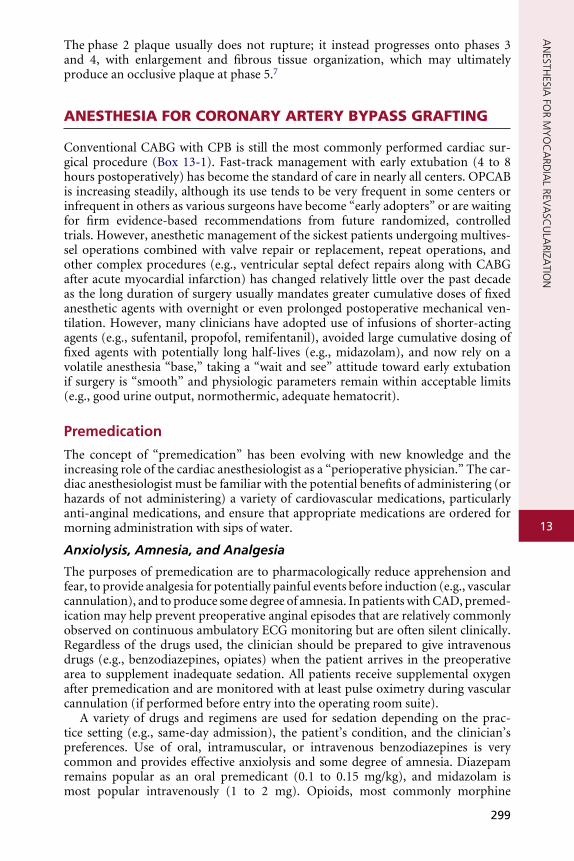
The phase 2 plaque usually does not rupture; it instead progresses onto phases 3 and 4, with enlargement and fibrous tissue organization, which may ultimately produce an occlusive plaque at phase 5.7
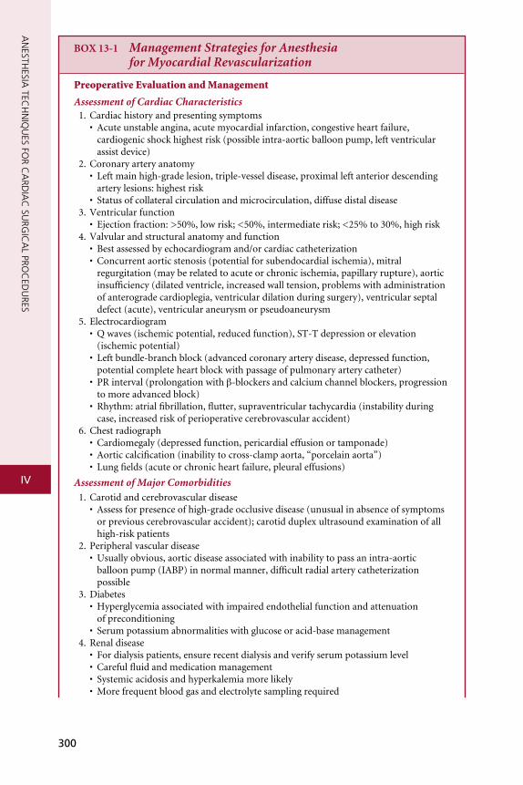
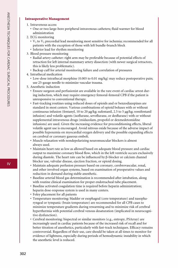
ANESTHESIA FOR CORONARY ARTERY BYPASS GRAFTING
Conventional CABG with CPB is still the most commonly performed cardiac sur-gical procedure (Box 13-1). Fast-track management with early extubation (4 to 8 hours postoperatively) has become the standard of care in nearly all centers. OPCAB is increasing steadily, although its use tends to be very frequent in some centers or infrequent in others as various surgeons have become “early adopters” or are waiting for firm evidence-based recommendations from future randomized, controlled trials. However, anesthetic management of the sickest patients undergoing multives-sel operations combined with valve repair or replacement, repeat operations, and other complex procedures (e.g., ventricular septal defect repairs along with CABG after acute myocardial infarction) has changed relatively little over the past decade as the long duration of surgery usually mandates greater cumulative doses of fixed anesthetic agents with overnight or even prolonged postoperative mechanical ven-tilation. However, many clinicians have adopted use of infusions of shorter-acting agents (e.g., sufentanil, propofol, remifentanil), avoided large cumulative dosing of fixed agents with potentially long half-lives (e.g., midazolam), and now rely on a volatile anesthesia “base,” taking a “wait and see” attitude toward early extubation if surgery is “smooth” and physiologic parameters remain within acceptable limits (e.g., good urine output, normothermic, adequate hematocrit).
Premedication
The concept of “premedication” has been evolving with new knowledge and the increasing role of the cardiac anesthesiologist as a “perioperative physician.” The car-diac anesthesiologist must be familiar with the potential benefits of administering (or hazards of not administering) a variety of cardiovascular medications, particularly anti-anginal medications, and ensure that appropriate medications are ordered for morning administration with sips of water.
Anxiolysis, Amnesia, and Analgesia
The purposes of premedication are to pharmacologically reduce apprehension and fear, to provide analgesia for potentially painful events before induction (e.g., vascular cannulation), and to produce some degree of amnesia. In patients with CAD, premed-ication may help prevent preoperative anginal episodes that are relatively commonly observed on continuous ambulatory ECG monitoring but are often silent clinically. Regardless of the drugs used, the clinician should be prepared to give intravenous drugs (e.g., benzodiazepines, opiates) when the patient arrives in the preoperative area to supplement inadequate sedation. All patients receive supplemental oxygen after premedication and are monitored with at least pulse oximetry during vascular cannulation (if performed before entry into the operating room suite).
A variety of drugs and regimens are used for sedation depending on the prac-tice setting (e.g., same-day admission), the patient’s condition, and the clinician’s preferences. Use of oral, intramuscular, or intravenous benzodiazepines is very common and provides effective anxiolysis and some degree of amnesia. Diazepam remains popular as an oral premedicant (0.1 to 0.15 mg/kg), and midazolam is most popular intravenously (1 to 2 mg). Opioids, most commonly morphine
299
iv
AN
ESTHESIA
TECH
NIQ
UES FO
R CA
RDIA
C SU
RGIC
AL PRO
CED
URES
BOX 13-1 �Management�Strategies�for�Anesthesia��for�Myocardial�Revascularization
Preoperative Evaluation and Management
Assessment�of�Cardiac�Characteristics 1. Cardiac history and presenting symptoms • Acute unstable angina, acute myocardial infarction, congestive heart failure,
cardiogenic shock highest risk (possible intra-aortic balloon pump, left ventricular assist device)
2. Coronary artery anatomy • Left main high-grade lesion, triple-vessel disease, proximal left anterior descending
artery lesions: highest risk • Status of collateral circulation and microcirculation, diffuse distal disease 3. Ventricular function • Ejection fraction: >50%, low risk; <50%, intermediate risk; <25% to 30%, high risk 4. Valvular and structural anatomy and function • Best assessed by echocardiogram and/or cardiac catheterization • Concurrent aortic stenosis (potential for subendocardial ischemia), mitral
regurgitation (may be related to acute or chronic ischemia, papillary rupture), aortic insufficiency (dilated ventricle, increased wall tension, problems with administration of anterograde cardioplegia, ventricular dilation during surgery), ventricular septal defect (acute), ventricular aneurysm or pseudoaneurysm
5. Electrocardiogram • Q waves (ischemic potential, reduced function), ST-T depression or elevation
(ischemic potential) • Left bundle-branch block (advanced coronary artery disease, depressed function,
potential complete heart block with passage of pulmonary artery catheter) • PR interval (prolongation with β-blockers and calcium channel blockers, progression
to more advanced block) • Rhythm: atrial fibrillation, flutter, supraventricular tachycardia (instability during
case, increased risk of perioperative cerebrovascular accident) 6. Chest radiograph • Cardiomegaly (depressed function, pericardial effusion or tamponade) • Aortic calcification (inability to cross-clamp aorta, “porcelain aorta”) • Lung fields (acute or chronic heart failure, pleural effusions)
Assessment�of�Major�Comorbidities 1. Carotid and cerebrovascular disease • Assess for presence of high-grade occlusive disease (unusual in absence of symptoms
or previous cerebrovascular accident); carotid duplex ultrasound examination of all high-risk patients
2. Peripheral vascular disease • Usually obvious, aortic disease associated with inability to pass an intra-aortic
balloon pump (IABP) in normal manner, difficult radial artery catheterization possible
3. Diabetes • Hyperglycemia associated with impaired endothelial function and attenuation
of preconditioning • Serum potassium abnormalities with glucose or acid-base management 4. Renal disease • For dialysis patients, ensure recent dialysis and verify serum potassium level • Careful fluid and medication management • Systemic acidosis and hyperkalemia more likely • More frequent blood gas and electrolyte sampling required
300

13
AN
ESTHESIA
FOR M
YO
CA
RDIA
L REVA
SCU
LARIZA
TION
5. Pulmonary disease • Bronchospasm, air trapping, high dead space with slower inhalation agent uptake,
atelectasis, segmental and lobar collapse possible
Preoperative�Medication�Management 1. β-Adrenergic blockers • Recommended for all patients unless contraindicated to reduce risk of ischemia 2. Calcium channel blockers • Higher incidence of heart block or need for pacing with concurrent β-blocker therapy • Efficacy in ischemia prevention controversial 3. Angiotensin-converting enzyme inhibitor • Clinical studies; increased propensity for hypotension during induction 4. Diuretics • Ensure adequate serum potassium levels 5. Statins • Beneficial anti-inflammatory effects and improved outcome independent of lipid
lowering effects 6. Aspirin • Strong data to suggest it should be continued despite increased bleeding, especially
for early and late graft patency and mortality 7. Other antiplatelet agents or glycoprotein IIb/IIIa inhibitors • Associated with increased bleeding; hold as appropriate, and treat with platelets, or
recombinant factor VII as necessary 8. Heparin • Usually discontinued 4 hours preoperatively for stable patients, continued up to
and through pre-CPB period for critical left main disease or acutely unstable angina patients
9. Insulin • Continuous infusion for poorly controlled hyperglycemia; insulin resistance may be
encountered during hypothermic CPB 10. Oral hypoglycemic agents • Metformin is associated with lactic acidosis in patients developing perioperative low
cardiac output; it is optimal to hold administration several half-lives preoperatively. • Glyburide experimentally blocks preconditioning by closure of ATP-mediated potas-
sium channels. 11. Antibiotic prophylaxis • Cephazolin (1 g IV) or cefuroxime (1.5 g IV) less than 60 minutes before incision;
vancomycin (1 g by slow infusion to avoid hypotension and flushing) or clindamycin (600 to 900 mg IV) for penicillin or cephalosporin allergy; adjust as appropriate for renal failure.
• Repeat: cefazolin every 2 to 5 hours, cefuroxime every 3 to 4 hours, vancomycin every 6 to 12 hours, clindamycin every 3 to 6 hours; adjust as appropriate for renal failure.
12. Anxiolytic or analgesic premedication • Oral diazepam (5 to 10 mg) at least 1 hour before induction; supplement with intra-
venous fentanyl or midazolam as needed. • Intramuscular lorazepam (1 to 2 mg) or morphine (approximately 0.15 mg/kg) at
least 1 hour before induction, depending on age, comorbidities, and anxiety level • Supplemental oxygen to avoid desaturation and ischemia 13. Preoperative epidural insertion • Uncommonly used in the United States, but it may reduce stress response, preserve
adrenoreceptor function, and decrease time to extubation, pulmonary complications and pain scores.
• Use for conscious off-pump coronary artery bypass has been reported. • Optimal to place at least 6 to 8 hours preoperatively to ensure adequate hemostasis
Box continued on following page
301
iv
AN
ESTHESIA
TECH
NIQ
UES FO
R CA
RDIA
C SU
RGIC
AL PRO
CED
URES
Intraoperative Management
1. Intravenous access • One or two large-bore peripheral intravenous catheters; fluid warmer for blood
administration 2. ECG monitoring • V3 to V5 precordial lead monitoring most sensitive for ischemia; recommended for all
patients with the exception of those with left bundle-branch block • Inferior lead for rhythm monitoring 3. Blood pressure monitoring • Radial artery catheter: right arm may be preferable because of potential effects of
retraction for left internal mammary artery dissection (with newer surgical retractors, this is likely less problematic).
• Backup cuff for arterial monitoring failure and correlation of pressures 4. Intrathecal medication • Low-dose intrathecal morphine (0.005 to 0.01 mg/kg) may reduce postoperative pain;
use 25-gauge needle to minimize vascular trauma. 5. Anesthetic induction • Ensure surgeon and perfusionist are available in the rare event of cardiac arrest dur-
ing induction, which may require emergency femoral-femoral CPB if the patient is unresponsive to conventional therapy.
• Fast-tracking routines using reduced doses of opioids and or benzodiazepines are standard in most centers. Various combinations of opioid boluses with or without continuous infusion (fentanyl, 10 to 20 μg/kg; sufentanil, 2.5 to 5 μg/kg; remifentanil infusion) and volatile agents (isoflurane, sevoflurane, or desflurane) with or without supplemental intravenous drugs (midazolam, propofol or dexmedetomidine infusions) are used. Given the increasing evidence for preconditioning effects, liberal volatile agent use is encouraged. Avoid nitrous oxide because of the adverse impact of possible hypoxemia on myocardial oxygen delivery and the possible expanding effects on cerebral or coronary gaseous emboli.
• Muscle relaxation with nondepolarizing neuromuscular blockers is almost always used.
• Maintain heart rate as low as allowed based on adequate blood pressure and cardiac output to maximize coronary blood flow, which in the left ventricle occurs exclusively during diastole. The heart rate can be influenced by β-blocker or calcium channel blocker use, valvular disease, ejection fraction, or opioid dosing.
• Maintain adequate perfusion pressure based on coronary, cerebrovascular, renal, and other involved organ systems, based on examination of preoperative values and reduction in demand during stable anesthetic.
• Baseline arterial blood gas determination is recommended after intubation, along with routine clinical examination for proper endotracheal tube placement.
• Baseline activated coagulation time is required before heparin administration; heparin dose-response system is used in many centers.
• Foley placement for all patients • Temperature monitoring: bladder or esophageal (core temperature) and nasopha-
ryngeal or tympanic (brain temperature) are recommended for all CPB cases to minimize temperature gradients during rewarming and to minimize risk of cerebral hyperthermia with potential cerebral venous desaturation (implicated in neurocogni-tive dysfunction).
• Cerebral monitoring: bispectral or similar monitors (e.g., entropy, PSArray) are increasingly used in cardiac patients because of the increased risk of recall and for better titration of anesthetics, particularly with fast-track techniques. Efficacy remains controversial. Regardless of their use, care should be taken at all times to monitor for evidence of lightness, especially during periods of hemodynamic instability in which the anesthetic level is reduced.
302
13
AN
ESTHESIA
FOR M
YO
CA
RDIA
L REVA
SCU
LARIZA
TION
6. Pulmonary artery catheter or central venous pressure catheter placement • Central venous pressure catheter is considered standard of care; pulmonary artery
catheter use is very center specific. • Need for placement of pulmonary artery catheter before induction is controversial,
but severe concurrent valve disease and a low ejection fraction are strong indications. • When using pulmonary artery catheter, ensure the catheter does not wedge inad-
vertently during surgical manipulation of the heart (closes the loop, advancing it outward). Transesophageal echocardiography (TEE) can easily verify positioning; otherwise, withdrawing the catheter several centimeters before institution of CPB is usually effective.
• Avoid pulmonary artery catheter balloon inflation, particularly when the patient is heparinized or hypothermic.
7. Pharmacologic adjuncts • Antifibrinolytic therapy – Used primarily for CPB cases. • ε-Aminocaproic acid (Amicar): continuous infusion from skin incision and bolus
dosing (5 g) after heparin administration and after CPB weaning are used with variable efficacy.
• Tranexamic acid: strong evidence in the literature for efficacy • Corticosteroid administration – High dose before CPB; methylprednisolone (2 to 3 g)
is used in some centers to block some components of inflammatory response. 8. Insertion of TEE • Use influenced by surgeon’s preference, center’s resources, anesthesia or cardiology
expertise, and availability; routine use common in many centers for all patients. • Can be of particular value in guiding anesthetic (fluid administration, detection
of new ischemia, verification of pulmonary artery catheter in proximal pulmonary artery) and surgery (verification of retrograde cardioplegia cannula, IABP tip relative to arch vessels, presence of aortic root and arch calcification, ulceration, or mobile components, placement of left ventricular vent, ischemic changes).
• Stomach decompression before and after placement of probe is recommended to decrease risk of postoperative passive aspiration.
• Esophageal perforation is rare but increasingly recognized as a complication that must be considered, especially with development of septic changes with new bilateral pleural effusions postoperatively.
9. Sternotomy • It is a very stimulating stress; ensure adequate anesthesia, especially if preparation and
drape period is prolonged. • “Let down the lungs” to avoid potential pleural tear from sternal saw. • Repeat sternotomy • Uses different technique: oscillating saw on outer sternal table, wires removed
manually, and blunt dissection with scissor used on the inner table of the sternum. • Risks include right ventricular perforation, damage to existing vein grafts,
and ventricular fibrillation from electrocautery energy transmitted by a sternal wire • Use external defibrillation pads on all repeat sternotomy cases. • Ensure that blood (preferably 2 units checked) is close at hand during opening. • Usual presenting sign of right ventricular perforation is ventricular fibrillation,
often with little obvious bleeding; emergent femoral-femoral CPB is usually required.
Management�before�Revascularization 1. Maintain “favorable hemodynamics,” with low heart rate and maintenance of systemic
blood pressure depending on the preoperative baseline, age, coronary anatomy, and presence of other compromised organs (e.g., brain, kidney). Hypotension with surgical manipulation of the heart and aorta is unavoidable but is usually treated with phenyl-ephrine, volume supplementation, and Trendelenburg position. Close communication with the surgeon is essential.
Box continued on following page
303
iv
AN
ESTHESIA
TECH
NIQ
UES FO
R CA
RDIA
C SU
RGIC
AL PRO
CED
URES
2. Left internal mammary artery dissection: elevate table, rotate to left, and reduce tidal volumes (increase rate) to facilitate surgeon’s exposure. Similar maneuvers are done for the less commonly performed right internal mammary artery dissection. Heparin is usually administered before clamping of the left internal mammary artery pedicle to avoid thrombosis. Papaverine is often injected retrograde by the surgeon, which occasionally causes hypotension.
3. Conventional coronary artery bypass grafting (CABG) • Passive hypothermia is likely protective, given the reduction in myocardial oxygen
demands and cerebral protective effects. • Pre-bypass phlebotomy is popular in certain centers, although evidence of its efficacy
is controversial. Blood may be sequestered before heparinization into storage bags, usually by central venous pressure catheter access, which may require additional fluid administration or by the right atrial cannula just before institution of bypass, usually with the assistance of phenylephrine.
• Retrograde cardioplegia may be used alone or, more commonly, in combination with anterograde techniques. Perfusion of the right ventricle is limited by this technique. TEE can assist in placement and confirmation of the catheter in the coronary sinus.
4. Repeat cases • Atheroma in prior vein grafts is sandy and often loose. Sudden, profound ischemia
may occur with surgical manipulation of grafts due to distal embolization. This may require emergent institution of CPB.
• Adequate conduits are essential for performing anastomoses. This can be problematic in repeat cases.
• Exposure for cannulation and of the left ventricle is often more complex because of adhesions and accounts for increased blood loss.
5. Off-pump CABG • Maintenance of normothermia is desirable to reduce bleeding and propensity toward
ventricular fibrillation, and for facilitating intraoperative extubation if planned. Forced air and water blankets can be helpful.
6. Heparin administration • Administer 300 to 400 IU/kg. Higher dose required with heparin resistance (most
commonly associated with prolonged preoperative administration). Resistance is usu-ally easily treated with 1 unit of fresh frozen plasma or recombinant antithrombin III. Activated coagulation time between 450 and 500 seconds is required for institution of CPB.
• Dosing of heparin for off-pump CABG cases is controversial, with centers using full- or low-dose regimens.
• Only use kaolin-activated coagulation time tubes with concurrent aprotinin adminis-tration.
• Mild hypotension due to vasodilation can occur with heparin administration. • For patients with heparin-induced thrombocytopenia, direct thrombin inhibitors
have been used with success. 7. Cannulation • Aortic cannula • Examination of cannulation site by epicardial imaging may be used particularly if
surgical palpation is suspicious for calcium or TEE examination reveals prominent distal arch or proximal aortic root disease. Surgical response to severe disease ranges from no change to “no touch” techniques to potential circulatory arrest.
• Ensure that systemic pressure is lowered to lowest safe level (90 to 110 mm Hg systolic) during cannulation to minimize risk of dissection.
• Adequate eye protection is required at all times and particularly during this period due to splash hazard.
• Surgeon inspects lines for air bubbles before hooking up circuit. • Hypotension can be effectively treated by perfusionist infusing fluid.
304
13
AN
ESTHESIA
FOR M
YO
CA
RDIA
L REVA
SCU
LARIZA
TION
• Retrograde cannula: Inserted in the coronary sinus with insertion site near the right atrial appendage. TEE can guide approximately but is limited by its two-dimensional imaging. Proper placement can be verified by TEE, surgical palpation in great cardiac vein, and dark color of the coronary sinus blood. Pressure monitoring is used to ensure adequate infusion (if low, it is likely in the right ventricle; if excessively high, rupture could occur). Insertion can be difficult and cause major hemodynamic compromise and arrhythmias. For sick repeat or low ejection fraction, insertion after institution of CPB or preparation for rapid institution of CPB may occasionally be required because arrest may occur.
• Venous cannula: For isolated CABG, a single large-bore venous cannula is inserted through the right atrium into the inferior vena cava. It can be partially imaged by TEE in the bicaval view.
• Left ventricular vent: Inserted after institution of CPB via the left superior pulmonary vein in many but not all cases. Can be visualized on TEE short-axis view. Verification of absence of thrombus in the LV is essential in patient with a recent anterior wall MI or aneurysm.
Revascularization�Management 1. Conventional CABG • On institution of CPB with full flow, discontinue mechanical ventilation. Passive
insufflation of oxygen (200 mL/min) is usually continued. Discontinue all main-tenance intravenous fluid. Turn off power to the TEE transducer if present. Verify pulmonary artery catheter tip position to avoid inadvertent wedging. Drain urine bag or mark value before CPB.
• Myocardial preservation by surgeon and perfusionist involves anterograde or retro-grade cardioplegia (or both), arrest with high-potassium cardioplegia, and in most centers, hypothermia (systemic, topical, and by cardioplegia).
2. Off-pump CABG • Left anterior descending and diagonal vessels • Only mild left ventricular displacement is required (packs under left ventricle with
mild rotation of the left ventricle upward and to the left); hemodynamic effects are usually minimal in the absence of ischemia or a very low ejection fraction. Diagonal visualization requires additional rotation. Stabilizer placement is usually well tolerated. Hemodynamic changes (hypotension) are usually easily treated with fluids, Trendelenburg position, or phenylephrine.
• Posterior descending and circumflex vessels • Moderate to severe left ventricular displacement (verticalization), most pronounced
for circumflex anastomoses, is required. Application of an apical suction device is com-mon, although other, simpler methods are used. With proper placement, hypotension is usually mild but more common than with left anterior descending or diagonal grafts. With improper placement or a large left ventricle or right ventricle, compressive effects on the right ventricle can be severe, leading to a major reduction in cardiac output. Opening of the right pleural space may accommodate the right ventricle, relieving the compression with hemodynamic improvement. Trendelenburg positioning is helpful for maintaining hemodynamics and for the surgeon’s visualization.
• Mechanical ischemic preconditioning (5-minute occlusion followed by 5-minute reperfusion) is used by some surgeons. However, evidence for efficacy, especially with concurrent volatile agent use, is weak.
• Some clinicians use “reperfusion prophylaxis,” similar to the cardiac catheterization lab setting during acute MI with lidocaine, thrombolytics, and magnesium sulfate. With routine off-pump CABG there is little evidence for this because most patients have native and collateral flow.
• Ischemia during anastomosis can be treated with insertion of an intracoronary shunt. Some surgeons use these routinely, although there is concern regarding endothelial damage with their use; and many surgeons avoid them unless absolutely necessary.
Box continued on following page
305
iv
AN
ESTHESIA
TECH
NIQ
UES FO
R CA
RDIA
C SU
RGIC
AL PRO
CED
URES
3
• TEE can be helpful. However, with verticalization and application of a stabilizer, visu-alization of all walls and interpretation of wall motion can be difficult. ECG ampli-tude is often markedly diminished with verticalization for posterior descending artery and circumflex anastomoses. Detection of new mitral regurgitation strongly suggests new ischemia.
Management after Revascularization
1. CPB weaning • Plan ahead for potential inotrope and other vasoactive drug use based on presence of
known predictors of difficult weaning, particularly low ejection fraction before CPB. • Epicardial leads (atrial and ventricular) should be placed. Atrioventricular block and
bradycardia are common in patients with severe ischemia and those on β-blockers and calcium channel antagonists. Impaired diastolic relaxation is common, reduc-ing the effectiveness of the Frank-Starling mechanism for augmentation of cardiac output. Heart rate plays a larger role, and maintaining heart rate at 80 to 90 beats per minute for several hours avoids ventricular distention. Frequent evaluation for return of the patient’s underlying sinus rhythm is optimal because it may be associated with the best cardiac output.
• Augmentation of cardiac index with catecholamines (epinephrine, dopamine, dobu-tamine) and/or phosphodiesterase inhibitors (amrinone, milrinone) may be required for low cardiac index (generally <2.0 L/min/m2) or elevation of pulmonary artery occlusion pressure (generally >18 to 20 mmHg).
• Intra-aortic balloon counterpulsation is used when inotrope therapy alone is insuf-ficient. IABP augments diastolic coronary perfusion and forward flow (“suction effect”). Aortic insufficiency is a relative contraindication to IABP. Evaluate with TEE to avoid ventricular distention, and check placement below the level of the left subcla-vian and carotid vessels.
2. Off-pump CABG • Assuming adequate anastomoses have been performed, cardiac index returns to
normal levels immediately. Function may deteriorate if significant ischemia has occurred, although this is unusual.
3. Reversal of heparin and hemostasis • Protamine is usually administered empirically in a 1:1 ratio in the absence of use
of automated systems such as the Heparin Dose Response System (Medtronic, Inc., Minneapolis, MN).
• Avoid a rapid bolus. Exact infusion rates remain controversial. 4. Chest closure and transport • Remove TEE, and decompress stomach with orogastric tube. • Monitor blood pressure, ECG, and SaO2 for transport. • Ensure chest tubes (if present) are on water seal because tension pneumothorax
is a known hazard during transport. • Ensure patency of mediastinal drainage because tamponade is a known hazard.
(0.1 to 0.15 mg/kg) given by the intramuscular route, and fentanyl (50 to 75 μg), administered intravenously, are recommended to provide analgesia, particularly during radial artery cannulation. Scopolamine, usually given intramuscularly (0.2 to 0.4 mg) but occasionally intravenously, has been commonly used for its potent amnestic effects. Given its potential to induce delirium and disorientation, particu-larly in the elderly, it is less frequently used today. Acute toxicity associated with overdosage can be effectively reversed with physostigmine.
06
13
AN
ESTHESIA
FOR M
YO
CA
RDIA
L REVA
SCU
LARIZA
TION
Management of Antianginal and Antihypertensive Medications
β-adrenergic receptor antagonists
For the newly trained anesthesiologist who is told to provide “perioperative β-block-ade” to all patients with known CAD, or those with multiple risk factors undergoing major noncardiac (particularly vascular) surgery, it may come as a surprise that in 1972, a case series from the prestigious Cleveland Clinic admonished clinicians to withdraw such medication at least 2 weeks before CABG surgery reporting that 5 such patients died within 24 hours of surgery. Several years later, Kaplan and co-workers found that it was safe to continue β-blockade and that operative mortality was similar in patients in whom propranolol had been continued within 24 to 48 hours of surgery. Randomized trials evaluating the safety of administration of pro-pranolol within 12 hours of surgery showed a significantly greater increase in the incidence of pre-CPB ischemia in patients withdrawn from propranolol (within 24 to 72 hours); they also recommended continuation of therapy up until the time of surgery. Further work in the 1980s documented the efficacy of continuation of β-blockers through surgery with regard to reducing pre-CPB ischemia. These studies were instrumental in laying the groundwork for the subsequent noncardiac surgery studies in the late 1980s. These led to the contemporary randomized perioperative β-blocker trials, which despite ongoing controversy regarding the exact efficacy of this therapy resulted in its becoming a clinical routine.8 Several contempo-rary observational studies have documented associations of β-blocker therapy with reduction in perioperative mortality in CABG patients. The largest of these by Ferguson and colleagues9 considered 629,877 patients in the STS database (1996 to 1999) in which a modest but statistically significant reduction in 30-day risk-adjusted mortality was reported (OR = 0.94, 95% CI = 0.91 to 0.97). This treatment effect was observed in many high-risk subgroups, although a trend toward increased mortality was seen in patients with an ejection fraction (EF) less than 30% (OR = 1.13; 95% CI, 0.96 to 1.33). Considerable efforts are being expended by major organizations (STS, ACC) in increasing compliance with existing guidelines for use of β-blockers at the time of hospital discharge (along with use of aspirin, statins, and angiotensin-converting enzyme [ACE] inhibitors).
other medications
Aspirin (and other platelet inhibitors such as dipyridamole) have long been recognized to have strong efficacy in the prevention of early graft thrombosis after CABG, and are a well-recognized component of primary and secondary preven-tion strategies for all patients with ischemic heart disease. A large observational anal-ysis reported substantial reduction in overall mortality (1.3% vs. 4.0%) and ischemic complications of the heart, brain, kidneys, and gastrointestinal tract when aspirin was administered within 48 hours after surgery. However, patients receiving aspirin immediately before surgery have more mediastinal bleeding and may receive more blood products. A consensus conference of the American College of Chest Physicians on antithrombotic and thrombolytic therapies recommended institution of aspirin within 6 hours after CABG surgery over continuation of preoperative therapy (level of evidence Ia).10
ACE inhibitors and statins are receiving attention as agents given a variety of important “pleiotropic” effects (e.g., effects independent of their primary actions of antihypertensive effects or lipid-lowering effects, respectively).11 Potent anti-inflammatory effects and beneficial effects on endothelial function have been reported for both agents, as well as less clear effects on angiogenesis. Both agents are commonly admin-istered acutely during PCI for their purported benefits. ACE inhibitors are widely
307
iv
AN
ESTHESIA
TECH
NIQ
UES FO
R CA
RDIA
C SU
RGIC
AL PRO
CED
URES
considered to be vasculoprotective, particularly with regard to ventricular remodel-ing after acute myocardial infarction, and they appear to reduce damage after ischemic reperfusion (likely related to reduction in ischemic-induced vasoconstriction and reduction in leukocyte adhesion). Statins have been reported to reduce cir-culating levels of adhesion molecules, which have been implicated in endothelial dysfunction after CPB. Both agents appear to have direct effects on platelet aggrega-tion and plasminogen activator inhibitors. Of the two classes of drugs, the greatest interest appears to be on the statins because of the greater number of publications and intense concurrent interest with regard to cardioprotection for noncardiac (particularly vascular) surgery. Several investigators have published similar reports of efficacy in observational cohorts of CABG patients.
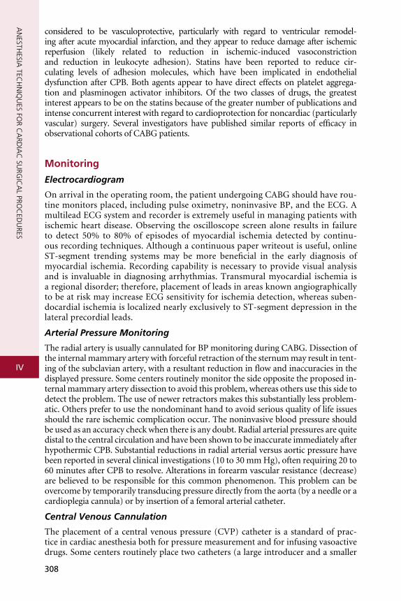
Monitoring
Electrocardiogram
On arrival in the operating room, the patient undergoing CABG should have rou-tine monitors placed, including pulse oximetry, noninvasive BP, and the ECG. A multilead ECG system and recorder is extremely useful in managing patients with ischemic heart disease. Observing the oscilloscope screen alone results in failure to detect 50% to 80% of episodes of myocardial ischemia detected by continu-ous recording techniques. Although a continuous paper writeout is useful, online ST-segment trending systems may be more beneficial in the early diagnosis of myocardial ischemia. Recording capability is necessary to provide visual analysis and is invaluable in diagnosing arrhythmias. Transmural myocardial ischemia is a regional disorder; therefore, placement of leads in areas known angiographically to be at risk may increase ECG sensitivity for ischemia detection, whereas suben-docardial ischemia is localized nearly exclusively to ST-segment depression in the lateral precordial leads.
Arterial Pressure Monitoring
The radial artery is usually cannulated for BP monitoring during CABG. Dissection of the internal mammary artery with forceful retraction of the sternum may result in tent-ing of the subclavian artery, with a resultant reduction in flow and inaccuracies in the displayed pressure. Some centers routinely monitor the side opposite the proposed in-ternal mammary artery dissection to avoid this problem, whereas others use this side to detect the problem. The use of newer retractors makes this substantially less problem-atic. Others prefer to use the nondominant hand to avoid serious quality of life issues should the rare ischemic complication occur. The noninvasive blood pressure should be used as an accuracy check when there is any doubt. Radial arterial pressures are quite distal to the central circulation and have been shown to be inaccurate immediately after hypothermic CPB. Substantial reductions in radial arterial versus aortic pressure have been reported in several clinical investigations (10 to 30 mm Hg), often requiring 20 to 60 minutes after CPB to resolve. Alterations in forearm vascular resistance (decrease) are believed to be responsible for this common phenomenon. This problem can be overcome by temporarily transducing pressure directly from the aorta (by a needle or a cardioplegia cannula) or by insertion of a femoral arterial catheter.
Central Venous Cannulation
The placement of a central venous pressure (CVP) catheter is a standard of prac-tice in cardiac anesthesia both for pressure measurement and for infusing vasoactive drugs. Some centers routinely place two catheters (a large introducer and a smaller
308
13
AN
ESTHESIA
FOR M
YO
CA
RDIA
L REVA
SCU
LARIZA
TION
CVP catheter) in the central circulation to facilitate volume infusion and vasoactive or inotropic drug administration.
Pulmonary Artery Catheterization
The efficacy of PA catheterization in medical and surgical settings has evolved over the past 20 years from steadily increasing use in the 1980s and 1990s to distinctly lower use now. Increasing literature evidence from a variety of experimental designs has strongly suggested that despite the substantial amount of physiologic information obtained, major clini-cal outcomes are little influenced. Earlier studies suggested that clinicians were unable to accurately judge filling pressures based on clinical signs and that therapy could be influ-enced by these data in the surgical and medical settings. Intraoperatively, PA catheterization was also shown to detect decreased left ventricular (LV) compliance associated with myo-cardial ischemia. It also appeared better suited for monitoring high-risk patients.
Based on the existing literature it is not possible to give precise criteria for use of a PA catheter in CABG.12 The higher the patient risk (based primarily on established preoperative clinical predictors), the more favorable is the risk-benefit ratio. Risk factors include the following:
1. Significant impairment of ventricular function (EF < 40%, evidence of acute or chronic congestive heart failure, known elevation of left ventricular end-diastolic pressure (LVEDP) on preoperative catheterization, need for preoperative intra-aortic balloon pump (IABP), acute or chronic severe mitral regurgitation due to ischemia, ventricular septal defect after myocardial infarction, or other mechanical complications).
2. High risk for intraoperative ischemia or difficult revascularization (i.e., recent, large myocardial infarction or severe unstable angina, known poor revascular-ization targets or severe microcirculatory disease, reoperation, catheterization laboratory PCI “crash”).
3. Severe comorbidities (e.g., renal failure, on or approaching need for dialysis; severe chronic obstructive pulmonary disease).
4. Combined procedures that significantly lengthen duration of surgery or add significant blood loss (e.g., CABG-carotid, other vascular procedures).
Although most of the recent clinical reports of patients undergoing OPCAB have used, and many recommend use of PA catheterization, it is not possible to give firm recommendations on this because of the lack of evidence-based data.
Transesophageal Echocardiography
It is appreciated that the earliest signs of myocardial ischemia include diastolic dysfunc-tion followed by systolic segmental wall motion abnormalities that occur within seconds of acute coronary occlusion. Comparison of TEE with continuous (Holter) ECGs have shown a greater incidence of segmental wall motion abnormalities than ECG changes in patients with CAD. However, it is thought that new segmental wall motion abnormalities detected in the intraoperative period may frequently occur due to nonischemic causes, particularly changes in loading conditions, and alteration in electrical conduction in the heart. The use of inotropic agents or elevations of catecholamine levels can aggravate (by increased oxygen demand) or improve wall motion. Changes in preload and afterload are likely the most important factors in the CABG setting; transient reversible segmen-tal wall motion abnormalities are commonly related to acute myocardial stunning due to ischemia before or during weaning from CPB. TEE is highly sensitive but not spe-cific for myocardial ischemia. The short-axis midpapillary muscle view, commonly used because of its inclusion of myocardium supplied by the three major coronary arteries,
309
iv
AN
ESTHESIA
TECH
NIQ
UES FO
R CA
RDIA
C SU
RGIC
AL PRO
CED
URES
may entirely miss segmental wall motion abnormalities occurring in the basal or api-cal portions of the heart. These changes necessitate interrogation of additional compo-nents of the comprehensive TEE examination recommended by the ASE/SCA Task Force before and after CPB or after completion of revascularization in OPCAB. The ASA Prac-tice Guidelines for TEE (which have not been revised since their initial publication in 1996) list the perioperative uses in patients with increased risk of myocardial ischemia or infarction as a category II indication (supported by weaker evidence and expert con-sensus). The indication is strengthened when ECG monitoring cannot be used to diag-nose ischemia (e.g., in patients with left bundle-branch block, extensive Q waves, or ST-T abnormalities on the baseline ECG), and it is weakened when baseline segmental wall motion abnormalities are present (particularly akinesis or dyskinesis due to fibrotic, calci-fied, or aneurysmal myocardium). Use of perioperative TEE to evaluate myocardial per-fusion, coronary anatomy, or graft patency is listed as a category III indication, but newer technology will upgrade this indication. Evaluation of ischemic mitral regurgitation by TEE may even influence the surgical management during CABG.13
Induction and Maintenance
Induction of anesthesia should take place in a calm and relaxed manner, preferably in a quiet operating room. Attention should be paid to the ambient room tempera-ture because entry into an excessively cold operating room can elicit a sympathetic response increasing blood pressure and sometimes heart rate, particularly in the elderly and thin patients. Allaying the patient’s anxiety with premedication and calm, reassuring verbal interaction is also critical. Preoxygenation should be used and monitoring should be in place, including PA catheterization in patients at very high risk, whose condition may be unstable during or after induction.
There are two main considerations in choosing an induction technique for patients undergoing CABG. The first is LV function. Patients with good LV function often have a strong sympathetic response to surgical stimulation and may require supranormal doses of anesthetics, plus the addition of β-blockers with or without vasodilators, to control these responses. Patients with poor LV function often do not tolerate normal doses of anesthetics and are unable to produce a significant hemodynamic response to sympa-thetic stimulation or the response may precipitate major reductions in cardiac output.
The second consideration is the desirability of early extubation. Time spent in the intensive care unit (ICU) is one of the most expensive aspects of hospital care for CABG and is heavily influenced by postoperative ventilator management. The patient with normal preoperative LV function, assuming an uneventful intraoperative course, will have recovered 90% of baseline LV function by 4 hours postoperatively and can usually be extubated within 4 to 6 hours postoperatively if attention is paid to adequate rewarming and postoperative analgesia and if high doses of respiratory depressant anesthetics (particularly opioids and benzodiazepines) have been avoided. With the routine application of fast-track techniques, some centers have adopted immediate extubation in the operating room. However, this remains relatively uncommon and requires the close cooperation of a highly coordinated team. Because this is not always the case (particularly in teaching institutions in which residents and fellows rotate for short intervals or in some private institutions in which staffing at night is relatively low), many centers take a more measured, less aggressive approach.
After the induction of anesthesia, the pre-bypass period (for conventional CABG) may last less than an hour (e.g., only one or two saphenous vein grafts harvested) or several hours (e.g., for dissection of the left internal mammary artery, right internal mammary artery, or radial or gastroepiploic arteries after a repeat sternotomy). Surgical stimulus may be severe, such as during sternotomy or dissection around
310
13
AN
ESTHESIA
FOR M
YO
CA
RDIA
L REVA
SCU
LARIZA
TION
the ascending aorta. Between 50% and 70% of patients in most series presenting for CABG have normal LV function (when not ischemic) and are capable of mounting significant blood pressure and heart rate responses to noxious stimuli, whereas others with poor LV function may require pharmacologic support of the blood pres-sure for such stimuli. It is evident that no single approach to anesthesia for CABG procedures is suitable for all patients. Most hypnotics, opioids, and volatile agents have been used in different combinations for the induction and maintenance of anesthesia with good results in the hands of experienced clinicians.
Primary Induction Agents
Considerations for choice of induction agent in the patient undergoing CABG are based on theoretical and practical clinical considerations. Desirable goals include avoidance of hypertension and tachycardia, which is most likely to occur in the patient with normal ventricular function, hypertension, and LV hypertrophy; avoid-ance of hypotension and excessive myocardial depression in a patient with depressed ventricular function or with severe flow-dependent stenoses; and provision of smooth intubating conditions with a lack of effect on airway resistance.
Thiopental has been used for decades for induction in this setting. Its predominant he-modynamic effects include reduction in mean arterial pressure and cardiac output accom-panied by a modest increase in heart rate. These are believed to result from a combination of direct myocardial depression, venodilation, and a decrease in central sympathetic out-flow. The use of thiopental in most centers has declined substantially in favor of propofol. Adverse effects on airway resistance, a greater propensity to elicit bronchospasm, and a greater association with postoperative nausea and vomiting are other potential factors.
The clinical effects of propofol are in general similar to those of thiopental. How-ever, it has numerous advantages over thiopental based on its predictable pharma-cokinetics and dynamics. Based on these, it has been widely adopted in the operating room for anesthesia delivery by computer-controlled devices and for postoperative ICU sedation. It is often used for sedation after CABG surgery.14
Benzodiazepines
Benzodiazepines are commonly used in combination with a narcotic to induce anesthesia for CABG. In most settings, midazolam has replaced diazepam given its numerous advantages (particularly water solubility, a shorter half-life, and absence of metabolites capable of accumulation prolonging sedative effects). Numerous clinical series of widely different sizes and designs, reporting on the efficacy of diazepam or, more commonly, midazolam used with high-dose opioids, were subsequently pub-lished. Moderate degrees of hypotension were reported in most studies primarily attributed to a reduction in systemic vascular resistance.
Opioids
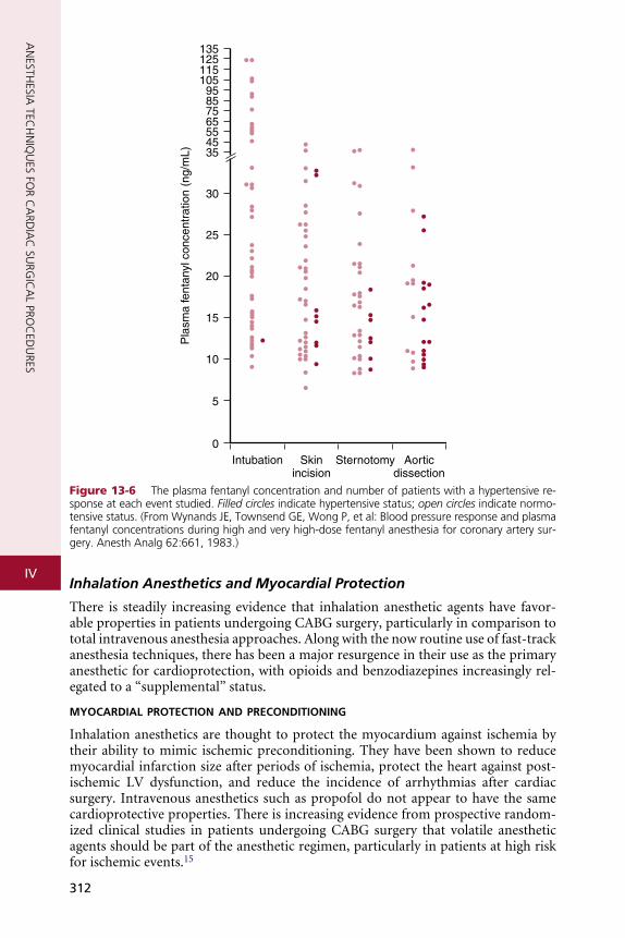
These drugs are pure opioid agonists and none provides complete anesthesia as defined by predictable dose-response relations for suppression of the stress response and release of endogenous catecholamines even with high serum concentrations. Hypertension and tachycardia have been commonly reported in response to induction/intubation and surgical stimuli (particularly with sternotomy) in older studies of high-dose opioid anesthesia with fentanyl or sufentanil. Figure 13-6 dem-onstrates this lack of association of serum levels with hemodynamic responses.
To provide complete anesthesia if high-dose opioids are used, the usual practice is to supplement with inhaled or other intravenous agents (e.g., midazolam). This permits a reduction in the total dose of opioid and, particularly with volatile agents, more rapid return of respiratory drive facilitating early extubation.
311
iv
AN
ESTHESIA
TECH
NIQ
UES FO
R CA
RDIA
C SU
RGIC
AL PRO
CED
URES
13512511510595857565554535
30
25
20
15
10
5
0Intubation Skin
incisionSternotomy Aortic
dissection
Pla
sma
fent
anyl
con
cent
ratio
n (n
g/m
L)
Figure 13-6 The plasma fentanyl concentration and number of patients with a hypertensive re-sponse at each event studied. Filled circles indicate hypertensive status; open circles indicate normo-tensive status. (From Wynands JE, Townsend GE, Wong P, et al: Blood pressure response and plasma fentanyl concentrations during high and very high-dose fentanyl anesthesia for coronary artery sur-gery. Anesth Analg 62:661, 1983.)
Inhalation Anesthetics and Myocardial Protection
There is steadily increasing evidence that inhalation anesthetic agents have favor-able properties in patients undergoing CABG surgery, particularly in comparison to total intravenous anesthesia approaches. Along with the now routine use of fast-track anesthesia techniques, there has been a major resurgence in their use as the primary anesthetic for cardioprotection, with opioids and benzodiazepines increasingly rel-egated to a “supplemental” status.
myocardial protection and preconditioning
Inhalation anesthetics are thought to protect the myocardium against ischemia by their ability to mimic ischemic preconditioning. They have been shown to reduce myocardial infarction size after periods of ischemia, protect the heart against post-ischemic LV dysfunction, and reduce the incidence of arrhythmias after cardiac surgery. Intravenous anesthetics such as propofol do not appear to have the same cardioprotective properties. There is increasing evidence from prospective random-ized clinical studies in patients undergoing CABG surgery that volatile anesthetic agents should be part of the anesthetic regimen, particularly in patients at high risk for ischemic events.15
312
13
AN
ESTHESIA
FOR M
YO
CA
RDIA
L REVA
SCU
LARIZA
TION
The exact mechanisms of preconditioning are still actively under investigation. After the administration of a preconditioning signal such as ischemia, inhalation anesthetics, opioids, bradykinin, or nitroglycerin, membrane-bound receptors (adenosine A1, adrenergic, bradykinin, muscarinic, delta-1 opioid) coupled to inhibitory G-proteins are activated. Consequently, products of intracellular transduction pathways (e.g., protein kinase C, tyrosine kinases, mitogen-activated protein kinases) mediate the opening and stabilization of adenosine triphosphate (ATP)-sensitive mitochondrial KATP channels, the effectors thought to be mainly responsible for the preconditioning phenomenon. Increased formation of nitric oxide, free oxygen radicals, and enzymes such as cyclooxygenase-2 are also involved in the preconditioning process.
There is increasing evidence that the choice of anesthetic in patients at risk for cardiac events may have a significant effect on myocardial protection. Inhalation anesthetics have multiple cardioprotective effects, including triggering the precon-ditioning cascade and blunting of reperfusion injury. The mode of administration, dose, timing, differences between various inhalation agents, patient selection, and the impact on cardiac morbidity and mortality remain to be more precisely elucidated by larger randomized clinical trials.
sevoflurane
Sevoflurane has been increasingly used during cardiac surgery owing to its favor-able hemodynamic effects and cardioprotective properties. It is a potent trigger of the preconditioning cascade.16 It has beneficial effects on intraoperative myocardial function after CPB, and it may also favorably influence long-term morbidity and mortality after CABG.
desflurane
Desflurane has unique hemodynamic effects particularly in relation to sevoflurane. These have resulted in controversy regarding its use in patients with coronary artery disease. Desflurane decreases systemic vascular resistance, blood pressure, and LV systolic and diastolic function in a dose-dependent fashion. However, desflurane causes a significant increase in heart rate, PA pressure, and pulmonary capillary wedge pressure. This increased sympathetic activity is most pronounced when desflurane concentrations are increased rapidly and with higher absolute concentrations. The resulting increase in heart rate could possibly lead to myocardial ischemia in patients presenting with ischemic heart disease.
Desflurane has been shown to have preconditioning-like cardioprotective effects in vitro and in vivo.17 It preconditions human myocardium by activation of KATP channels, stimulation of adenosine A1 receptors, and nitric oxide release.
Desflurane has been used safely and effectively in CABG surgery worldwide. However, the clinician must remain aware of its sympathetic stimulating properties, particularly with rapid increases in concentration.
isoflurane
Preconditioning with isoflurane has been documented in multiple studies. Its hemo-dynamic properties and its effect on the preconditioning cascade are similar to those of sevoflurane. When isoflurane was compared with sevoflurane in patients with cardiac disease undergoing noncardiac surgery, there was no significant difference in perioperative ischemic events, adverse cardiac outcomes, intraoperative hemody-namic stability, and inotropic support between the two groups. Isoflurane mimics ischemic preconditioning through activation of mitochondrial KATP channels and the generation of free radicals. Its preconditioning effect appears to be potentiated by concurrent opioid administration.
313
iv
AN
ESTHESIA
TECH
NIQ
UES FO
R CA
RDIA
C SU
RGIC
AL PRO
CED
URES
Neuromuscular Blocking Agents
All of the available neuromuscular blocking agents have been used to produce adequate intubation conditions and relaxation during CABG surgery. Traditionally, pancuronium had been advocated for use with high-dose narcotic techniques, because it offsets opioid-induced bradycardia. However, it has long been recognized that clin-ically significant tachycardia resulting in myocardial ischemia could occur during induction of anesthesia with high-dose fentanyl and pancuronium. With the increas-ing popularity of fast-track cardiac surgery, early extubation is now most desirable and the longer duration of action of pancuronium is a potential disadvantage. Sev-eral studies have compared the duration of action of pancuronium and rocuronium in patients undergoing cardiac surgery. Irrespective of a single intubating dose or a continuous infusion, patients receiving rocuronium had significantly less residual neuromuscular blockade and shorter time to extubation. Especially in fast-track car-diac surgery, shorter-acting neuromuscular blocking agents such as rocuronium are recommended to avoid residual paralysis and to allow for early extubation and ICU discharge. Neuromuscular transmission monitoring to assess for residual blockade and use of pharmacologic reversal is advisable especially if a fast-track anesthesia technique is used.
Intraoperative Awareness and Recall
Patients undergoing cardiac surgery have always been considered to be at increased risk due to anesthetic regimens intentionally devoid of cardiodepressant inhalation anesthetics and due to frequent periods of light anesthesia in the presence of hemody-namic instability resulting from surgical manipulation of the heart and great vessels, depressed contractility after CPB, or bleeding. The published incidence of aware-ness in patients undergoing cardiac surgery is significantly higher than that reported for general surgery with older reports of up to 23%. However, the introduction of fast-track anesthesia techniques and the recognition that inhalation agents are useful in intraoperative preconditioning before CPB have changed anesthetic regimens for cardiac surgery. It is now recognized that use of inhalation agents during cardiac surgery (including during CPB) reduces the risk of awareness.18
Choice of Technique
A wide variety of techniques have been used for anesthetic induction and mainte-nance for CABG. Hemodynamic alterations such as hypotension after induction, or hypertension and tachycardia at intubation, are not infrequent. They can be read-ily treated with small doses of vasopressors, such as phenylephrine or ephedrine for hypotension, or deepening anesthesia or adding β-blockade to treat hyperdynamic responses. No single technique was demonstrated to be superior in terms of reduced intraoperative ischemia, postoperative myocardial infarction, or death.
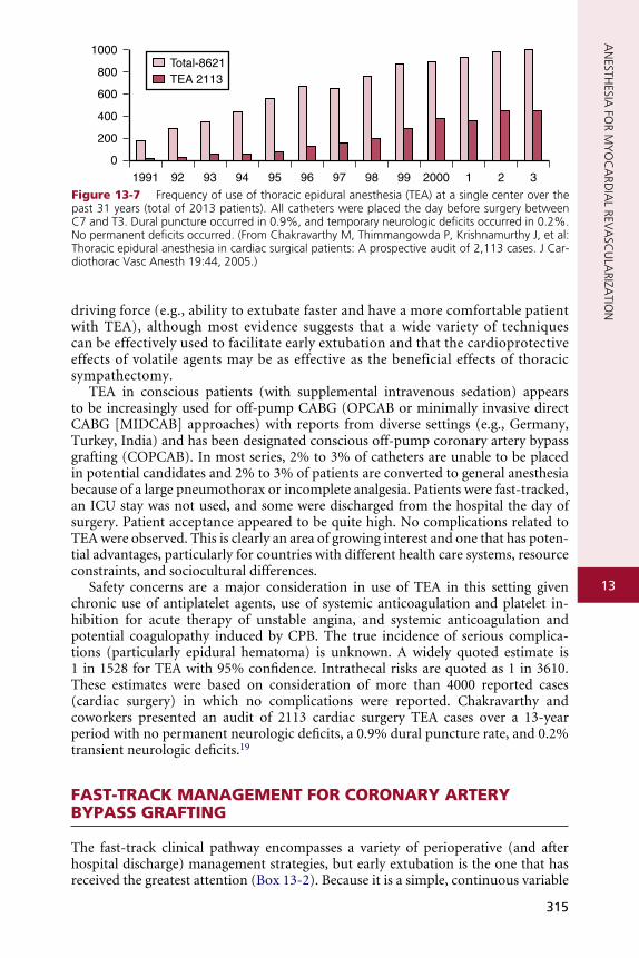
Interest in the use of thoracic epidural anesthesia (TEA) for cardiac surgery has steadily increased over the past 15 years. It has been long appreciated that thoracic sympathectomy has favorable effects on the heart and coronary circula-tion. Its coronary vasodilating effects have been well documented and it has been used to treat unstable angina for many years. There has been a resurgence in interest, and it is frequently used as a supplement to general anesthesia for car-diac surgery, particularly in Europe and Asia (Fig. 13-7). However, in the United States, medicolegal concerns about the rare but present danger of a devastating neurologic injury and the substantial logistical issues regarding placement the night before surgery, increased time to place relative to inducing general anesthe-sia, and the potential for cancellation of a case in event of bloody return are ma-jor limiting factors. The advent of fast-tracking could be considered a potential
314
AN
ESTHESIA
FOR M
YO
CA
RDIA
L REVA
SCU
LARIZ
1000
800
600
400
200
0
1991 92 93 94 95 96 97 98 99 2000 1 2 3
Total-8621TEA 2113
Figure 13-7 Frequency of use of thoracic epidural anesthesia (TEA) at a single center over the past 31 years (total of 2013 patients). All catheters were placed the day before surgery between C7 and T3. Dural puncture occurred in 0.9%, and temporary neurologic deficits occurred in 0.2%. No permanent deficits occurred. (From Chakravarthy M, Thimmangowda P, Krishnamurthy J, et al: Thoracic epidural anesthesia in cardiac surgical patients: A prospective audit of 2,113 cases. J Car-diothorac Vasc Anesth 19:44, 2005.)
13
ATIO
N
driving force (e.g., ability to extubate faster and have a more comfortable patient with TEA), although most evidence suggests that a wide variety of techniques can be effectively used to facilitate early extubation and that the cardioprotective effects of volatile agents may be as effective as the beneficial effects of thoracic sympathectomy.TEA in conscious patients (with supplemental intravenous sedation) appears to be increasingly used for off-pump CABG (OPCAB or minimally invasive direct CABG [MIDCAB] approaches) with reports from diverse settings (e.g., Germany, Turkey, India) and has been designated conscious off-pump coronary artery bypass grafting (COPCAB). In most series, 2% to 3% of catheters are unable to be placed in potential candidates and 2% to 3% of patients are converted to general anesthesia because of a large pneumothorax or incomplete analgesia. Patients were fast-tracked, an ICU stay was not used, and some were discharged from the hospital the day of surgery. Patient acceptance appeared to be quite high. No complications related to TEA were observed. This is clearly an area of growing interest and one that has poten-tial advantages, particularly for countries with different health care systems, resource constraints, and sociocultural differences.
Safety concerns are a major consideration in use of TEA in this setting given chronic use of antiplatelet agents, use of systemic anticoagulation and platelet in-hibition for acute therapy of unstable angina, and systemic anticoagulation and potential coagulopathy induced by CPB. The true incidence of serious complica-tions (particularly epidural hematoma) is unknown. A widely quoted estimate is 1 in 1528 for TEA with 95% confidence. Intrathecal risks are quoted as 1 in 3610. These estimates were based on consideration of more than 4000 reported cases (cardiac surgery) in which no complications were reported. Chakravarthy and coworkers presented an audit of 2113 cardiac surgery TEA cases over a 13-year period with no permanent neurologic deficits, a 0.9% dural puncture rate, and 0.2% transient neurologic deficits.19
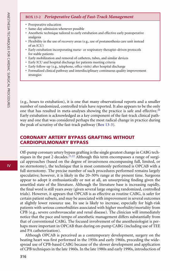
FAST-TRACK MANAGEMENT FOR CORONARY ARTERY BYPASS GRAFTING
The fast-track clinical pathway encompasses a variety of perioperative (and after hospital discharge) management strategies, but early extubation is the one that has received the greatest attention (Box 13-2). Because it is a simple, continuous variable
315
iv
AN
ESTHESIA
TECH
NIQ
UES FO
R CA
RDIA
C SU
RGIC
AL PRO
CED
URES
BOX 13-2 �Perioperative�Goals�of�Fast-Track�Management
• Preoperative education • Same-day admission whenever possible • Anesthetic technique tailored to early extubation and effective early postoperative
analgesia • Flexibility in the use of recovery areas (e.g., use of postanesthesia care unit instead
of an ICU) • Early extubation incorporating nurse- or respiratory therapist–driven protocols
for stable patients • Early mobilization and removal of catheters, tubes, and similar devices • Early ICU and hospital discharge for patients meeting criteria • Early follow-up (e.g., telephone, office visits) after hospital discharge • Formalized clinical pathway and interdisciplinary continuous quality improvement
strategies
(e.g., hours to extubation), it is one that many observational reports and a smaller number of randomized, controlled trials have reported. It also appears to be the only one that has resulted in meta-analyses showing the practice is safe and effective.20 Early extubation is acknowledged as a key component of the fast-track clinical path-way and one that was considered perhaps the most radical change in practice during the peak of scrutiny of the fast-track pathway (Box 13-3).
CORONARY ARTERY BYPASS GRAFTING WITHOUT CARDIOPULMONARY BYPASS
Off-pump coronary artery bypass grafting is the single greatest change in CABG tech-niques in the past 2 decades.21,22 Although this term encompasses a range of surgi-cal approaches (based on the degree of invasiveness encompassing full, limited, or no sternotomy), the technique that is most commonly performed is OPCAB with a full sternotomy. The precise number of such procedures performed remains largely speculative; however, it is likely in the 20–30% range at the present time. Surgeons appear to adopt it enthusiastically or not at all, an unsurprising finding given the unsettled state of the literature. Although the literature base is increasing rapidly, the final word is still years away (given several large ongoing randomized, controlled trials). However, it appears that OPCAB is as effective as routine CABG, is safer for certain patient subsets, and may be associated with improvement in several outcomes at slightly lower resource use. Its use is likely to increase, especially for high-risk patients with serious comorbidities associated with higher morbidity/mortality from CPB (e.g., severe cerebrovascular and renal disease). The clinician will immediately notice that the pace and tempo of anesthetic management differs substantially from that of conventional CABG. The focused involvement of the anesthesiologist is per-haps more important in OPCAB than during on-pump CABG (including use of TEE and PA catheterization).
Although OPCAB is perceived as a contemporary development, surgery on the beating heart was first performed in the 1950s and early 1960s, preceding the wide-spread use of CPB-based CABG because of the slower development and application of CPB techniques in the late 1960s. In the late 1980s and early 1990s, introduction of
316
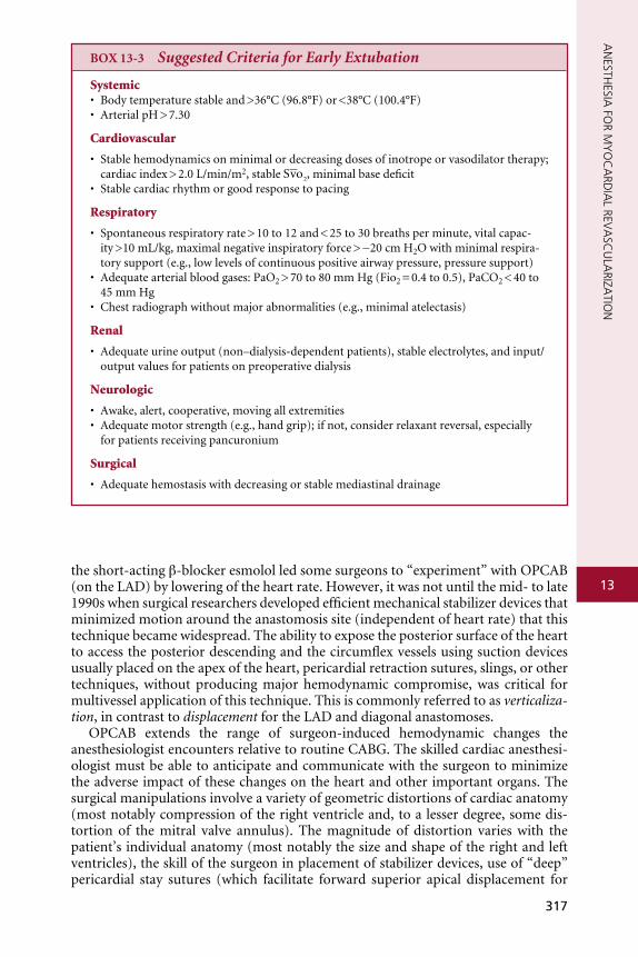
AN
ESTHESIA
FOR M
YO
CA
RDIA
L REVA
SCU
LARIZA
TION
BOX 13-3 �Suggested�Criteria�for�Early�Extubation
Systemic • Body temperature stable and >36°C (96.8°F) or <38°C (100.4°F) • Arterial pH > 7.30
Cardiovascular
• Stable hemodynamics on minimal or decreasing doses of inotrope or vasodilator therapy; cardiac index > 2.0 L/min/m2, stable Svo2, minimal base deficit
• Stable cardiac rhythm or good response to pacing
Respiratory
• Spontaneous respiratory rate > 10 to 12 and < 25 to 30 breaths per minute, vital capac-ity >10 mL/kg, maximal negative inspiratory force > −20 cm H2O with minimal respira-tory support (e.g., low levels of continuous positive airway pressure, pressure support)
• Adequate arterial blood gases: PaO2 > 70 to 80 mm Hg (Fio2 = 0.4 to 0.5), PaCO2 < 40 to 45 mm Hg
• Chest radiograph without major abnormalities (e.g., minimal atelectasis)
Renal
• Adequate urine output (non–dialysis-dependent patients), stable electrolytes, and input/output values for patients on preoperative dialysis
Neurologic
• Awake, alert, cooperative, moving all extremities • Adequate motor strength (e.g., hand grip); if not, consider relaxant reversal, especially
for patients receiving pancuronium
Surgical
• Adequate hemostasis with decreasing or stable mediastinal drainage
13
the short-acting β-blocker esmolol led some surgeons to “experiment” with OPCAB (on the LAD) by lowering of the heart rate. However, it was not until the mid- to late 1990s when surgical researchers developed efficient mechanical stabilizer devices that minimized motion around the anastomosis site (independent of heart rate) that this technique became widespread. The ability to expose the posterior surface of the heart to access the posterior descending and the circumflex vessels using suction devices usually placed on the apex of the heart, pericardial retraction sutures, slings, or other techniques, without producing major hemodynamic compromise, was critical for multivessel application of this technique. This is commonly referred to as verticaliza-tion, in contrast to displacement for the LAD and diagonal anastomoses.OPCAB extends the range of surgeon-induced hemodynamic changes the anesthesiologist encounters relative to routine CABG. The skilled cardiac anesthesi-ologist must be able to anticipate and communicate with the surgeon to minimize the adverse impact of these changes on the heart and other important organs. The surgical manipulations involve a variety of geometric distortions of cardiac anatomy (most notably compression of the right ventricle and, to a lesser degree, some dis-tortion of the mitral valve annulus). The magnitude of distortion varies with the patient’s individual anatomy (most notably the size and shape of the right and left ventricles), the skill of the surgeon in placement of stabilizer devices, use of “deep” pericardial stay sutures (which facilitate forward superior apical displacement for
317
iv
AN
ESTHESIA
TECH
NIQ
UES FO
R CA
RDIA
C SU
RGIC
AL PRO
CED
URES
LAD anastomosis), and manipulation of the pleural space (e.g., right pleural inci-sion to create space for the compressed right ventricle). With a skilled surgeon, the changes are usually modest or easily treated with the Trendelenburg position, use of vasoconstrictors or inotropes, and judicious volume expansion. However, severe changes due to acute ischemia, mitral regurgitation, or unrecognized right ventricu-lar compression may occur, necessitating emergent conversion to CPB.
Monitoring for Off-Pump Coronary Artery Bypass Grafting
Hemodynamic changes with OPCAB vary with location of the anastomosis and type of stabilizer or verticalizer system used, although most of the available hemodynamic data was obtained from low-risk patients. At present, there is no clear consensus or guidelines with regard to the type of monitoring recommended for OPCAB. Most observational studies have used fairly extensive monitoring with PA catheterization. TEE appears to be widely used, despite problems with imaging during the anasto-motic period, particularly when the heart is verticalized. As with on-pump CABG, it is likely that as this procedure is more widely adopted and clinicians gain increasing familiarity, that monitoring recommendations, particularly for low-risk patients, will become less sophisticated.
Outcomes in Off-Pump Coronary Artery Bypass Grafting
Several investigators and working groups have comprehensively reviewed outcomes in patients undergoing OPCAB. Investigators have analyzed 37 randomized trials of 3369 patients with comparable treatment groups with the exception of a mar-ginal difference in number of grafts performed (2.6 OPCAB versus 2.8 CABG).21 All but one of the studies specifically excluded “high-risk” patients. Although various definitions were used, most excluded patients with low ejection fractions, re-peat procedures, and renal failure, and several studies excluded patients with dis-eased circumflex vessels. As expected, not all studies reported on all outcomes. The investigators found no significant differences in 30-day or 1- to 2-year mor-tality, myocardial infarction, stroke (30 day and 1 to 2 years), renal dysfunction, need for IABP, wound infection, or reoperation for bleeding or reintervention (for ischemia). OPCAB was associated with a significant reduction in atrial fibrillation (OR = 0.58), numbers of patients transfused (OR = 0.43), respiratory infections (OR = 0.41), need for inotropes (OR = 0.48), duration of ventilation (weighted mean difference [WMD] of 3.4 hours), ICU length of stay (WMD of 0.3 day), and hospital length of stay (WMD of 1.0 days). Changes in neurocognitive dysfunction were not different in the immediate postoperative period; they were significantly improved at 2 to 6 months (OR = 0.57), but there were no differences seen at 12 months. The critical issue of graft patency was addressed in only four studies, and these varied substantially with regard to when this was assessed (3 months in two and 12 months in two). Only one study reported a difference (reduction in cir-cumflex patency with OPCAB). Because of the small numbers of patients, the overall data for this category were considered inadequate for meta-analysis. A large-scale, randomized, controlled trial is being conducted by the Department of Veterans Af-fairs with evaluation of graft patency at 1 year postoperatively. Four randomized, controlled trials have analyzed quality of life. Various methods precluded inclusion in the meta-analysis, but it generally appears there is little difference between opera-tions. Of the 20 trials reporting conversion rates, 8% of OPCAB patients required
318
13
AN
ESTHESIA
FOR M
YO
CA
RDIA
L REVA
SCU
LARIZA
TION
conversion to CABG, whereas only 1.7% were converted from CABG to OPCAB. The conversion rate for OPCAB in these low- to medium-risk patients is substantial and would be expected to be even higher in higher-risk patients with greater disease burdens, more complex lesions, or impaired ventricular function in whom toler-ance of stabilization and verticalization may be less. The anesthesiologist must be prepared for rapid institution of CPB at all times.
The ultimate adoption of OPCAB resides in demonstration of similar rates of long-term graft patency. Given the longevity of most grafts, particularly the internal mammary artery (>15 years), this will take some time to be conclusively established. With ongoing technologic advances, it is likely this approach will continue to expand in numbers of patients and surgical complexity.
MYOCARDIAL ISCHEMIA DURING REVASCULARIZATION
Incidence of Myocardial Ischemia in Patients Undergoing Revascularization Surgery
In addition to providing anesthesia, a major concern of the anesthesiologist is the prevention and treatment of myocardial ischemia. Numerous laboratory and clini-cal studies have examined the incidence of myocardial ischemia in the perioperative period, as it relates to the administration of anesthetic drugs. Although the incidence of pre-bypass ischemia in patients appears to be between 10% and 50%, it is not at all clear that the anesthetic drug combination per se is a determinant of this incidence. Several studies have addressed whether the anesthetic technique (e.g., fast-track or early extubation, high thoracic epidural) is related to perioperative morbidity and myocardial ischemic events. An extensive meta-analysis of more than 30 random-ized, controlled trials of patients undergoing cardiac surgery showed no difference in the relative risk of postoperative myocardial ischemia when early extubation proto-cols were compared with conventional extubation management.22
Hemodynamic Changes Related to Myocardial Ischemia
Besides ECG abnormalities, some hemodynamic changes should alert the anesthe-siologist to the possibility of intraoperative myocardial ischemia. The association of tachycardia with hypotension or increased LV filling pressure (both of which reduce coronary perfusion pressure [CPP] is a particularly undesirable combination jeopardizing the oxygen supply-demand relationship. Figure 13-8 demonstrates how hypertension in the absence of tachycardia, in response to surgical stress (skin inci-sion), can be associated with pulmonary hypertension, elevated pulmonary capillary wedge pressure, and prominent A and V waves on the PCWP waveform. Although ECG changes occurred later, the early hemodynamic abnormalities almost certainly were the result of ischemic LV dysfunction. Treatment included deepening anesthe-sia and administering a nitroglycerin infusion.
LV diastolic dysfunction detected with TEE is the earliest change identified after coronary artery occlusion, and it often precedes the development of abnormal sys-tolic function. Regional wall motion abnormalities also have been described as early signs of ischemia. They occur within seconds of inadequate blood flow or oxygen supply. Regional wall motion abnormalities detected by TEE have been shown to be a more sensitive method of detecting myocardial ischemia in patients undergoing CABG compared with ST-segment changes. Myocardial ischemia on repositioning
319
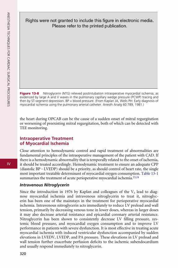
iv
AN
ESTHESIA
TECH
NIQ
UES FO
R CA
RDIA
C SU
RGIC
AL PRO
CED
URES
Figure 13-8 Nitroglycerin (NTG) relieved postintubation intraoperative myocardial ischemia, as evidenced by large A and V waves in the pulmonary capillary wedge pressure (PCWP) tracing and then by ST-segment depression. BP = blood pressure. (From Kaplan JA, Wells PH: Early diagnosis of myocardial ischemia using the pulmonary arterial catheter. Anesth Analg 60:789, 1981.)
the heart during OPCAB can be the cause of a sudden onset of mitral regurgitation or worsening of preexisting mitral regurgitation, both of which can be detected with TEE monitoring.
Intraoperative Treatment of Myocardial Ischemia
Close attention to hemodynamic control and rapid treatment of abnormalities are fundamental principles of the intraoperative management of the patient with CAD. If there is a hemodynamic abnormality that is temporally related to the onset of ischemia, it should be treated accordingly. Hemodynamic treatment to ensure an adequate CPP (diastolic BP − LVEDP) should be a priority, as should control of heart rate, the single most important treatable determinant of myocardial oxygen consumption. Table 13-1 summarizes the treatment of acute perioperative myocardial ischemia.23,24
Intravenous Nitroglycerin
Since the introduction in 1976 by Kaplan and colleagues of the V5 lead to diag-nose myocardial ischemia and intravenous nitroglycerin to treat it, nitroglyc-erin has been one of the mainstays in the treatment for perioperative myocardial ischemia. Intravenous nitroglycerin acts immediately to reduce LV preload and wall tension, primarily by decreasing venous tone in lower doses, whereas in larger doses it may also decrease arterial resistance and epicardial coronary arterial resistance. Nitroglycerin has been shown to consistently decrease LV filling pressure, sys-temic blood pressure, and myocardial oxygen consumption and to improve LV performance in patients with severe dysfunction. It is most effective in treating acute myocardial ischemia with induced ventricular dysfunction accompanied by sudden elevations in LVEDV, LVEDP, and PA pressure. These elevations in LV preload and wall tension further exacerbate perfusion deficits to the ischemic subendocardium and usually respond immediately to nitroglycerin.
320
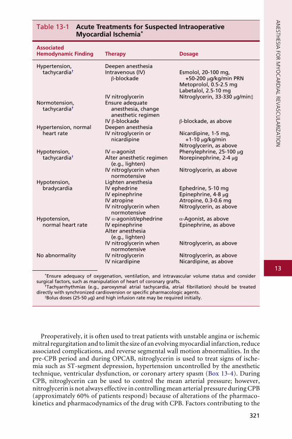
13
AN
ESTHESIA
FOR M
YO
CA
RDIA
L REVA
SCU
LARIZA
TION
Associated Hemodynamic Finding Therapy Dosage
Hypertension, tachycardia†
Deepen anesthesiaintravenous (iv)
β-blockade
iv nitroglycerin
Esmolol, 20-100 mg, +50-200 μg/kg/min PRN
Metoprolol, 0.5-2.5 mgLabetalol, 2.5-10 mgNitroglycerin, 33-330 μg/min‡
Normotension, tachycardia†
Ensure adequate anesthesia, change anesthetic regimen
iv β-blockade β-blockade, as aboveHypertension, normal
heart rateDeepen anesthesiaiv nitroglycerin or
nicardipineNicardipine, 1-5 mg,
+1-10 μg/kg/minNitroglycerin, as above
Hypotension, tachycardia†
iv α-agonistAlter anesthetic regimen
(e.g., lighten)iv nitroglycerin when
normotensive
Phenylephrine, 25-100 μgNorepinephrine, 2-4 μg
Nitroglycerin, as above
Hypotension, bradycardia
Lighten anesthesiaiv ephedrineiv epinephrineiv atropineiv nitroglycerin when
normotensive
Ephedrine, 5-10 mgEpinephrine, 4-8 μgAtropine, 0.3-0.6 mgNitroglycerin, as above
Hypotension, normal heart rate
iv α-agonist/ephedrineiv epinephrineAlter anesthesia
(e.g., lighten)iv nitroglycerin when
normotensive
α-Agonist, as aboveEpinephrine, as above
Nitroglycerin, as above
No abnormality iv nitroglyceriniv nicardipine
Nitroglycerin, as aboveNicardipine, as above
*Ensure adequacy of oxygenation, ventilation, and intravascular volume status and consider surgical factors, such as manipulation of heart of coronary grafts.
†Tachyarrhythmias (e.g., paroxysmal atrial tachycardia, atrial fibrillation) should be treated directly with synchronized cardioversion or specific pharmacologic agents.
‡Bolus doses (25-50 μg) and high infusion rate may be required initially.
Table 13-1 Acute Treatments for Suspected Intraoperative Myocardial Ischemia*
321
Preoperatively, it is often used to treat patients with unstable angina or ischemic mitral regurgitation and to limit the size of an evolving myocardial infarction, reduce associated complications, and reverse segmental wall motion abnormalities. In the pre-CPB period and during OPCAB, nitroglycerin is used to treat signs of ische-mia such as ST-segment depression, hypertension uncontrolled by the anesthetic technique, ventricular dysfunction, or coronary artery spasm (Box 13-4). During CPB, nitroglycerin can be used to control the mean arterial pressure; however, nitroglycerin is not always effective in controlling mean arterial pressure during CPB (approximately 60% of patients respond) because of alterations of the pharmaco-kinetics and pharmacodynamics of the drug with CPB. Factors contributing to the
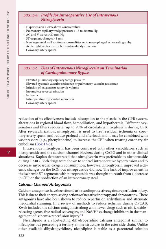
iv
AN
ESTHESIA
TECH
NIQ
UES FO
R CA
RDIA
C SU
RGIC
AL PRO
CED
URES
BOX 13-4 �Profile�for�Intraoperative�Use�of�Intravenous��Nitroglycerin
• Hypertension > 20% above control values • Pulmonary capillary wedge pressure > 18 to 20 mm Hg • AC and V waves > 20 mm Hg • ST-segment changes > 1 mm • New segmental wall motion abnormalities on transesophageal echocardiography • Acute right ventricular or left ventricular dysfunction • Coronary artery spasm
BOX 13-5 �Uses�of�Intravenous�Nitroglycerin�on�Termination�of�Cardiopulmonary�Bypass
• Elevated pulmonary capillary wedge pressure • Elevated systemic vascular resistance or pulmonary vascular resistance • Infusion of oxygenator reservoir volume • Incomplete revascularization • Ischemia • Intraoperative myocardial infarction • Coronary artery spasm
reduction of its effectiveness include adsorption to the plastic in the CPB system, alterations in regional blood flow, hemodilution, and hypothermia. Different oxy-genators and filters sequester up to 90% of circulating nitroglycerin during CPB. After revascularization, nitroglycerin is used to treat residual ischemia or coro-nary artery spasm and reduce preload and afterload, and it may be combined with vasopressors (e.g., phenylephrine) to increase the CPP when treating coronary air embolism (Box 13-5).
Intravenous nitroglycerin has been compared with other vasodilators such as nitroprusside and the calcium channel blockers during CABG and in other clinical situations. Kaplan demonstrated that nitroglycerin was preferable to nitroprusside during CABG. Both drugs were shown to control intraoperative hypertension and to decrease myocardial oxygen consumption; however, nitroglycerin improved isch-emic changes on the ECG but nitroprusside did not. The lack of improvement in the ischemic ST segments with nitroprusside was thought to result from a decrease in CPP or the production of an intracoronary steal.
Calcium Channel Antagonists
Calcium antagonists have been found to be cardioprotective against reperfusion injury. This is due to their energy-saving actions of negative inotropy and chronotropy. These antagonists have also been shown to reduce reperfusion arrhythmias and attenuate myocardial stunning. In a review of methods to reduce ischemia during OPCAB, Kwak included the calcium antagonists along with newer drugs such as nitric oxide–releasing agents, free radical scavengers, and Na+/H+ exchange inhibitors in the man-agement of ischemia-reperfusion injury.23
Nicardipine is a short-acting dihydropyridine calcium antagonist similar to nifedipine but possessing a tertiary amine structure in the ester side chain. Unlike other available dihydropyridines, nicardipine is stable as a parenteral solution
322
13
AN
ESTHESIA
FOR M
YO
CA
RDIA
L REVA
SCU
LARIZA
TION
and therefore can be administered intravenously. It has highly specific modes of action, which include coronary antispasmodic and vasodilatory effects and sys-temic vasodilation. Among the calcium antagonists, nicardipine is unique in its consistent augmentation of coronary blood flow and its ability to induce potent and more selective vasodilator responses in the coronary bed than in the systemic vascular bed. Other important hemodynamic effects include reductions in blood pressure and systemic vascular resistance and increases in myocardial contractil-ity and cardiac output. Nicardipine also produces minimal myocardial depression and significant improvement in diastolic function in patients with ischemic heart disease. Intravenous doses of 5 to 10 mg of nicardipine administered to patients with CAD produce therapeutic plasma levels. Plasma concentrations decline in a biphasic manner, with an initial half-life of 14 minutes and a terminal half-life of 4.75 hours. Clearance of nicardipine results mainly from its metabolism by the liver, and excretion is primarily through bile and the feces. It undergoes rapid and extensive first-pass hepatic metabolism with the production of inac-tive metabolites. Nicardipine’s rapid onset and cessation of action make it an attractive drug for the perioperative management of hypertension or myocardial ischemia. It has been administered to control hemodynamics during and after noncardiac vascular surgery and CABG.
Esmolol
Hypertension, tachycardia, arrhythmias, and myocardial ischemia from sympa-thetic stimulation are common occurrences in the perioperative period. Despite the benefits of early use of β-blockers in the treatment of myocardial ischemia, the relatively long half-life and prolonged duration of action of previously avail-able β-blockers have limited their usefulness during surgery and the immediate postoperative period. The introduction of esmolol, an ultra-short-acting car-dioselective β1-blocker with a half-life of 9 minutes because of rapid esterase metabolism, provides a β-blocker that is extremely useful in the perioperative period. Esmolol has been shown to be effective in treating patients with acute unstable angina or during acute coronary occlusion. During percutaneous trans-luminal coronary angioplasty, esmolol was also found to reduce the amount of ST-segment elevation and the onset of segmental wall motion abnormalities. Esmolol has been used during CABG in a prophylactic manner to prevent hy-pertension, tachycardia, and myocardial ischemia. Before the introduction of newer stabilizing mechanical devices, it had been used frequently during OPCAB procedures to slow the heart rate during the surgical procedure. Esmolol has also been used to treat intraoperative hypertension, tachycardia, and myocardial ischemia. Bolus doses of 1.5 mg/kg have been found to be effective in treating ST-segment changes in patients with CAD. More commonly, a smaller bolus dose is used and is combined with an infusion of esmolol. Bolus doses ranging from 0.5 to 1.0 mg/kg have been used, followed by infusions of 50 to 300 μg/kg/min. These doses have been found to effectively treat increases in heart rate that occur during surgery and to block the β-adrenergic effects of catecholamines associ-ated with surgical stress.
Coronary Artery and Arterial Conduit Spasm
Spasm has usually been associated with profound ST-segment elevation on the ECG, hypotension, severe dysfunction of the ventricles, and myocardial irritability. Many hypotheses have been put forward to explain the origin of coronary artery
323
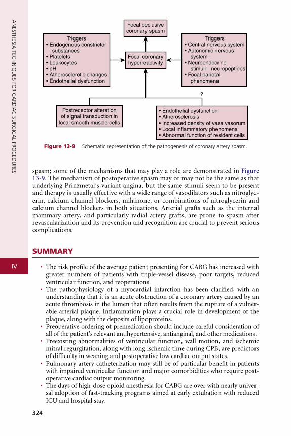
iv
AN
ESTHESIA
TECH
NIQ
UES FO
R CA
RDIA
C SU
RGIC
AL PRO
CED
URES
Triggers• Endogenous constrictor substances• Platelets• Leukocytes• pH• Atherosclerotic changes• Endothelial dysfunction
Postreceptor alterationof signal transduction in
local smooth muscle cells
Focal occlusivecoronary spasm
Focal coronaryhyperreactivity
Triggers• Central nervous system• Autonomic nervous system • Neuroendocrine stimuli—neuropeptides• Focal parietal phenomena
• Endothelial dysfunction• Atherosclerosis• Increased density of vasa vasorum• Local inflammatory phenomena• Abnormal function of resident cells
?
Figure 13-9 Schematic representation of the pathogenesis of coronary artery spasm.
spasm; some of the mechanisms that may play a role are demonstrated in Figure 13-9. The mechanism of postoperative spasm may or may not be the same as that underlying Prinzmetal’s variant angina, but the same stimuli seem to be present and therapy is usually effective with a wide range of vasodilators such as nitroglyc-erin, calcium channel blockers, milrinone, or combinations of nitroglycerin and calcium channel blockers in both situations. Arterial grafts such as the internal mammary artery, and particularly radial artery grafts, are prone to spasm after revascularization and its prevention and recognition are crucial to prevent serious complications.
SUMMARY
• The risk profile of the average patient presenting for CABG has increased with greater numbers of patients with triple-vessel disease, poor targets, reduced ventricular function, and reoperations.
• The pathophysiology of a myocardial infarction has been clarified, with an understanding that it is an acute obstruction of a coronary artery caused by an acute thrombosis in the lumen that often results from the rupture of a vulner-able arterial plaque. Inflammation plays a crucial role in development of the plaque, along with the deposits of lipoproteins.
• Preoperative ordering of premedication should include careful consideration of all of the patient’s relevant antihypertensive, antianginal, and other medications.
• Preexisting abnormalities of ventricular function, wall motion, and ischemic mitral regurgitation, along with long ischemic time during CPB, are predictors of difficulty in weaning and postoperative low cardiac output states.
• Pulmonary artery catheterization may still be of particular benefit in patients with impaired ventricular function and major comorbidities who require post-operative cardiac output monitoring.
• The days of high-dose opioid anesthesia for CABG are over with nearly univer-sal adoption of fast-tracking programs aimed at early extubation with reduced ICU and hospital stay.
324
13
AN
ESTHESIA
FOR M
YO
CA
RDIA
L REVA
SCU
LARIZA
TION
RE
1
2
3
4
5
6 7
8
9
10
11
12
13
14
15
16
17
18
19
20
• All contemporary inhalation agents have been shown to have potent ischemic preconditioning effects, and newer randomized trials have demonstrated reduc-tions in postoperative troponin release and improved ventricular function rela-tive to exclusively intravenous techniques.
• Intraoperative recall is a risk in all cardiac surgical procedures, although its inci-dence appears to have declined substantially with the increased continuous use of inhalation anesthetics associated with fast-tracking.
• Off-pump CABG is the greatest change in surgical revascularization technique in the past 2 decades.
• The hemodynamic changes associated with OPCAB include reduction in cardiac output, stroke volume, and mean arterial pressure.
• Intravenous nitroglycerin and short-acting β-blockers, such as esmolol, remain the mainstay of treatment for perioperative myocardial ischemia.
FERENCES
. Myles PS, Daly DJ, Djaiani G, et al: A systematic review of the safety and effectiveness of fast-track cardiac anesthesia. Anesthesiology 99:982, 2003
. Cheng DC, Bainbridge D, Martin JE, et al: Does off-pump coronary artery bypass reduce mortal-ity, morbidity, and resource utilization when compared with conventional coronary artery bypass? A meta-analysis of randomized trials. Anesthesiology 102:188, 2005
. Chakravarthy M, Thimmangowda P, Krishnamurthy J, et al: Thoracic epidural anesthesia in cardiac surgical patients: A prospective audit of 2,113 cases. J Cardiothorac Vasc Anesth 19:44, 2005
. Shroyer AL, Coombs LP, Peterson ED, et al: The Society of Thoracic Surgeons: 30-day operative mortality and morbidity risk models. Ann Thorac Surg 75:1856, 2003
. Nashef SA, Roques F, Hammill BG, et al: Validation of European System for Cardiac Operative Risk Evaluation (EuroSCORE) in North American cardiac surgery. Eur J Cardiothorac Surg 22:101, 2002
. Wei K, Kaul S: The coronary microcirculation in health and disease Cardiol Clin 22:221, 2004
. Fuster V, Moreno PR, Fayad ZA, et al: Atherothrombosis and high-risk plaque. J Am Coll Cardiol 46:937, 2005
. London MJ, Zaugg M, Schaub MC, et al: Perioperative beta-adrenergic receptor blockade: Physi-ologic foundations and clinical controversies. Anesthesiology 100:170, 2004
. Ferguson TB Jr, Coombs LP, Peterson ED: Preoperative beta-blocker use and mortality and morbid-ity following CABG surgery in North America. JAMA 287:2221, 2002
. Stein PD, Schunemann HJ, Dalen JE, et al: Antithrombotic therapy in patients with saphenous vein and internal mammary artery bypass grafts: The Seventh ACCP Conference on Antithrombotic and Thrombolytic Therapy. Chest 126:600S, 2004
. O’Neil-Callahan K, Katsimaglis G, Tepper MR, et al: Statins decrease perioperative cardiac complica-tions in patients undergoing noncardiac vascular surgery: The Statins for Risk Reduction in Surgery (StaRRS) study. J Am Coll Cardiol 45:336, 2005
. Murphy GS, Vender JS: Is the pulmonary artery catheter dead? con position. J Cardiothorac Vasc Anesth 21:147, 2007
. Minhaj M, Patel K, Muzic D, et al: The effect of routine intraoperative transesophageal echocardio-graphy on surgical management. J Cardiothorac Vasc Anesth 21:800, 2007
. Herr DL, Sum-Ping ST, England M: ICU sedation after coronary artery bypass graft surgery: Dexmedetomidine based versus propofol-based sedation regimens. J Cardiothorac Vasc Anesth 17:576, 2003
. Zaugg M, Lucchinetti E, Garcia C, et al: Anaesthetics and cardiac preconditioning: II. Clinical impli-cations. Br J Anaesth 91:566, 2003
. De Hert SG, Van der Linden PJ, Cromheecke S, et al: Cardioprotective properties of sevoflurane in patients undergoing coronary surgery with cardiopulmonary bypass are related to the modalities of its administration. Anesthesiology 101:299, 2004
. Piriou V, Chiari P, Gateau-Roesch O, et al: Desflurane-induced preconditioning alters calcium- induced mitochondrial permeability transition. Anesthesiology 100:581, 2004
. Ranta SO, Herranen P, Hynynen M: Patients’ conscious recollections from cardiac anesthesia. J Cardiothorac Vasc Anesth 16:426, 2002
. Chakravarthy M, Thimmangowda P, Krishnamurthy J, et al: Thoracic epidural anesthesia in cardiac surgical patients: A prospective audit of 2,113 cases. J Cardiothorac Vasc Anesth 19:44, 2005
. Hawkes CA, Dhileepan S, Foxcroft D: Early extubation for adult cardiac surgical patients. Cochrane Database Syst Rev 4:CD003587, 2003
325
iv
AN
ESTHESIA
TECH
NIQ
UES FO
R CA
RDIA
C SU
RGIC
AL PRO
CED
URES
21. Raja SG, Dreyfus GD: Off-pump coronary artery bypass surgery: To do or not to do? Current best available evidence. J Cardiothorac Vasc Anesth 18:486, 2004
22. Sellke FW, DiMaio JM, Caplan LR, et al: Comparing on-pump and off-pump coronary artery bypass grafting: Numerous studies but few conclusions: A scientific statement from the American Heart Association council on cardiovascular surgery and anesthesia in collaboration with the interdiscipli-nary working group on quality of care and outcomes research. Circulation 111:2858, 2005
23. Kwak YL: Reduction of ischemia during off-pump coronary artery bypass graft surgery. J Cardiothorac Vasc Anesth 19:667, 2005
24. Hannan EL, Wu C, Walford G, et al: Drug-eluting stents versus coronary-artery bypass grafting in multivessel coronary disease. N Engl J Med 358:331, 2008
326