Embed Size (px)
Citation preview
1
Anesthetic Considerations in Porphyrias
Niels F. Jensen, MD*
Daniel S. Fiddler, MD**
Volker Striepe, MBBCh, DA, FFA(SA)***
*Assistant Professor, Department of Anesthesiology, University of Iowa
**Anesthesia Associates of Medford, Medford, Oregon
***Assistant Professor, Department of Anesthesiology, Vanderbilt University
Address correspondence to Dr. Jensen, Department of Anesthesiology, University of Iowa
College of Medicine, Iowa City, Iowa 52242. (319) 356-2633 No reprints available.

2
Key Words: Complications: porphyria; Acute intermittent porphyria (AIP); ALA
dehydratase deficiency porphyria (PLP); Hereditary coproporphyria (HCP); Porphyria
cutanea tarda (PCT); Variegate porphyria (VP)
3
Outline
I. Introduction
II. Pathophysiology
III. Classification and Incidence
IV. Clinical features: acute attack
V. Preoperative evaluation
a. Acute abdomen and porphyria
b. Known acute porphyria
VI. Intraoperative management
a. Regional anesthesia
b. Induction agents
c. Maintenance agents
d. Treatment of complications
e. Monitoring
VII. Postoperative management
VIII. Treatment: acute crisis
IX. Further investigation
X. Summary
4
Introduction
Porphyrias present special anesthetic challenges, including preoperative assessment of
a patient with acute abdominal pain, intraoperative management of known porphyria, and
respiratory and cardiovascular management of acute porphyric crisis. To meet these
challenges, a current and thorough understanding of porphyria is essential. Several years have
elapsed since there has been a comprehensive review of the spectrum of issues related to
porphyria of concern to the anesthesiologist: presentation, pathophysiology, monitoring, and
relevant pharmacology. The last significant review of this subject provided a pharmacologic
perspective but much relevant anesthetic information was not addressed, such as new methods
of detection and the evolving role of hematin and heme arginate in treatment. (1) DNA
analysis of suspected porphyric patients promises earlier, definitive diagnosis of the disease
and thereby the opportunity for safer anesthetic management. This management may soon
include heme arginate, the most stable form of heme, which currently lacks FDA approval but
is soon to be tested in clinical trials in the United States. Safe anesthetic management of
porphyria demands far more today than an understanding of appropriate pharmacologic
therapy. It demands a thorough, current understanding of many other aspects of the disease.
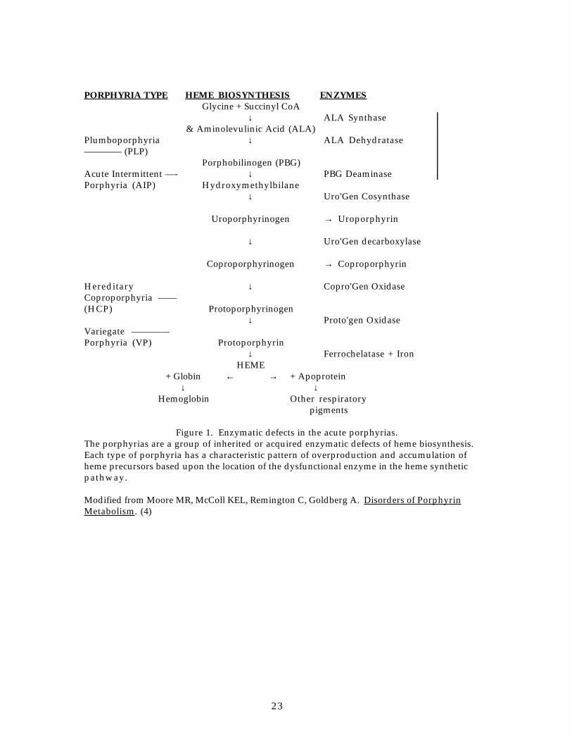
Pathophysiology
The porphyrias are a group of inherited or acquired enzymatic defects of heme
biosynthesis. Each type of porphyria has a characteristic pattern of overproduction and
accumulation of heme precursors based upon the location of the dysfunctional enzyme in the
heme synthetic pathway (Fig. 1).
The rate limiting step in heme synthesis is the condensation of succinyl CoA and glycine
to form delta-amino levulinic acid (ALA), (2, 3) catalyzed by the mitochondrial enzyme ALA
synthetase. The basal activity of ALA synthetase is substantially lower than that of
subsequent enzymes in the synthetic pathway, and therefore changes in ALA synthetase
activity are rate limiting, controlling the rate of heme synthesis. Heme, the end product of the
synthetic pathway, exerts negative feedback regulation on ALA synthetase activity. (4-6)
The specific enzyme deficit in each type of porphyria results in a partial block in heme
biosynthesis and lower intramitochondrial heme levels (see Fig. 1). Decreased negative
feedback from heme contributes to the elevated “baseline” ALA synthetase activity which is
characteristic of the porphyrias. (4-6)
The manifestions of the disease are thought to be due to increased ALA synthetase
activity, increased porphyrin accumulation in the tissues, or decreased heme production.(4-6)
The increased ALA synthetase activity results in elevated levels of heme precursors proximal
to the site of the specific enzyme deficiency. These precursors are colorless and nonfluorescent
5
porphyrinogens. Irreversible oxidation of these porphyrinogens causes the formation of
porphyrins, which have no known physiologic function but are highly reactive oxidants. The
accumulation of porphyrins in the epidermal skin layers leads to cutaneous photosensitivity.
(7)
Acute porphyria often causes severe neuropathy, the basis for multisystem impairment.
Changes in autonomic ganglia, anterior horns of the spinal cord, peripheral nerves, brainstem
nuclei, cerebellar axons, schwann cells and myelin sheaths have been demonstrated. (6, 8-12)
Neuronal damage and axonal degeneration may be the primary pathologic lesions, with later
axonal changes leading to secondary demyelination. (4, 6)
Many hypotheses have been proposed to explain the mechanism of porphyric
neuropathy. (4, 9) Two of the most plausible attribute the neuronal dysfunction to direct
neurotoxicity of ALA [not porphobilinogen (PBG)], or to diminished intraneuronal heme level or
both. (4, 6, 11) In addition, there may be a significant relationship between tryptophan
metabolites and/or folate deficiency and clinical expression of the disease. (13, 14) Recent
reviews provide additional detail. (4, 6, 9)
Classification and Incidence
The porphyrias may be classified according to three characteristics:
1. The major site of abnormal porphyrin production (hepatic versus erythropoietic)
2. Acute or nonacute presentation
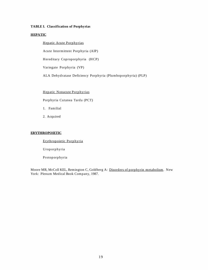
3. Pattern of enzyme deficiency in heme production (Table I) (4)
Heme is a component of microsomal and mitochondrial cytochrome systems and is
synthesized and utilized in all cells. The two major quantitative sites of heme synthesis are
erythropoeitic and hepatic cells where heme is incorporated into hemoglobin and hepatic
cytochromes. Erythropoeitic porphyrias cause extreme skin sensitivity but lack neurologic
involvement and are not associated with drug precipitated crises.
Porphyria cutanea tarda (PCT) is the only hepatic porphyria without neurologic
sequelae. PCT is usually associated with hepatic disease but not acute neurologic crisis. Other
hepatic porphyrias are associated with abdominal pain, peripheral neuropathy, and mental
status changes, with crisis frequently precipitated by "triggering" drugs. Barbiturates are the
most frequent "trigger".(9, 15) There is little difference in the neurologic syndrome exhibited
during an attack among the four acute hepatic porphyrias. Hereditary coproporphyria (HCP)
and variegate porphyria (VP) manifest skin photosensitivity and extreme skin fragility,
whereas acute intermittent porphyria (AIP) does not. (4-6)
Once diagnosed, AIP is associated with a relatively good prognosis. Symptoms occur in
less than one third of genetically susceptible patients but rarely before puberty. Acute attacks

6
are associated with a significant risk of mortality, particularly if the diagnosis is delayed and
neurologic involvement progresses. Although autosomal dominant, clinical expression is more
common in females. (16)
Most patients with HCP are asymptomatic and clinical onset may be associated with
intercurrent hepatic disease. Presentation has occurred between the ages of 7 and 75 years.
Prognosis is generally good. (17)
VP has a prognosis as good as acute intermittent porphyria. Systemic effects are more
common in women, while cutaneous manifestations are more common in men. (17)
Treatment of known hepatic porphyria consists of prophylaxis and treatment of the
acute attack.
Factors known to precipitate acute porphyric crisis include fasting/dehydration,
infection, psychological stress, physiological hormone variation, excessive alcohol intake, and
administration of specific drugs. Many drugs cause porphyric crisis (see Table II). Most do so by
decreasing heme levels, thus decreasing negative feedback and thereby increasing ALA
synthetase activity. (4-6)
Many commonly used drugs trigger porphyric crisis by decreasing heme. (2)
Barbiturates induce the cytochrome P450 system; this incorporates more heme into the new
cytochromes, thereby decreasing heme levels. Oral contraceptives cause destruction of the
heme group in cytochromes, requiring new heme for incorporation into cytochromes.
Griseofulvin converts heme into N-methylated derivatives, which further inhibit heme
synthesis.
Some endogenous steroid hormones are thought to trigger porphyria by increasing the
synthesis of new ALA synthetase enzyme. (2) Factors known to decrease synthetase activity
include high carbohydrate (glucose) loading, propranolol, and increased negative feedback
from heme. (3-6, 18) Propranolol is used during the acute porphyric attack to control
hypertension and tachycardia. This drug increases heme synthesis in vitro, which exerts an
inhibitory effort upon ALA synthetase activity through negative feedback. (19)
The acute porphyrias are associated with hereditary enzyme deficits. (20-24) AIP,
HCP, and VP exhibit autosomal dominant transmission with variable expression. The
frequency of AIP is estimated to be 1/20,000 in Europe, with a high of 1/10,000 in Northern
Sweden. (4, 25) ALA dehydratase deficiency porphyria, also known as plumboporphyria
(PLP), has an autosomal dominant pattern. Since PLP has only recently been described, no
estimate of prevalence has been established. (26-28) The frequency of HCP is also difficult to
estimate since greater than half of affected individuals are asymptomatic (variable
expression) and the number of reported cases is small. (4) VP is particularly common in certain
7
population groups, such as white South Africans, where prevalence has been estimated at
1/250-500. (4, 29)
Pregnancy may exacerbate or provoke an acute attack. Avoidance of planned pregnancy
until a one year latent period has elapsed is recommended. The mortality rate from acute
attack among pregnant patients has been reported to be as high as 42%.(30)
Clinical Features : Acute Attack
Acute attacks only occur in four types of porphyria: AIP, HCP, VP, and PLP. (7)
The signs and symptoms of acute porphyric crisis are well-known and quite consistent:
severe abdominal pain, vomiting, anxiety, confusion, autonomic instability manifested by
hypertension and tachycardia, dehydration, and electrolyte disturbances such as
hyponatremia, hypokalemia, and hypocalcemia. (22, 24, 31) (Table III)
AIP, HCP, and VP may be clinically indistinguishable during acute attacks. (32)
Central to each is neurologic dysfunction (Table III) (9, 29) , with significant impairment of
both sympathetic and parasympathetic nervous systems occurring during an acute attack. (33)
During remissions function improves, but parasympathetic dysfunction can persist. (33)
Tachycardia is often an indicator of disease state progression. (25, 34, 35) As heart
rate increases, the patient's condition generally worsens and with clinical improvement
tachycardia usually resolves. Most of the clinical features subside within the approximate
time course of the acute crises, but residual paresis may persist for years in the absence of
further attacks. (34) This paresis, per se, does not have specific implications for the use of
neuromuscular relaxants. The use of muscle relaxants in the setting of porphyria is discussed
below. Recovery of mental function often lags behind physical recovery, and some patients
report anxiety, emotional instability, or other functional disturbances indefinitely. (29)
Electrolyte abnormalities occur secondary to dehydration, vomiting, and diarrhea and
may entail serious hyponatremia and hypochloremia. (6) Stein et al performed radioisotope
studies to measure blood volume in nine AIP patients. (35) All had low blood volumes, ranging
from 67% to 97% of normal, despite normal electrolytes in some patients. Though the syndrome
of inappropriate secretion of anti-diuretic hormone (siADH) is well described in acute
porphyrias, presumably due to hypothalamic involvement, hyponatremia with appropriate
ADH levels occurs more often. (34-36)
Laboratory diagnosis of porphyric crisis can involve fecal analysis (32) but most
frequently involves quantification of urinary porphyrin and porphyrinogen precursors. (37)
These can be markedly elevated during an attack, but may return to normal during remission.
This normality can create a difficulty in early and accurate diagnosis in high risk groups, such
8
as in patients with a strong family history for porphyria. Many carriers of the trait can thus
remain asymptomatic unless exposed to precipitants.
One technique known as gene linkage analysis offers a new approach to the diagnosis of
acute intermittent porphyria and relies on direct cDNA sequencing. (20, 22) It does not depend
on urinalysis but rather on polymorphic markers within the porphobilingen deaminase gene.
(21) This permits unequivocal and early detection of carriers. Early and accurate detection of
the disease in high risk patients is of obvious benefit to safer anesthetic management of those
affected and constitutes a major breakthrough in terms of perioperative anesthetic
management.
Preoperative Evaluation
A. Acute Abdomen and porphyria : The following symptoms should raise suspicion of
porphyria in patients with acute abdominal pain: mental status changes (confusion, hysteria),
peripheral neuropathy (motor > sensory), dark colored (red to purple) urine, and known family
history of porphyria. (38) Of special concern is the parturient with acute abdominal pain.
Greater than 50% of pregnant women who have porphyria will experience a crisis during
pregnancy or puerperium, (39) probably due to ALA sythetase induction by hormonal changes
of pregnancy. If the patient with an acute abdomen, pregnant or not, does not have suggestive
symptoms of porphyria, anesthetic drugs and therapies should not be modified. (40)
B. Known acute porphyria : In the setting of known acute porphyria, perhaps the most
difficult situation is when an acute attack is caused by and is concurrent with a disease process
which mandates surgical intervention; i.e. the infection, pyrexia, and anorexia of acute
appendicitis inducing ALA synthetase and precipitating crisis.
Neurologic evaluation should focus on mental status and peripheral neuropathy. If an
acute crisis is suspected, attention to cranial dysfunction and bulbar symptomatology may
predict impending respiratory failure.
Premedication is important, as psychological stress alone has been reported to
precipitate crises. (18, 34, 36) Numerous reports have implicated benzodiazepines, (4, 41, 42)
and their use is discussed below. Narcotics are safe in porphyria, with the exception of
pentazocine, a partial agonist. Scoplamine and atropine are considered safe. Acceptable non-
narcotic sedative include droperidol, promethazine, chloral hydrate, and diphenhydramine.
Intraoperative Management
A. Regional anesthesia
Acute porphyria is not an absolute contraindication to regional anesthesia but in the
setting of peripheral neuropathy detailed preoperative examination and documentation is
9
essential. Potential mental status changes and patient cooperation is especially important in
this setting. Bupivacaine is considered safe for regional anesthesia. While some evidence
suggests that lidocaine may increase ALA synthetase activity in animal tissue culture cells, no
clinical exacerbations have been reported after the administration of ester or amide local
anesthetics. (1, 43) Procaine decreases ALA synthase activity in the rat liver experimental
model. (43)
Regional anesthesia should probably be avoided in the setting of acute porphyric
crisis. Associated neuropathy may be rapid in onset, clouding the differentiation between
regional anesthetic onset and progressive porphyric neuropathy. In addition, mental status
changes often make porphyric patients uncooperative. Finally, hypovolemia and a labile
autonomic response, characteristic of acute porphyric crisis, increase the risk of hemodynamic
instability in the setting of a sympathectomy. In fact, however, there are no studies specific to
this issue--probably secondary to the ethical and medicolegal issues surrounding the institution
of regional anesthesia while acute neurologic deterioration is occuring.
B. Induction of anesthesia
Thiopental has accounted for the majority of drug-precipitated attacks(15, 35) but the
multifactorial nature of porphyric crisis makes interpretation of isolated cases difficult. (18)
Since dehydration, infection, fever, and endogonous steroid hormones themselves induce ALA
synthetase, virtually any drug administered to a patient entering a porphyric crisis implicates
that drug as a "trigger".(18)
Interestingly, even a known trigger may not induce an attack. (5, 44) For example,
Ward reviewed 36 cases of barbiturate induction of general anesthesia in patients with
porphyria. None had a postoperative porphyric crisis. (40) In another study, thiopental was
administered to 27 patients with an acute porphyria but not in crisis. (5) None of these
patients developed an attack postoperatively. Of 10 patients who were in acute crisis prior to
anesthetic induction with thiopental, however, seven had worsening of porphyric symptoms.
(5) These results suggest that administration of porphyrinogenic drugs does not, by itself,
determine if an attack will occur. Administration of such drugs is therefore probably only one
factor which may precipitate crisis. Therefore, although thiopental will not always
precipitate crisis, barbiturates are contraindicated in known porphyric patients. (9)
Benzodiazepines vary in their porphyrinogenic potential. Diazepam has been
implicated as a "trigger" as have chlordiazepoxide, flunitrazepam and nitrazepam. (1, 6)
However, diazepam has been safely used during porphyric crises. (41) Midazolam has been
used safely for induction of anesthesia in patients with confirmed VP, as has (45) lorazepam.
(45)
10
Etomidate is porphyrinogenic in animal models. (46) One case has been reported of its
use for induction in a latent porphyria patient with an uneventful clinical course , but at least
one human porphyric crises has been reported after its use. (47)
Ketamine has been implicated as triggering porphyric crisis. (18) Laboratory
investigation of tissue culture results are controversial, as ketamine appears to be
porphyrinogenic at higher concentrations, but not at clinical levels. (46, 48-50) The validity of
extrapolating such in vivo or in vitro animal studies to the clinical human setting is
questionable. Many consider ketamine to be safe in porphyrias. (18, 51, 52)
Propofol has been suggested as an alternative drug to induce anesthesia. Many
porphyric patients have received propofol with no clinical evidence of resultant acute
crisis,(46, 53-62) ALA synthetase is not induced in animal models. (63) (64) A prospective
clinical trial of 13 VP subjects showed no evidence of porphyrinogenicity when propofol was
used for the induction of anesthesia. (7) A single recent case report of a patient with VP noted
modest elevations of urinary porphyrins after a propofol infusion. (33) However, as was
pointed out by Harrison et al this increase occured after the third consecutive anesthetic and
was not accompanied by any symptoms. (1, 7) Propofol is therefore considered "probably safe"
although careful monitoring for porphyrinogenesis after anesthesia is suggested. (1)
B. Maintenance of anesthesia
Volatile anesthetics are generally considered safe in porphyric patients. Reports of a
possible association of halothane with crises contradict both experimental and clinical
experience. (3, 18) Neither isoflurane nor enflurane exacerbate porphyric crisis in humans. (3,
18) However, enflurane has been classified as porphyrinogenic based on animal data. (43, 65)
Nitrous oxide and opioids are considered safe. (9)
d-Tubocurarine and succinylcholine have been used extensively and are safe. (9)
Whether there is any degree of hyperkalemic response to succinylcholine is not known, but,
again, succinylcholine has been used extensively and is considered safe in porphyrics. Other
muscle relaxants reported as safe include vecuronium and atracurium. (1, 66) It is interesting to
note that some steroids are considered unsafe in porphyria. Vecuronium and pancuronium share
a steroid structure , but only the latter has been incriminated as unsafe based on data obtained
from animals. (1, 25, 41) Long-term experience and definitive data are lacking.
C. Treatment of complications
Hypertension and tachycardia are common features of an acute attack and beta
adrenergic blockers are the most appropriate therapy. (38, 67-69) Propranolol decreases ALA
synthetase activity and increases intracellular heme levels in tissue culture. (19) It is not
known definitely if these effects occur with other beta adrenergic blockers, such as labetalol or
11
esmolol, but atenolol, a selective β-1 adrenoceptor blocker, is safe. (6) Alpha-methyldopa is
contraindicated--with both clinical and laboratory reports implicating it in porphyric crisis.
(9) Hydralazine is also associated with laboratory evidence of porphyinogenicity. (9)
Since 20% of patients in porphyric crisis develop generalized convulsions, safe
anticonvulsant therapy is necessary. Standard anticonvulsants including dilantin,
barbiturates, and diazepam are safe. (41, 70 ) Bromides are effective oral treatment for
chronic management but are not available for acute seizure management. Diazepam has been
recommended for acute seizures despite reports of porphyria crises after its administration. (3 ,
4, 41, 71) Magnesium sulfate is effective in hypomagnesemic porphyria patients. (72)
D. Monitoring
Hypovolemia and autonomic neuropathy with labile hypertension suggests the value
of invasive blood pressure monitoring during an acute crisis. Central venous pressure monitoring
may be warranted in the setting of ischemic cardiac disease, clinical findings consistent with
heart failure, and procedures where excessive blood loss or fluid shifts are anticipated. There
is no documentation of a primary cardiomyopathy associated with this disease. The
cardiovascular manifestations seen are secondary to its autonomic nervous system
manifestations aggravated by electrolyte disturbances described above. These may have
greater implications in the face of co-existing cardiac disease.
Postoperative Management
Monitoring for the potential onset of porphyric crisis should be continued for up to five
days, since onset may be delayed. The onset of crisis may be heralded by either neurologic signs
or autonomic nervous system stimulation. (16) In such patients, appropriate cardiovascular
monitoring may include electrocardiography, as well as a pulmonary artery catheter, to
evaluate cardiac function and aid in the diagnosis and treatment of cardiac failure.
Cardiovascular monitoring in the setting of an acute attack should be continued
postoperatively--the extent determined by clinical circumstances. The incidence of post-
operative cardiac failure following acute porphyric attack is unknown. The institution of
invasive monitoring of volume status should be guided by the type, duration, and extent of
surgery and blood loss and the clinical status of the patient postoperatively. Neurologic status
should be assessed frequently postoperatively. If anti-emetic therapy is indicated,
metoclopramide should probably be avoided, but promethazine, droperidol, or chlorpramazine
are acceptable (see below). (6)
Treatment: acute crisis

12
Management of acute porphyric crisis involves specific attempts to reverse factors
which increase ALA synthetase activity, withdrawal of offending drugs, treatment of
symptoms with appropriate medications, and appropriate patient monitoring. Primary
treatment, directed at reversing the disease process includes hydration, electrolyte monitoring,
administration of glucose (20g/hr), propranolol (which may decrease enzyme activity as well
as control tachycardia), treatment of underlying infection and heme--which directly increases
negative feedback to ALA synthetase. (4-6)
Hematin, the preparation used most often in the treatment of porphyria, is standard
therapy, but probably not the best. Hematin has produced dramatic clinical improvement,
with marked decreases in urinary amino-levulinic acid and prophobilinogen excretion. (67)
Associated problems with hematin, however, are significant and include renal failure,
thrombophlebitis, and dose related coagulopathy. (11, 73, 74) These negative side effects are
largely secondary to the inherent instability of the compound in the infusate. The shelf-life of
hematin is only about three months.
These problems and this instability has led to the introduction of another heme
compound, heme arginate. Heme arginate is more stable in solution, has a shelf-life of
approximately two years, and does not to the share the undesirable side effects profile of
hematin. (75)
The response to heme therapy usually occurs within 2-4 days after the start of infusion.
Hematin suppresses endogenous heme synthesis and decreases significantly the excretion of
ALA and PBG. The only commercial preparation in the United States is Panhematin (Abbott
Laboratories, Chicago, IL) at a dose 3-4 mg/kg/day. (73) Parenthetically, it is interesting to
note that this was the first drug approved through the Orphan Drug Act.
Up to 48 hours of initial treatment without hematin has been recommended by some,
withholding its use for refractory or rapidly progressing cases. (76) Recent information
however, suggests that the early administration of heme arginate shortens hospitalization
and improves outcome. (75) At this time however, heme arginate, the most stable form of the
compound, lacks FDA approval and is not available except for research purposes in the United
States. Clinical trials are scheduled to begin at the University of Texas Medical Branch at
Galveston in the future.
Cimetidine may have a role in the treatment of acute intermittent porphyria by
inhibiting heme oxidase activity, decreasing heme consumption, and inhibiting ALA
synthetase through a negative feedback mechanism. (77) This apparent efficacy led to the
supposition that cimetidine may be useful as a prophylactic agent, stabilizing and prolonging
periods of remission. Initial laboratory and animal experiments were positive, but human
studies have not been confirmatory. While cimetidine does not worsen the clinical course and
13
can be safely used as an H2 antagonist in patients with AIP, it does not appear to be effective
prophylactic therapy. (78)
Pain associated with an acute attack may require opioid therapy. Nausea and
vomiting should be treated with promazine, chlorpromazine, or prochloroperazine, rather
than metaclopramide, which is porphyrinogenic. (6) Should bulbar symptoms appear,
frequent scrutiny for respiratory failure should be undertaken. Arterial blood gas analysis and
serial FVC measurements are often important adjuncts. In patients with a history of coronary
artery disease, tachycardia and hypertension characteristic of the acute crisis increase
myocardial oxygen demand and should be avoided.
Mortality in porphyric crises is about 10% with current treatment regimens and is
primarily due to two factors: underlying infectious processes and respiratory failure secondary
to depression of central respiratory drive or respiratory muscle paralysis. (4) Cardiac arrest
has been reported in severely affected patients with flaccid quadriplegia, coma, and bulbar
symptoms. (18) The presence of severe dysrhythmias may precede refractory cardiac arrest.
(12, 35)
Further Investigation
Large clinical studies on the use of ketamine, proprofol, etomidate, and volatile
anesthetics are needed. Only anecdotal clinical reports attest to the safety of these
anesthetics. Neither laboratory nor clinical investigation of the use of atracurium or
vecuronium in porphyria is available. The lack of significant hemodynamic alterations with
these drugs makes their availability particularly desirable in this group of patients.
Summary
Four hereditary types of porphyria are now classified as acute porphyrias. Enzymatic
defects result in accumulation of porphyrin precursors (usually ALA and PGB). The quantity of
these precursors may be normal or slightly elevated in latent periods but increase to toxic levels
during a porphyric crisis. Iatrogenic induction of ALA synthetase by administration of certain
triggers (classically barbiturates) is only one of several factors which contribute to porphyric
crisis. Signs and symptoms of acute porphyric attack consist primarily of neurologic
dysfunction, which occurs secondary to neurotoxicity of ALA or diminished intraneuronal heme
levels.
Appropriate anesthetic management of porphyria requires a knowledge of the type of
porphyria (acute vs. non-acute), assessment of latent versus active (crisis) phase, awareness of
clinical features of porphyric attack, and knowledge of safe pharmacologic intervention.
14
1. Harrison GG, Meissner PN, Hift RJ. Anaesthesia for the porphyric patient. Anaesthesia1993; 48: 417-421.
2. Elder GH, Path FRC. Enzymatic defects in porphyria: An overview. Seminars in LiverDisease 1982; 2: 87-99.
3. Moore MR, Disler PB. Drug induction of the acute porphyrias. Adv Drug React Ac Pois Rev1983; 2: 149-89.
4. Moore MR, McColl KEL, Remington C, Goldberg A. Disorders of porphyrin metabolism. In:ed. New York: Plenum Medical Book Company, 1987;
5. Mustajoki P, Heinonen J. General anesthesia in "inducible" porphyrias. Anesthesiology1980; 53: 15-20.
6. Yeung Laiwah AC, McColl KEL. Management of attacks of acute porphyria. Drugs 1987; 34:604-16.
7. Meissner PN, Jarrison GG, Hift RJ. Propofol as an I.V. anaesthetic induction agent invariegate porphyria. Br J Anaesth 1991; 66: 60-65.
8. Becker DM, Kramer S. The neurological manifestations of porphyria: A review. Medicine1977; 56: 411-423.
9. Bonkowsky HL, Schady W. Neurologic manifestations of acute porphyria. Semin Liver Dis1982; 2: 108-24.
10. Cavanagh JB, Ridley AR. The nature of the neuropathy complicating acute intermittentporphyria. The Lancet 1967; November 11: 1023-1023.
11. Pierach CA, Watson CJ. Treatment of acute hepatic porphyria. Lancet 1978; 1: 1361.
12. Yeung Laiwah AC. Autonomic neuropathy in acute intermittent porphyria. J NeurolNeurosurg Psychiatry 1985; 48: 1025-30.
13. Puy H, Deyback J, Baudry P, et al. Decreased nocturnal plasma melatonin levels in patientswith recurrent acute intermittent porphyria attacks. Life Sciences 1993; 53: 621-627.
14. DiMario Jr. FJ, Quinn JJ, Zalneraitis EL, et al. Folate deficiency and acute intermittentporphyria in a 12-year-old boy. Neurology 1993; 43: 1438-1439.
15. Eales L. Porphyria and the dangerous life-threatening drugs. SA Medical Journal 1979;November 24: 914-917.
16. Goldbery A, Moore MR, McColl KE, Brody, MJ. Porphyrin metabolism and the porphyrias.In: Oxford Textbook of Medicine , 2nd edition, vol. 1, Weatherall DJ, Ledingham JGG, WarrellDA ed. Oxford Medical Publications, Oxford, 1987: 9.136-9.144.
17. Meyer, UA. Porphyria. In: Harrison's Principles of Internal Medicine , Wilson, JD, ed.McGraw Hill, New York, 1991: 1829-1834.
18. Disler PB, Eales L. The acute attack of porphyria. S Afr Med J 1982; 61: 82-4.
15
19. Epstein O, Schoenfeld N, Greenblat Y, et al. The influence of propranolol on theconcentration of heme and on the activity of d-aminolevulinate synthase in monolayers of chickembryo liver cells. Biochemical Pharmacology 1982; 31: 485-489.
20. Mgone CS, Lanyon WG, Moore MR, Connor JM. Detection of seven point mutations in theporphobilinogen deaminase gene in patients with acute intermittent porphyria, by directsequencing of in vitro amplified cDNA. Human Genetics 1992; 90: 12-16.
21. Schreiber WE, Jamani A, Ritchie B. Detection of a t/c polymorphism in theporphobilinogen deaminase gene by polymerase chain reaction amplification of specificalleles. Clinical Chemistry 1992; 38: 2153-2155.
22. Bjersing L, Andersson C, Lithner F. Easy detection of mutations in acute intermittentporphyria and hepatocellular carcinoma on paraffin-embedded tissue. Journal of InternalMedicine 1993; 234: 339-340.
23. Llewellyn DH, Whatley S, Elder GH. Acute intermittent porphyria caused by an arginineto histidine substitution (R26H) in the cofactor-binding cleft of porphobilinogen deaminase.Human Molecular Genetics 1993; 2: 1315-1316.
24. Kauppinen R, Peltonen L, Pihlaja H, Mustajoki P. CRIM-Positive mutations of acuteintermittent porphyria in Finland. Human Mutation 1992; 1: 392-396.
25. Tschudy DP, Valsamis M, Magnussen CR. Acute intermittent porphyria: clinical andselected research aspects. Ann Int Med 1975; 83: 851-64.
26. Bird TD, Hamernyik P, Nutter JY, Labbe TR. Inherited deficiency of delta-aminolevulinicacid dehydratase. Am J Hum Genet 1979; 31: 662-8.
27. Doss M, Schneider J, VonTiepermann R, Brandt A. New type of acute porphyria withporphobilinogen synthase (d-Aminolevulinic acid dehydratase) defect in the homozygousstate. Clin Biochem 1982; 15: 52-55.
28. Labbe RF, Bird TJ. More on identifying inherited deficiency of porphobilinogen synthase.Clin Chem 1985; 31: 162.
29. Taddeini L, Watson CJ. The clinical porphyrias. Semin Hematol 1968; 5: 335-69.
30. Kanaan C, Veille JC, Lakin M. Pregnancy and acute intermittent porphyria. Obstet GynecolSurv 1989; 44: 244-9.
31. Hift RJ, Meissner PN, Meissner DM. Porphyria: A guide for people with porphyria andtheir doctors. MRC/UCT Liver Research Centre 1991; 1-45.
32. Minder EI. Coproporphyrin isomers in acute-intermittent porphyria. Scand J Clin Lab Invest1993; 53: 87-90.
33. Weir PM, Hodkinson BP. Is propofol a safe agent in porphyria? Anaesthesia 1988; 43: 1022-23.
34. Eales L, Linder GC. Porphyria - the acute attack: analysis of 80 cases. S Afr Med J 1962;284-92.
16
35. Stein JA, Tschudy DP. Acute intermittnet porphyria: a clinical and biochemical study of 46patients. Medicine 1970; 49: 1-16.
36. Eales L, Dowdle EB. Electrolyte abnormalities in porphyria. Lancet 1969; 1: 51.
37. Hernandez A, Sepulveda P, Fernandez-Cuartero B, DeSalamanca RE. Urinaryporphyrinogens in normal subjects and in patients with porphyria cutanea tarda and acuteintermittent porphyria. Horm. metab. Res. 1993; 25: 454-455.
38. Srugo I, Said E, Korman S, Jaffe M. Acute intermittent porphyria - an unusual case of"surgical abdomen". Eur J Pediatr 1987; 146: 305-8.
39. Brodie MJ, Moore MR, Thompson GG, et al. Pregnancy and the acute porphyrias. B J Ob Gyn1977; 84: 726-731.
40. Ward RJ. Porphyria and its relation to anesthesia. Anesthesiology 1965; 26: 212-5.
41. Moore MR. International review of drugs in acute porphyrias - 1980. Int J Biochem 1980; 12:1089-97.
42. Stone DR, Munson ES. Anaesthetics and porphyria. Br J Anaesth 1979; 51: 808.
43. Parikh RK, Moore MR. Effect of certain anesthetic agents on the activity of rat hepaticdelta -aminolaevulinate synthase. Br J Anaesth 1978; 50: 1099.
44. Slavin SA, Christoforides C. Thiopental administration in acute intermittent porphyriawithout adverse effect. Anesthesiology 1976; 44: 77-9.
45. Freedman M, Ingram HJ, Smuts JHL. Midazolam for the induction of anesthesia in patientswith porphyria. SAMJ 1985; 68: 212.
46. Harrison GG, Moore MR, Meissner PN. Porphyrinogenicity of etomidate and ketamine ascontinuous infusions. Br J Anaesth 1985; 57: 420-423.
47. Famewo CE. Induction of anesthesia with etomidate in a patient with acute intermittentporphyria. Can Anaesth Soc J 1985; 32: 171-3.
48. Kostrzewska E, Gregor A, Lipinska D. Ketamine in acute intermittent porphyria -dangerous or safe? Anesthesiology 1979; 51: 184.
49. Rizk SF. Ketamine is safe in acute intermittent porphyria. Anesthesiology 1979; 51: 184.
50. Rizk SF, Jacobson JH, Silvay G. Ketamine as an induction agent for acute intermittentporphyria. Anesthesiology 1977; 46: 305-6.
51. Capouet V, Dernovoi B, Azagra JS. Induction of anaesthesia with ketamine during an acutecrisis of hereditary coproporphyria. Can J Anaesth 1987; 34: 388-390.
52. Silvay G, Miller R, Tausk C. Safety of ketamine in patients with acute intermittentporphyria. Acta Anaesth Scand 1979; 23: 329-30.
53. Hodkinson B. Porphyria and propofol. Anaesthesia 1989; 89: 613.
54. Parr MJA, Hayden Smith J. Propofol, porphyria and epilepsy. Anaesthesia 1990; 45: 594.
17
55. Meissner PN, Hift RJ, G HG. Porphyria and propofol. Anaesthesia 1989; 44: 612-613.
56. Richard T, Haberer JP. Use of propofol (Diprivan) in a patient with intermittentporphyria. Ann Fr Anesth Reanim 1988; 7: 772.
57. McLoughlin C. Use of propofol in a patient with porphyria. Br J Anaesth 1989; 62: 114.
58. Cooper R. Anaesthesia for porphyria using propofol. Anaesthesia 1988; 43: 611.
59. Mitterschiffthaler G, Theiner A, Hetzel H. Safe use of propofol in a patient with acuteintermittent porphyria. Br J Anaesth 1988; 60: 109-11.
60. Malthe R, Fouilloux P. Using propofol in a female patient with acute intermittentporphyria. Ann Fr Anesth Reanim 1989; 8: 297.
61. Montange F, Thomas B, Truffa Bachi J. Use of propofol in a patinet suffering from acuteintermittent porphyria. Ann Fr Anesth Reanim 1989; 8:
62. Hughes PJ. Propofol in acute porphyrias. Anaesthesia 1990; 45: 415-16.
63. Parikh RK, Moore MR. A comparison of the porphyrinogenicity of di-isopropylphenol(propofol) and phenobarbitone. Biochem Soc Trans 1986; 14: 726-27.
64. Bohrer H, Schmidt H, Augenstein T, et al. Porphyrinogenicity and metabolic effects ofpropofol in am AIA-primed rat model. European Journal of Anaesthesiology 1991; 8: 486.
65. Buzaleh A, De Salamanca R, Del C Battle A. Porphyrinogenic properties of the anestheticenflurane. Gen Pharmac 1992; 23: 665-669.
66. Bleckenhorst GH, Cook ES, Eales L. Drug safety in porphyria. Lancet 1980; 1: 1366-1367.
67. Brezis M, Ghanem J, Weiler-Ravell D, et al. Hematin and propranolol in acute intermittentporphyria: full recovery from quadriplegic coma and respiratory failure. Eur Neurol 1979; 18:289-94.
68. Douer D, Weinberger A, Pinkhas J, Atsmon A. Treatment of acute intermittent porphyriawith large doses of propranolol. JAMA 1978; 240: 766-768.
69. Menewat AS, Kochar DK, Panwar RB, Joshi CK. Propranolol in acute intermittentporphyria. Postgrad Med J 1979; 55: 546-7.
70. Magnussen CR, Doherty JM, Hess RA, Tschudy DP. Grand mal seizures and acuteintermittent porphyria - the problem of differential diagnosis and treatment. Neurology 1975;25: 1121-5.
71. Moore MR, McColl KEL, Goldberg A. Drugs and the acute porphyrias. Trends In PharmacolSci 1981; 2: 330-4.
72. Taylor RL, Bonkowsky HL. Magnesium sulfate for AIP seizures. Neurology 1981; 31: 1371-1372.
73. Bissel DM. Treatment of acute hepatic porphyria with hematin. J Hepatol 1988; 6: 1-7.
18
74. Morris DL, Dudley MD, Pearson RD. Coagulopathy associated with hematin treatment foracute intermittent porphyria. Ann Int Med 1981; 95: 700-1.
75. Mustajoki P, Nordmann Y. Early administration of heme arginate for acute porphyricattacks. Arch Intern Med 1993; 153: 2004-2008.
76. Mustajoki P. Prevention and treatment of acute porphyric attacks. Ann Clin Res 1985; 17:289-91.
77. Baccino E, Wah LSHLC, Bressollette L, Mottier D. Cimetidine in the treatment of acuteintermittent porphyria. JAMA 1989; 262: 3000.
78. Siepman M, Stolzel U, Sieg I, et al. Cimetidin in der behandlung der akutenintermittierenden porphyrie. Zeitschrift Fur Gastroenterologie 1993; 31: 246-249.
19
TABLE I. Classification of Porphyrias
HEPATIC
Hepatic Acute Porphyrias
Acute Intermittent Porphyria (AIP)
Hereditary Coproporphyria (HCP)
Variegate Porphyria (VP)
ALA Dehydratase Deficiency Porphyria (Plumboporphyria) (PLP)
Hepatic Nonacute Porphyrias
Porphyria Cutanea Tarda (PCT)
1. Familial
2. Acquired
ERYTHROPOIETIC
Erythropoietic Porphyria
Uroporphyria
Protoporphyria
Moore MR, McColl KEL, Remington C, Goldberg A: Disorders of porphyrin metabolism . NewYork: Plenum Medical Book Company, 1987.
20
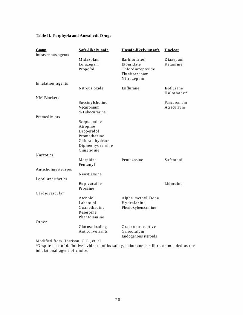
Table II. Porphyria and Anesthetic Drugs
Group Safe-likely safe Unsafe-likely unsafe Unclear Intravenous agents
MidazolamLorazepamPropofol
BarbituratesEtomidateChlordiazepoxideFlunitrazepamNitrazepam
DiazepamKetamine
Inhalation agentsNitrous oxide Enflurane Isoflurane
Halothane*NM Blockers
SuccinylcholineVecuroniumd-Tubocurarine
PancuroniumAtracurium
PremedicantsScopolamineAtropineDroperidolPromethazineChloral hydrateDiphenhydramineCimetidine
NarcoticsMorphineFentanyl
Pentazosine Sufentanil
AnticholinesterasesNeostigmine
Local anestheticsBupivacaineProcaine
Lidocaine
CardiovascularAtenololLabetololGuanethadineReserpinePhentolamine
Alpha methyl DopaHydralazinePhenoxybenzamine
OtherGlucose loadingAnticonvulsants
Oral contraceptiveGriseofulvinEndogenous steroids
Modified from Harrison, G.G., et. al.*Despite lack of definitive evidence of its safety, halothane is still recommended as theinhalational agent of choice.
21
Table III. Features of the acute porphyric attack
Nervous system dysfunction Approx % Involvement Notes
Autonomic Neuropathy
Abdominal Pain
Vomiting
Tachycardia
Hypertension
Postural Hypotension
95
46
80
36
21
C.
Peripheral Neuropathy
Paresis to paralysis of
muscle groups or
extremities
Flaccid quadriplegia
Respiratory paralysis
60
Bulbar Involement
Vagal-Cranial N.
Dysphagia
Dysphonia
Respiratory dysfunction
30
Hypothalmic Involvement
siADH(syn. inappropriate
secretion ADH)
Pyrexia
12
9
Cerebral Involvement-
Mental status changes
Anxiety, confusion,
hysteria, depression,
psychosis
Seizures
Coma
55
20
10
B .

22
Laboratory
Dark urine
Hypochloremia
Hyponatremia
Hypokalemia
siADH
Leukocytosis
Hypomagnesemia
74
50
41
29
12
11
9
B .
B .
A. All data from Stein and Tschudy(35) except as notedB . Percentages taken from Eales and Linder(34) due to unclear or unavailable data from
Stein.C. Other reviews have reported values up to 55%.Modified from Taddeini and Watson(29)
23
PORPHYRIA TYPE HEME BIOSYNTHESIS ENZYMES Glycine + Succinyl CoA
↓ ALA Synthase& Aminolevulinic Acid (ALA)
Plumboporphyria———— (PLP)
↓ ALA Dehydratase
Porphobilinogen (PBG)Acute Intermittent —- ↓ PBG DeaminasePorphyria (AIP) Hydroxymethylbilane
↓ Uro'Gen Cosynthase
Uroporphyrinogen → Uroporphyrin
↓ Uro'Gen decarboxylase
Coproporphyrinogen → Coproporphyrin
Hereditary ↓ Copro'Gen OxidaseCoproporphyria ——(HCP) Protoporphyrinogen
↓ Proto'gen OxidaseVariegate ————Porphyria (VP) Protoporphyrin
↓ Ferrochelatase + IronHEME
+ Globin ← → + Apoprotein↓ ↓
Hemoglobin Other respiratorypigments
Figure 1. Enzymatic defects in the acute porphyrias.The porphyrias are a group of inherited or acquired enzymatic defects of heme biosynthesis.Each type of porphyria has a characteristic pattern of overproduction and accumulation ofheme precursors based upon the location of the dysfunctional enzyme in the heme syntheticpathway.
Modified from Moore MR, McColl KEL, Remington C, Goldberg A. Disorders of PorphyrinMetabolism . (4)