Embed Size (px)
Citation preview
ANIMAL NUTRITION
CHAPTER 41
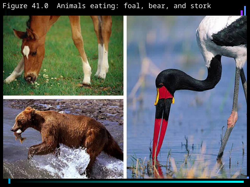
Figure 41.0 Animals eating: foal, bear, and stork
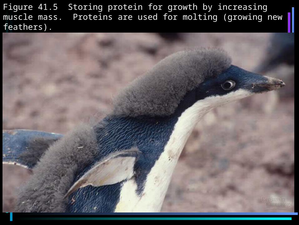
Figure 41.5 Storing protein for growth by increasing muscle mass. Proteins are used for molting (growing new feathers).
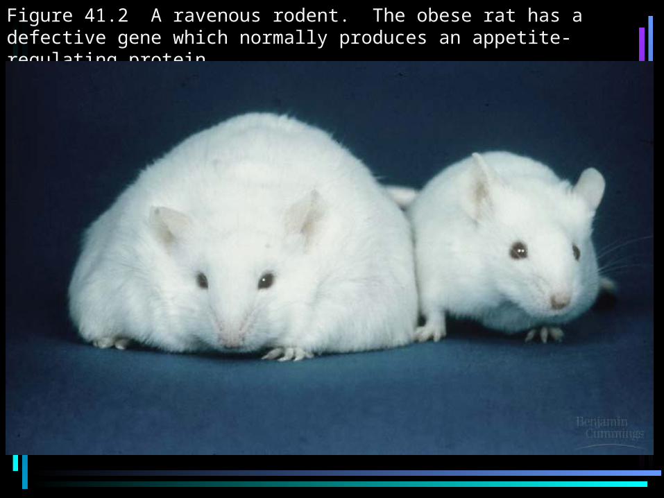
Figure 41.2 A ravenous rodent. The obese rat has a defective gene which normally produces an appetite-regulating protein.
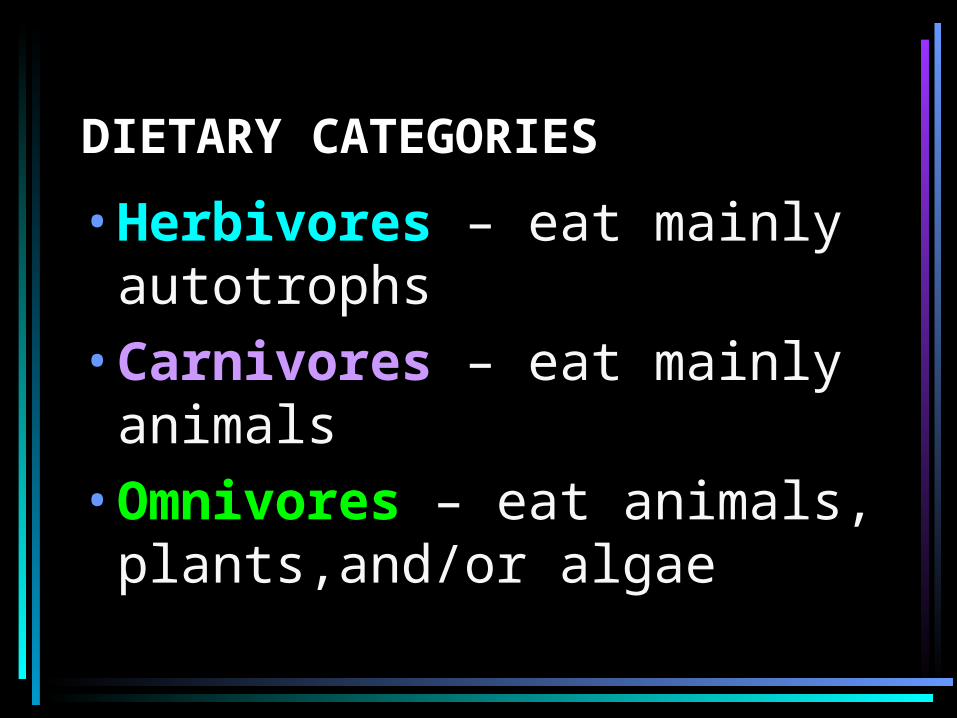
DIETARY CATEGORIES
•Herbivores – eat mainly autotrophs
•Carnivores – eat mainly animals
•Omnivores – eat animals, plants,and/or algae
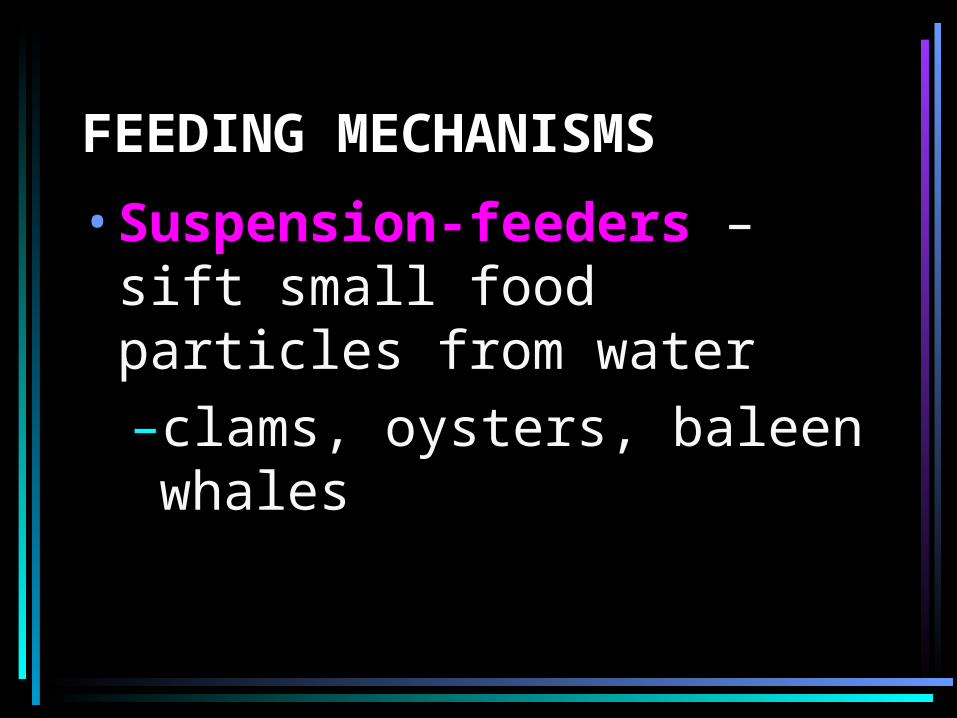
FEEDING MECHANISMS
•Suspension-feeders – sift small food particles from water–clams, oysters, baleen whales
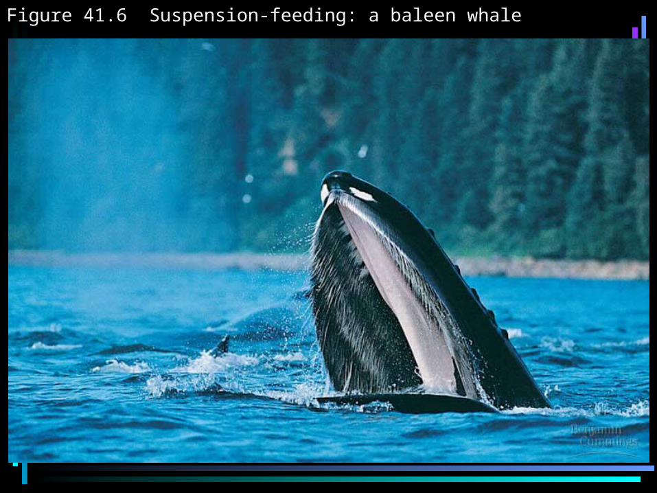
Figure 41.6 Suspension-feeding: a baleen whale
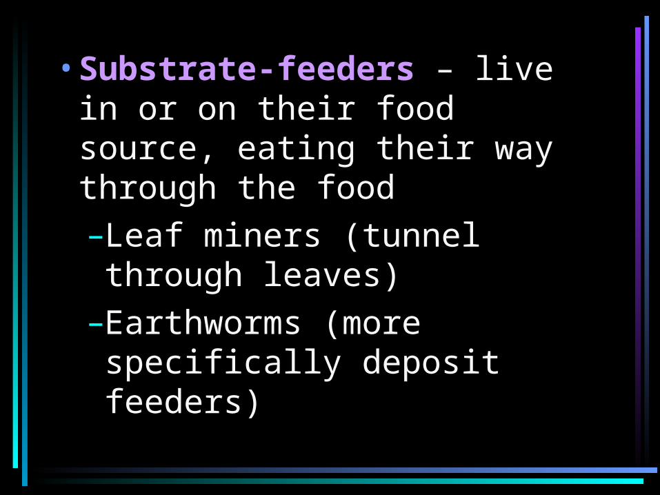
•Substrate-feeders – live in or on their food source, eating their way through the food–Leaf miners (tunnel through leaves)
–Earthworms (more specifically deposit feeders)
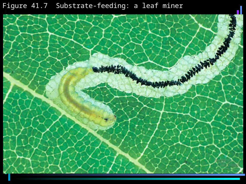
Figure 41.7 Substrate-feeding: a leaf miner
•Fluid-feeders – sucking nutrients from living hosts–Mosquitoes, leeches, aphids
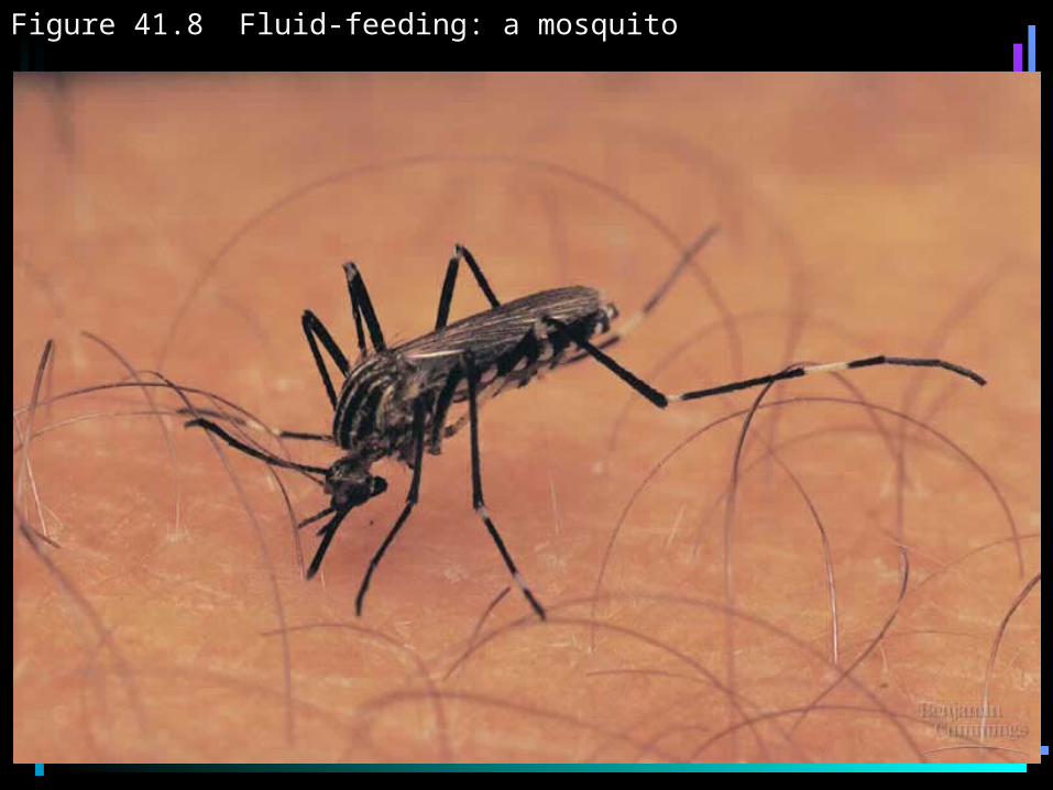
Figure 41.8 Fluid-feeding: a mosquito
•Bulk-feeders – eat relatively large pieces of food–Most animals
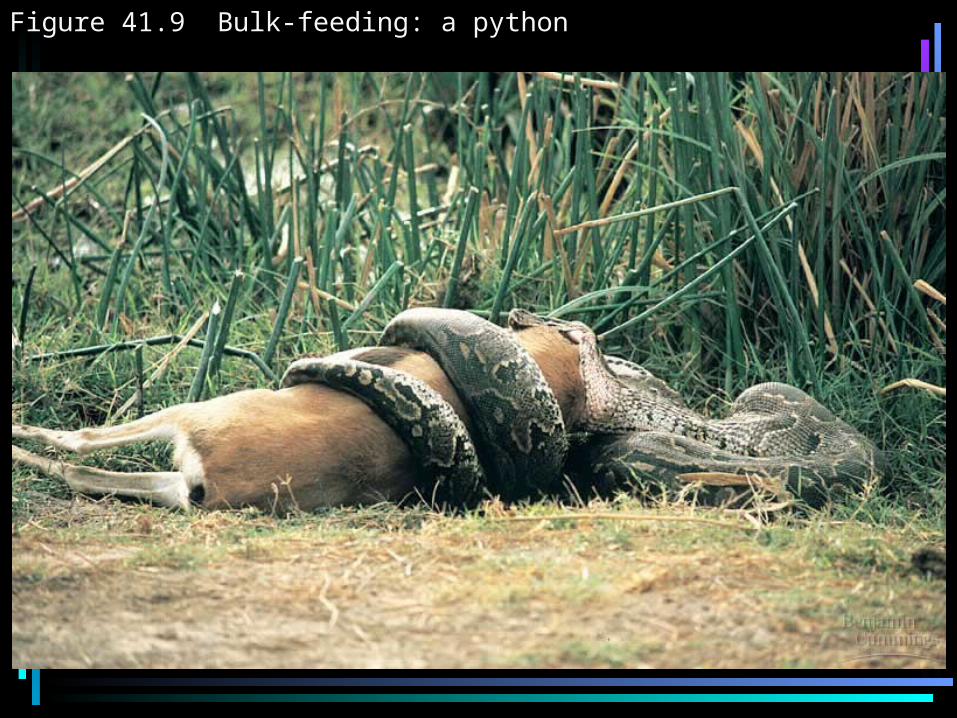
Figure 41.9 Bulk-feeding: a python
FOUR MAIN STAGES OF FOOD PROCESSING
• Ingestion•Digestion
–Enzymatic hydrolysis•Absorption•Elimination
INTRACELLULAR DIGESTION•Food vacuoles fuse with
lysosomes that have hydrolytic enzymes to digest food–Sponges (entirely intracellular digestion)
–Paramecium – oral groove leads to making food vacuole
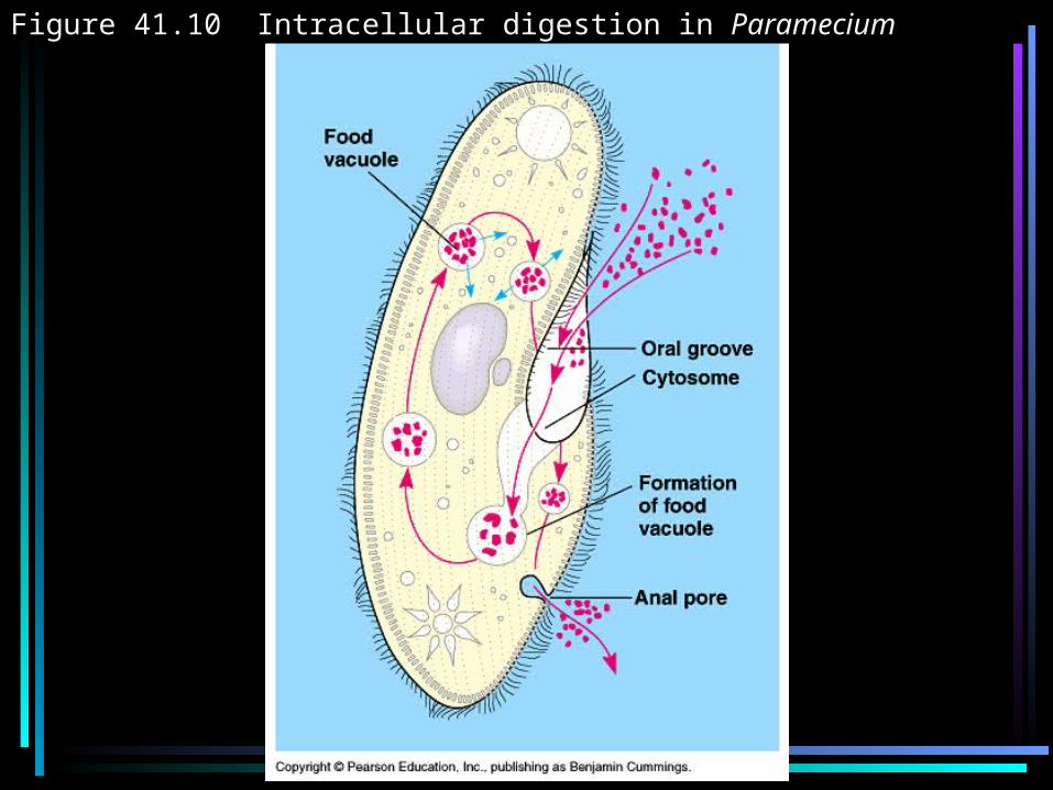
Figure 41.10 Intracellular digestion in Paramecium
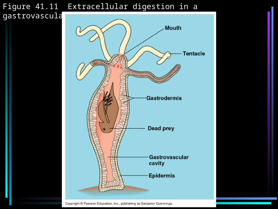
Figure 41.11 Extracellular digestion in a gastrovascular cavity
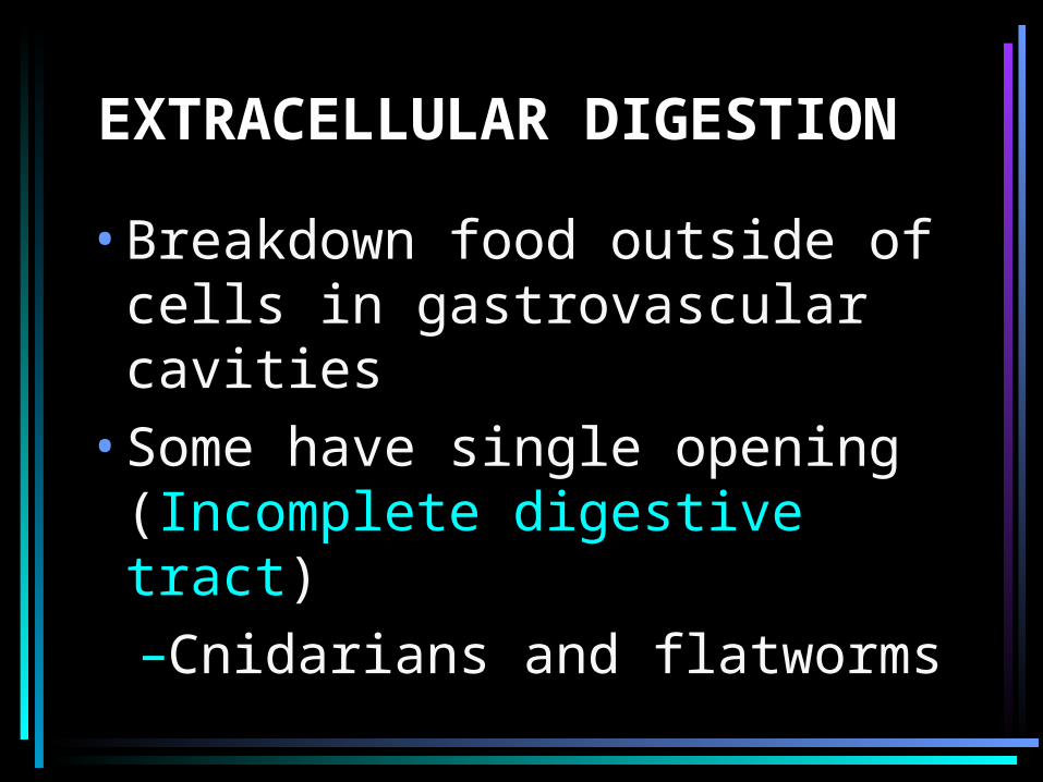
EXTRACELLULAR DIGESTION
•Breakdown food outside of cells in gastrovascular cavities
•Some have single opening (Incomplete digestive tract)–Cnidarians and flatworms
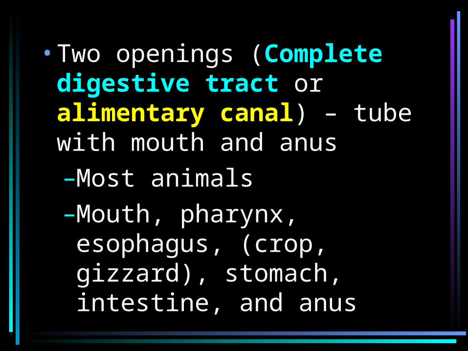
•Two openings (Complete digestive tract or alimentary canal) – tube with mouth and anus–Most animals–Mouth, pharynx, esophagus, (crop, gizzard), stomach, intestine, and anus
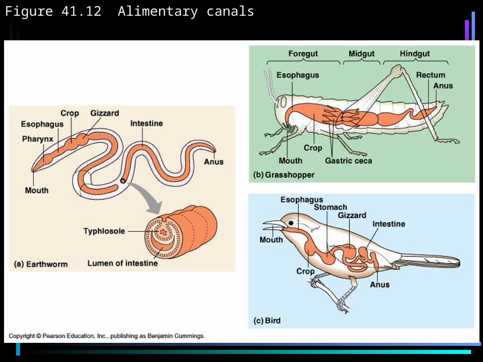
Figure 41.12 Alimentary canals
MAMMALIAN DIGESTIVE SYSTEM
•Peristalsis – contraction of smooth muscles in wall of canal
•Sphincters – ring-like valves
•Salivary glands, pancreas, liver, gall bladder
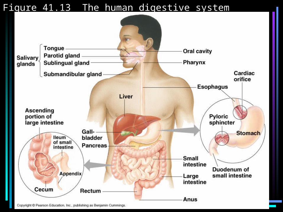
Figure 41.13 The human digestive system
HUMAN DIGESTIVE SYSTEM
•Oral Cavity–Mucin (glycoprotein) – protects soft lining
–Salivary amylase – hydrolyzes starch
–Bolus- food ball that is swallowed
•The Pharynx–Epiglottis (cartilaginous flap)
–Esophageal sphincter contracts, epiglottis up
–Esophageal sphincter relaxed, epiglottis covers trachea, food moves into esophagus
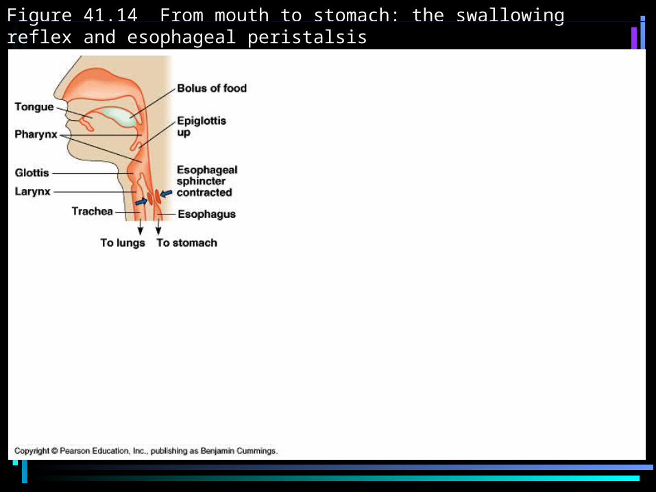
Figure 41.14 From mouth to stomach: the swallowing reflex and esophageal peristalsis
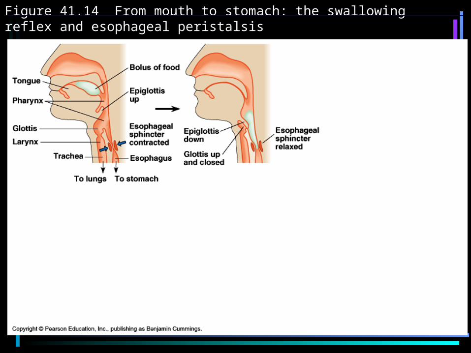
Figure 41.14 From mouth to stomach: the swallowing reflex and esophageal peristalsis
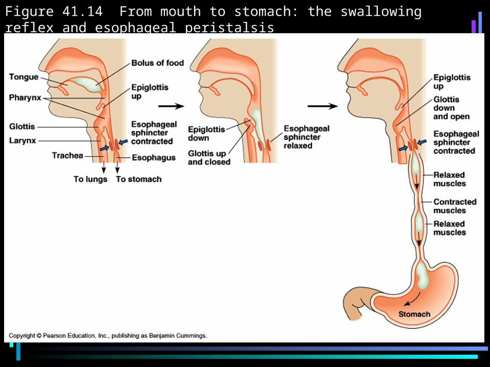
Figure 41.14 From mouth to stomach: the swallowing reflex and esophageal peristalsis
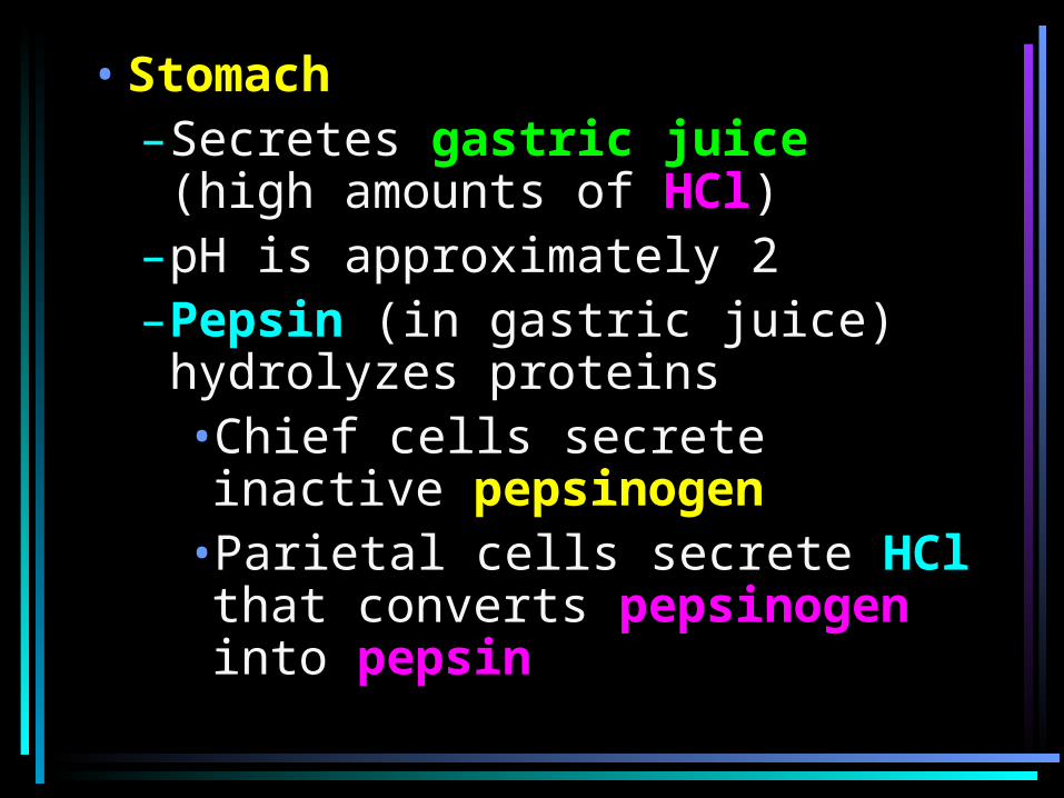
•Stomach–Secretes gastric juice (high amounts of HCl)
–pH is approximately 2–Pepsin (in gastric juice) hydrolyzes proteins•Chief cells secrete inactive pepsinogen
•Parietal cells secrete HCl that converts pepsinogen into pepsin
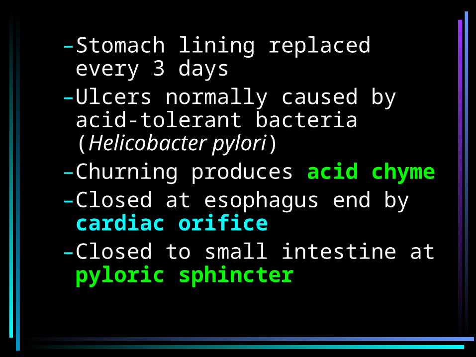
–Stomach lining replaced every 3 days
–Ulcers normally caused by acid-tolerant bacteria (Helicobacter pylori)
–Churning produces acid chyme–Closed at esophagus end by cardiac orifice
–Closed to small intestine at pyloric sphincter
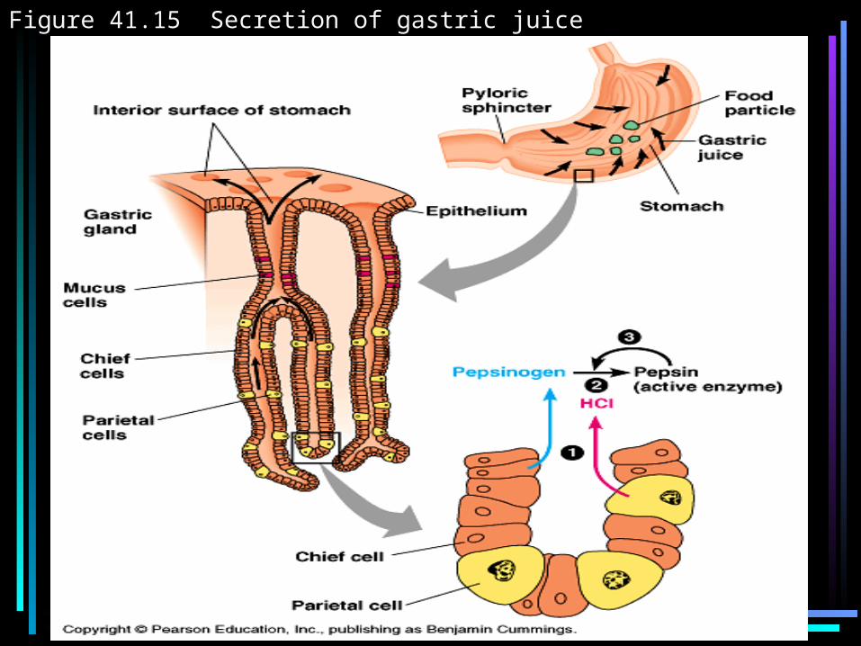
Figure 41.15 Secretion of gastric juice
•Small Intestine –Longest section of canal–Where most absorption occurs
–First 25 cm = duodenum–In duodenum
•digestive juices from pancreas, liver, gall bladder, and gland cells enter
•Pancreas–Produces hydrolytic enzymes and bicarbonate (to reduce acidity)
•Liver–Production of bile (with bile salts) that aids in digestion of fats
•Gall bladder–Stores bile
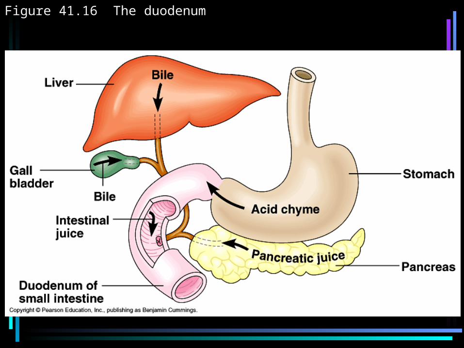
Figure 41.16 The duodenum
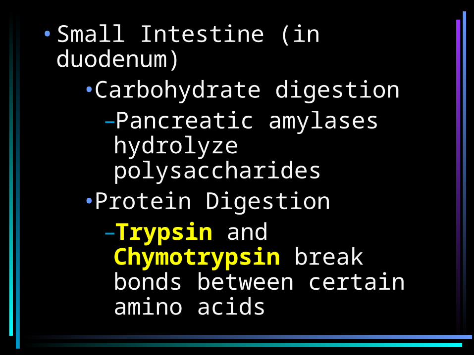
•Small Intestine (in duodenum)•Carbohydrate digestion
–Pancreatic amylases hydrolyze polysaccharides
•Protein Digestion–Trypsin and Chymotrypsin break bonds between certain amino acids
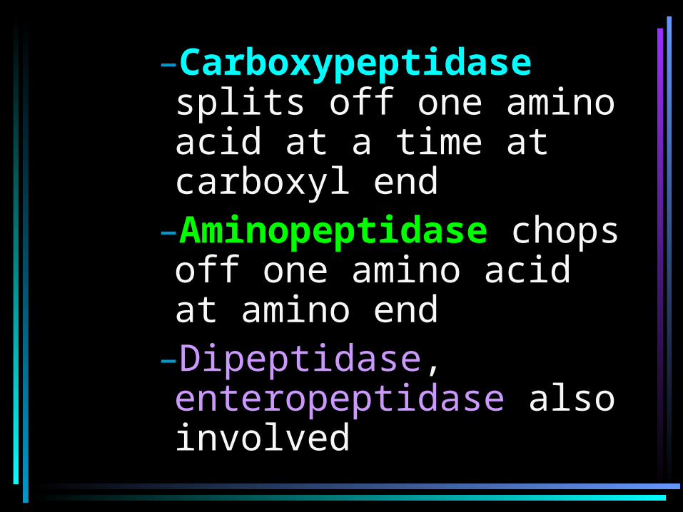
–Carboxypeptidase splits off one amino acid at a time at carboxyl end
–Aminopeptidase chops off one amino acid at amino end
–Dipeptidase, enteropeptidase also involved
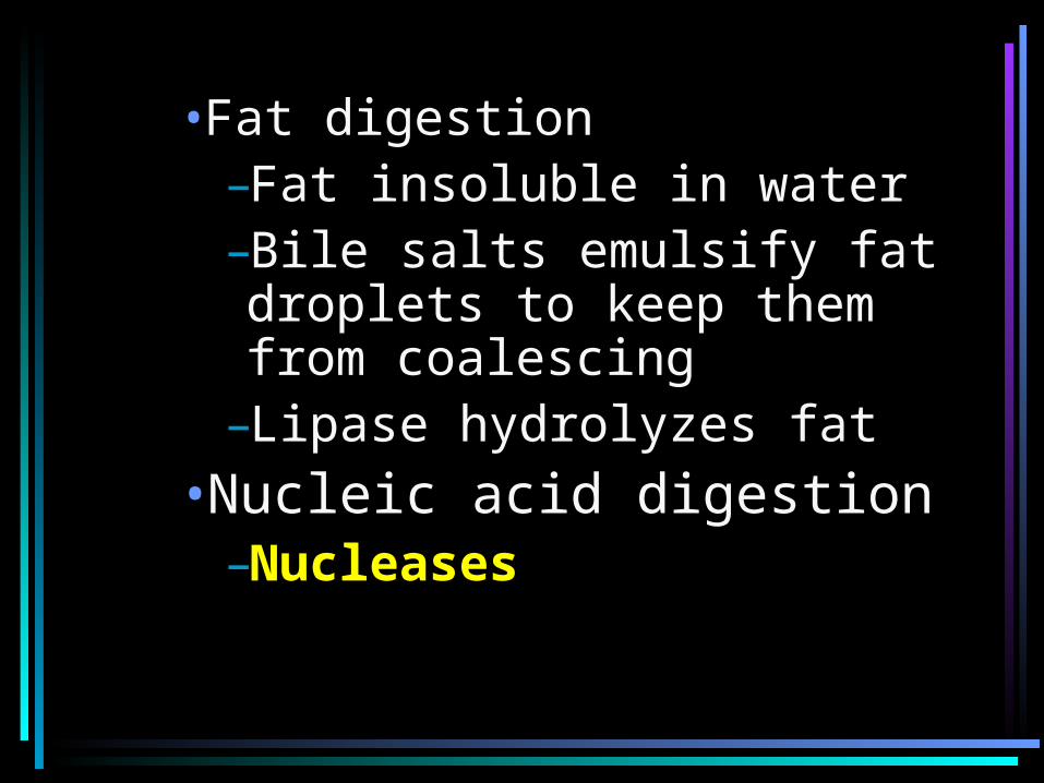
•Fat digestion–Fat insoluble in water–Bile salts emulsify fat droplets to keep them from coalescing
–Lipase hydrolyzes fat•Nucleic acid digestion
–Nucleases
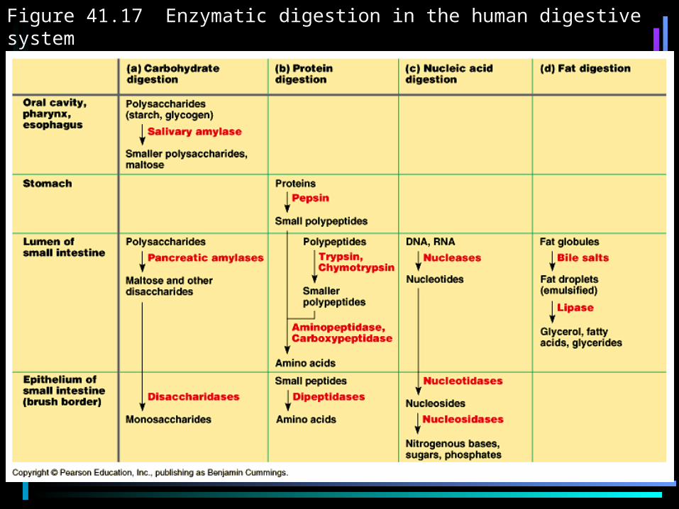
Figure 41.17 Enzymatic digestion in the human digestive system
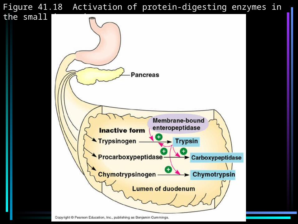
Figure 41.18 Activation of protein-digesting enzymes in the small intestine
– Absorption of nutrients (mostly in jejunum and ileum)
– Increased surface area by villi and microvilli
– Each villi contains capillaries and lacteal (small lymphatic vessel); each only one cell thick
– Nutrients move via diffusion and active transport
– Fats move into lacteal– All other nutrients empty into
capillaries and eventually move into hepatic vessel to liver
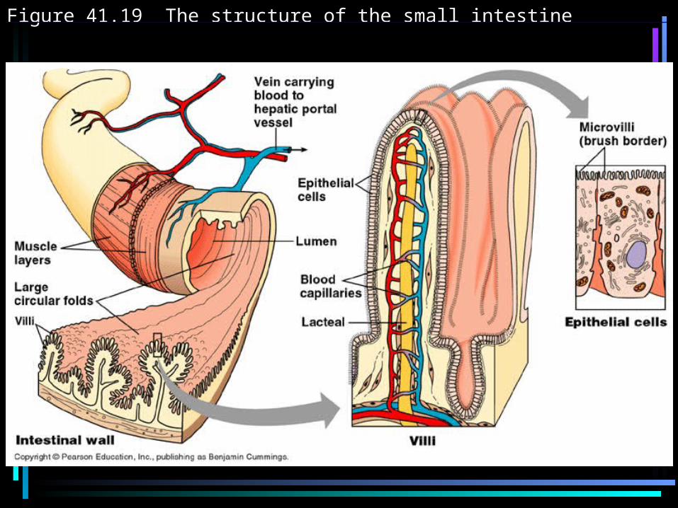
Figure 41.19 The structure of the small intestine
•Large Intestine (colon) –Connected to small intestine at T-shaped junction•One arm is cecum that leads to appendix in humans
•Other arm is the colon–Reabsorbs water–Leftover waste is feces–Feces stored in rectum and released through anus
–E. coli in colon
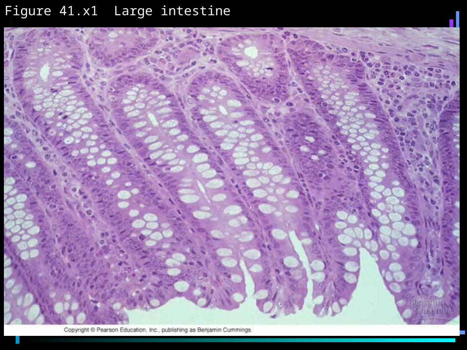
Figure 41.x1 Large intestine
EVOLUTIONARY ADAPTIONS
•Teeth vary according to what the animal eats–Fangs, incisors, canines, “grinders”
•Herbivores and omnivores longer alimentary canals than carnivores due to cellulose digestion
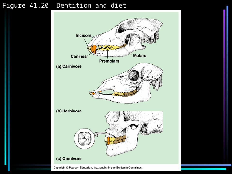
Figure 41.20 Dentition and diet
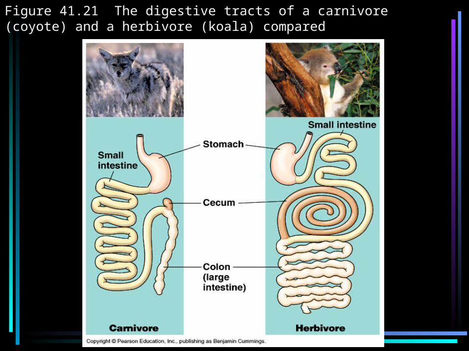
Figure 41.21 The digestive tracts of a carnivore (coyote) and a herbivore (koala) compared
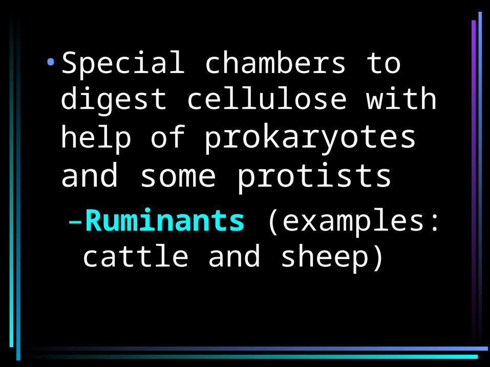
•Special chambers to digest cellulose with help of prokaryotes and some protists –Ruminants (examples: cattle and sheep)
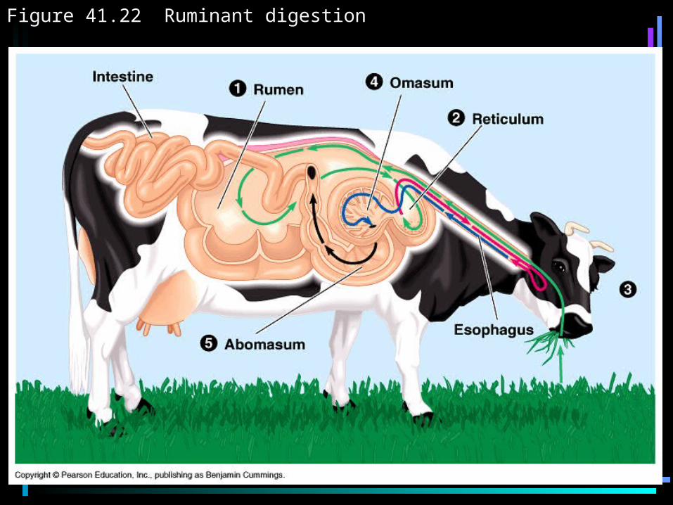
Figure 41.22 Ruminant digestion
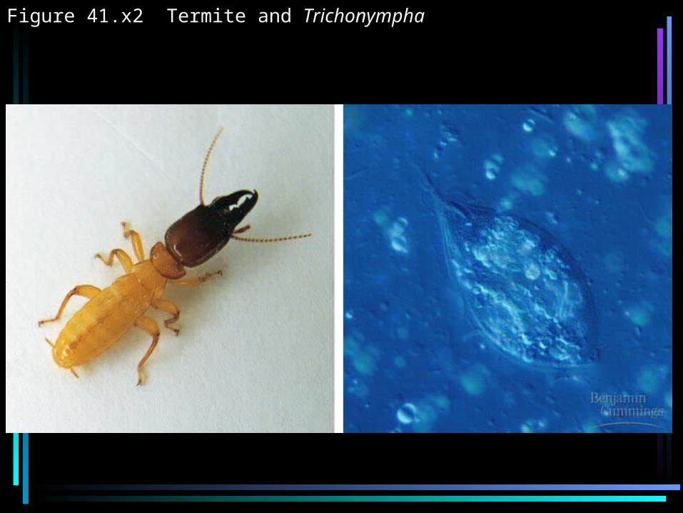
Figure 41.x2 Termite and Trichonympha