Embed Size (px)
Citation preview
A. James Barkovich 1.2
David Norman2
This article appears in the May/June 1988 issue of AJNR and the July 1988 issue of AJR.
Received June 18. 1987; accepted after revision October 15. 1987.
The views expressed in this article are those of the authors and do not reflect the official policy or position of the Department of the Army, Department of Defense, or U.S. Government.
Presented in part at the annual meeting of the American Society of Neuroradiology, New York City, May 1987.
, Department of Radiology, Letterman Army Medical Center, Presidio of San Francisco, CA 94129-6700. Address reprint requests to Technical Publications Editor HSHH-CSD-TP.
2 Department of Radiology, Neuroradiology Section, University of California School of Medicine, San Francisco, CA 94143.
AJNR 9:493-501, May/June 1988 0195-6108/88/0903-0493 © American Society of Neuroradiology
Anomalies of the Corpus Callosum: Correlation with Further Anomalies of the Brain
493
The MR imaging studies of 68 patients who had brain anomalies were reviewed retrospectively to evaluate specific anatomic abnormalities of the corpus callosum, The corpus callosum was abnormal in 32 (47%) of the 68 patients. Excluding patients with the Chiari I malformation, callosal anomalies were present in 30 (68%) of 44 patients. Callosal dysgenesis was most common, followed by callosal atrophy or hypoplasia and complete agenesis. The anterior commissure was present in all patients. On the basis of the known temporal sequence of brain and callosal embryogenesis, we deduced the following regarding the pathogenesis of developmental anomalies: (1) callosal dysgenesis occurs as a result of insults during the formation of its precursors, not during formation of the corpus callosum itself; (2) the Dandy-Walker malformation sometimes occurs as a result of an insult in the eighth week of gestation, several weeks later than has been generally accepted; (3) sphenoidal encephaloceles probably occur as a result of faulty disjunction of neuroectoderm and cutaneous ectoderm at the anterior neuropore; and (4) a complete but atrophic corpus callosum results from an insult to the cortex or white matter after formation of the corpus callosum is complete (18-20 weeks).
Callosal anomalies, easily identified on MR, are an important indicator of additional brain anomalies. Analysis of the corpus callosum provides important information about the embryogenesis of brain anomalies and may assist in distinguishing between in utero and perinatal brain insults.
The corpus callosum is the major pathway of association fibers between the two cerebral hemispheres. The anatomy and embryology of this commissure have been studied extensively. Development occurs between approximately 8 and 17 weeks gestation [1], contemporaneous with many other major cerebral structures. Corpus callosum anomalies are commonly associated with other congenital brain anomalies [2, 3], suggesting the possibility of a causal relationship .
The corpus callosum is seen in great detail with MR imaging [4]. It is of interest, therefore, from both the embryologic and diagnostic point of view, to investigate both the types and degrees of callosal anomalies associated with developmental abnormalities of the brain and to use the known embryology of the corpus callosum to speculate on the timing and nature of the related malformations.
Subjects and Methods
Sixty-eight patients with 70 congenital anomalies of the brain were evaluated over a 20-month period at the University of California, San Francisco, and the San Francisco Magnetic Resonance Center. The anomalies and their distribution are listed in Table 1. These diagnoses were determined on the basis of a characteristic clinical and radiographic presentation, biochemical data when appropriate, and, in one case, biopsy of the brain .
The patients were 8 months to 61 years old (average, 14.4 years) . Patients with Chiari I malformations averaged 32.1 years of age, while the remaining patients averaged 5.1 years of age.
The MR scans of 57 patients were obtained with a 1.5-T General Electric Signa imager. The routine imaging sequences consisted of sagittal spin-echo 600/20 (TRfTE) images with

494 BARKOVICH AND NORMAN AJNR:9, May/June 1988
TABLE 1: Distribution of 70 Congenital Anomalies in 68 Patients
Anomaly
Chiari I malformation
Chiari II malformation
Anomalies of neuronal migration: Lissencephaly Pachygyria Gray-matter heterotopias Polymicrogyria Schizencephaly
Subtotal
Encephaloceles
Dandy-Walker malformation
Holoprosencephaly
In utero infarcts
Metabolic diseases: Pelizaeus-Merzbacher disease Alexander disease Citrullinemia
Subtotal
Olivopontocerebellar degeneration
Aicardi syndrome
Isolated agenesis of the corpus callosum
Total
No. Studied
24
14
2 1 2 1 4
10 6
4
2
3
2
70
5-mm slice thickness and 1-mm interslice gap, supplemented by axial 2000/35-70 images of 5-mm slice thickness with 2.5-mm interslice gap. Coronal images were obtained occasionally for additional information. This plane was especially helpful in the evaluation of patients with holoprosencephaly, basilar encephaloceles, and migration anomalies. Eight patients were scanned on a Diasonics MT/S 0.35-T imager. Routine sequences on this scanner included a sagittal 500/ 40 sequence with 5-mm slices (1-mm gap) and axial 2000/40-80 sequence with 5-mm slices (2.5-mm gap). Three scans were obtained at outside institutions, two on a Technicare 0.6-T imager and one on a Siemens 0.5-T imager. Standard T1- and T2-weighted sagittal and axial images were obtained in these patients.
The scans were evaluated retrospectively for the presence or absence of and size of the anterior commissure and portions of the corpus callosum (genu, body, splenium, and rostrum). Findings were compared with age-matched controls. The midline sagittal T1-weighted image was the most useful. If present, the size of each portion of the corpus callosum was graded as either normal or small. The rest of the brain was then evaluated for coexistent anomalies.
Two patients with Chiari II malformations had marked hydrocephalus at the time of MR. In both instances, it was not possible to evaluate the corpus callosum because of considerable stretching and thinning by the distended lateral ventricles. These two patients were therefore excluded from the MR study.
The data were analyzed from two perspectives. The patients were categorized by type of brain anomaly and by type(s) of callosal anomaly in that category. The embryogenesis of the anomaly was considered in light of the known developmental sequence of the corpus callosum. In addition , on each scan, the presence or absence of the anterior commissure was noted. The hippocampal commissure is difficult to identify on MR and, therefore, was not evaluated, nor
was the posterior commissure, which has a separate embryologic development.
The various pathoanatomic and radiographic features of the brain resulting from dysgenic corpus callosum have been well defined [3-6] and were not a part of this study.
Results
Chiari /I Malformation
Fourteen patients with the Chiari " malformation were imaged. Two patients had marked hydrocephalus and were eliminated from the MR study. In two patients, the splenium and rostrum were absent-the anterior two-thirds of the corpus was present in one and the anterior one-third was present in the other. In seven patients, the splenium was hypoplastic and the rostrum was absent (Fig. 1). In two patients, the corpus callosum was normal. There was no correlation between the degrees of deformity of the hindbrain and corpus callosum.
Chiari I Malformation
Twenty-two of the 24 patients with the Chiari I malformation had a normal corpus callosum. In two patients the splenium and rostrum were absent; one of the two also had marked hypoplasia of the genu and body. The latter patient also had marked atrophy of the white matter; he had a clinical diagnosis of cerebral palsy.
Migration Disorders
One patient with lissencephaly and the patient with focal left frontal pachygyria had a hypoplastic splenium and absent rostrum. The second patient with lissencephaly had less severe gyral anomalies and a normal corpus callosum. Four patients had schizencephaly. In three patients with narrowing of the corpus callosum, the degree and extent of narrowing were related to the size of the clefts. Two had bilateral clefts in the frontoparietal regions; both had marked hypoplasia/ atrophy of the genu and body of the corpus callosum (Fig. 2). One had a large unilateral cleft in the right frontoparietal region; this patient had a small anterior body. The patient with a normal corpus callosum had a small unilateral cleft with fused lips. One of the two patients with ectopic gray matter had a heterotopia located exclusively in the subependymal region bilaterally; the gyral pattern was normal. This patient had a nearly complete agenesis of the corpus callosum, with only a poorly defined genu present. The second patient had heterotopia located in the white matter of the left cerebral hemisphere as well as an abnormal gyral pattern in the overlying cortex, representing pachygyria; the corpus callosum was intact.
Encephaloceles
Two patients who had previous repair of occipital encephaloceles had hypoplasia of the splenium but an otherwise

AJNR:9, May/June 1988 ANOMALIES OF THE CORPUS CALLOSUM 495
Fig. 1.-Chiari II malformation. Sagittal MR image, 600/20. Corpus callosum is markedly dysgenic; the genu (long straight arrow) and body (short straight arrows) are present, but splenium and rostrum are absent. Anterior commissure (curved arrow) is present and of normal size.
Fig. 2.-Schizencephaly. Midline sagittal MR image, 500/40, reveals hypoplasia of genu and body of corpus callosum (arrows) in patient with bilateral frontoparietal clefts. Degree and iocation of thinning of corpus callosum corresponded to size and location of hemispheric clefts in all patients with schizencephaly.
Fig. 3.-Sphenoidal encephalocele and complete callosal agenesis. Midline sagittal MR image, 600/20. Encephalocele sac protrudes into nasopharynx (arrowheads) . Layer of fat (long arrows) lines lamina terminalis, suggesting premature disjunction of cutaneous ectoderm from neuroectoderm in region of anterior neuropore. Extension of sulci into third ventricle results from lack of inversion of cingulate gyrus. Curvilinear high-intensity signal above pineal gland results from entry-slice phenomenon in vein. Anterior commissure (short arrow) is small , as it was in all patients with complete callosal agenesis.
Fig. 4.-Dandy-Walker malformation. Midline sagittal MR image, 600/20. Only genu and most anterior portion of body of corpus callosum have formed. Anteriorly, cingulate sulcus is formed by inversion of cingulate gyrus (arrows) , but, posterior to formed corpus, cingulate gyrus remains everted and sulci extend all the way into third ventricle.
Fig. 5.-Cerebral palsy and hypoplasia of parietal and occipital corona radiata bilaterally. Midline sagittal MR image, 600/20. Corpus callosum is generally hypoplasic, with thinning most marked in posterior body and splenium (arrows). Generalized thinning of corpus callosum was observed in all patients with degenerative and destructive lesions of the cerebrum.
4
normal-appearing corpus callosum; of note, the rostrum was present in both patients. One neonate with a small frontal encephalocele had an intact corpus callosum. One patient with a frontonasal encephalocele and two patients with sphenoidal encephaloceles had complete agenesis of the corpus callosum (Fig. 3). One of the two patients with a sphenoidal encephalocele had a thin layer of fat lining the anterior aspect of the lamina terminalis.
Dandy-Walker Syndrome
In two of the four patients who had the Dandy-Walker malformation, only the genu of the corpus callosum was
5
present (Fig. 4). In the other two patients, the corpus callosum was intact.
Holoprosencephaly
The patient with alobar holoprosencephaly had total absence of the corpus callosum. The patient with a mild semilobar holoprosencephaly had a spleniumlike structure posteriorly but no genu, body, or rostrum. In this case, a small falx cerebri was present posteriorly along with a partial posterior interhemispheric fissure . The cerebral hemispheres were fused in their rostral two-thirds , and there was continuity of the white matter across the midline, without formation of a callosumlike structure.

496 BAR KOVICH AND NORMAN AJNR:9, May/June 1988
Degenerative Disorders
In the two patients who had olivopontocerebellar atrophy, the corpus callosum was normal. The two patients with dysmyelinating disease (Alexander disease and PelizaeusMerzbacher disease) had marked atrophy of the centrum semiovale and the corpus callosum; all the components of the corpus callosum were present. The corpus callosum was atrophic but complete in a patient who had citrullinemia and in the three patients who had severe in utero or perinatal infarcts (Fig. 5).
Aicardi Syndrome and Isolated Agenesis
One patient with Aicardi syndrome had complete agenesis of the corpus callosum. This patient had, in addition, a large, left-sided arachnoid cyst. Another patient had complete agenesis of the corpus callosum with no apparent associated anomalies.
Anterior Commissure
A discrete anterior commissure was identified in all patients other than those with holoprosencephaly. In the two patients with holoprosencephaly, a definite anterior commissure could not be resolved within the massive number of fibers crossing the midline in the undivided forebrain . The commissure was subjectively small in the five patients who had complete agenesis of the corpus callosum (this group included three patients with basilar encephaloceles, the patient with Aicardi syndrome, and the patient with isolated agenesis of the corpus callosum). The commissure was seen only on the midline sagittal image in the three patients with encephaloceles and the patient with Aicardi syndrome; the configuration , therefore, could not be assessed. It was small but had a normal configuration in the patient with isolated callosal agenesis. The anterior commissure was in a normal location in all patients in the study.
Discussion
Embryogenesis of the Corpus Callosum
After closure of the neural tube during the fourth week of gestation, the thin rostral wall of the telencephalon (the primitive lamina terminalis) extends from the optic chiasm to the velum transversum. Between 6 and 8 weeks of gestation, when the fetus has attained a 15- to 30-mm crown-rump length (CRL), there is a rapid increase in thickness in the dorsal end of the rostral wall near the paraphysis (Fig. 6). This thickened, densely cellular dorsal part represents the lamina reuniens of His (7); as this region develops, axons from both hemispheres will grow into it, forming the cerebral commissures. The ventral end of the primitive wall will remain unchanged and become the anatomic lamina terminalis, forming the anterior wall of the third ventricle.
At 8- 9 weeks gestation (25- to 30-mm CRL), fiber bundles are seen growing medially from the ventrolateral wall of each
Paraphysis
Lamina reuniens
Lamina terminalis
Chiasmatic plate
Fig. 5.-Rostral midline telencephalon at approximately 7 weeks gestational age. Thickening of dorsal aspect of thin rostral wall of telencephalon (primitive lamina terminalis) represents lamina reuniens of His, which will eventually form precursors of corpus callosum and anterior commissure.
hemispheric vesicle. At 10 weeks (40-mm CRL), the two bundles meet and cross in the midline within the lamina reuniens, forming the anterior commissure [1].
During the eighth week of gestation, the dorsal part of the lamina reuniens begins to fold into a median groove called the sulcus medianus telencephali medii (SMTM). Initially, only a single fold of mesenchyme (meninx primativa) lies between the banks of the groove. During the ninth week, however, cells from the lamina reuniens migrate into the median groove (SMTM), which becomes filled by these cell masses. Eventually the groove is obliterated as fusion of the superior banks occurs during the tenth week of gestation (Fig. 7). The large cell mass remaining within the obliterated SMTM is known as the massa commissuralis; this grows in a dorsal direction along the SMTM over 5-7 weeks and becomes the bed for the ingrowth of commissural fibers of the corpus callosum [1 ].
At 11-12 weeks of gestation (50- to 60-mm CRL), the pioneer callosal fibers begin to enter the massa commissuralis. By 12-13 weeks, a definite corpus callosum is formed in the region of the commissural plate, which will become the genu of the corpus callosum. Growth continues over the next 5-7 weeks in a caudal direction, reflecting the rapid rate of caudal growth of the cerebral hemispheres. Growth of the corpus callosum is primarily from anterior to posterior with the genu forming first, then the anterior body, posterior body, and splenium (Fig. 7). The exception to this orderly ante rioposterior development is the rostrum, which forms somewhere between 18 and 20 weeks gestational age [1]. Although all the parts of the corpus callosum have formed at this time, the structure is not yet complete. As the cortical plates of the hemispheres enlarge, they extend axons as association fibers to distant regions of the brain. The majority of those crossing to the contralateral hemisphere do so through the corpus callosum with resultant thickening and

AJNR:9, May/June 1988 ANOMALIES OF THE CORPUS CALLOSUM 497
5
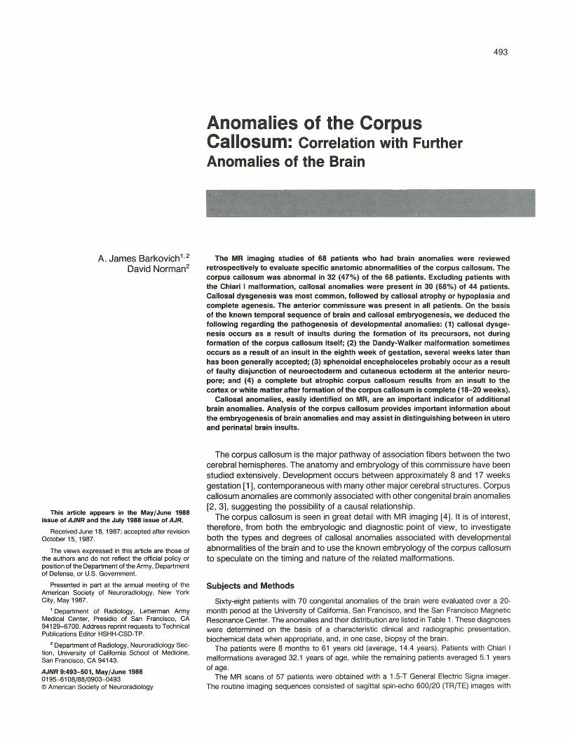
Fig. 7.-Formation of corpus callosum. Initially, there is thickening of lamina terminalis posteriorly, forming lamina reuniens (1). Depression develops in superior aspect of lamina reuniens, forming the sulcus medianus telencephali medii (SMTM) (2 and 3). Cells from lamina reuniens on either side migrate into SMTM (4), which is filled by these cell masses and eventually obliterated as superior banks fuse to form massa commissuralis. Massa (also known as commissural plate) becomes the bed for ingrowing fibers of developing cerebral hemispheres (5), forming corpus callosum. An important concept is that these steps occur simultaneously in different parts of the developing corpus callosum as it forms in a ventralto-dorsal direction. Thus, an insult to the developing corpus will result in dysgenesis of portions dorsal to those formed (or almost formed, as explained in the text) at the time of insult.
enlarging of the corpus callosum. The genu and splenium become particularly bulbous. This process continues until growth has ceased .
When attempting to correlate the degree of dysgenesis of the corpus callosum with associated anomalies of known chronology (such as the migration anomalies), it soon became apparent that the timing did not coincide if the chronology of actual callosal formation was used. However, the correlation was nearly exact if we related the timing of the insult to the formation of the SMTM or the massa commissuralis, as is described later in this article. It therefore appears that the crucial steps in the development of the corpus callosum are probably the formation of the SMTM and the massa commissuralis. Once these are formed, the axons from any developing cortical neurons will be induced to cross through the massa commissuralis , forming at least a thin corpus callosum at that point. The presence or absence of insults occurring during the formation of the SMTM and massa commissuralis are therefore the critical factors in determining whether or not association fibers will cross to the other hemisphere through the massa commissuralis 3-4 weeks later.
During its formation, the corpus callosum causes inversion of the cingulate gyri, giving the medial surface of the brain its characteristic pattern. If the corpus callosum does not form
Fig. a.-Formation of lateral callosal bundles (of Probst). Because of lack of induction by massa commissuralis, axonal fibers from cerebral hemispheres fail to cross midline. Instead, these fibers reach medial hemispheric wall and turn to course parallel to interhemispheric fissure, indenting medial walls of lateral ventricles. Broken lines represent fibers of corpus callosum in normal brain. Solid lines represent fibers that fail to cross where dysgenic corpus callosum is present. (Adapted from [1].)
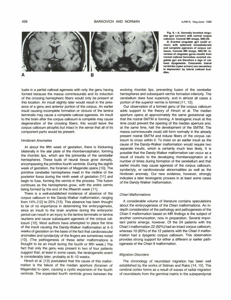
in its entirety, the cingulum remains everted at those points of agenesis, and the sulci of the medial brain surface extend all the way into the third ventricle (Figs. 3 and 4). Moreover, the axons destined for the corpus callosum instead turn parallel to the interhemispheric fissure and form the longitudinal callosal bundles of Probst (Figs. 8 and 9). These fiber bundles indent the superomedial borders of the lateral ventricles, giving them a characteristic crescentic shape. The findings of an everted cingulate gyrus and longitudinal callosal bundles are strong evidence for a primary dysgenesis as opposed to a secondary destruction of the corpus callosum.
Congenital brain anomalies result from an insult to the developing brain. The nature of the insult (vascular, infectious, metabolic, etc.) is far less important than the timing of the insult (i .e., which structures are forming at the time) and the genetic susceptibility of the embryo or fetus [8]. The insults can be localized or diffuse; if localized, the remaining brain may be completely normal. If the insult is diffuse, as is commonly the case, many of the intracranial structures developing at the time may be affected. Insults that have been shown in laboratory animals to cause callosal anomalies include irradiation as well as riboflavin, folic acid , and niacin deficiencies. In humans, maternal rubella, congenital toxoplasmosis, fetal alcohol syndrome, and maternal diabetes are reported to cause callosal anomalies [9] .
The corpus callosum forms in a ventral to dorsal direction. The fibers from the developing hemispheres are apparently induced to grow into and across the massa commissuralis by chemotaxis or perhaps by a nerve growth factor as yet to be identified. The important concept is that the SMTM forms in a caudal direction from the lamina terminalis , followed ·by conversion to massa commissuralis and finally crossing of the callosal fibers (Fig . 7). An insult that arrests formation of the SMTM early in its caudal progression , therefore, would even-
498 BARKOVICH AND NORMAN AJNR:9, May/June 1988
A B
tuate in a partial callosal agenesis with only the genu having formed because the massa commissuralis and its induction of the crossing hemispheric fibers would only be present at this location. An insult slightly later would result in the presence of a genu and anterior portion of the corpus. An earlier insult causing incomplete formation or closure of the lamina terminalis may cause a complete callosal agenesis. An insult to the brain after the corpus callosum is complete may cause degeneration of the crossing fibers; this would leave the corpus callosum atrophic but intact in the sense that all of its component parts would be present.
Hindbrain Anomalies
At about the fifth week of gestation, there is thickening bilaterally in the alar plate of the rhombencephalon, forming the rhombic lips, which are the primordia of the cerebellar hemispheres. These buds of neural tissue grow dorsally, encompassing the primitive fourth ventricle. During the eighth week of gestation, the foramen of Magendie opens [10]. The primitive cerebellar hemispheres meet in the midline of the posterior fossa during the ninth week of gestation [11] and begin to fuse, forming the vermis in the process. This fusion continues as the hemispheres grow, with the entire vermis being formed by the end of the fifteenth week [11].
There is a well-established incidence of absence of the corpus callosum in the Dandy-Walker malformation, ranging from 14% [12] to 25% [13]. This absence has been thought to be of no importance in determining the embryogenesis, since an insult to the brain anytime during the embryonic period can result in an injury to the lamina terminalis or lamina reuniens and cause subsequent agenesis of the corpus callosum [10]. Most authors have attempted to place the time of the insult causing the Dandy-Walker malformation at 4-5 weeks of gestation on the basis of the fact that cardiovascular anomalies and syndactyly of the fingers are sometimes noted [10]. (The pathogenesis of these latter malformations is thought to be an insult during the fourth or fifth week.) The fact that only the genu was present in two of four patients suggest that, at least in some cases, the teratogenetic event is considerably later, probably at 8-10 weeks.
Hirsch et al. [13] postulated that the cause of this malformation is the failure of the median aperture (foramen of Magendie) to open, causing a cystic expansion of the fourth ventricle. The expanded fourth ventricle grows between the
Fig. 9.-A, Normally inverted cingulate gyri (arrows) with normal corpus callosum. Coronal MR image, 600/20.
B, Everted cingulate gyri (solid ar· rows) with sphenoid encephalocele and complete agenesis of corpus callosum. Coronal MR image, 600/20. Inversion of cingulate gyrus results from normal callosal formation; everted cingulate gyri are therefore a sign of callosal dysgenesis. Crescentic lateral ventricles (open arrows) are secondary to impression by lateral callosal bundles.
evolving rhombic lips, preventing fusion of the cerebellar hemispheres and subsequent vermis formation inferiorly. The cerebellum does fuse superiorly, and in almost all cases a portion of the superior vermis is formed [11, 12].
Our observation of a formed genu of the corpus callosum adds support to the theory of Hirsch et al. The median aperture opens at approximately the same gestational age that the rostral SMTM is forming. A teratogenic insult at this time could prevent the opening of the median aperture and, at the same time, halt the development of the SMTM. The massa commissuralis could still form normally in the already present rostral SMTM and induce fibers of the corpus callosum to cross within it. To insist on an earlier insult as the cause of the Dandy-Walker malformation would require two separate insults, which is certainly much less likely. It is possible that the Dandy-Walker malformation can occur as a result of insults to the developing rhombencephalon at a number of times during formation of the cerebellum and that earlier insults may cause agenesis of the corpus callosum, syndactyly, or cardiovascular abnormalities as well as the hindbrain anomaly. Our new evidence, however, strongly indicates a later teratogenic process in at least some cases of the Dandy-Walker malformation.
Chiari Malformations
A considerable volume of literature contains speculations about the embryogenesis of the Chiari malformations. An indepth consideration of the pathology and pathogenesis of the Chiari II malformation based on MR findings is the subject of another communication, now in preparation. Several important points emerge, however. Of the 24 patients with the Chiari I malformation 22 (92%) had an intact corpus callosum, whereas 10 (83%) of the 12 patients with the Chiari II malformation had a dysgenic corpus callosum. This observation provides strong support for either a different or earlier pathogenesis of the Chiari II malformation.
Migration Disorders
The chronology of neuroblast migration has been well established by the works of Sidman and Rakic [14, 15] . The cerebral cortex forms as a result of waves of radial migration of neuroblasts from the germinal matrix in the subependymal
AJNR:9, May/June 1988 ANOMALIES OF THE CORPUS CALLOSUM 499
zones of the lateral ventricle. These migrations begin at about the eighth week of gestation and continue until approximately the end of the second trimester (weeks 25-26). The two major waves of migration occur between weeks 8 and 16.
Lissencephaly is the most severe of the anomalies of neuroblast migration. It is characterized by a thickened cortex, which is composed largely of neurons that have been arrested during their migration. It has been proposed that a vascular insult at 12 or 13 weeks of gestation may be the causal factor in this disorder [16]. Callosal anomalies have not been mentioned in association with lissencephaly; however, several authors have discussed the presence of colpocephaly [11, 17, 18]. (Colpocephaly refers to enlargement of the atria and occipital horns of the lateral ventricles, which is thought to result from poor organization of the forceps major. This is seen most often with callosal dysgenesis.)
The two patients with lissencephaly had pachygyria in the frontal and anterior temporal cortex and agyria in the posterior parietal region . One of these two patients also had hypoplasia of the splenium and absence of the rostrum. In the latter case, the cortex was thicker and the gyri flatter and broader. From an embryologic point of view, this more severe case presumably resulted from an earlier insult. Since fewer neuroblasts have migrated to their final cortical position, a thicker layer of heterotopic neuroblasts is present, resulting in a thicker cortex. The earlier insult is also consistent with the incomplete formation of the corpus callosum in the patient with the severe deformity; in this case, the insult occurred during the formation of the dorsal SMTM or massa commissuralis. The degree of the callosal anomaly would indicate that the insult occurs at 13-14 weeks, in excellent agreement with estimates from other methods [16]. In the case in which the corpus callosum was complete, the insult occurred after completion of these precursors, resulting in less severe developmental delay in this patient.
The patient with bilateral subependymal heterotopias had an extremely poorly developed corpus callosum with only a small portion of the genu formed. Bergeron [19] reported a high incidence of callosal anomalies in patients with subependymal heterotopias. A diffuse insult to the telencephalon during the eighth week of gestation would explain the association. This would arrest migration of the first wave of neuroblasts from the germinal matrix and development of the SMTM near its origin, resulting in the developmental abnormalities in this patient. Since the radial glial guides that direct later waves of migrating cells to their final positions have not formed at this point [15] , they will develop normally; subsequent waves of neuroblasts can migrate normally along the radial glial guides and the cortex will develop normally. In the second patient with ectopic gray matter, the heterotopias were unilateral, the overlying cortex was mildly pachygyric, and the corpus callosum was intact. The unilaterality suggests a more focal process; the fact that heterotopias were in the subcortical white matter (as opposed to the subependymal region) suggests a later insult than in the previous case. The mild pachygyric overlying cortex suggests an insult sometime between 14 and 16 weeks of gestation. The fact that the corpus callosum is intact is not helpful in this case since it
may have been unaffected because of either the focal nature of the insult or the timing of the insult.
Schizencephaly refers to the presence of holohemispheric clefts within the cerebral hemispheres, which are lined with gray matter. The fact that the entire corpus callosum was present in all four of the cases of schizencephaly is an indication that the event causing this malformation occurs either before or after the formation of the SMTM and massa commissuralis. Since the second major wave of neuroblast migration is nearing its end and by the time the corpus callosum is complete, it is unlikely that such a late insult could cause this anomaly. It is far more likely that the insult occurs during the seventh week of gestation as the germinal matrix forms along the lateral ventricular wall and before the SMTM begins to form.
The narrowing of the corpus callosum was in the region of the callosal fibers crossing from the cortical regions involved and was proportional to the volume of the hemisphere involved. Since the various regions of the brain send interhemispheric association fibers through specific regions within the corpus callosum [20] , absence of these regions, for whatever reason, should then be reflected in diminution in the number of transcallosal fibers, as was seen. In schizencephaly, the absence of a portion of the cortex and the resulting lack of development of interhemispheric fibers is the likely cause of the observed callosal narrowings.
The normal corpus callosum in the patient with polymicrogyria is compatible with the generally accepted theory that this anomaly results from an insult at 24-25 weeks gestational age [21].
Cephaloceles
Cephaloceles are extracranial extensions of intracranial structures through defects within the cranium. If there is no brain tissue within the sac, they are called meningoceles; if brain tissue is present within the sac, they are called encephaloceles. The embryology of these malformations has not been well defined. The embryologic events leading to the development of basilar encephaloceles, in which the herniation is through the endochondral bone forming the skull base, are probably different from those leading to the occipital, parietal, and interfrontal cephaloceles, in which the defect is in membranous bone [22]. Although our series is small, our results verify previous reports of a high incidence (80%) of agenesis of the corpus callosum in sphenoid encephaloceles [22, 23] . The high incidence of midline facial anomalies in basilar cephaloceles [22, 23] suggests an anomaly of induction of mesenchymal tissues. This induction is normally the result of a poorly understood influence by the underlying neural tissue, which in this region is the primitive lamina terminalis . A malformation of the anterior neuropore (later to become the lamina terminalis), such as incomplete disjunction from the cutaneous ectoderm or lack of fusion of the neural folds into a neural tube at this point, could result in this incomplete midline fusion of bony structures. This would be similar to the development of spina bifida resulting from

500 BAR KOVICH AND NORMAN AJNR:9. May/June 1988
incomplete closure of the caudal neural tube in myelomeningocele [24]. The presence of fat along the lamina terminalis gives further support to an anomaly of neural tube closure and disjunction of cutaneous ectoderm from neuroectoderm as the cause of the encephalocele. Fat is believed to form as a result of abnormal contact between mesoderm and the primitive ependyma of the neural tube [24] . Formation of fat occurs with premature disjunction of cutaneous ectoderm from neuroectoderm, as in lipomyelomeningoceles. These anomalies also have incomplete fusion of midline bony structures caused by the neural tube anomaly. The callosal agenesis is then easily explained on the basis of malformation of its precursor, the lamina terminalis.
Occipital encephaloceles are most commonly associated with the Chiari III and Dandy-Walker malformations. In the Chiari III malformation, the cephaloceles are almost always immediately adjacent to or continuous with the foramen magnum [22]. These may be caused by incomplete closure of the neural tube at that point or, more likely, by progressive thinning and resorption of bone resulting from pressure on the occipital vault from the compressed cerebellar tissue. Progressive thinning and resorption of the occipital vault have been demonstrated by in utero sonography of patients with the Dandy-Walker malformation [25], in which the cephalocele is usually higher in the occipital region. In both of these anomalies, the callosal dysgenesis is most likely associated with the underlying hindbrain anomaly, as discussed earlier. Occipital encephaloceles in the absence of underlying anomalies have no known increase in the incidence of callosal anomalies [5, 22].
Degenerative and Destructive Anomalies
In this group, the disorders have in common the degeneration or destruction of large portions of the brain. PelizaeusMerzbacher and Alexander diseases are disorders of myelination in which white-matter tracts initially form normally, but subsequently degenerate. Citrullinemia is a disorder of urea metabolism, in which toxic metabolites accumulate within the body; large areas of demyelination are seen in the brain. The olivopontocerebellar atrophies are a group of disorders in which the structures of the posterior fossa are preferentially affected. The supratentorial white matter and corpus callosum generally are spared . Three patients had intrauterine or perinatal infarcts in which the cortex and white-matter tracts initially formed but were later destroyed.
As expected, all patients in this group had a fully developed corpus callosum, since the degenerative or destructive processes occurred long after the callosal formation was complete. The full development could be demonstrated even in the extremely atrophic corpus callosum of the patients with Alexander and Pelizaeus-Merzbacher diseases, since the bulbous enlargement of the splenium and genu and the presence of the rostrum were all apparent. The fact that this is atrophy as opposed to dysgenesis was also apparent from the presence of a normally inverted cingulate gyrus.
Holoprosencephaly
The holoprosencephalies are a complex group of malformations in which the primitive prosencephalon fails to separate into diencephalon and telencephalon, often with associated facial malformations. The underlying cause is not entirely clear but the basic malfunction seems to be a lack of induction of the rostral portions of the brain and the premaxillary portion of the face [26-28]. This results in an abnormal lamina terminalis (which arises from the rostral-most portion of the neural tube). As has been discussed, a malformation of the lamina terminalis results in lack of formation of the corpus callosum, so it is not surprising that the corpus callosum is almost always absent in this group of anomalies. The presence of a splenium like structure in a mild semilobar or severe lobar form of holoprosencephaly has been described [29, 30]. The exact nature of this bundle of transhemispheric fibers is unclear. The differentiation of a nonspecific bundle of transcallosal fibers from a true corpus callosum may be solely of academic interest. However, further investigation into the mode of formation of these deep-association fibers in mild cases of holoprosencephaly may provide further insight into the development of both the corpus callosum and the holoprosencephalies.
Isolated Dysgenesis of the Corpus Callosum
The fact that only one patient with an apparently isolated dysgenesis was identified may be explained by the fact that so many other structures are developing at the same time. It is unlikely that an insult would be so focal that no other developing structures would be affected. Moreover, patients with isolated callosal defects can be asymptomatic [31]. and they are therefore less likely to be imaged. However, since the number of asymptomatic patients is thought to be small [3], our results suggest that isolated anomalies of the corpus callosum are unusual. The easy assessment of this structure by MR, therefore, makes it extremely useful as an indicator of further cerebral malformations.
Anterior Commissure
It was difficult to be certain whether the anterior commissure was present in the patients with holoprosencephaly because of the distorted architecture of their brains; the presence of so many axons crossing the midline of the poorly developed, undivided forebrain could obscure such a small tract. It would not be surprising, however, if the commissure were absent in these patients in view of the previously discussed anomalous lamina terminalis (from which the commissure derives) in these patients.
The finding of small anterior commissures in the patients with callosal agenesis in this series is in agreement with the results of Atlas et al. [32] , who found this commissure to be hypoplastic or absent in six of seven patients who had callosal agenesis. This association is most likely the result of either an anomaly of the primitive lamina terminalis (such as incom-

AJNR:9, May/June 1988 ANOMALIES OF THE CORPUS CALLOSUM 501
plete closure of the anterior neuropore, as discussed earlier) or an insult to the rostral lamina reuniens early in its formation . Either of these situations would inhibit the formation of the beds of tissue into which both the commissural and callosal fibers are induced to grow. The presence of normal anterior commissures in those patients with a partially formed corpus callosum suggests that the insult to the brain that disrupts callosal formation occurs after the bed for ingrowth of the anterior commissure is formed.
Conclusions
The corpus callosum was abnormal in 32 of 68 patients who had brain anomalies; it was dysgenic in 27 of 60 who had congenital anomalies, and atrophic in six of eight who had degenerative anomalies. If the patients with Chiari I malformation are excluded, the corpus callosum was abnormal in 30 of 44 patients. Isolated agenesis of the corpus callosum was seen in only one patient. Callosal dysgenesis, easily detected by MR, strongly suggests the presence of additional brain abnormality.
By using the known chronology and sequence of callosal formation and correlating it with other anomalies of known chronology, it is apparent that callosal dysgenesis probably occurs as a result of an insult to the brain during the formation of the callosal precursors (the lamina terminalis, SMTM, or massa commissuralis) . The degree of corpus callosum development can be used as a practical indicator of the timing of teratogenesis. This approach to teratogenesis has provided evidence that the Dandy-Walker malformation can occur as a result of an insult during the eighth week of gestation, several weeks later than the generally accepted timing, and that sphenoidal encephaloceles may result from abnormal closure of the neural tube at the anterior neuropore.
REFERENCES
1. Rakic P, Yakovlev PI. Development of the corpus callosum and cavum septi in man. J Comp Neuro/1968;132:45-72
2. Parrish ML, Roessmann U, Levinsohn MW. Agenesis of the corpus callosum: a study of the frequency of associated malformations. Ann Neurol 1979;6:349-354
3. Kendall BE. Dysgenesis of the corpus callosum. Neuroradiology 1983;25:239-256
4. Davidson HD, Abraham R, Steiner RE. Agenesis of the corpus callosum: magnetic resonance imaging. Radiology 1985;155:371-373
5. Probst FP. Congenital defects of the corpus callosum-morphology and encephalographic appearances. Acta Radiol [Diagn] [Suppl] (Stock h) 1973;331 :1-152
6. Ettlinger G. Agenesis of the corpus callosum. In: Vinken PJ , Bruyn GW, eds. Handbook of clinical neurology , vol. 30. Amsterdam: Elsevier/NorthHolland Biomedical , 1977:285-297
7. His W. Die Formentwicklung des mensch lichen Vorderhirns vom Ende des ersten bis zum Beginne des dritten Monates. Abhandl d Math, Phy KI Kon Sachs Akad d Wissensch , Bd, 1889;15:675-735, cited by Ravie P, Yakov-
lev PI. Development of the corpus callosum and cavum septi in man. J Comp Neuro/1968;132:45-72
8. Kemeyama Y. Comparative developmental pathology of malformations of the central nervous system. Prog Clin Bioi Res 1985;163A:143-156
9. Andermann E. Agenesis of the corpus callosum. In: Vinken PJ, Bruyn GW, eds. Handbook of clinical neurology , vol. 42. Section I. Malformations. Amsterdam: Elsevier/North-Holland Biomedical , 1981:6-9
10. Gardner E, O'Rahilly R, Prolo D. The Dandy-Walker and Arnold-Chiari malformations. Clinical, developmental, and teratological considerations. Arch Neurol 1975;32:393-407
11. Lemire RJ , Loeser JD, Leech RW, Alvord EC. Normal and abnormal development of the human nervous system. Hagerstown, MD: Harper & Row, 1975:144-165
12. Hart MN, Malamud N, Ellis WG. The Dandy-Walker syndrome. A clinicopathological study based on 28 cases. Neurology 1972;22:771-780
13. Hirsch J-F, Pierre-Kahn A, Renier 0 , Sainte-Rose C, Hoppe-Hirsch E. The Dandy-Walker malformation. A review of 40 cases. J Neurosurg 1984;61 :515-522
14. Sidman RL, Rakic P. Neuronal migration, with special reference to developing human brain: a review. Brain Res 1973;62:1-35
15. Rakic P. Neuronal migration and contact guidance in the primate telencephalon. Postgrad Med J 1978;54[suppI1]:25-37
16. Jellinger K, Rett A. Agyria-pachygyria (lissencephaly syndrome). Neuropaediatrie 1976;7:66-91
17. Daube JR, Chou SM. Lissencephaly: two cases. Neurology 1966;16:179-191
18. Garcia CA, Dunn 0 , Trevor R. The lissencephaly (agyria) syndrome in siblings. Arch Neuro/1978;35:608-611
19. Bergeron RT. Pneumographic demonstration of subependymal heterotropic cortical gray matter in children. AJR 1967;101 :168-177
20. De Lacoste MC, Kirkpatrick JB, Ross ED. Topography of the human corpus callosum. J Neuropathol Exp Neuro/1985;44:578-591
21. Stewart RM , Richman DP, Caviness VS Jr. Lissencephaly and pachygyria: an architectonic and topographical analysis. Acta Neuropathol (Berl) 1975;31 :1-12
22. Diebler C, Dulac O. Cephaloceles: clinical and neuroradiological appearance. Associated cerebral malformations. Neuroradiology 1983;25 :199-216
23. Yokota A, Matsukado Y, Fuwa I, Moroki K, Nagahiro S. Anterior basal encephalocele of the neonatal and infantile period. Neurosurgery 1986;19:468-478
24. Naidich TP, McLone DG , Harwood-Nash DC. Spinal dysraphism. In: Newton TH, Potts DG, eds. Computed tomography of the spine and spinal cord. San Anselmo, CA: Clavadel, 1983:299-305
25. Evrad P, Belpaire MC, Boog G, et al. Diagnostic prenatal des affections du systeme nerveux de /'enfant. Lausanne, Switzerland: Societe Internat de Neurollnfantile, 1982
26. Yakovlev PI. Pathoarchitectonic studies of cerebral malformations. III. Arrhinencephalies (holotelencephalies). J Neuropathol Exp Neurol 1959;18:22-25
27. DeMyer W. Holoprosencephaly, (cyclopia-arhinencephaly). In: Vinken PI , Bruyn GW, eds. Handbook of clinical neurology, vol. 30. Amsterdam: Elsevier/North-Holland Biomedical , 1977:431-478
28. Fitz CR . Holoprosencephaly and related entities. Neuroradiology 1983;25:225-238
29. Altman NR, Altman DH , Sheldon JJ , Leborgne J. Holoprosencephaly classified by computed tomography. AJNR 1984;5:433-437
30. Probst FP. The prosencephalies. Serlin: Springer-Verlag, 1979:46 31. Possy JG. Morphological study of a case of completely isolated and
asymptomatic agenesis of the corpus callosum. Arch Anat Histol Embryol (Strasb) 1970;53:289-340
32. Atlas SW, Zimmerman RA, Bilaniuk L T, et al. Corpus callosum and limbic system: neuroanatomic MR evaluation of developmental anomalies. Radiology 1986;160:355-362