Embed Size (px)
Citation preview
6 CPOI — Vol. 3 No. 1 — Winter 2012
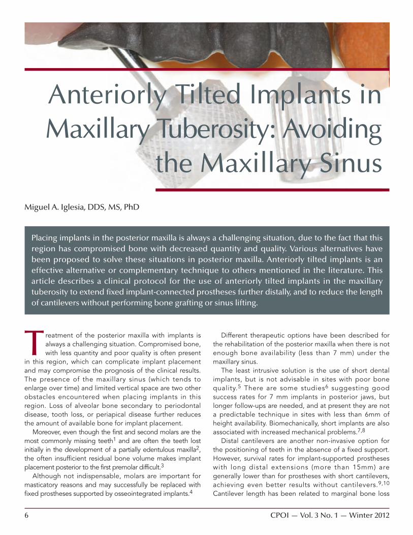
Anteriorly Tilted Implants inMaxillary Tuberosity: Avoiding
the Maxillary SinusMiguel A. Iglesia, DDS, MS, PhD
T reatment of the posterior maxilla with implants isalways a challenging situation. Compromised bone,with less quantity and poor quality is often present
in this region, which can complicate implant placementand may compromise the prognosis of the clinical results.The presence of the maxillary sinus (which tends toenlarge over time) and limited vertical space are two otherobstacles encountered when placing implants in thisregion. Loss of alveolar bone secondary to periodontaldisease, tooth loss, or periapical disease further reducesthe amount of available bone for implant placement.
Moreover, even though the first and second molars are themost commonly missing teeth1 and are often the teeth lostinitially in the development of a partially edentulous maxilla2,the often insufficient residual bone volume makes implantplacement posterior to the first premolar difficult.3
Although not indispensable, molars are important formasticatory reasons and may successfully be replaced withfixed prostheses supported by osseointegrated implants.4
Different therapeutic options have been described forthe rehabilitation of the posterior maxilla when there is notenough bone availability (less than 7 mm) under themaxillary sinus.
The least intrusive solution is the use of short dentalimplants, but is not advisable in sites with poor bonequality.5 There are some studies6 suggesting goodsuccess rates for 7 mm implants in posterior jaws, butlonger follow-ups are needed, and at present they are nota predictable technique in sites with less than 6mm ofheight availability. Biomechanically, short implants are alsoassociated with increased mechanical problems.7,8
Distal cantilevers are another non-invasive option forthe positioning of teeth in the absence of a fixed support.However, survival rates for implant-supported prostheseswith long distal extensions (more than 15mm) aregenerally lower than for prostheses with short cantilevers,achieving even better results without cantilevers.9,10
Cantilever length has been related to marginal bone loss
Placing implants in the posterior maxilla is always a challenging situation, due to the fact that thisregion has compromised bone with decreased quantity and quality. Various alternatives havebeen proposed to solve these situations in posterior maxilla. Anteriorly tilted implants is aneffective alternative or complementary technique to others mentioned in the literature. Thisarticle describes a clinical protocol for the use of anteriorly tilted implants in the maxillarytuberosity to extend fixed implant�connected prostheses further distally, and to reduce the lengthof cantilevers without performing bone grafting or sinus lifting.
CPOI_V3N1_Winter 2012:Spectrum 2/24/2012 9:27 AM Page 6
CPOI — Vol. 3 No. 1 — Winter 2012 7
around implants and mechanical failure of thecomponents11, including screw loosening, mechanicalfracture of implants or prosthetic components.
Bone compacting by the use of osteotomes is anothertreatment approach1 but has limitations in the possibleamount of bone volume to be gained. The maximumheight gain is between 2 and 4.6 mm, depending on theprevious residual bone height.12
Sinus lift grafting is another procedure that is wellsupported in the literature13, but patient acceptance isrelatively low due to the risk of increased site morbidity,the graft choice dilemma, postoperative discomfort,extended healing periods and costs.
Several authors have documented the clinical efficacyof tilting distal implants, placing them parallel to theanterior sinus wall and positioning the implant platform ina more posterior position3,14,15. The tilted implants can beanchored in the bone pyramid anterior to the maxillarysinus, where no anatomic vital structures, such as arteriesor nerves, are present. Implantation following thisapproach makes it possible to extend the prostheticsupport posteriorly, thus reducing cantilever arms. Thefavorable clinical outcomes in retrospective andprospective analyses of this technique imply that tiltingdoes not negatively affect the outcome of implanttherapy; rather, it appears that tilting allows for betterprosthetic support due to larger inter-implant distances.Finite element analysis data regarding rehabilitation of theposterior maxilla reveals that tilting distal implants, rigidlysplinted with a fixed denture, does not increase stress inthe peri-implant bone and frameworks.16
One attractive approach when treating the posteriormaxilla is to use anteriorly tilted implants into thetuberosity and the areas of the pterygoid process toovercome the sinus antrum obstacle.15,17-19 In theliterature, tilting of implants for engaging the pterygoidplate in the posterior maxilla is reported, indicating thatthis is a predictable procedure for establishing endsupport for a maxillary prosthetic restoration.17,20-22
The use of tilted implants moves implant supportposteriorly and permits a longer distance between implants,allowing for the elimination of cantilevers in the prosthesis,which results in a better load distribution situation andprovides satisfactory molar support for a fixed prosthesis.Angled implants also permit the use of significantly longerimplants, which increases the degree of implant-to-bonecontact area and also the implant primary stability. Anotheradvantage is the placement of implants in residual bone,avoiding more complex techniques, such as sinus lifting andother grafting procedures.
The anatomy of the maxillary tuberosity23,24 has itsposterior boundary in the pyramidal process of the palatalbone. This process intervenes between the posterior-inferiorsurface of the maxilla and the anterior-inferior surface of the
pterygoid laminae of the sphenoid bone. The medialportion of the process contains the lesser palatine canal andforamen, and immediately adjacent to the anterior edge isthe greater palatine canal and foramen, which thus lielingual and lateral to the tuberosity. The bone in this area isvery cancellous, and when there is tooth loss secondary toperiodontal disease, the bone is reabsorbed in a palataldirection, thus narrowing the tuberosity. The cortical bone isvery thin and irregular, and it sometimes merges into thecancellous bone, which has an open and irregulardistribution of the lamellae.
The clinical results15 indicate that implant tilting doesnot induce any biological disadvantage. On the contrary,it seems to be both clinically and biologicallyadvantageous, and a tilted implant as a member of aprosthesis configuration can be well justified from abiomechanical point of view.
The tilted implant must be placed in combination withat least one more implant in cases of partial edentulismand in combination with at least two more implants withcross-arch stabilization in cases of total edentulism.
The purpose of this article is to describe a clinicalprotocol for the use of anteriorly tilted implants in themaxillary tuberosity to extend fixed implant-connectedprostheses further distally, and to reduce the length ofcantilevers without performing bone grafting or sinus lifting.
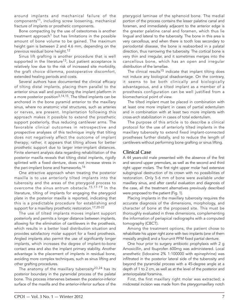
Clinical CaseA 44 years-old male presented with the absence of the firstand second upper premolars, as well as the second and thirdright upper molars. The first right upper molar had extensivesubgingival destruction of its crown with no possibilities ofrestoration. Only 5-6 mm of bone were available undermaxillary sinus, and after careful evaluation and diagnosis ofthe case, all the treatment alternatives previously describedwere proposed to the patient (Fig. 1).
Placing implants in the maxillary tuberosity requires theaccurate diagnosis of the dimensions, morphology, andcharacter of bone at the proposed site. This must bethoroughly evaluated in three dimensions, complementingthe information of periapical radiographs with a computedtomography (CBCT).
Among the treatment options, the patient chose torehabilitate his upper right zone with two implants (one of themmesially angled) and a four-unit PFM fixed partial denture.
One hour prior to surgery antibiotic prophylaxis with 2 gAmoxicillin, and Ibuprofen 600mg was administered. Localanesthetic (lidocaine 2% 1:100000 with epinephrine) wasinfiltrated in the posterior lateral side of the tuberosity andbeyond the pyramidal process with a 45-degree angle at adepth of 1 to 2 cm, as well as at the level of the posterior andanteriorpalatal foramina.
First, the first maxillary right molar was extracted; amidcrestal incision was made from the pterygomaxillary notch
CPOI_V3N1_Winter 2012:Spectrum 2/24/2012 9:27 AM Page 7
8 CPOI — Vol. 3 No. 1 — Winter 2012
Figure 1 — Preoperatory radiograph. Note 5-6 mm of bone availableunder maxillary sinus. Enough bone availability in maxillary tuberosity.
Figure 2 — Periapical intraoperative radiograph. The initial drill is notangled enough to avoid maxillary sinus.
Figure 3 — Periapical intraoperative radiograph. The axis obtainedwith the initial drill is corrected.
Figure 4 — Periapical intraoperative radiograph. Implant placed inmaxillary tuberosity.
Figure 5 — Occlusal view after 3 months. Figure 6 — Implants. Occlusal view.
to the premolar area. Small releasing vertical (buccal andpalatal) incisions were also made at both ends of the crestalincision. Then the buccal and palatal flaps were carefully raised.
Implant sites were prepared in the place of upper firstright premolar and mesially tilted in the maxillary rightsecond molar. The mesial implant was placed before thedistal one. The correct inclination of this tilted implant was
evaluated intra-operatively with a periapical radiograph afterthe preparation of the first 5 mm depth with the initial drill(Fig. 2), correcting the angle if necessary until this first drillhad the correct angulation (Fig. 3). Angulation is the mostdifficult aspect of the technique described, because anattempt is made to minimize the angulation as much aspossible, but at the same time maintaining the drill within
CPOI_V3N1_Winter 2012:Spectrum 2/24/2012 9:27 AM Page 8
10 CPOI — Vol. 3 No. 1 — Winter 2012
the bone and avoiding the maxillary sinus, simulating thecorrect inclination of a natural third molar.
Bone infra-preparation is essential when implants are placedin compromised areas such as the tuberosity. The implant sitemust be as narrow as possible to allow implant insertion butprevent micromovements thereafter; otherwise, the implanthas a decreased chance to integrate. To achieve stabilization,minimal and precise manipulation with the fewest possibleentries is required. The tilted implant site in the tuberosity wasslightly underprepared in full length to ensure high implantstability. The depth of the drilled site was measured with anappropriate depth gauge, and the integrity of the sinusmembrane was verified. Countersinking was avoided in orderto engage as much of the crestal bone as possible and toavoid damaging the cortical bone. Tapping was also avoidedto allow the implant to achieve good initial stability. Two rough-surfaced acid-etched self-tapping tapered implants (OsseotiteNT; Biomet 3i, Palm Beach Gardens, FL, USA) were placedwith good primary stability after placement. Both of them were4 mm wide, with the mesial having a 10 mm length, and thedistal angled implant with a length of 13 mm (Fig. 4).
Flaps were then adapted and sutured around healingabutments. No provisional prosthesis was delivered. After3 months osseointegration was achieved and the
prosthetic phase began (Fig. 5). The mesial angulation ofthe distal implant will have a better passive fit than thedistal one, so careful impressions of the implant platforms
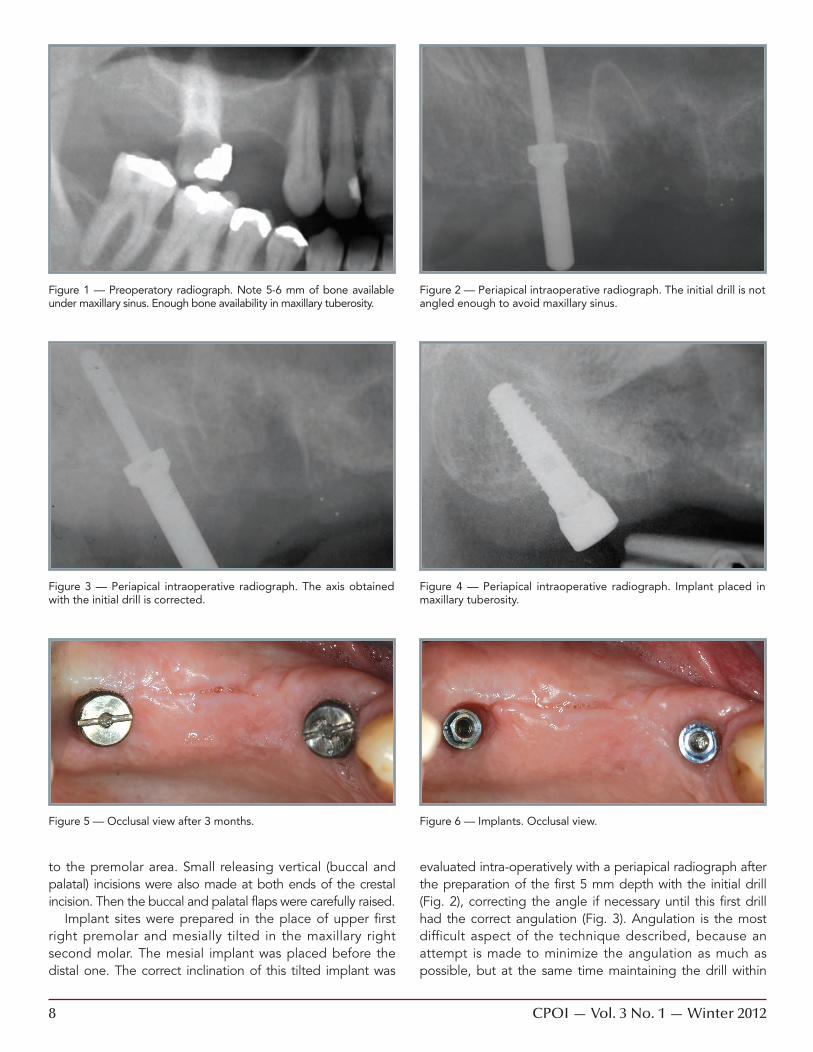
Figure 7 — Pre-welded cast titanium framework. Occlusal view. Figure 8 — Pre-welded cast titanium framework. Palatal view.
Figure 9 — Pre-welded cast titanium framework. Buccal view. Notethe screwdrivers showing the axis of the implants.
Figure 10 — Pre-welded cast titanium framework. Buccal view.
Figure 11 — Pre-welded cast titanium framework. Apical view.
Figure 12 — Clinical fit test of the pre-welded cast titaniumframework. Occlusal view.
CPOI_V3N1_Winter 2012:Spectrum 2/24/2012 9:28 AM Page 10
12 CPOI — Vol. 3 No. 1 — Winter 2012
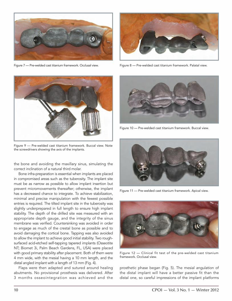
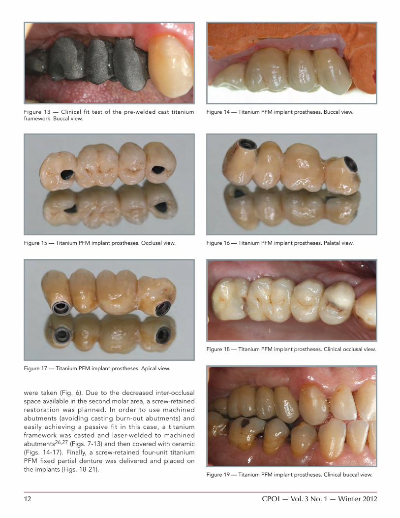
were taken (Fig. 6). Due to the decreased inter-occlusalspace available in the second molar area, a screw-retainedrestoration was planned. In order to use machinedabutments (avoiding casting burn-out abutments) andeasily achieving a passive fit in this case, a titaniumframework was casted and laser-welded to machinedabutments26,27 (Figs. 7-13) and then covered with ceramic(Figs. 14-17). Finally, a screw-retained four-unit titaniumPFM fixed partial denture was delivered and placed onthe implants (Figs. 18-21).
Figure 13 — Clinical fit test of the pre-welded cast titaniumframework. Buccal view.
Figure 15 — Titanium PFM implant prostheses. Occlusal view.
Figure 17 — Titanium PFM implant prostheses. Apical view.
Figure 14 — Titanium PFM implant prostheses. Buccal view.
Figure 16 — Titanium PFM implant prostheses. Palatal view.
Figure 18 — Titanium PFM implant prostheses. Clinical occlusal view.
Figure 19 — Titanium PFM implant prostheses. Clinical buccal view.
CPOI_V3N1_Winter 2012:Spectrum 2/24/2012 9:28 AM Page 12
14 CPOI — Vol. 3 No. 1 — Winter 2012
Conclusions1. The results in the literature indicate that tilted implants
are an effective alternative to the maxillary sinus bonegrafting procedure. The method of tilting implantsdescribed represents an alternative or complementarytechnique to others mentioned in the literature.
2. More patients can be successfully treated with dentalimplants without more complex techniques, placingimplants in the pre-existing bone. Anteriorly tilteddental implants placed in the maxillary tuberosity canavoid the compromised bone of the sinus antrum.Additional clinical advantages of this approach are thepossibility of avoiding cantilever arms, and creatinglarger interimplant distances than the posteriorly tiltedimplants technique.
3. The treatment principle is to make the maximum use ofthe available bone, which simplif ies treatmentprocedures, reduces surgical invasion and shortenstreatment time compared to sinus lift procedures.
4. Surgical planning must be precise. The occlusal schememust be carefully designed and executed, and theprosthetic phase must be previously planned to easilyachieve passive fit, solving the different angulation ofthe implants.
5. The site preparation technique must adapt to the bonein the tuberosity: low-speed drilling technique andinfra-preparation of the site for the implant must beperformed, in order to respect the cancellous bone inthis area, and to achieve good primary stability. �
Acknowledgments:The author wants to recognize the artistry of the ceramist Alicia Tomé (CDT).
References:1. Marcus SE, Drury TF, Brown LJ, Zion GR. Tooth retention and tooth
loss in the permanent dentition of adults: United States, 1988-1991.J Dent Res 1996; 75 (Special Isuue): S684-S695.
2. Hirschfield L, Wasserman B. A long-term survey of tooth loss in 600treated periodontal treatments. J Periodontol 1978; 49: 225-237.
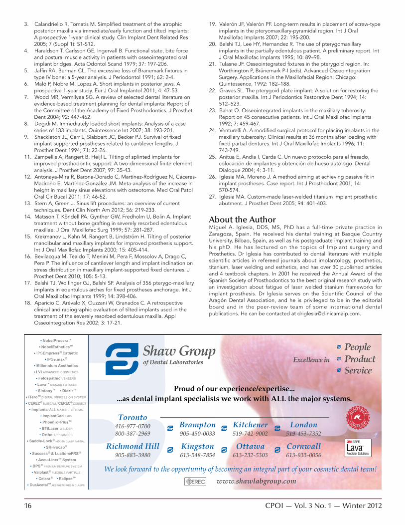
Figure 21 — Three-year control. Clinical buccal view.
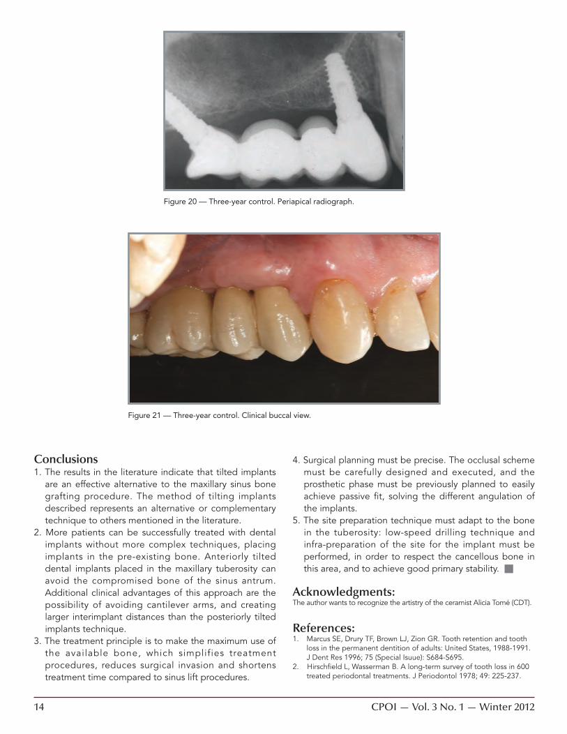
Figure 20 — Three-year control. Periapical radiograph.
CPOI_V3N1_Winter 2012:Spectrum 2/24/2012 9:28 AM Page 14
16 CPOI — Vol. 3 No. 1 — Winter 2012
3. Calandriello R, Tomatis M. Simplified treatment of the atrophicposterior maxilla via immediate/early function and tilted implants:A prospective 1-year clinical study. Clin Implant Dent Related Res2005; 7 (Suppl 1): S1-S12.
4. Haraldson T, Carlsson GE, Ingervall B. Functional state, bite forceand postural muscle activity in patients with osseointegrated oralimplant bridges. Acta Odontol Scand 1979; 37: 197-206.
5. Jaffin RA, Berman CL. The excessive loss of Branemark fixtures intype IV bone: a 5-year analysis. J Periodontol 1991; 62: 2-4.
6. Maló P, Nobre M, Lopez A. Short implants in posterior jaws. Aprospective 1-year study. Eur J Oral Implantol 2011; 4: 47-53.
7. Wood MR, Vermilyea SG. A review of selected dental literature onevidence-based treatment planning for dental implants: Report ofthe Committee of the Academy of Fixed Prosthodontics. J ProsthetDent 2004; 92: 447-462.
8. Degidi M. Immediately loaded short implants: Analysis of a caseseries of 133 implants. Quintessence Int 2007; 38: 193-201.
9. Shackleton JL, Carr L, Slabbert JC, Becker PJ. Survival of fixedimplant-supported prostheses related to cantilever lengths. JProsthet Dent 1994; 71: 23-26.
11. Zampellis A, Rangert B, Heijl L. Tilting of splinted implants forimproved prosthodontic support: A two-dimensional finite elementanalysis. J Prosthet Dent 2007; 97: 35-43.
12. Antonaya-Mira R, Barona-Dorado C, Martínez-Rodríguez N, Cáceres-Madroño E, Martínez-González JM. Meta-analysis of the increase inheight in maxillary sinus elevations with osteotome. Med Oral PatolOral Cir Bucal 2011; 17: 46-52.
13. Stern A, Green J. Sinus lift procedures: an overview of currenttechniques. Dent Clin North Am 2012; 56: 219-233.
14. Matsson T, Köndell PA, Gynther GW, Fredholm U, Bolin A. Implanttreatment without bone grafting in severely resorbed edentulousmaxillae. J Oral Maxillofac Surg 1999; 57: 281-287.
15. Krekmanov L, Kahn M, Rangert B, Lindström H. Tilting of posteriormandibular and maxillary implants for improved prosthesis support.Int J Oral Maxillofac Implants 2000; 15: 405-414.
16. Bevilacqua M, Tealdo T, Menini M, Pera F, Mossolov A, Drago C,Pera P. The influence of cantilever length and implant inclination onstress distribution in maxillary implant-supported fixed dentures. JProsthet Dent 2010; 105: 5-13.
17. Balshi TJ, Wolfinger GJ, Balshi SF. Analysis of 356 pterygo-maxillaryimplants in edentulous arches for fixed prostheses anchorage. Int JOral Maxillofac Implants 1999; 14: 398-406.
18. Aparicio C, Arévalo X, Ouzzani W, Granados C. A retrospectiveclinical and radiographic evaluation of tilted implants used in thetreatment of the severely resorbed edentulous maxilla. ApplOsseointegration Res 2002; 3: 17-21.
19. Valerón JF, Valerón PF. Long-term results in placement of screw-typeimplants in the pteryomaxillary-pyramidal region. Int J OralMaxillofac Implants 2007; 22: 195-200.
20. Balshi TJ, Lee HY, Hernandez R. The use of pterygomaxillaryimplants in the partially edentulous patient. A preliminary report. IntJ Oral Maxillofac Implants 1995; 10: 89–98.
21. Tulasne JF. Osseointegrated fixtures in the pterygoid region. In:Worthington P, Brånemark P-I (eds). Advanced OsseointegrationSurgery. Applications in the Maxillofacial Region. Chicago:Quintessence, 1992: 182–188.
22. Graves SL. The pterygoid plate implant: A solution for restoring theposterior maxilla. Int J Periodontics Restorative Dent 1994; 14:512–523.
23. Bahat O. Osseointegrated implants in the maxillary tuberosity:Report on 45 consecutive patients. Int J Oral Maxillofac Implants1992; 7: 459-467.
24. Venturelli A. A modified surgical protocol for placing implants in themaxillary tuberosity: Clinical results at 36 months after loading withfixed partial dentures. Int J Oral Maxillofac Implants 1996; 11:743-749.
25. Anitua E, Andía I, Carda C. Un nuevo protocolo para el fresado,colocación de implantes y obtención de hueso autólogo. DentalDialogue 2004; 4: 3-11.
26. Iglesia MA, Moreno J. A method aiming at achieving passive fit inimplant prostheses. Case report. Int J Prosthodont 2001; 14:570-574.
27. Iglesia MA. Custom-made laser-welded titanium implant prostheticabutment. J Prosthet Dent 2005; 94: 401-403.
About the AuthorMiguel A. Iglesia, DDS, MS, PhD has a full-time private practice inZaragoza, Spain. He received his dental training at Basque CountryUniversity, Bilbao, Spain, as well as his postgraduate implant training andhis phD. He has lectured on the topics of Implant surgery andProsthetics. Dr Iglesia has contributed to dental literature with multiplescientific articles in refereed journals about implantology, prosthetics,titanium, laser welding and esthetics, and has over 30 published articlesand 4 textbook chapters. In 2001 he received the Annual Award of theSpanish Society of Prosthodontics to the best original research study withan investigation about fatigue of laser welded titanium frameworks forimplant prosthesis. Dr Iglesia serves on the Scientific Council of theAragón Dental Association, and he is privileged to be in the editorialboard and in the peer-review team of some international dentalpublications. He can be contacted at [email protected].
CPOI_V3N1_Winter 2012:Spectrum 2/24/2012 9:28 AM Page 16