Embed Size (px)
Citation preview
Received on: 21-12-2014 Accepted on: 10-01-2015 Published on: 21-01-2015
B. Jayanthi Department of Chemistry, A.M.Jain College, Meenambakkam, Chennai - 114, Tamilnadu, India.
QR Code for Mobile users
DOI: 10.15272/ajbps.v4i40.654
Anti cancer activity of silver nano particles bio-
synthesized using stingless bee propolis (Tetragonula iridipennis) of Tamilnadu
S.Kothai a, B. Jayanthi b*
aDepartment of Chemistry, Ethiraj College for Women (Autonomous), Chennai – 8, Tamilnadu, India.
bDepartment of Chemistry, A.M.Jain College, Meenambakkam, Chennai - 114, Tamilnadu, India.
Abstract Global efforts to reduce hazardous wastes and alarming environmental issues urge the need to develop more and more eco-friendly processes and bio-compatible products. In this study, silver nano particles were synthesized using stingless bee propolis (Tetragonula iridipennis) reared from Tamilnadu. The bio-reduction process was further intensified by ultrasonics irradiation. The synthesized silver nano particles were well characterized by UV-visible spectrophotometer, Energy dispersive X-ray analysis, Scanning electron Microscope, Dynamic light scattering measurement. The active functional groups were identified by Fourier Transform Infra-red spectroscopy. The crystalline nature was established by XRD technique. The anticancer potential of the synthesized silver nano particles was evaluated by MTT assay and it was found to show significant activity against A549 human lung cancer cells. IC 50 value is 38 µg/ml. Keywords: Stingless bee propolis, silver nano particles, process intensification, ultrasonication, A549 cancer cells, anticancer activity.
Cite this article as:
S.Kothai and B. Jayanthi. Anti cancer activity of silver nano particles bio- synthesized using stingless bee propolis (Tetragonula iridipennis) of Tamilnadu. Asian Journal of Biomedical and Pharmaceutical Sciences; 04 (40); 2014, 30-37.
S.Kothai and B. Jayanthi.: Asian Journal of Biomedical and Pharmaceutical Sciences; 4(40) 2014, 30-37.
© Asian Journal of Biomedical and Pharmaceutical Sciences, all rights reserved. Volume 4, Issue 40, 2014. 31
INTRODUCTION Nano biotechnology is a promising field of nano science, which extends the horizon of nano sized systems for various newer applications both in the field of biotechnology as well as in the field of Nano medicine. Nano particles exhibit distinct physical and chemical properties compared to their bulk counterparts due to their small size and large surface area. Metal nano particles are found to be potential therapeutic alternatives for the treatment of various diseases including cancer (1). Cancer - a life threatening disease has nowadays become a common disease due to the vast changes in the life style of people and its accounts for second major cause of mortality rate in the world(2). Existing anti-cancer treatments including chemotherapeutic drugs, radiations, surgery are serving their purpose to some extent, but their side effects are much more than it, targeted treatment of disease(3). Nano particles enabled targeted drug delivery(4) is found to be efficient for cancer treatment as nano particulate can cross some of the biological barriers and achieve therapeutic concentration in tumours even with less dosage of drug administration and spares the surrounding normal tissues from toxic effect. So, a separate branch of Nanoscience, Nano Oncology has emerged, which deals with the ways and means of developing potential alternatives to manage the dreadful disease. Among all the metal nano particles, silver nano particles have always attracted researchers due to its widespread applications in various fields(5) such as catalysis, sensors, food industries, agriculture, textile industries and more particularly in the field of biomedical applications(6) as antimicrobial(7), antifungal(8),antiviral(9) and anti-angiogenic agent(10) and now emerging as a potential therapeutic agent for cancer treatment(11-17). Though various conventional Physical and chemical methods(18-22) are used for the synthesis of silver nano particles, they are always associated with one or more limitations(23,24) such as defective surface formation, low production rate, high cost of manufacturing, large energy requirement, formation of hazardous byproducts etc,. Limitation of these methods has made the recent research to focus on the development of clean, eco-friendly and cost effective synthesis protocols. Apart from the above said advantages, use of Biogenic method leads to the production of silver nano particles coated with a lipid layer that gives physiological solubility and stability which is critical for any biomedical applications (25) Various natural resources such as plants (26-28) insect origin (29), bacteria
(30), algae (31) fungi (32) were reported for the synthesis of silver nano particles.
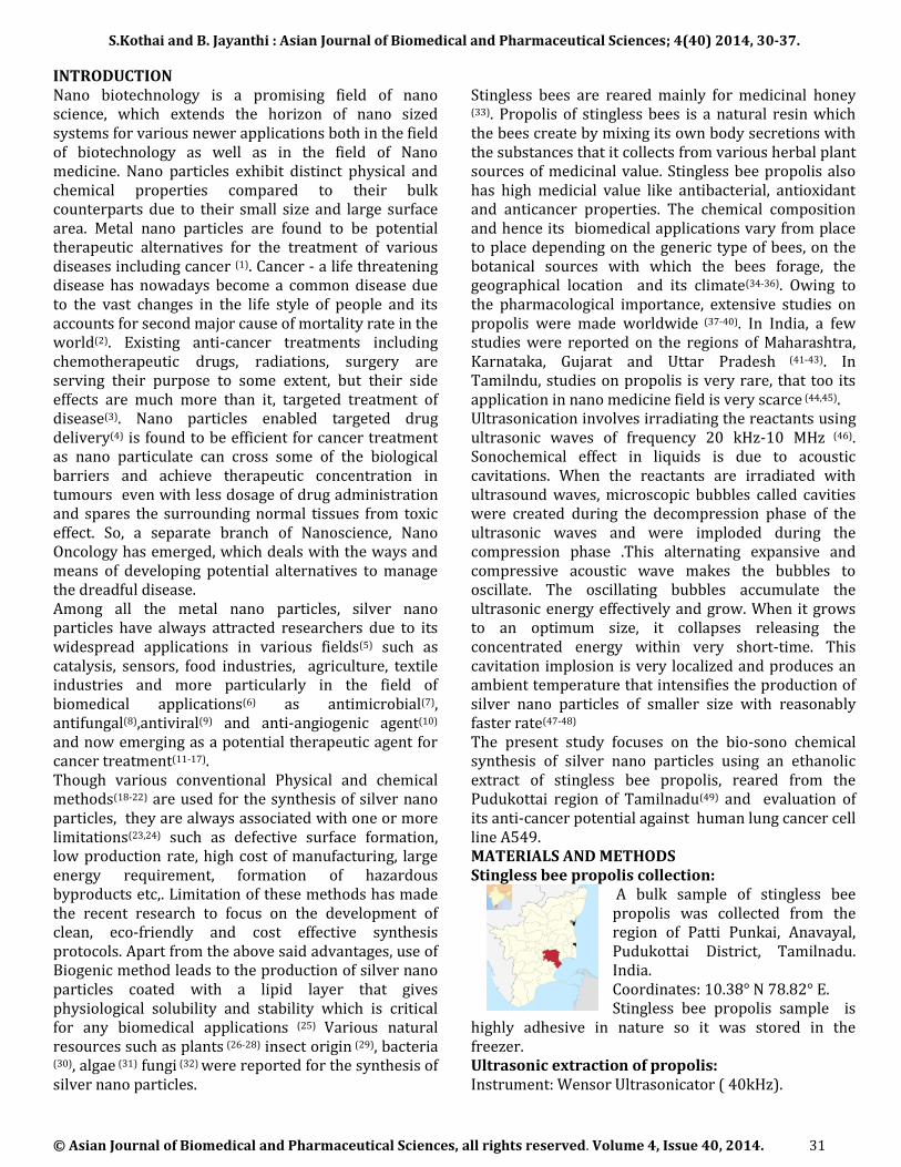
Stingless bees are reared mainly for medicinal honey
(33). Propolis of stingless bees is a natural resin which the bees create by mixing its own body secretions with the substances that it collects from various herbal plant sources of medicinal value. Stingless bee propolis also has high medicial value like antibacterial, antioxidant and anticancer properties. The chemical composition and hence its biomedical applications vary from place to place depending on the generic type of bees, on the botanical sources with which the bees forage, the geographical location and its climate(34-36). Owing to the pharmacological importance, extensive studies on propolis were made worldwide (37-40). In India, a few studies were reported on the regions of Maharashtra, Karnataka, Gujarat and Uttar Pradesh (41-43). In Tamilndu, studies on propolis is very rare, that too its application in nano medicine field is very scarce (44,45). Ultrasonication involves irradiating the reactants using ultrasonic waves of frequency 20 kHz-10 MHz (46). Sonochemical effect in liquids is due to acoustic cavitations. When the reactants are irradiated with ultrasound waves, microscopic bubbles called cavities were created during the decompression phase of the ultrasonic waves and were imploded during the compression phase .This alternating expansive and compressive acoustic wave makes the bubbles to oscillate. The oscillating bubbles accumulate the ultrasonic energy effectively and grow. When it grows to an optimum size, it collapses releasing the concentrated energy within very short-time. This cavitation implosion is very localized and produces an ambient temperature that intensifies the production of silver nano particles of smaller size with reasonably faster rate(47-48) The present study focuses on the bio-sono chemical synthesis of silver nano particles using an ethanolic extract of stingless bee propolis, reared from the Pudukottai region of Tamilnadu(49) and evaluation of its anti-cancer potential against human lung cancer cell line A549. MATERIALS AND METHODS Stingless bee propolis collection:
A bulk sample of stingless bee propolis was collected from the region of Patti Punkai, Anavayal, Pudukottai District, Tamilnadu. India. Coordinates: 10.38° N 78.82° E. Stingless bee propolis sample is
highly adhesive in nature so it was stored in the freezer. Ultrasonic extraction of propolis: Instrument: Wensor Ultrasonicator ( 40kHz).
S.Kothai and B. Jayanthi.: Asian Journal of Biomedical and Pharmaceutical Sciences; 4(40) 2014, 30-37.
© Asian Journal of Biomedical and Pharmaceutical Sciences, all rights reserved. Volume 4, Issue 40, 2014. 32
20g of propolis was cut into small pieces and grounded well. To this 200 ml of a solvent mixture containing 140ml of ethanol and 60ml of distilled water in the ratio (7:3) was added and subjected to ultrasonication for about an hour. This is then filtered through Whatman 41 filter paper. Materials: Silver Nitrate, Sodium hydroxide, Minimum essential medium (MEM), Fetal bovine serum (FBS), Trypsin, 3-(4,5-dimethyl-2-thiazolyl)-2,5-diphenyl--tetrazolium bromide (MTT), and Dimethyl sulfoxide (DMSO) were purchased from Hi media & Sigma Aldrich. Characterization of Silver nano particles: UV-Visible Spectral Analysis: The initial characterization of the synthesized silver nano particles was carried out using Shimadzu Dual Beam (UV-1650PC) spectrophotometer of resolution 1nm. To avoid errors due to high optical density, the solution (0.5 ml) was diluted ten times with double distilled water. Fourier Transform Infrared Spectral Analysis (FT-IR): The bio reduced solution was centrifuged at 10,000 rpm for 20 minutes, twice. The sample was grinded with KBr, dried in infra-red light and the spectra were recorded in the spectrum range of 4000- 400 cm-1.using Shimadzu FT-IR Spectrophotometer. Energy Dispersive X-Ray Analysis (EDAX): The elemental composition of the synthesized silver nano solution was determined by using Philips XL-30 Energy Dispersive Analysis X-ray spectrometer. SEM Analysis: The sample was drop coated on a tiny piece of glass substrate, dried at 40°C and then subjected to SEM analysis using Teftan Vega 3-SBU instrument. X-ray diffraction: The synthesized silver nano particles were drop coated on a 1cm2 glass piece, dried and its crystalline nature was examined on Rigaku Mini Flex diffractometer with Cu Kα radiation. Dynamic Light Scattering Analysis: The average particle size distribution (PSD) of the synthesized silver nanoparticles, poly dispersity index (PI) were determined using Dynamic Light Scattering Instrument- Malbern Vetasizer Nano-S-Series. In vitro anticancer studies of synthesized Silver nano particles: Cell Culture: A549 cell lines were obtained from National Centre for cell sciences (NCCS) Pune, India. The cells were maintained in Minimal Essential Media supplemented with 10% FBS, penicillin (100 U/ml) and streptomycin (100 μg/ml) in a humidified atmosphere of 50 μg/ml CO2 at 37 °C.
Cell Viability: The anticancer activity of the synthesized silver nano particles on A549 human cancer cells was determined by the MTT assay (50). Cultured A549 human lung cancer cells (1 × 10⁵ cells)) were plated in 0.2 ml of the medium in 96 flat bottomed well plates. Incubated at 5 % CO2 atmosphere for 72 hours. Then, various concentrations of the synthesized silver nano particles in 0.1% DMSO were added to the cells and maintained at 5% CO2 incubator for 24hrs. After incubation and washing with phosphate-buffered saline (pH 7.4), 20µl of 0.5% 3-(4, 5-dimethyl-2-thiazolyl)-2, 5-diphenyl--tetrazolium bromide (MTT) in phosphate- buffered saline solution was added to the incubated cells and further incubated for four more hours at 37°C and at 5% CO2 atmosphere. After 4hrs of incubation, 1ml of DMSO was added. Viable cells at various concentrations (dilution method) were determined by measuring the absorbance at 540nm. The concentration required for 50% inhibition (IC50) was determined graphically. The effect of the synthesized silver nano particles on the proliferation of A549 cells was expressed as the % cell viability. % cell viability = A549 of treated cells / A549 of control cells × 100%
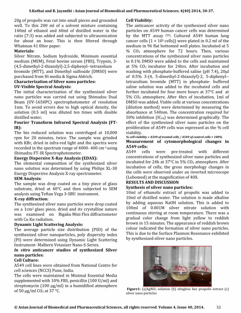
Measurement of cytomorphological changes in A549 cells: A549 cells were pre-treated with different concentrations of synthesized silver nano particles and incubated for 24h at 37°C in 5% CO2 atmosphere. After incubation of cells, the gross morphology changes in the cells were observed under an inverted microscope (Lobomed) at the magnification of 40X. RESULTS AND DISCUSSION Synthesis of silver nano particles: 10ml of ethanolic extract of propolis was added to 10ml of distilled water. The solution is made alkaline by adding aqueous NaOH solution. This is added to 100ml of 0.001M silver nitrate solution with continuous stirring at room temperature. There was a gradual color change from light yellow to reddish brown in 15 minutes. The appearance of reddish brown colour indicated the formation of silver nano particles. This is due to the Surface Plasmon Resonance exhibited by synthesized silver nano particles.
Figure1: (a)AgNO3 solution (b) stingless bee propolis extract (c) silver nano particles
S.Kothai and B. Jayanthi.: Asian Journal of Biomedical and Pharmaceutical Sciences; 4(40) 2014, 30-37.
© Asian Journal of Biomedical and Pharmaceutical Sciences, all rights reserved. Volume 4, Issue 40, 2014. 33
When the same amount of the precursor and the stingless bee propolis extract were irradiated with ultrasonic waves, it has taken only five minutes of time for the appearance of reddish brown color. This may be due to the fact that in addition to the active reducing groups present in the extract, some of the free radicals like H. and OH. generated during sonolysis of the solution, also involved in the bio- reduction of Ag+ ions to Ag(0) (48) resulting in the quick formation of silver nano particles. Thus Ultrasonication- a process intensification technique plays a major role in the formation of silver nano particles. The possible mechanism is
Table 1: Sono Chemical Reduction Figure 2 : Polyphenols giving e-- to Ag+
Characterization of Biosynthesized silver nano particles by spectral methods: UV-VIS spectral analysis:
Figure 3a: UV-VIS Spectrum of stingless bee propolis Figure.3b: UV-VIS Spectrum of AgNPs
Figure. 3a and 3b show the UV-VIS spectra of ethanolic extract of stingless bee propolis and silver nano particles synthesized using it. An intense absorption peak at 265nm in Fig.3a indicates the presence of polyphenols and flavonoids in the raw propolis sample
(49). A strong absorption peak in Figure 3b, around 413nm is characteristic of silver nano particles and is due to the Surface Plasmon Resonance (SPR) exhibited by the synthesized silver nano particles. Surface plasmon resonance is due to the collective oscillations of the free electrons of silver nano particles in resonance with the light waves. Availability of free electrons could be possible only if Ag+ ions were reduced to elemental silver nano particles. This shows
the efficiency of Stingless bee propolis in reducing Ag+ ions to Ag (0). Fourier Transform-Infra red Spectroscopic Analysis:
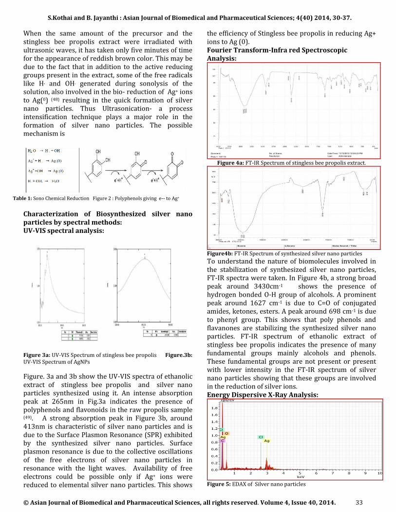
Figure 4a: FT-IR Spectrum of stingless bee propolis extract.
Figure4b: FT-IR Spectrum of synthesized silver nano particles
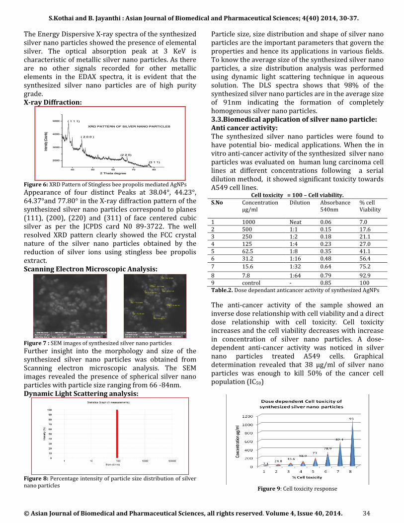
To understand the nature of biomolecules involved in the stabilization of synthesized silver nano particles, FT-IR spectra were taken. In Figure 4b, a strong broad peak around 3430cm-1 shows the presence of hydrogen bonded O-H group of alcohols. A prominent peak around 1627 cm-1 is due to C=O of conjugated amides, ketones, esters. A peak around 698 cm-1 is due to phenyl group. This shows that poly phenols and flavanones are stabilizing the synthesized silver nano particles. FT-IR spectrum of ethanolic extract of stingless bee propolis indicates the presence of many fundamental groups mainly alcohols and phenols. These fundamental groups are not present or present with lower intensity in the FT-IR spectrum of silver nano particles showing that these groups are involved in the reduction of silver ions. Energy Dispersive X-Ray Analysis:
Figure 5: EDAX of Silver nano particles
S.Kothai and B. Jayanthi.: Asian Journal of Biomedical and Pharmaceutical Sciences; 4(40) 2014, 30-37.
© Asian Journal of Biomedical and Pharmaceutical Sciences, all rights reserved. Volume 4, Issue 40, 2014. 34
The Energy Dispersive X-ray spectra of the synthesized silver nano particles showed the presence of elemental silver. The optical absorption peak at 3 KeV is characteristic of metallic silver nano particles. As there are no other signals recorded for other metallic elements in the EDAX spectra, it is evident that the synthesized silver nano particles are of high purity grade. X-ray Diffraction:
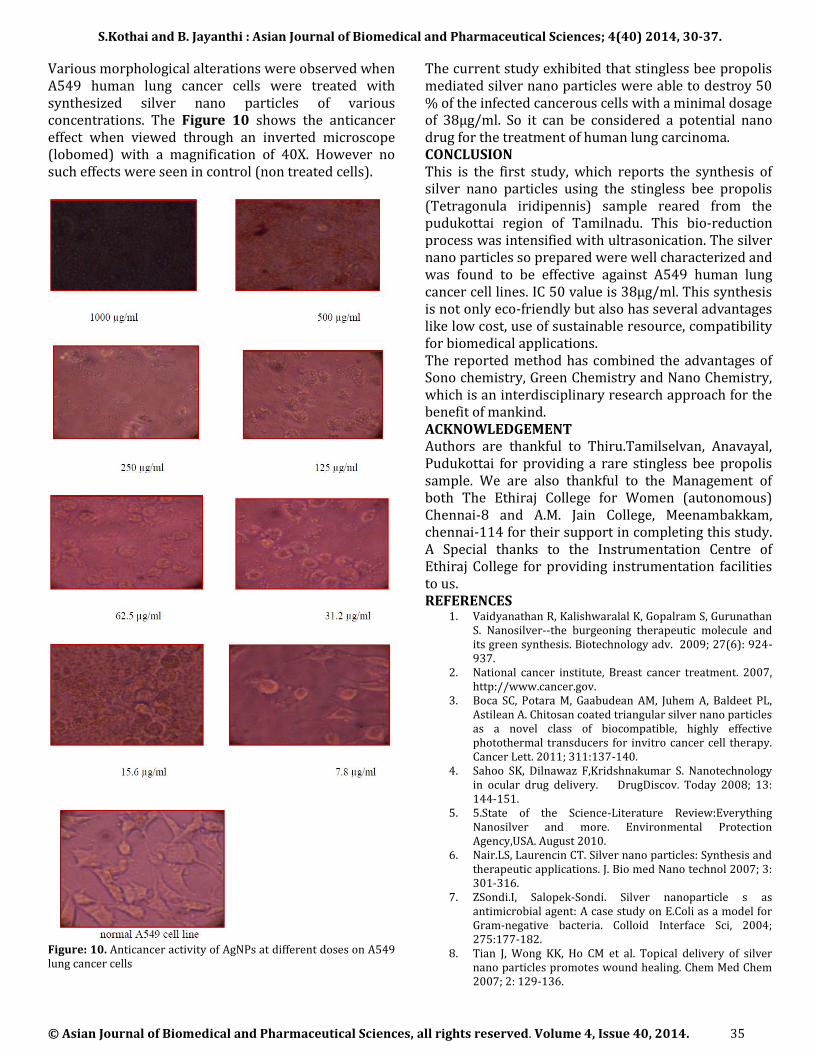
Figure 6: XRD Pattern of Stingless bee propolis mediated AgNPs
Appearance of four distinct Peaks at 38.04°, 44.23°, 64.37°and 77.80° in the X-ray diffraction pattern of the synthesized silver nano particles correspond to planes (111), (200), (220) and (311) of face centered cubic silver as per the JCPDS card N0 89-3722. The well resolved XRD pattern clearly showed the FCC crystal nature of the silver nano particles obtained by the reduction of silver ions using stingless bee propolis extract. Scanning Electron Microscopic Analysis:
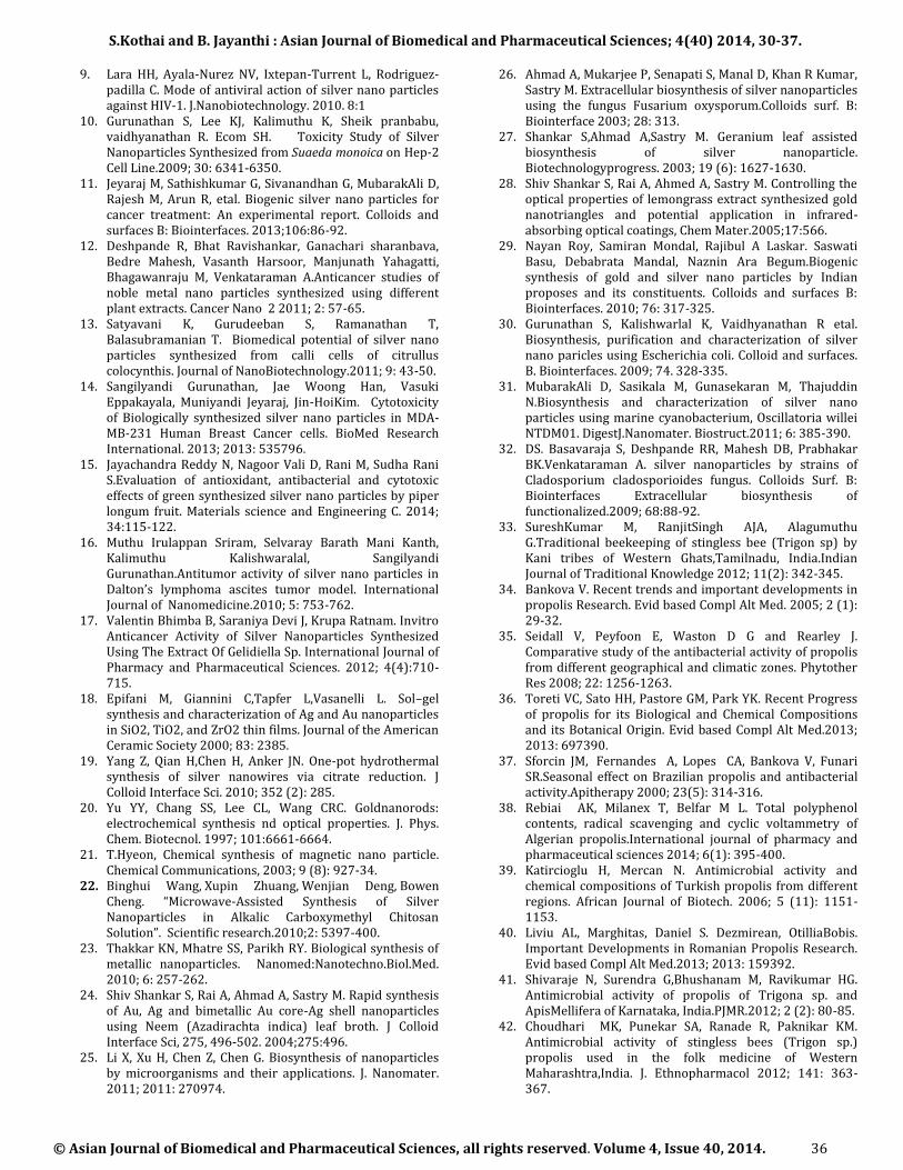
Figure 7 : SEM images of synthesized silver nano particles
Further insight into the morphology and size of the synthesized silver nano particles was obtained from Scanning electron microscopic analysis. The SEM images revealed the presence of spherical silver nano particles with particle size ranging from 66 -84nm. Dynamic Light Scattering analysis:
Figure 8: Percentage intensity of particle size distribution of silver nano particles
Particle size, size distribution and shape of silver nano particles are the important parameters that govern the properties and hence its applications in various fields. To know the average size of the synthesized silver nano particles, a size distribution analysis was performed using dynamic light scattering technique in aqueous solution. The DLS spectra shows that 98% of the synthesized silver nano particles are in the average size of 91nm indicating the formation of completely homogenous silver nano particles. 3.3.Biomedical application of silver nano particle: Anti cancer activity: The synthesized silver nano particles were found to have potential bio- medical applications. When the in vitro anti-cancer activity of the synthesized silver nano particles was evaluated on human lung carcinoma cell lines at different concentrations following a serial dilution method, it showed significant toxicity towards A549 cell lines.
Cell toxicity = 100 – Cell viability. S.No Concentration
µg/ml Dilution Absorbance
540nm % cell Viability
1 1000 Neat 0.06 7.0 2 500 1:1 0.15 17.6 3 250 1:2 0.18 21.1 4 125 1:4 0.23 27.0 5 62.5 1:8 0.35 41.1 6 31.2 1:16 0.48 56.4
7 15.6 1:32 0.64 75.2
8 7.8 1:64 0.79 92.9 9 control - 0.85 100 Table.2. Dose dependant anticancer activity of synthesized AgNPs
The anti-cancer activity of the sample showed an inverse dose relationship with cell viability and a direct dose relationship with cell toxicity. Cell toxicity increases and the cell viability decreases with increase in concentration of silver nano particles. A dose-dependent anti-cancer activity was noticed in silver nano particles treated A549 cells. Graphical determination revealed that 38 µg/ml of silver nano particles was enough to kill 50% of the cancer cell population (IC50)
Figure 9: Cell toxicity response
S.Kothai and B. Jayanthi.: Asian Journal of Biomedical and Pharmaceutical Sciences; 4(40) 2014, 30-37.
© Asian Journal of Biomedical and Pharmaceutical Sciences, all rights reserved. Volume 4, Issue 40, 2014. 35
Various morphological alterations were observed when A549 human lung cancer cells were treated with synthesized silver nano particles of various concentrations. The Figure 10 shows the anticancer effect when viewed through an inverted microscope (lobomed) with a magnification of 40X. However no such effects were seen in control (non treated cells).
Figure: 10. Anticancer activity of AgNPs at different doses on A549 lung cancer cells
The current study exhibited that stingless bee propolis mediated silver nano particles were able to destroy 50 % of the infected cancerous cells with a minimal dosage of 38µg/ml. So it can be considered a potential nano drug for the treatment of human lung carcinoma. CONCLUSION This is the first study, which reports the synthesis of silver nano particles using the stingless bee propolis (Tetragonula iridipennis) sample reared from the pudukottai region of Tamilnadu. This bio-reduction process was intensified with ultrasonication. The silver nano particles so prepared were well characterized and was found to be effective against A549 human lung cancer cell lines. IC 50 value is 38µg/ml. This synthesis is not only eco-friendly but also has several advantages like low cost, use of sustainable resource, compatibility for biomedical applications. The reported method has combined the advantages of Sono chemistry, Green Chemistry and Nano Chemistry, which is an interdisciplinary research approach for the benefit of mankind. ACKNOWLEDGEMENT Authors are thankful to Thiru.Tamilselvan, Anavayal, Pudukottai for providing a rare stingless bee propolis sample. We are also thankful to the Management of both The Ethiraj College for Women (autonomous) Chennai-8 and A.M. Jain College, Meenambakkam, chennai-114 for their support in completing this study. A Special thanks to the Instrumentation Centre of Ethiraj College for providing instrumentation facilities to us. REFERENCES
1. Vaidyanathan R, Kalishwaralal K, Gopalram S, Gurunathan S. Nanosilver--the burgeoning therapeutic molecule and its green synthesis. Biotechnology adv. 2009; 27(6): 924-937.
2. National cancer institute, Breast cancer treatment. 2007, http://www.cancer.gov.
3. Boca SC, Potara M, Gaabudean AM, Juhem A, Baldeet PL, Astilean A. Chitosan coated triangular silver nano particles as a novel class of biocompatible, highly effective photothermal transducers for invitro cancer cell therapy. Cancer Lett. 2011; 311:137-140.
4. Sahoo SK, Dilnawaz F,Kridshnakumar S. Nanotechnology in ocular drug delivery. DrugDiscov. Today 2008; 13: 144-151.
5. 5.State of the Science-Literature Review:Everything Nanosilver and more. Environmental Protection Agency,USA. August 2010.
6. Nair.LS, Laurencin CT. Silver nano particles: Synthesis and therapeutic applications. J. Bio med Nano technol 2007; 3: 301-316.
7. ZSondi.I, Salopek-Sondi. Silver nanoparticle s as antimicrobial agent: A case study on E.Coli as a model for Gram-negative bacteria. Colloid Interface Sci, 2004; 275:177-182.
8. Tian J, Wong KK, Ho CM et al. Topical delivery of silver nano particles promotes wound healing. Chem Med Chem 2007; 2: 129-136.
S.Kothai and B. Jayanthi.: Asian Journal of Biomedical and Pharmaceutical Sciences; 4(40) 2014, 30-37.
© Asian Journal of Biomedical and Pharmaceutical Sciences, all rights reserved. Volume 4, Issue 40, 2014. 36
9. Lara HH, Ayala-Nurez NV, Ixtepan-Turrent L, Rodriguez-padilla C. Mode of antiviral action of silver nano particles against HIV-1. J.Nanobiotechnology. 2010. 8:1
10. Gurunathan S, Lee KJ, Kalimuthu K, Sheik pranbabu, vaidhyanathan R. Ecom SH. Toxicity Study of Silver Nanoparticles Synthesized from Suaeda monoica on Hep-2 Cell Line.2009; 30: 6341-6350.
11. Jeyaraj M, Sathishkumar G, Sivanandhan G, MubarakAli D, Rajesh M, Arun R, etal. Biogenic silver nano particles for cancer treatment: An experimental report. Colloids and surfaces B: Biointerfaces. 2013;106:86-92.
12. Deshpande R, Bhat Ravishankar, Ganachari sharanbava, Bedre Mahesh, Vasanth Harsoor, Manjunath Yahagatti, Bhagawanraju M, Venkataraman A.Anticancer studies of noble metal nano particles synthesized using different plant extracts. Cancer Nano 2 2011; 2: 57-65.
13. Satyavani K, Gurudeeban S, Ramanathan T, Balasubramanian T. Biomedical potential of silver nano particles synthesized from calli cells of citrullus colocynthis. Journal of NanoBiotechnology.2011; 9: 43-50.
14. Sangilyandi Gurunathan, Jae Woong Han, Vasuki Eppakayala, Muniyandi Jeyaraj, Jin-HoiKim. Cytotoxicity of Biologically synthesized silver nano particles in MDA-MB-231 Human Breast Cancer cells. BioMed Research International. 2013; 2013: 535796.
15. Jayachandra Reddy N, Nagoor Vali D, Rani M, Sudha Rani S.Evaluation of antioxidant, antibacterial and cytotoxic effects of green synthesized silver nano particles by piper longum fruit. Materials science and Engineering C. 2014; 34:115-122.
16. Muthu Irulappan Sriram, Selvaray Barath Mani Kanth, Kalimuthu Kalishwaralal, Sangilyandi Gurunathan.Antitumor activity of silver nano particles in Dalton’s lymphoma ascites tumor model. International Journal of Nanomedicine.2010; 5: 753-762.
17. Valentin Bhimba B, Saraniya Devi J, Krupa Ratnam. Invitro Anticancer Activity of Silver Nanoparticles Synthesized Using The Extract Of Gelidiella Sp. International Journal of Pharmacy and Pharmaceutical Sciences. 2012; 4(4):710-715.
18. Epifani M, Giannini C,Tapfer L,Vasanelli L. Sol–gel synthesis and characterization of Ag and Au nanoparticles in SiO2, TiO2, and ZrO2 thin films. Journal of the American Ceramic Society 2000; 83: 2385.
19. Yang Z, Qian H,Chen H, Anker JN. One-pot hydrothermal synthesis of silver nanowires via citrate reduction. J Colloid Interface Sci. 2010; 352 (2): 285.
20. Yu YY, Chang SS, Lee CL, Wang CRC. Goldnanorods: electrochemical synthesis nd optical properties. J. Phys. Chem. Biotecnol. 1997; 101:6661-6664.
21. T.Hyeon, Chemical synthesis of magnetic nano particle. Chemical Communications, 2003; 9 (8): 927-34.
22. Binghui Wang, Xupin Zhuang, Wenjian Deng, Bowen Cheng. “Microwave-Assisted Synthesis of Silver Nanoparticles in Alkalic Carboxymethyl Chitosan Solution”. Scientific research.2010;2: 5397-400.
23. Thakkar KN, Mhatre SS, Parikh RY. Biological synthesis of metallic nanoparticles. Nanomed:Nanotechno.Biol.Med. 2010; 6: 257-262.
24. Shiv Shankar S, Rai A, Ahmad A, Sastry M. Rapid synthesis of Au, Ag and bimetallic Au core-Ag shell nanoparticles using Neem (Azadirachta indica) leaf broth. J Colloid Interface Sci, 275, 496-502. 2004;275:496.
25. Li X, Xu H, Chen Z, Chen G. Biosynthesis of nanoparticles by microorganisms and their applications. J. Nanomater. 2011; 2011: 270974.
26. Ahmad A, Mukarjee P, Senapati S, Manal D, Khan R Kumar, Sastry M. Extracellular biosynthesis of silver nanoparticles using the fungus Fusarium oxysporum.Colloids surf. B: Biointerface 2003; 28: 313.
27. Shankar S,Ahmad A,Sastry M. Geranium leaf assisted biosynthesis of silver nanoparticle. Biotechnologyprogress. 2003; 19 (6): 1627-1630.
28. Shiv Shankar S, Rai A, Ahmed A, Sastry M. Controlling the optical properties of lemongrass extract synthesized gold nanotriangles and potential application in infrared-absorbing optical coatings, Chem Mater.2005;17:566.
29. Nayan Roy, Samiran Mondal, Rajibul A Laskar. Saswati Basu, Debabrata Mandal, Naznin Ara Begum.Biogenic synthesis of gold and silver nano particles by Indian proposes and its constituents. Colloids and surfaces B: Biointerfaces. 2010; 76: 317-325.
30. Gurunathan S, Kalishwarlal K, Vaidhyanathan R etal. Biosynthesis, purification and characterization of silver nano paricles using Escherichia coli. Colloid and surfaces. B. Biointerfaces. 2009; 74. 328-335.
31. MubarakAli D, Sasikala M, Gunasekaran M, Thajuddin N.Biosynthesis and characterization of silver nano particles using marine cyanobacterium, Oscillatoria willei NTDM01. DigestJ.Nanomater. Biostruct.2011; 6: 385-390.
32. DS. Basavaraja S, Deshpande RR, Mahesh DB, Prabhakar BK.Venkataraman A. silver nanoparticles by strains of Cladosporium cladosporioides fungus. Colloids Surf. B: Biointerfaces Extracellular biosynthesis of functionalized.2009; 68:88-92.
33. SureshKumar M, RanjitSingh AJA, Alagumuthu G.Traditional beekeeping of stingless bee (Trigon sp) by Kani tribes of Western Ghats,Tamilnadu, India.Indian Journal of Traditional Knowledge 2012; 11(2): 342-345.
34. Bankova V. Recent trends and important developments in propolis Research. Evid based Compl Alt Med. 2005; 2 (1): 29-32.
35. Seidall V, Peyfoon E, Waston D G and Rearley J. Comparative study of the antibacterial activity of propolis from different geographical and climatic zones. Phytother Res 2008; 22: 1256-1263.
36. Toreti VC, Sato HH, Pastore GM, Park YK. Recent Progress of propolis for its Biological and Chemical Compositions and its Botanical Origin. Evid based Compl Alt Med.2013; 2013: 697390.
37. Sforcin JM, Fernandes A, Lopes CA, Bankova V, Funari SR.Seasonal effect on Brazilian propolis and antibacterial activity.Apitherapy 2000; 23(5): 314-316.
38. Rebiai AK, Milanex T, Belfar M L. Total polyphenol contents, radical scavenging and cyclic voltammetry of Algerian propolis.International journal of pharmacy and pharmaceutical sciences 2014; 6(1): 395-400.
39. Katircioglu H, Mercan N. Antimicrobial activity and chemical compositions of Turkish propolis from different regions. African Journal of Biotech. 2006; 5 (11): 1151-1153.
40. Liviu AL, Marghitas, Daniel S. Dezmirean, OtilliaBobis. Important Developments in Romanian Propolis Research. Evid based Compl Alt Med.2013; 2013: 159392.
41. Shivaraje N, Surendra G,Bhushanam M, Ravikumar HG. Antimicrobial activity of propolis of Trigona sp. and ApisMellifera of Karnataka, India.PJMR.2012; 2 (2): 80-85.
42. Choudhari MK, Punekar SA, Ranade R, Paknikar KM. Antimicrobial activity of stingless bees (Trigon sp.) propolis used in the folk medicine of Western Maharashtra,India. J. Ethnopharmacol 2012; 141: 363-367.

S.Kothai and B. Jayanthi.: Asian Journal of Biomedical and Pharmaceutical Sciences; 4(40) 2014, 30-37.
© Asian Journal of Biomedical and Pharmaceutical Sciences, all rights reserved. Volume 4, Issue 40, 2014. 37
43. Mathivanan V. Nabi Shah GH, Manzoor M, Mir GM, Selvi Sabhanayakam. A Review on propolis- As a novel Folkmedicine. Indian journal of science, 2013; 2 (30): 23-30.
44. Thirugnana sampandan R, Raveedran SB, Jayakumar.R. Analysis of chemical composition and bio active property of evaluation of indian propolis.Asian pac.J.trop.Biomed. 2012; 2 (8): 651-654.
45. Kumar N, Ahmad M, Dang R and Husain A. Antioxidant and antimicrobial activity of propolis from Tamil Nadu Zone. J.Med Plant Res 2008; 2(12): 361-364.
46. Vimal Kumar, Krishna Deo Prasad Nigam. Process intensification in green synthesis. Green process synth. 2012; 1:79-107.
47. A. Gendaken. Sonoochemistry and its application to nanochemistry. Current Science 2003; 85 (12):1720-1722.
48. Jin Ho Bang, Kenneth, Suslick S, Applications of Ultrasound to the synthesis of Nanostructured Materials. Advanced materials .2010; 22:1039-1059.
49. Kothai. S, Jayanthi B. Evaluation of Antioxidant and Antimicrobial Activity of Stingless bee propolis (Tetragonula iridipennis) of TamilNadu. International Journal of Pharmacy and Pharmaceutical Sciences. 2014; 6 (8): 81-85.
50. Mosmann T. Rapid colorimetric assay for cellular growth and survival: application to proliferation and cytotoxicity assays. J Immunol Methods.1983; 65: 55–63.