Embed Size (px)
Citation preview
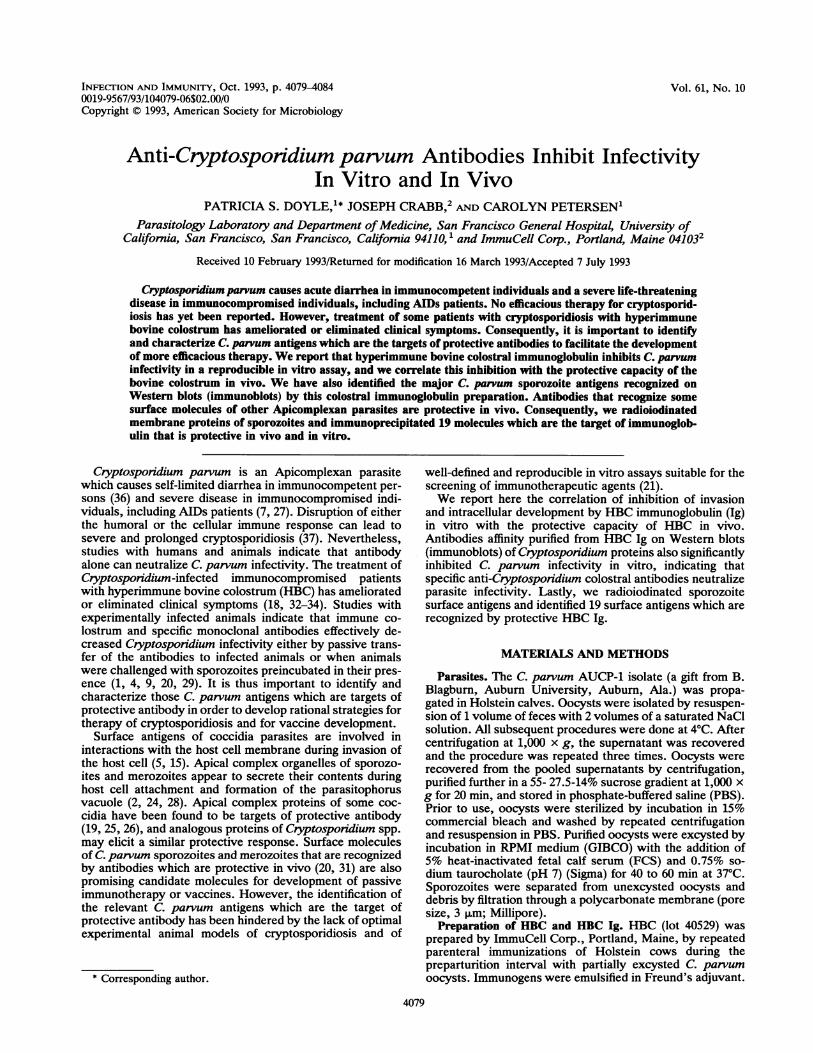
INFECrION AND IMMUNITY, OCt. 1993, p. 4079-40840019-9567/93/104079-06$02.00/0Copyright © 1993, American Society for Microbiology
Vol. 61, No. 10
Anti-Cryptosporidium parvum Antibodies Inhibit InfectivityIn Vitro and In Vivo
PATRICIA S. DOYLE,1* JOSEPH CRABB,2 AND CAROLYN PETERSEN'Parasitology Laboratory and Department ofMedicine, San Francisco General Hospital, University of
California, San Francisco, San Francisco, California 94110,1 and ImmuCell Corp., Portland, Maine 041032Received 10 February 1993/Returned for modification 16 March 1993/Accepted 7 July 1993
Cryptosporidium parvum causes acute diarrhea in immunocompetent individuals and a severe life-threateningdisease in immunocompromised individuals, including AIDs patients. No efficacious therapy for cryptosporid-iosis has yet been reported. However, treatment of some patients with cryptosporidiosis with hyperimmunebovine colostrum has ameliorated or eliminated clinical symptoms. Consequently, it is important to identifyand characterize C. parnum antigens which are the targets of protective antibodies to facilitate the developmentof more efficacious therapy. We report that hyperimmune bovine colostral immunoglobulin inhibits C. parvuminfectivity in a reproducible in vitro assay, and we correlate this inhibition with the protective capacity of thebovine colostrum in vivo. We have also identified the major C. parvum sporozoite antigens recognized onWestern blots (immunoblots) by this colostral immunoglobulin preparation. Antibodies that recognize somesurface molecules of other Apicomplexan parasites are protective in vivo. Consequently, we radioiodinatedmembrane proteins of sporozoites and immunoprecipitated 19 molecules which are the target of immunoglob-ulin that is protective in vivo and in vitro.
Cryptosporidium parvum is an Apicomplexan parasitewhich causes self-limited diarrhea in immunocompetent per-sons (36) and severe disease in immunocompromised indi-viduals, including AIDs patients (7, 27). Disruption of eitherthe humoral or the cellular immune response can lead tosevere and prolonged cryptosporidiosis (37). Nevertheless,studies with humans and animals indicate that antibodyalone can neutralize C. parvum infectivity. The treatment ofCryptosporidium-infected immunocompromised patientswith hyperimmune bovine colostrum (HBC) has amelioratedor eliminated clinical symptoms (18, 32-34). Studies withexperimentally infected animals indicate that immune co-lostrum and specific monoclonal antibodies effectively de-creased Cryptosporidium infectivity either by passive trans-fer of the antibodies to infected animals or when animalswere challenged with sporozoites preincubated in their pres-ence (1, 4, 9, 20, 29). It is thus important to identify andcharacterize those C. parvum antigens which are targets ofprotective antibody in order to develop rational strategies fortherapy of cryptosporidiosis and for vaccine development.
Surface antigens of coccidia parasites are involved ininteractions with the host cell membrane during invasion ofthe host cell (5, 15). Apical complex organelles of sporozo-ites and merozoites appear to secrete their contents duringhost cell attachment and formation of the parasitophorusvacuole (2, 24, 28). Apical complex proteins of some coc-cidia have been found to be targets of protective antibody(19, 25, 26), and analogous proteins of Cryptosponidium spp.may elicit a similar protective response. Surface moleculesof C. parvum sporozoites and merozoites that are recognizedby antibodies which are protective in vivo (20, 31) are alsopromising candidate molecules for development of passiveimmunotherapy or vaccines. However, the identification ofthe relevant C parvum antigens which are the target ofprotective antibody has been hindered by the lack of optimalexperimental animal models of cryptosporidiosis and of
* Corresponding author.
well-defined and reproducible in vitro assays suitable for thescreening of immunotherapeutic agents (21).We report here the correlation of inhibition of invasion
and intracellular development by HBC immunoglobulin (Ig)in vitro with the protective capacity of HBC in vivo.Antibodies affinity purified from HBC Ig on Western blots(immunoblots) of Cryptosporidium proteins also significantlyinhibited C. parvum infectivity in vitro, indicating thatspecific anti-Cryptosporidium colostral antibodies neutralizeparasite infectivity. Lastly, we radioiodinated sporozoitesurface antigens and identified 19 surface antigens which arerecognized by protective HBC Ig.
MATERIALS AND METHODS
Parasites. The C. parvum AUCP-1 isolate (a gift from B.Blagburn, Auburn University, Auburn, Ala.) was propa-gated in Holstein calves. Oocysts were isolated by resuspen-sion of 1 volume of feces with 2 volumes of a saturated NaClsolution. All subsequent procedures were done at 4°C. Aftercentrifugation at 1,000 x g, the supernatant was recoveredand the procedure was repeated three times. Oocysts wererecovered from the pooled supernatants by centrifugation,purified further in a 55- 27.5-14% sucrose gradient at 1,000 xg for 20 min, and stored in phosphate-buffered saline (PBS).Prior to use, oocysts were sterilized by incubation in 15%commercial bleach and washed by repeated centrifugationand resuspension in PBS. Purified oocysts were excysted byincubation in RPMI medium (GIBCO) with the addition of5% heat-inactivated fetal calf serum (FCS) and 0.75% so-dium taurocholate (pH 7) (Sigma) for 40 to 60 min at 37°C.Sporozoites were separated from unexcysted oocysts anddebris by filtration through a polycarbonate membrane (poresize, 3 ,um; Millipore).
Preparation of HBC and HBC Ig. HBC (lot 40529) wasprepared by ImmuCell Corp., Portland, Maine, by repeatedparenteral immunizations of Holstein cows during thepreparturition interval with partially excysted C. parvumoocysts. Immunogens were emulsified in Freund's adjuvant.
4079
4080 DOYLE ET AL.
Sham HBC (lot 41038) was prepared after immunization witha commercial herd health vaccine which was also given tocows immunized with C. parvum to prepare HBC (lot40529). Colostra were collected by using standard dairypractices and frozen. A filtered (0.45-,um-pore-size filter),lyophilized colostral whey Ig preparation, free of low-mo-lecular-weight solutes, was prepared from pooled colostrafrom several immunized animals. Colostra were partiallypurified to obtain antibody products highly enriched for IgG(HBC Ig and sham HBC Ig). Colostral Ig concentrates wereprepared by using large-scale production methods developedat ImmuCell Corp. Briefly, a pasteurized whey preparationof colostrum was prepared to eliminate the majority ofcaseins and fat. This preparation was then subjected to aseries of ultrafiltration and microfiltration steps to removelow-molecular-weight solutes such as lactose and somewhey proteins and peptides as well as particulate matter,including residual fat and caseins. The resulting concentratewas filtered, dried, and shown to be stable at room temper-ature. These preparations were greater than 85% protein byweight and greater than 55% IgG on a protein basis. Thedegree of IgG purification was at least twofold.HBC (lot 40529) was used in the animal protection studies,
and HBC Ig (50 mg of IgG per ml) of the same lot was usedin the in vitro inhibition of development studies. Anti-Cryptospordium antibody titers were determined indepen-dently several times for each colostrum preparation. HBC Ig(lot 40529) had an average anti-Cryptosporidium antibodytiter of 1/176,000 U/ml for a 43-,ug/pl IgG concentration byenzyme-linked immunosorbent assay (ELISA). Sham HBCIg (lot 41038) had an approximately 10-fold-lower averageantibody titer to Cryptosporidium antigens by ELISA(17,000 U/ml for a 45-ug/,l IgG concentration), probably asa result of natural infection of the animals in the field.
Anti-C. parvum antibodies eluted from Western blots. Forsodium dodecyl sulfate (SDS)-polyacrylamide gel electro-phoresis (PAGE), 2 x 10 oocysts were lysed by five cyclesof freezing-thawing in 1% Triton buffer (150 mM NaCl, 100mM EDTA, 1% Triton X-100) in the presence of proteaseinhibitors (100 ,uM each E64, chymotrypsin, pepstatin, andleupeptin, 1.6 mM phenylmethylsulfonyl fluoride) and boiledin sample buffer (SB). Proteins were electrophoresed in 5 to15% gradient gels (16) and blotted onto nitrocellulose at 0.7A for 8 h (22). Western blots were incubated with HBC Ig(lot 40529) (dilution of 1/500) in 20 ml of PBS for 3 h at 4°Cand rinsed three times with PBS, and antibodies were elutedwith 10 ml of glycine buffer (pH 2.6) for 3 min; then a 1/10volume of 2 M Tris buffer (pH 8) (13) was added. Elutedantibodies were filter sterilized and concentrated to a finalvolume of 1 ml in a Centriprep 10 concentrator (Amicon,Lexington, Mass.).
In vitro inhibition assay. We have modified an in vitro cellculture system (12) in order to quantify the effect of antibodyon the infection of epithelial cells by C parvum. MDCKcells were maintained in RPMI 1640 with the addition of 5%heat-inactivated FCS. Two-milliliter aliquots containing 2 x10⁵ MDCK cells per ml were seeded in eight-well tissueculture plates and allowed to attach to 20-mm cover glassesfor 24 h at 37°C in a 5% CO2-95% air atmosphere. Cells werethen rinsed in RPMI without FCS for 30 min, exposed to 2 x10⁶ purified oocysts resuspended in RPMI medium contain-ing HBC Ig (lot 40529; 1,000, 500, 200, 100, and 50 p,g of IgGper ml), sham HBC Ig (lot 41038; 1,500, 250, 150, and 75 p,gof IgG per ml), eluted antibodies (50 to 100 jig of IgG per ml),or 5% FCS (300 to 500 ,ug of IgG per ml). In someexperiments, controls also consisted of MDCK cells infected
with C. parvum oocysts resuspended in RPMI medium withthe addition of glycine buffer at the same concentration usedfor the cultures treated with eluted antibody as describedabove. Cultures were incubated for 2 h at 37°C, rinsed fourtimes with RPMI to remove extracellular sporozoites andunexcysted oocysts, and reincubated for an additional 21-hperiod in the presence of the respective antibody reagents asdescribed above. Monolayers were subsequently fixed in3.7% formaldehyde in PBS, rinsed, and stained in PBS withthe addition of 1 ,uM Hoescht 33258 dye (Sigma) for 1 h at37°C (lla, 17). The number of intracellular parasites per 200to 400 cells was quantified in three independent experimentsin coded slides (n = 3 to 4) by fluorescence microscopy.Differences in the mean number of intracellular parasites percell were statistically analyzed. Data were expressed as theE/C ratio (+ standard error of the mean), where E was themean number of intracellular parasites in the treated cultureand C was the mean number of intracellular parasites in theuntreated or sham HBC Ig-treated controls (6, 8).Assessment of in vivo efficacy of HBC (lot 40529). Four
newborn, colostrum-deprived Holstein calves were fed 100ml of HBC plus 2 qt (ca. 1.9 liters) of commercial milkreplacer at 4 h of age (treated group). Similarly, four calveswere fed nonimmune colostrum (control group), and allother parameters were equal. All eight animals were chal-lenged at 12 h of age with 5 x 10⁶ oocysts of C. parvum.Animals were fed 100 ml of sham HBC or HBC every 24 hand 2 qt of milk replacer every 12 h. Clinical observations ofdiarrhea and dehydration were made every 12 h over a 7-dayinterval on all calves, and fecal samples were taken every 12h. Fecal and dehydration scores were tabulated from days 5to 7, the days of peak patency. Oocyst shedding wastabulated over days 5 to 9 postinfection in three of fourtreated animals and two of four controls. Samples from theremaining animals were not available. Oocyst shedding wasmeasured by mixing 1 volume of fecal sample with 4 volumesof Sheather's solution and enumerating the refractiveoocysts in a hemocytometer. Confirmation of the oocystcounts was performed with a commercial immunofluores-cence kit, using a monoclonal antioocyst antibody (Meri-fluor, Meridian Diagnostics, Cincinnati, Ohio). Data werestatistically analyzed by the Student t test.Western blots. To identify the molecular targets of protec-
tive antibody, total C. parvum sporozoite and sporozoite-oocyst proteins were boiled in sample buffer, resolved in 5 to15% gradient gels by SDS-PAGE, and Western blotted withHBC Ig. In addition, sporozoite-oocyst proteins solubilizedin Triton-X 100 were immunoprecipitated with HBC Ig atdilutions of 1/1,000, 1/5,000, 1/10,000, 1/50,000, and1/100,000. Controls were C. parvum proteins immunoprecip-itated under the same conditions but with sham HBC Ig atdilutions of 1/1,000 to 1/10,000. Immunoprecipitates werealso resolved by SDS-PAGE and Western blotted. Westernblots of HBC Ig immunoprecipitates were developed withHBC Ig (dilution of 1/1,000), and sham immunoprecipitateswere developed with sham HBC Ig (dilution of 1/1,000).After incubation with 10 ,uCi of 125I-protein G for 1 h at roomtemperature, blots were dried and exposed for autoradiog-raphy.
Surface iodination and immunoprecipitation. Sporozoiteswere washed twice by resuspension in Dulbecco PBS withthe addition of 1% glucose, taken to a final concentration of109 cells per ml, and radiolabeled with 300 p,Ci of 125INa(Amersham, Arlington Heights, Ill.) in an lodogen (Pierce,Rockford, Ill.)-coated glass vial as previously described (11)except that addition of 5 mM KI was omitted. Radiolabeled
INFECT. IMMUN.
COLOSTRAL ANTIBODIES INHIBIT INFECTION BY C. PARVUM 4081
sporozoites were subsequently washed three times in RPMImedium with protease inhibitors as described above. Afterdisruption of the labeled sporozoites by five cycles offreezing-thawing, the membrane pellet and the soluble frac-tion containing cytoplasmic proteins were collected. Analiquot of the membrane pellet was directly boiled in SB andstored at -70°C (sporozoite membrane proteins). The re-maining membrane material was divided into two radiola-beled samples. Prior to immunoprecipitation, one aliquotwas extracted by boiling in SB as described above, afterwhich 9 volumes of NETT (0.15 M NaCl, 5 mM EDTA, 0.5M Tris, 0.5% Triton X-100 [pH 7.4]) with 1% bovine serumalbumin (BSA; Sigma), 1% Triton-X 100, and proteaseinhibitors (SDS-solubilized membranes) was added; theother radiolabeled membrane sample was extracted directlywith 3 volumes of NETT with 1% BSA, 1% Triton X-100,and protease inhibitors (Triton X-100-solubilized mem-branes). Both samples were precleared by addition of 1 ,ul ofsham HBC Ig followed by overnight incubation at 4°C.Subsequently, 200 ,u of protein G-Sepharose 4B beads wasadded, and the samples were rocked for 1 h at roomtemperature. After centrifugation at 10,000 x g, HBC Igaffinity bound to protein G-Sepharose 4B beads (300 ,ul) wasadded to the supernatants, and the samples were rocked foran additional 2 h at 37°C. Immunoprecipitates were washedsequentially with NETT buffer alone and NETT containing1% BSA (Sigma) or 500 mM NaCl, then boiled in SB, andstored at -70°C. Proteins were separated in 5 to 15%gradient gels by SDS-PAGE and processed for autoradiog-raphy using X-Omat film (Kodak). Iodination controls con-sisted of trichloroacetic acid precipitates of the solublefraction containing sporozoite cytoplasmic proteins whichwere also processed as described above.
RESULTS
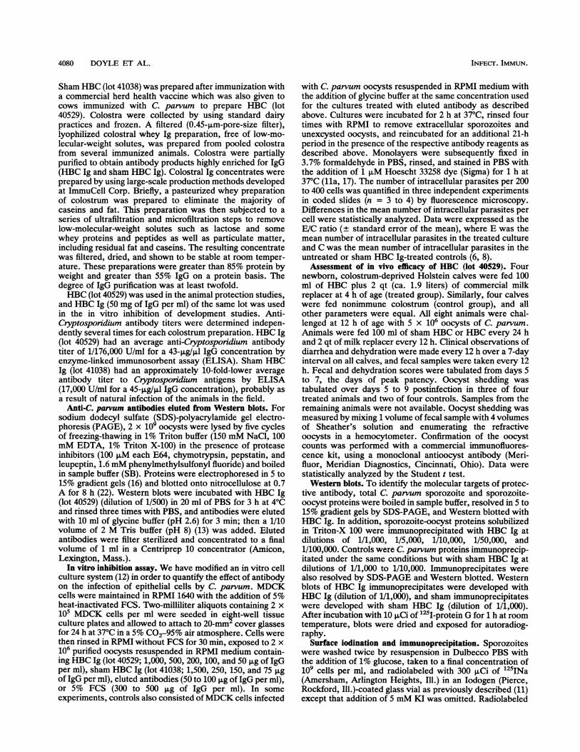
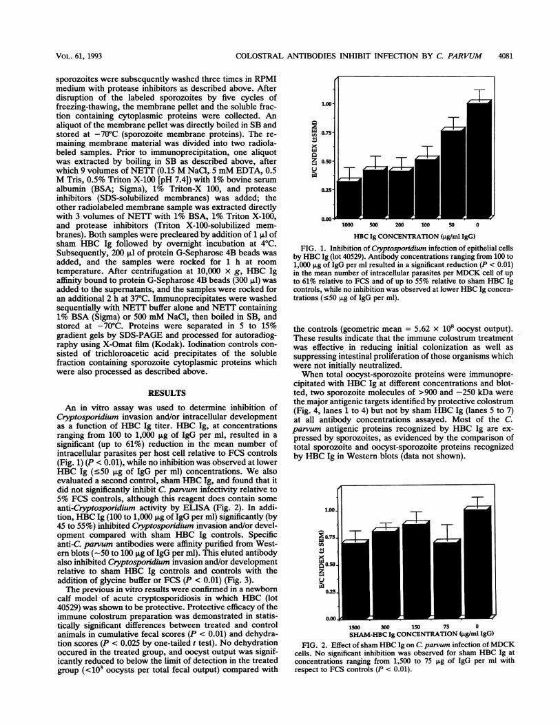
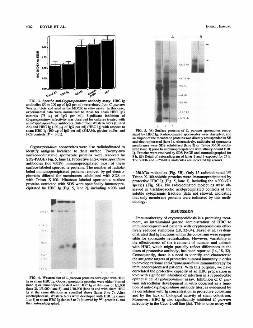
An in vitro assay was used to determine inhibition ofCryptosporidium invasion and/or intracellular developmentas a function of HBC Ig titer. HBC Ig, at concentrationsranging from 100 to 1,000 pg of IgG per ml, resulted in asignificant (up to 61%) reduction in the mean number ofintracellular parasites per host cell relative to FCS controls(Fig. 1) (P < 0.01), while no inhibition was observed at lowerHBC Ig (<50 ,g of IgG per ml) concentrations. We alsoevaluated a second control, sham HBC Ig, and found that itdid not significantly inhibit C parvum infectivity relative to5% FCS controls, although this reagent does contain someanti-Cryptosporidium activity by ELISA (Fig. 2). In addi-tion, HBC Ig (100 to 1,000 p,g of IgG per ml) significantly (by45 to 55%) inhibited Cryptosporidium invasion and/or devel-opment compared with sham HBC Ig controls. Specificanti-C. parvum antibodies were affinity purified from West-ern blots (-50 to 100 F±g of IgG per ml). This eluted antibodyalso inhibited Cryptosporidium invasion and/or developmentrelative to sham HBC Ig controls and controls with theaddition of glycine buffer or FCS (P < 0.01) (Fig. 3).The previous in vitro results were confirmed in a newborn
calf model of acute cryptosporidiosis in which HBC (lot40529) was shown to be protective. Protective efficacy of theimmune colostrum preparation was demonstrated in statis-tically significant differences between treated and controlanimals in cumulative fecal scores (P < 0.01) and dehydra-tion scores (P < 0.025 by one-tailed t test). No dehydrationoccured in the treated group, and oocyst output was signif-icantly reduced to below the limit of detection in the treatedgroup (<103 oocysts per total fecal output) compared with
LOO-
' 0.75-CA
Z 0.50-0
0.2
0.00-
-T1
1000 500 200 100 50 0
HBC Ig CONCENTRATION (jig/ml IgG)FIG. 1. Inhibition of Cryptosporidium infection of epithelial cells
by HBC Ig (lot 40529). Antibody concentrations ranging from 100 to1,000 jIg of IgG per ml resulted in a significant reduction (P < 0.01)in the mean number of intracellular parasites per MDCK cell of upto 61% relative to FCS and of up to 55% relative to sham HBC Igcontrols, while no inhibition was observed at lower HBC Ig concen-trations (<50 jig of IgG per ml).
the controls (geometric mean = 5.62 x 10⁸ oocyst output).These results indicate that the immune colostrum treatmentwas effective in reducing initial colonization as well assuppressing intestinal proliferation of those organisms whichwere not initially neutralized.When total oocyst-sporozoite proteins were immunopre-
cipitated with HBC Ig at different concentrations and blot-ted, two sporozoite molecules of >900 and -250 kDa werethe major antigenic targets identified by protective colostrum(Fig. 4, lanes 1 to 4) but not by sham HBC Ig (lanes 5 to 7)at all antibody concentrations assayed. Most of the C.parvum antigenic proteins recognized by HBC Ig are ex-pressed by sporozoites, as evidenced by the comparison oftotal sporozoite and oocyst-sporozoite proteins recognizedby HBC Ig in Western blots (data not shown).
1.00.
0.75CoX±1
0.50.zu
0.25.
0.00.I,J1500 300 150 75 0SHAM-HBC Ig CONCENTRATION (jig/ml IgG)
FIG. 2. Effect of sham HBC Ig on C. parvum infection ofMDCKcells. No significant inhibition was observed for sham HBC Ig atconcentrations ranging from 1,500 to 75 jig of IgG per ml withrespect to FCS controls (P < 0.01).
00
VOL. 61, 1993
I
4082 DOYLE ET AL.
A B
1.00
uh 0.75:
xL0 0.50z
Lu
0.00
Nb
FIG. 3. Specific anti-Cryptosporidium antibody assay. HBC Igantibodies (50 to 100 ,ug of IgG per ml) were eluted from C. parvumWestern blots and used in the MDCK in vitro assay. In this case,experimental data were normalized to those for sham HBC IgGcontrols (75 ,ug of IgG per ml). Significant inhibition ofCryptosporidium infectivity was observed for cultures treated withanti-Cryptosporidium antibodies eluted from Western blots (ElutedAb) and HBC Ig (100 ,g of IgG per ml) (HBC Ig) with respect tosham HBC Ig (100 ,ug of IgG per ml) (SHAM), glycine buffer, andFCS controls (P < 0.01).
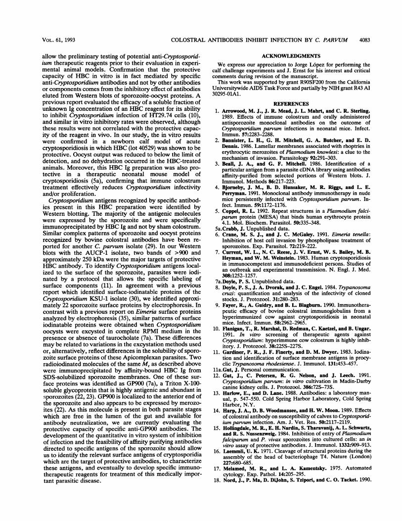
Cryptosponidium sporozoites were also radioiodinated toidentify antigens localized to their surface. Twenty-twosurface-iodinatable sporozoite proteins were resolved bySDS-PAGE (Fig. 5, lane 1). Protective anti-Cryptosporidiumantibodies (lot 40529) immunoprecipitated most of thesesurface-labeled sporozoite proteins. The number of radiola-beled immunoprecipitated proteins resolved by gel electro-phoresis differed for membranes solubilized with SDS orwith Triton X-100. Nineteen labeled sporozoite surfaceproteins extracted with SDS were specifically immunopre-cipitated by HBC Ig (Fig. 5, lane 2), including >900- and
P.
.2f L!
I4 ._
FIG. 4. Western blot of C. parvum proteins developed with HBCIg or sham HBC Ig. Oocyst-sporozoite proteins were either blotted(lane 1) or immunoprecipitated with HBC Ig at dilutions of 1/1,000(lane 2), 1/5,000 (lane 3), and 1/10,000 (lane 4) and with sham HBCIg at the same dilutions as specified above (lanes 5 to 7). Afterelectrophoresis, Western blots were developed with HBC Ig (lanes1 to 4) or sham HBC Ig (lanes 5 to 7) followed by "2I-protein G andthen autoradiographed.
-200 kD
-97.4 kD
-69 kD
-46 kD
-30 kO
-2 1.5 kD
t-14.3 kD
1 2 3
FIG. 5. (A) Surface proteins of C. parvum sporozoites recog-nized by HBC Ig. Radioiodinated sporozoites were disrupted, andan aliquot of the membrane proteins was directly resuspended in SBand electrophoresed (lane 1). Alternatively, radiolabeled sporozoitemembranes were SDS solubilized (lane 2) or Triton X-100 solubi-lized (lane 3) prior to immunoprecipitation with affinity-bound HBCIg. Proteins were resolved by SDS-PAGE and autoradiographed for8 h. (B) Detail of autoradiogram of lanes 2 and 3 exposed for 24 h.The >900- and -250-kDa molecules are indicated by arrows.
-250-kDa molecules (Fig. 5B). Only 13 radioiodinated 1%Triton X-100-soluble proteins were immunoprecipitated byprotective HBC Ig (Fig. 5, lane 3), including the >900-kDaspecies (Fig. 5B). No radioiodinated molecules were ob-served in trichloroacetic acid-precipitated controls of thesoluble cytoplasmic fraction (data not shown), indicatingthat only membrane proteins were iodinated by this meth-odology.
DISCUSSION
Immunotherapy of cryptosporidiosis is a promising treat-ment, as intraluminal gastric administration of HBC toimmunocompromised patients with cryptosporidiosis effec-tively reduced symptoms (18, 32-34). Fayer et al. (9) dem-onstrated that Ig fractions within the colostrum were respon-sible for sporozoite neutralization. However, variability inthe effectiveness of the treatment of humans and animalswith HBC, which might partially reflect differences in thetiters of protective antibody, has been reported (14, 18, 32).Consequently, there is a need to identify and characterizethe antigenic targets of protective humoral immunity in orderto develop rational anti-Cryptosporidium immunotherapy forimmunocompromised patients. With this purpose, we havecorrelated the protective capacity of an HBC preparation invivo with significant inhibition of infection in a reproducibleepithelial cell-Cryptospondium assay. Inhibition of C. par-vum intracellular development in vitro occurred as a func-tion of anti-Cryptosporidium antibody titer, as evidenced byits correlation with Ig concentration in protective colostrumand by the lack of biological activity of sham colostrum.Moreover, HBC Ig also significantly inhibited C. parvuminfectivity in the Caco-2 cell line (Sa). This in vitro assay will
INFECT. IMMUN.
0.25
COLOSTRAL ANTIBODIES INHIBIT INFECTION BY C. PARVUM 4083
allow the preliminary testing of potential anti-Cryptosporid-ium therapeutic reagents prior to their evaluation in experi-mental animal models. Confirmation that the protectivecapacity of HBC in vitro is in fact mediated by specificanti-Cryptosporidium antibodies and not by other antibodiesor components comes from the inhibitory effect of antibodieseluted from Western blots of sporozoite-oocyst proteins. Aprevious report evaluated the efficacy of a soluble fraction ofunknown Ig concentration of an HBC reagent for its abilityto inhibit Cryptosporidium infection of HT29.74 cells (10),and similar in vitro inhibitory rates were observed, althoughthese results were not correlated with the protective capac-ity of the reagent in vivo. In our study, the in vitro resultswere confirmed in a newborn calf model of acutecryptosporidiosis in which HBC (lot 40529) was shown to beprotective. Oocyst output was reduced to below the limit ofdetection, and no dehydration occurred in the HBC-treatedanimals. Moreover, this HBC Ig preparation was also pro-tective in a therapeutic neonatal mouse model ofcryptosporidiosis (5a), confirming that immune colostrumtreatment effectively reduces Cryptosporidium infectivityand/or proliferation.Cryptosporidium antigens recognized by specific antibod-
ies present in this HBC preparation were identified byWestern blotting. The majority of the antigenic moleculeswere expressed by the sporozoite and were specificallyimmunoprecipitated by HBC Ig and not by sham colostrum.Similar complex patterns of sporozoite and oocyst proteinsrecognized by bovine colostral antibodies have been re-ported for another C. parvum isolate (29). In our Westernblots with the AUCP-1 isolate, two bands of >900 andapproximately 250 kDa were the major targets of protectiveHBC antibody. To identify Cryptosporidium antigens local-ized to the surface of the sporozoite, parasites were iodi-nated by a protocol that allows the specific labeling ofsurface components (11). In agreement with a previousreport which identified surface-iodinatable proteins of theCryptosporidium KSU-1 isolate (30), we identified approxi-mately 22 sporozoite surface proteins by electrophoresis. Incontrast with a previous report on Eimeria surface proteinsanalyzed by electrophoresis (35), similar patterns of surfaceiodinatable proteins were obtained when Cryptosporidiumoocysts were excysted in complete RPMI medium in thepresence or absence of taurocholate (7a). These differencesmay be related to variations in the excystation methods usedor, alternatively, reflect differences in the solubility of sporo-zoite surface proteins of these Apicomplexan parasites. Tworadioiodinated molecules of the same Mr as described abovewere immunoprecipitated by affinity-bound HBC Ig fromSDS-solubilized sporozoite membranes. One of these sur-face proteins was identified as GP900 (7a), a Triton X-100-soluble glycoprotein that is highly antigenic and abundant insporozoites (22, 23). GP900 is localized to the anterior end ofthe sporozoite and also appears to be expressed by merozo-ites (22). As this molecule is present in both parasite stageswhich are free in the lumen of the gut and available forantibody neutralization, we are currently evaluating theprotective capacity of specific anti-GP900 antibodies. Thedevelopment of the quantitative in vitro system of inhibitionof infection and the feasibility of affinity purifying antibodiesdirected to specific antigens of the sporozoite should allowus to identify the relevant surface antigens of cryptosporidiawhich are the target of protective antibodies, to characterizethese antigens, and eventually to develop specific immuno-therapeutic reagents for treatment of this medically impor-tant parasitic disease.
ACKNOWLEDGMENTSWe express our appreciation to Jorge L6pez for performing the
calf challenge experiments and J. Ernst for his interest and criticalcomments during revision of the manuscript.
This work was supported by grant R9OSF200 from the CaliforniaUniversitywide AIDS Task Force and partially by NIH grant R43 AI30295-OlAl.
REFERENCES1. Arrowood, M. J., J. R. Mead, J. L. Mahrt, and C. R. Sterling.
1989. Effects of immune colostrum and orally administeredantisporozoite monoclonal antibodies on the outcome ofCryptosporidium parvum infections in neonatal mice. Infect.Immun. 57:2283-2288.
2. Bannister, L. H., G. H. Mitchell, G. A. Butcher, and E. D.Dennis. 1986. Lamellar membranes associated with rhoptries inerythrocytic merozoites of Plasmodium knowlesi: a clue to themechanism of invasion. Parasitology 92:291-303.
3. Beall, J. A., and G. F. Mitchell. 1986. Identification of aparticular antigen from a parasite cDNA library using antibodiesaffinity-purified from selected portions of Western blots. J.Immunol. Methods 86:217-223.
4. Bjorneby, J. M., B. D. Hunsaker, M. R. Riggs, and L. E.Perryman. 1991. Monoclonal antibody immunotherapy in nudemice persistently infected with Cryptosporidium parvum. In-fect. Immun. 59:1172-1176.
5. Coppel, R. L. 1992. Repeat structures in a Plasmodium falci-parum protein (MESA) that binds human erythrocyte protein4.1. Mol. Biochem. Parasitol. 50:335-348.
5a.Crabb, J. Unpublished data.6. Crane, M. S. J., and J. C. McGaley. 1991. Eimeria tenella:
Inhibition of host cell invasion by phospholipase treatment ofsporozoites. Exp. Parasitol. 72:219-222.
7. Current, W. L., N. C. Reese, J. V. Ernst, W. S. Bailey, M. B.Heyman, and W. M. Weinstein. 1983. Human cryptosporidiosisin immunocompetent and immunodeficient persons. Studies ofan outbreak and experimental transmission. N. Engl. J. Med.308:1252-1257.
7a.Doyle, P. S. Unpublished data.8. Doyle, P. S., J. A. Dvorak, and J. C. Engel. 1984. Trypanosoma
cnrzi: quantification and analysis of the infectivity of clonedstocks. J. Protozool. 31:280-283.
9. Fayer, R., A. Guidry, and B. L. Blagburn. 1990. Immunothera-peutic efficacy of bovine colostral immunoglobulins from ahyperimmunized cow against cryptosporidiosis in neonatalmice. Infect. Immun. 58:2962-2965.
10. Flanigan, T., R. Marshal, D. Redman, C. Kaetzel, and B. Ungar.1991. In vitro screening of therapeutic agents againstCryptosporidium: hyperimmune cow colostrum is highly inhib-itory. J. Protozool. 38:225S-227S.
11. Gardiner, P. R., J. F. Finerty, and D. M. Dwyer. 1983. Iodina-tion and identification of surface membrane antigens in procy-clic Trypanosoma rhodesiense. J. Immunol. 131:453-457.
11a.Gut, J. Personal communication.12. Gut, J., C. Petersen, R. G. Nelson, and J. Leech. 1991.
Cryptosporidium parvum: in vitro cultivation in Madin-Darbycanine kidney cells. J. Protozool. 386:72S-73S.
13. Harlow, E., and D. Lane. 1988. Antibodies: a laboratory man-ual, p. 547-550. Cold Spring Harbor Laboratory, Cold SpringHarbor, N.Y.
14. Harp, J. A., D. B. Woodmansee, and H. W. Moon. 1989. Effectsof colostral antibody on susceptibility of calves to Cryptosporid-ium parvum infection. Am. J. Vet. Res. 50:2117-2119.
15. Hollingdale, M. R., E. H. Nardin, S. Tharavani, A. L. Schwartz,and R. S. Nussenzweig. 1984. Inhibition of entry of Plasmodiumfalciparum and P. vivax sporozoites into cultured cells: an invitro assay of protective antibodies. J. Immunol. 1332:909-913.
16. Laemmli, U. K. 1971. Cleavage of structural proteins during theassembly of the head of bacteriophage T4. Nature (London)227:680-685.
17. Melamed, M. R., and L. A. Kamentsky. 1975. Automatedcytology. Exp. Pathol. 14:205-295.
18. Nord, J., P. Ma, D. Dijohn, S. Tzipori, and C. 0. Tacket. 1990.
VOL. 61, 1993
4084 DOYLE ET AL.
Treatment with bovine hyperimmune bovine colostrum ofcryptosporidial diarrhea in AIDs patients. AIDS 4:581-584.
19. Perkdns, M. E. Rhoptry organelles of Apicomplexan parasites.Parasitol. Today 8:28-32.
20. Perryman, L. E., M. W. Riggs, P. H. Mason, and R. Fayer. 1990.Kinetics of Cryptosporidium parvum sporozoite neutralizationby monoclonal antibodies, immune bovine serum, and immunebovine colostrum. Infect. Immun. 58:257-259.
21. Petersen, C. 1992. Cryptosporidiosis in patients infected withthe human immunodeficiency virus. Clin. Infect. Dis. 15:903-909.
22. Petersen, C., J. Gut, P. S. Doyle, J. H. Crabb, R. G. Nelson, andJ. H. Leech. 1992. Characterization of an Mr->900,000Cryptosporidium parvum sporozoites glycoprotein recognizedby hyperimmune bovine colostral immunoglobulin. Infect. Im-mun. 60:5132-5138.
23. Petersen, C., J. Gut, J. Leech, and R. G. Nelson. 1992. Identi-fication and initial characterization of five Cryptosporidiumparvum sporozoite antigen genes. Infect. Immun. 60:2343-2348.
24. Sam-Yellowe, T. Y., H. Shio, and M. E. Perkins. 1988. Secretionof Plasmodium falciparum rhoptry protein into the plasmamembrane of host erythrocytes. J. Cell Biol. 106:1507-1513.
25. Schofield, L., G. R. Bushell, J. A. Cooper, A. J. Saul, J. A.Upcroft, and C. Kidson. 1987. A rhoptry antigen ofPlasmodiumfalciparum contains conserved and variable epitopes recognizedby inhibitory monoclonal antibodies. Mol. Biochem. Parasitol.18:183-195.
26. Schwartzman, J. D. 1986. Inhibition of a penetration-enhancingfactor of Toxoplasma gondii by monoclonal antibodies specificfor rhoptries. Infect. Immun. 51:760-764.
27. Soave, R., and W. D. Johnson. 1988. Cryptosporidium andIsospora belli infections. J. Infect. Dis. 157:225-229.
28. Stewart, M. J., S. Schulman, and J. P. Vanderberg. 1986.Rhoptry secretion of membraneous whorls by Plasmodium
falciparum merozoites. Am. J. Trop. Med. Hyg. 35:37-44.29. Tilley, M., R. Fayer, A. Guidry, S. J. Upton, and B. L. Blagburn.
1990. Cryptosporidium parvum (Apicomplexa: Cryptosporidi-idae) oocyst and sporozoite antigens recognized by bovinecolostral antibodies. Infect. Immun. 58:2966-2971.
30. Tilley, M., and S. J. Upton. 1990. Electrophoretic characteriza-tion of Cryptosporidiumparvum (KSU-1 isolate) (Apicomplexa:Cryptosporidiidae). Can. J. Zool. 68:1513-1519.
31. Tilley, M., S. J. Upton, R. Fayer, J. R. Barta, C. E. Chrisp, P. S.Freed, B. L. Blagburn, B. C. Anderson, and S. M. Barnard.1991. Identification of a 15-kilodalton surface glycoprotein onsporozoites of Cryptosporidium parvum. Infect. Immun. 59:1002-1007.
32. Tzipori, S., D. Robertson, and C. Chapman. 1986. Remission ofdiarrhea due to cryptosporidiosis in an immunodeficient childtreated with hyperimmune bovine colostrum. Br. Med. J. 293:1276-1277.
33. Tzipori, S., D. Robertson, D. A. Cooper, and L. White. 1987.Chronic cryptosporidiosis and hyperimmune cow colostrum.Lancet ii:344-345.
34. Ungar, B. L. P., D. J. Ward, R. Fayer, and C. A. Quinn.. 1990.Cessation of Cryptosporidium-associated diarrhea in an ac-quired immunodeficiency syndrome patient after treatment withhyperimmune bovine colostrum. Gastroenterology 98:486-489.
35. Wisher, M. H., and M. E. Rose. 1987. Eimeria tenella sporo-zoites: the method of excystation affects the surface membraneproteins. Parasitology 95:479-489.
36. Wolfson, J. A., J. M. Richter, M. A. Waldron, D. J. Weber,D. M. McCarthy, and C. C. Hopkins. 1985. Cryptosporidiosis inimmunocompromised patients. N. Engl. J. Med. 312:1278-1285.
37. Zu, S. X., G. D. Fang, R. Fayer, and R. L. Guerrant. 1992.Cryptosporidiosis: pathogenesis and immunology. Parasitol.Today 8:24-27.
INFEc-r. IMMUN.