Embed Size (px)
Citation preview
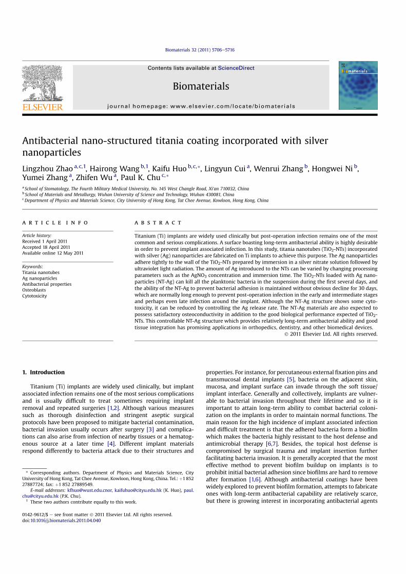
Antibacterial nano-structured titania coating incorporated with silvernanoparticles
Lingzhou Zhao a,c,1, Hairong Wang b,1, Kaifu Huo b,c,*, Lingyun Cui a, Wenrui Zhang b, Hongwei Ni b,Yumei Zhang a, Zhifen Wu a, Paul K. Chu c,*
a School of Stomatology, The Fourth Military Medical University, No. 145 West Changle Road, Xi’an 710032, Chinab School of Materials and Metallurgy, Wuhan University of Science and Technology, Wuhan 430081, ChinacDepartment of Physics and Materials Science, City University of Hong Kong, Tat Chee Avenue, Kowloon, Hong Kong, China
a r t i c l e i n f o
Article history:Received 1 April 2011Accepted 18 April 2011Available online 12 May 2011
Keywords:Titania nanotubesAg nanoparticlesAntibacterial propertiesOsteoblastsCytotoxicity
a b s t r a c t
Titanium (Ti) implants are widely used clinically but post-operation infection remains one of the mostcommon and serious complications. A surface boasting long-term antibacterial ability is highly desirablein order to prevent implant associated infection. In this study, titania nanotubes (TiO2-NTs) incorporatedwith silver (Ag) nanoparticles are fabricated on Ti implants to achieve this purpose. The Ag nanoparticlesadhere tightly to the wall of the TiO2-NTs prepared by immersion in a silver nitrate solution followed byultraviolet light radiation. The amount of Ag introduced to the NTs can be varied by changing processingparameters such as the AgNO3 concentration and immersion time. The TiO2-NTs loaded with Ag nano-particles (NT-Ag) can kill all the planktonic bacteria in the suspension during the first several days, andthe ability of the NT-Ag to prevent bacterial adhesion is maintained without obvious decline for 30 days,which are normally long enough to prevent post-operation infection in the early and intermediate stagesand perhaps even late infection around the implant. Although the NT-Ag structure shows some cyto-toxicity, it can be reduced by controlling the Ag release rate. The NT-Ag materials are also expected topossess satisfactory osteoconductivity in addition to the good biological performance expected of TiO2-NTs. This controllable NT-Ag structure which provides relatively long-term antibacterial ability and goodtissue integration has promising applications in orthopedics, dentistry, and other biomedical devices.
� 2011 Elsevier Ltd. All rights reserved.
1. Introduction
Titanium (Ti) implants are widely used clinically, but implantassociated infection remains one of the most serious complicationsand is usually difficult to treat sometimes requiring implantremoval and repeated surgeries [1,2]. Although various measuressuch as thorough disinfection and stringent aseptic surgicalprotocols have been proposed to mitigate bacterial contamination,bacterial invasion usually occurs after surgery [3] and complica-tions can also arise from infection of nearby tissues or a hematog-enous source at a later time [4]. Different implant materialsrespond differently to bacteria attack due to their structures and
properties. For instance, for percutaneous external fixation pins andtransmucosal dental implants [5], bacteria on the adjacent skin,mucosa, and implant surface can invade through the soft tissue/implant interface. Generally and collectively, implants are vulner-able to bacterial invasion throughout their lifetime and so it isimportant to attain long-term ability to combat bacterial coloni-zation on the implants in order to maintain normal functions. Themain reason for the high incidence of implant associated infectionand difficult treatment is that the adhered bacteria form a biofilmwhich makes the bacteria highly resistant to the host defense andantimicrobial therapy [6,7]. Besides, the topical host defense iscompromised by surgical trauma and implant insertion furtherfacilitating bacteria invasion. It is generally accepted that the mosteffective method to prevent biofilm buildup on implants is toprohibit initial bacterial adhesion since biofilms are hard to removeafter formation [1,6]. Although antibacterial coatings have beenwidely explored to prevent biofilm formation, attempts to fabricateones with long-term antibacterial capability are relatively scarce,but there is growing interest in incorporating antibacterial agents
* Corresponding authors. Department of Physics and Materials Science, CityUniversity of Hong Kong, Tat Chee Avenue, Kowloon, Hong Kong, China. Tel.: þ185227887724; fax: þ1 852 27889549.
E-mail addresses: [email protected], [email protected] (K. Huo), [email protected] (P.K. Chu).
1 These two authors contribute equally to this work.
Contents lists available at ScienceDirect
Biomaterials
journal homepage: www.elsevier .com/locate/biomateria ls
0142-9612/$ e see front matter � 2011 Elsevier Ltd. All rights reserved.doi:10.1016/j.biomaterials.2011.04.040
Biomaterials 32 (2011) 5706e5716
into coatings on biomedical implants. By optimizing the structureas well as antibacterial agent loading capacity and release rate,materials that possess more long-term antibacterial ability andpromote tissue integration and repair simultaneously may beproduced.
Since bone tissues are composed of hierarchical nano-composites, the proper nanotopography can promote osteo-conductivity from the biomimetic viewpoint. As a result, surfacemodification of biomaterials including surface nanotexturing hasbeen explored extensively [8e18]. In particular, highly orderedtitania nanotubes (TiO2-NTs) fabricated on Ti implants by electro-chemical anodization have attracted increasing attention [11e18].TiO2-NTs have a lower elastic modulus of approximately 36e43 GPa[17] which is much closer to that of natural bones and are expectedto have better biomechanical compatibility than other artificialbiomaterials. For instance, TiO2-NTs have been found to foster thegrowth of nano-structured hydroxyapatite in simulated body fluids[13] and induce extracellular matrix secretion, mineralization, andother functions of bone cells [12,14e16]. Moreover, our recent studyrevealed that the hierarchical hybrid TiO2 micropits/nanotubesstructures induced more balanced promotion of multiple cellfunctions [11]. Therefore, TiO2-NTs are promising bioactive coatingsthat can induce direct bone-implant bonding with enhanced hostdefense on the implant surface.
Another merit of TiO2-NTs is that they can serve as carriers fordrugs such as antibacterial agents [18,19] and it opens up thepossibility of long-term antibacterial functionality as aforemen-tioned. Various kinds of antibiotics have been studied but in spite ofscattered success, development of new antibiotics resistant strainsafter prolonged use reduces the effectiveness [20e24]. Silver (Ag) isone of the strong bactericides and has attracted increasing atten-tion because of other benefits such as a broad antibacterial spec-trum including antibiotic-resistant bacteria, non-cytotoxicity atsuitable doses, satisfactory stability, and smaller possibility todevelop resistant strains [1,3,24e29]. Since the required Ag dose istypically low, it is possible to fabricate coatings with long-termantibacterial characteristics by introducing and controlling Agrelease. In fact, it has been reported that a certain range of Agconcentrations can kill bacteria without impairing mammalian cellfunctions [24] and Ag-containing coatings that can resist biofilmformation and do not show cytotoxicity have been explored[3,24,26,28].
We believe that by optimizing the structure of TiO2-NTs fromthe perspective of tissue integration, introducing a sufficientamount of Ag, and regulating the Ag release rate in a controlledmanner, a surface that boasts a relatively long-term antibacterialability and simultaneously enhances cell functions can beproduced. In this study, we investigate whether Ag can be incor-porated into TiO2-NTs controllably and reliably and if the Ag loadedTiO2-NTs (NT-Ag) indeed possess long-term antibacterial capabilityand other desirable biological functionalities.
2. Materials and methods
2.1. Specimen preparation
Pure titanium foils (Aldrich, 10 � 10 � 1 mm3) underwent electrochemicalanodization to form a titania nanotubular layer. The Ti foils were polished by SiCsandpapers and then ultrasonically cleaned with acetone, ethanol, and deionizedwater sequentially. Anodizationwas carried out in a conventional two-electrode cellequipped with a direct current (DC) power supply (IT6834, ITECH, China). The Ti foilserved as the anode electrode and a graphite foil as the counter electrode (1 cmseparation). The electrolyte was ethylene glycol containing 0.5 wt % ammoniumfluoride (NH4F) and 5 vol % distilled (DI) water. After anodization at 60 V for 1 h,TiO2-NTs were formed on Ti foils. The samples were ultrasonically cleaned andannealed at 450 �C in air for 3 h to convert the amorphous TiO2-NTs into the anatasephase. The annealed samples were soaked in AgNO3 solutions with four differentconcentrations (0.5, 1, 1.5 and 2 M) for 10 min. Afterward, they were rinsed with
deionized water, dried, and irradiated with UV light from a high-pressure Hg lampfor 10 min. This process was carried out once for the 0.5 M group and twice for the 1,1.5 and 2 M samples to obtain NT-Ag samples with different Ag contents.Thesesamples were denoted as NT-Ag0.5, NT-Ag1.0, NT-Ag1.5 and NT-Ag2.0, respectively.After ultrasonic cleaning (3 times and 10 min each), both sides of the samples weresterilized by UV irradiation for 30 min before ensuing antibacterial and cell cultureexperiments.
2.2. Surface characterization
Field-emission scanning electron microscopy (FE-SEM, FEI Nova 400 Nano) andatomic force microscopy (AFM, Auto-Probe CP, Park Scientific Instruments) wereutilized to determine the surface topography of the specimens, and transmissionelectron microscopy (TEM, Philips CM20) was used to observe the microstructure ofthe Ag loaded nanotubes. The crystalline structure of the samples was determinedby X-ray diffraction (XRD, Philips X0 Pert Pro), and the chemical composition of thesurface layer was determined by X-ray photoelectron spectroscopy (XPS, ESCALABMK-II).
2.3. Sliver release
The amounts of Ag released from the NT-Ag samples were monitored in thephosphate buffered saline (PBS). The samples were immersed in 6 ml of PBS for 1day in dark, taken out, and then immersed again in 6 ml of fresh PBS. This processwas repeated for a total of 14 days to generate solutions at different time points inorder to determine the Ag release time profile. The PBS solution containing releasedsilver was analyzed by inductively-coupled plasma atomic emission spectrometry(ICP-AES, IRIS Advantage ER/S).
2.4. Antibacterial assay
The antibacterial ability was evaluated using Staphylococcus aureus (S. aureus,ATCC 6538) cultivated in a beef extract-peptone (BEP) medium at 37 �C for 12 h. Itwas adjusted to a concentration of 105 CFU/ml in the antibacterial assay. Eachspecimenwas incubated in 1 ml of the bacteria suspension in BEP at 37 �C for 1 day.At the end of the incubation period, the culture mediumwas sampled to determinethe viable counts of planktonic bacteria. The specimens were gently rinsed thricewith PBS in order to eliminate non-adherent bacteria, and the adhered bacteria oneach specimen were detached into 1 ml of BEP by ultrasonic vibration (40 W) for5 min with the resulting bacterial suspension sampled to count the viable bacteriaadhered on the specimens. Complete detachment of the adhered bacteria byultrasonic vibration was verified by fluorescence microscopy after fluorescencestaining. Afterward, the specimens were ultrasonically cleaned, dried, sterilized, andre-incubated as described above. This process was repeated daily for a total incu-bation time of 30 days. The viable bacteria in the sampled suspensions at days 1, 4, 7,10, 15, 20, and 30 were counted by serial dilution and the spread plate method. Theantibacterial rates with regard to planktonic bacteria in the culture medium and theantibacterial rates for adhered bacteria on the specimens were calculated based onthe following formulas: (1) Antibacterial rate for planktonic bacteria in the medium(Rp) (%) ¼ (B e A)/B � 100% and (2) Antibacterial rate for adherent bacteria on thespecimen (Ra) (%) ¼ (D e C)/D � 100%. Here, A indicates the average number ofviable bacteria in the culturemedium inoculatedwith the specimen, B is the averagenumber of viable bacteria in the culture medium inoculated with no specimen(blank control), C is the average number of viable bacteria on the NT-Ag specimens,and D is the average number of viable bacteria on the TiO2-NTs.
In order to perform fluorescence staining to show the viability of bacteria on thesamples, bacteria were inoculated on the samples mentioned above. The bacteriamedium was refreshed every 24 h for a total of 7 days. Afterward, a new bacteriasuspension was added and cultured for another 7 h. The culture medium was thenremoved and the samples were rinsed with PBS, stained using acridine orange andethidium bromide for 15 min in dark, and observed by fluorescence microscopy(Olympus). Ethidium bromide did not penetrate the plasma membrane in the viablecells and stained only the dead cells, whereas acridine orange penetrated the plasmamembrane without permeabilization and stained the viable and dead cells. Whenexamined by fluorescence microscopy, the living cells appeared green while thedead cells were orange.
2.5. Protein adsorption assay
A 1 ml droplet of the Dulbecco’s minimum essential medium (DMEM, Gibco)containing 10% bovine calf serum (BCS, Gibco) was pipetted onto each specimen.After incubation at 37 �C for 4 h, the disks were placed in new 24 well plates (onedisk per well) andwashed with PBS thrice. 500 ml of 1% sodium dodecyl sulfate (SDS)solution was added to these wells and shaken for 1 h to detach proteins from thedisk surfaces.The protein concentrations in the collected SDS solutions weredetermined using a MicroBCA protein assay kit (Pierce).
L. Zhao et al. / Biomaterials 32 (2011) 5706e5716 5707
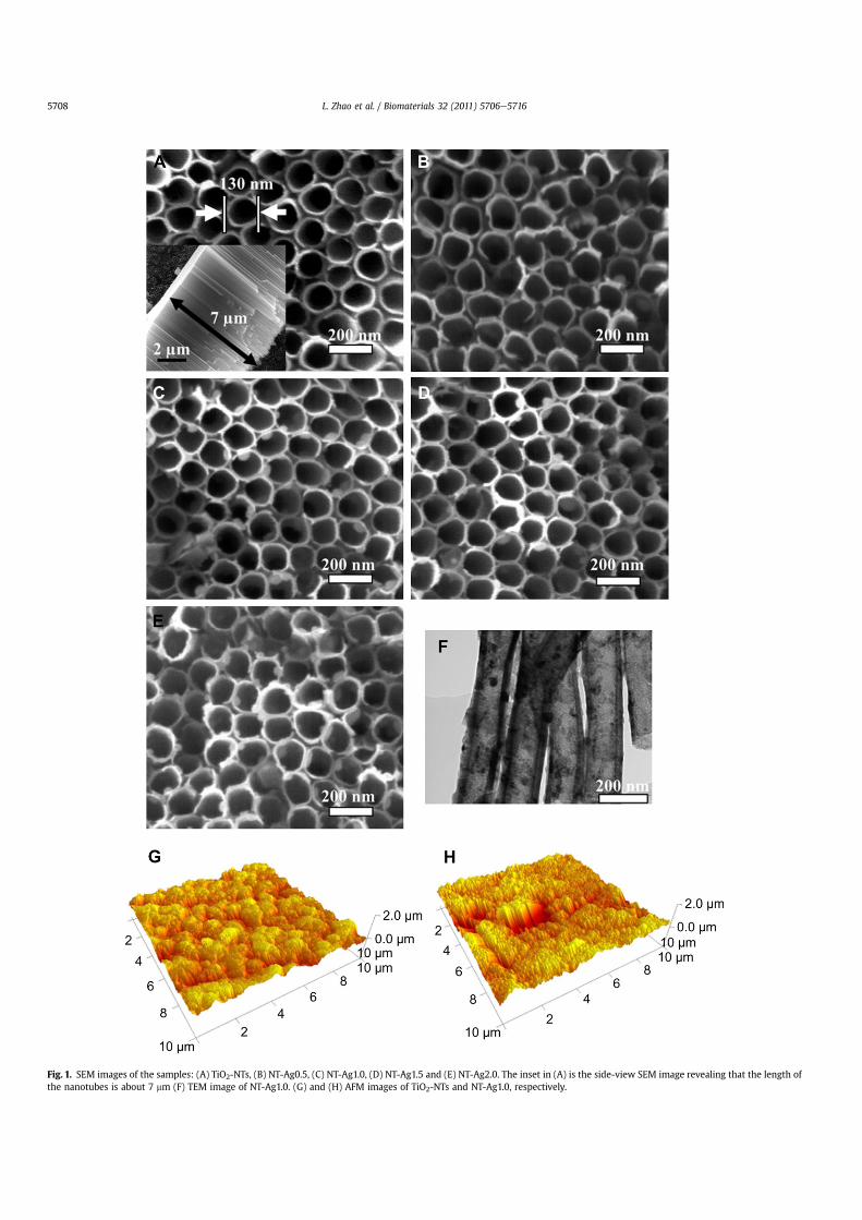
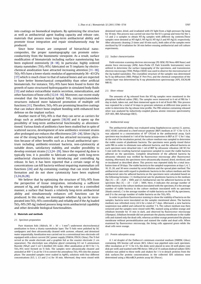
Fig. 1. SEM images of the samples: (A) TiO2-NTs, (B) NT-Ag0.5, (C) NT-Ag1.0, (D) NT-Ag1.5 and (E) NT-Ag2.0. The inset in (A) is the side-view SEM image revealing that the length ofthe nanotubes is about 7 mm (F) TEM image of NT-Ag1.0. (G) and (H) AFM images of TiO2-NTs and NT-Ag1.0, respectively.
L. Zhao et al. / Biomaterials 32 (2011) 5706e57165708
2.6. Cell culture
Primary rat osteoblasts were extracted by digesting the calvarial bone ofneonatal SpragueeDawley rats [30]. The cells were cultured in DMEM containing10% BCS and 1% penicillin/streptomycin and incubated in a humidified atmosphereof 5% CO2 at 37 �C. Passages 2e5 were used in the experiments.
2.7. Lactate dehydrogenase activity assay
The activity of lactate dehydrogenase (LDH) in the culture media released by thecells was used as an index of cytotoxicity. After incubation for 1 day, the culturemedia were sampled and centrifuged, and the supernatant was used for the LDHactivity assay. The LDH activity was determined spectrophotometrically according tothe manufacturer’s instructions (Sigma).
2.8. Cell number
The cells were seeded on the specimens placed in 24 well plates at a density of2� 104 cells/well. After culturing for 1 and 4 days, cell numbers were determined byDNA assay as previously reported [31]. Briefly, cells on samples were digested in500 ml buffer solution (0.5 mg/ml proteinase K (Beyotime), 0.2 mg/ml sodiumdodecyl sulfate (Solarbio), and 30 mM saline-sodium citrate (SSC)) at 56 �C over-night. The resulting mixture was subjected to centrifugation and aliquots (50 ml) ofthe supernatants after mixing with 150 ml Hoechst 33258 dye solution (1 mg/ml,Sigma) and transferred to 96-well plates. The DNA content was quantified spec-trofluorometrically using a Multi-Detection microplate reader (Bio-Rad) at a wave-length of 465 nm (excitation wavelength of 360 nm) by correlating with a DNAstandard curve which was generated by lysing serial dilution of a known concen-tration of osteoblasts.
2.9. Intracellular total protein synthesis and alkaline phosphatase activity
A 1ml cell suspensionwas seeded on each specimen at a density of 2�104 cells/ml. After culturing for 7 days, the cells were washed with PBS and lysed in 0.1 vol%Triton X-100 using the standard freezeethaw cycles. The alkaline phosphatase (ALP)activity in the lysis was determined by means of a colorimetric assay using an ALPreagent containing p-nitrophenyl phosphate (p-NPP) as the substrate. The absor-bance of the formed p-nitrophenol was measured at 405 nm. The intracellular totalprotein content was determined using the MicroBCA protein assay kit and the ALPactivity was normalized to it.
2.10. Statistical analysis
The assays were performed in triplicate and data were expressed asmeans � standard deviations. Each experiment was repeated three times with datafrom a typical experiment shown. A one-way ANOVA combined with a Student-Newman-Keuls (SNK) post hoc test was utilized to determine the level of signifi-cance. p < 0.05 was regarded to be significant and p < 0.01 was considered highlysignificant.
3. Results
3.1. Surface characterization
The SEM and AFM pictures of the TiO2-NTs and the NT-Agsamples as well as TEM images of the nanotubes taken from NT-Ag1.0 are depicted in Fig. 1. The TiO2-NTs formed by anodizationof a Ti foil at 60 V for 1 h have a typical diameter of about 130 nmand length of about 7 mm (Fig. 1A). Fig. 1BeE display the SEMimages of NT-Ag0.5, NT-Ag1.0, NT-Ag1.5, and NT-Ag2.0, respec-tively. The NT-Ag samples retain the nanotubular structure with Ag
Fig. 2. XRD patterns of samples: (a) TiO2-NTs, (b) NT-Ag0.5, (c) NT-Ag1.0, (d) NT-Ag1.5and (e) NT-Ag2.0.
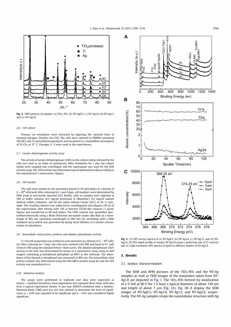
Fig. 3. (A) XPS survey spectra of (a) NT-Ag0.5, (b) NT-Ag1.0, (c) NT-Ag1.5, and (d) NT-Ag2.0, (B) XPS depth profile of sample NT-Ag1.0 using a sputtering rate of 21 nm/minand (C) high-resolution XPS spectra of Ag3d at different depths in NT-Ag1.0.
L. Zhao et al. / Biomaterials 32 (2011) 5706e5716 5709
nanoparticles incorporated into the nanotubes. The size of the Agnanoparticles is related to the AgNO3 concentration. The TEMpicture acquired from the nanotubes taken fromNT-Ag1.0 shown inFig.1F demonstrates that the Ag nanoparticles attached to the innerwall of the TiO2-NTs have a diameter of about 10e20 nm. The AFMimages show that the TiO2-NTs and NT-Ag surfaces have similarroughness values of several hundred nanometers. The AFM image
acquired from NT-Ag1.0 (Fig. 1H) is shown here as an example tocompare to the TiO2-NTs (Fig. 1G).
Fig. 2 exhibits the XRD patterns of the specimens. Only peakscorresponding to the anatase phase and Ti substrate can beobserved from the TiO2-NTs. In contrary, Ag peaks appear in theXRD patterns obtained from the NT-Ag samples. In addition, theintensities of the Ag peaks increase from NT-Ag0.5 to NT-Ag2.0,suggesting that the amounts of Ag incorporated into the nano-tubes increase with AgNO3 concentrations.
The XPS survey spectra acquired from NT-Ag0.5, NT-Ag1.0, NT-Ag1.5, and NT-Ag2.0 after 5 nm of the surface was sputtered offusing Arþ bombardment shown in Fig. 3A reveal peaks corre-sponding to Ti, Ag, O, Ar, and C. The C signal detected by XPS isbelieved to arise from adventitious contamination. The Ag concen-trations determined from the XPS survey spectra of NT-Ag0.5, NT-Ag1.0, NT-Ag1.5, and NT-Ag2.0 are 0.82, 1.24, 1.45, and 1.58 at%,respectively. TheXPSdepthprofile obtained fromNT-Ag1.0 in Fig. 3Bdiscloses that Ag is distributed along the entire NT length althoughthe absolute concentration decreases with depth from 1.24 at% nearthe surface to 0.2 at% at a depth of 3 mm. The high-resolution XPSspectra of Ag obtained from NT-Ag at different depths are shown inFig. 3C. The binding energies of the Ag 3d peak at 368.25 eV and374.25 eV can be assigned to 3d5/2 and 3d3/2 of metallic Ag0 [32] anddo not shift with depths, indicating that Ag mainly exists in the Ag0
state in the TiO2-NTs. SEM, TEM, XRD, and XPS provide unequivocalproof that metallic Ag nanoparticles have been successfully loadedinto the TiO2-NT along the entire length.
3.2. Ag release
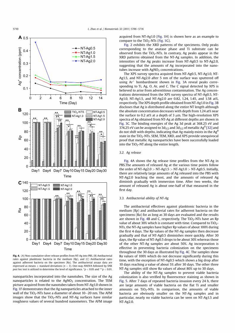
Fig. 4A shows the Ag release time profiles from the NT-Ag inPBS.The amounts of released Ag at the various time points followthe order of NT-Ag2.0 > NT-Ag1.5 > NT-Ag1.0 > NT-Ag0.5. Initially,there are relatively large amounts of Ag released into the PBS withNT-Ag2.0 leaching the most, and the amounts of released Agdiminish gradually with immersion time. After two weeks, theamount of released Ag is about one-half of that measured in thefirst day.
3.3. Antibacterial ability of NT-Ag
The antibacterial effectives against planktonic bacteria in themedium (Rp) and antibacterial rates for adherent bacteria on thespecimens (Ra) for as long as 30 days are evaluated and the resultsare shown in Fig. 4B and C, respectively. The TiO2-NTs have an Rpvalue of about 30% which is constant with time. Compared to TiO2-NTs, the NT-Ag samples have higher Rp values of about 100% duringthe first 4 days. The Rp values of the NT-Ag samples then decreasegradually and that of NT-Ag0.5 diminishes more quickly. After 30days, the Rp value of NT-Ag0.5 drops to be about 30%whereas thoseof the other NT-Ag samples are about 50%. Ag incorporation iseffective in preventing bacteria colonization on the specimensthroughout the 30 days as illustrated by Fig. 4C. The samples showRa values of 100% which do not decrease significantly during thistime, with the exception of NT-Ag0.5 which shows a big drop after10 days reaching a value of about 5% after 30 days. The other threeNT-Ag samples still show Ra values of about 80% up to 30 days.
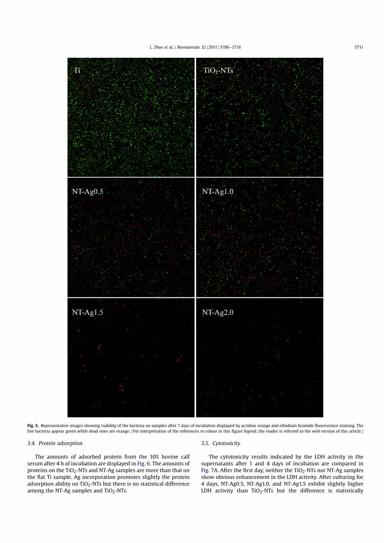
The ability of the NT-Ag samples to prevent viable bacteriacolonization is also verified by fluorescence staining as shown inFig. 5. After 7 days of repeated bacteria invasion every 24 h, thereare large amounts of viable bacteria on the flat Ti and smalleramounts on TiO2-NTs. In comparison, the amounts of viablebacteria are obviously smaller on the NT-Ag samples and inparticular, nearly no viable bacteria can be seen on NT-Ag1.5 andNT-Ag2.0.
Fig. 4. (A) Non-cumulative silver release profiles from NT-Ag into PBS, (B) Antibacterialrates against planktonic bacteria in the medium (Rp), and (C) Antibacterial ratesagainst adherent bacteria on the specimen (Ra). The antibacterial assays data areexpressed as means � standard deviations (n ¼ 3). One-way ANOVA followed by SNKpost hoc test is utilized to determine the level of significance. *p < 0.05 and **p < 0.01.
L. Zhao et al. / Biomaterials 32 (2011) 5706e57165710
3.4. Protein adsorption
The amounts of adsorbed protein from the 10% bovine calfserum after 4 h of incubation are displayed in Fig. 6. The amounts ofproteins on the TiO2-NTs and NT-Ag samples are more than that onthe flat Ti sample. Ag incorporation promotes slightly the proteinadsorption ability on TiO2-NTs but there is no statistical differenceamong the NT-Ag samples and TiO2-NTs.
3.5. Cytotoxicity
The cytotoxicity results indicated by the LDH activity in thesupernatants after 1 and 4 days of incubation are compared inFig. 7A. After the first day, neither the TiO2-NTs nor NT-Ag samplesshow obvious enhancement in the LDH activity. After culturing for4 days, NT-Ag0.5, NT-Ag1.0, and NT-Ag1.5 exhibit slightly higherLDH activity than TiO2-NTs but the difference is statistically
Fig. 5. Representative images showing viability of the bacteria on samples after 7 days of incubation displayed by acridine orange and ethidium bromide fluorescence staining. Thelive bacteria appear green while dead ones are orange. (For interpretation of the references to colour in this figure legend, the reader is referred to the web version of this article.)
L. Zhao et al. / Biomaterials 32 (2011) 5706e5716 5711
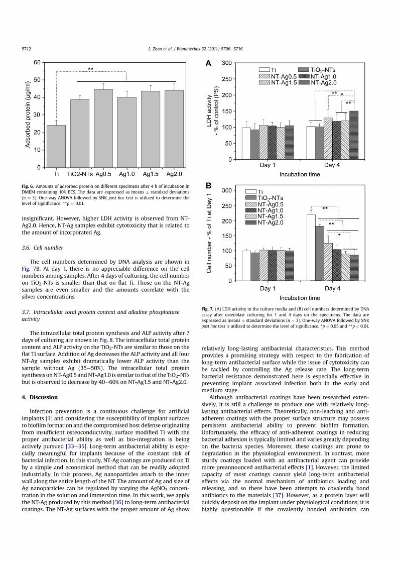
insignificant. However, higher LDH activity is observed from NT-Ag2.0. Hence, NT-Ag samples exhibit cytotoxicity that is related tothe amount of incorporated Ag.
3.6. Cell number
The cell numbers determined by DNA analysis are shown inFig. 7B. At day 1, there is no appreciable difference on the cellnumbers among samples. After 4 days of culturing, the cell numberon TiO2-NTs is smaller than that on flat Ti. Those on the NT-Agsamples are even smaller and the amounts correlate with thesilver concentrations.
3.7. Intracellular total protein content and alkaline phosphataseactivity
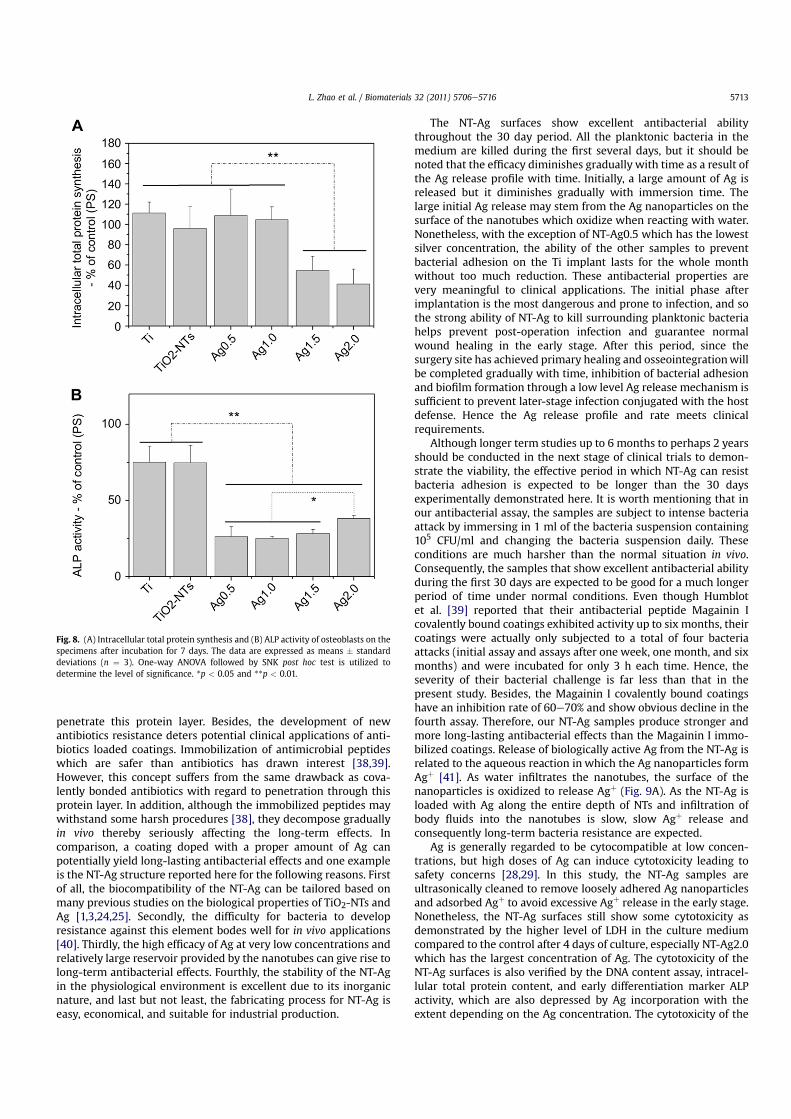
The intracellular total protein synthesis and ALP activity after 7days of culturing are shown in Fig. 8. The intracellular total proteincontent and ALP activity on the TiO2-NTs are similar to those on theflat Ti surface. Addition of Ag decreases the ALP activity and all fourNT-Ag samples exhibit dramatically lower ALP activity than thesample without Ag (35e50%). The intracellular total proteinsynthesis onNT-Ag0.5 andNT-Ag1.0 is similar to that of the TiO2-NTsbut is observed to decrease by 40e60% on NT-Ag1.5 and NT-Ag2.0.
4. Discussion
Infection prevention is a continuous challenge for artificialimplants [1] and considering the susceptibility of implant surfacesto biofilm formation and the compromised host defense originatingfrom insufficient osteoconductivity, surface modified Ti with theproper antibacterial ability as well as bio-integration is beingactively pursued [33e35]. Long-term antibacterial ability is espe-cially meaningful for implants because of the constant risk ofbacterial infection. In this study, NT-Ag coatings are produced on Tiby a simple and economical method that can be readily adoptedindustrially. In this process, Ag nanoparticles attach to the innerwall along the entire length of the NT. The amount of Ag and size ofAg nanoparticles can be regulated by varying the AgNO3 concen-tration in the solution and immersion time. In this work, we applythe NT-Ag produced by this method [36] to long-term antibacterialcoatings. The NT-Ag surfaces with the proper amount of Ag show
relatively long-lasting antibacterial characteristics. This methodprovides a promising strategy with respect to the fabrication oflong-term antibacterial surface while the issue of cytotoxicity canbe tackled by controlling the Ag release rate. The long-termbacterial resistance demonstrated here is especially effective inpreventing implant associated infection both in the early andmedium stage.
Although antibacterial coatings have been researched exten-sively, it is still a challenge to produce one with relatively long-lasting antibacterial effects. Theoretically, non-leaching and anti-adherent coatings with the proper surface structure may possesspersistent antibacterial ability to prevent biofilm formation.Unfortunately, the efficacy of anti-adherent coatings in reducingbacterial adhesion is typically limited and varies greatly dependingon the bacteria species. Moreover, these coatings are prone todegradation in the physiological environment. In contrast, moresturdy coatings loaded with an antibacterial agent can providemore preannounced antibacterial effects [1]. However, the limitedcapacity of most coatings cannot yield long-term antibacterialeffects via the normal mechanism of antibiotics loading andreleasing, and so there have been attempts to covalently bondantibiotics to the materials [37]. However, as a protein layer willquickly deposit on the implant under physiological conditions, it ishighly questionable if the covalently bonded antibiotics can
Fig. 6. Amounts of adsorbed protein on different specimens after 4 h of incubation inDMEM containing 10% BCS. The data are expressed as means � standard deviations(n ¼ 3). One-way ANOVA followed by SNK post hoc test is utilized to determine thelevel of significance. **p < 0.01.
Fig. 7. (A) LDH activity in the culture media and (B) cell numbers determined by DNAassay after osteoblast culturing for 1 and 4 days on the specimens. The data areexpressed as means � standard deviations (n ¼ 3). One-way ANOVA followed by SNKpost hoc test is utilized to determine the level of significance. *p < 0.05 and **p < 0.01.
L. Zhao et al. / Biomaterials 32 (2011) 5706e57165712
penetrate this protein layer. Besides, the development of newantibiotics resistance deters potential clinical applications of anti-biotics loaded coatings. Immobilization of antimicrobial peptideswhich are safer than antibiotics has drawn interest [38,39].However, this concept suffers from the same drawback as cova-lently bonded antibiotics with regard to penetration through thisprotein layer. In addition, although the immobilized peptides maywithstand some harsh procedures [38], they decompose graduallyin vivo thereby seriously affecting the long-term effects. Incomparison, a coating doped with a proper amount of Ag canpotentially yield long-lasting antibacterial effects and one exampleis the NT-Ag structure reported here for the following reasons. Firstof all, the biocompatibility of the NT-Ag can be tailored based onmany previous studies on the biological properties of TiO2-NTs andAg [1,3,24,25]. Secondly, the difficulty for bacteria to developresistance against this element bodes well for in vivo applications[40]. Thirdly, the high efficacy of Ag at very low concentrations andrelatively large reservoir provided by the nanotubes can give rise tolong-term antibacterial effects. Fourthly, the stability of the NT-Agin the physiological environment is excellent due to its inorganicnature, and last but not least, the fabricating process for NT-Ag iseasy, economical, and suitable for industrial production.
The NT-Ag surfaces show excellent antibacterial abilitythroughout the 30 day period. All the planktonic bacteria in themedium are killed during the first several days, but it should benoted that the efficacy diminishes gradually with time as a result ofthe Ag release profile with time. Initially, a large amount of Ag isreleased but it diminishes gradually with immersion time. Thelarge initial Ag release may stem from the Ag nanoparticles on thesurface of the nanotubes which oxidize when reacting with water.Nonetheless, with the exception of NT-Ag0.5 which has the lowestsilver concentration, the ability of the other samples to preventbacterial adhesion on the Ti implant lasts for the whole monthwithout too much reduction. These antibacterial properties arevery meaningful to clinical applications. The initial phase afterimplantation is the most dangerous and prone to infection, and sothe strong ability of NT-Ag to kill surrounding planktonic bacteriahelps prevent post-operation infection and guarantee normalwound healing in the early stage. After this period, since thesurgery site has achieved primary healing and osseointegrationwillbe completed gradually with time, inhibition of bacterial adhesionand biofilm formation through a low level Ag release mechanism issufficient to prevent later-stage infection conjugated with the hostdefense. Hence the Ag release profile and rate meets clinicalrequirements.
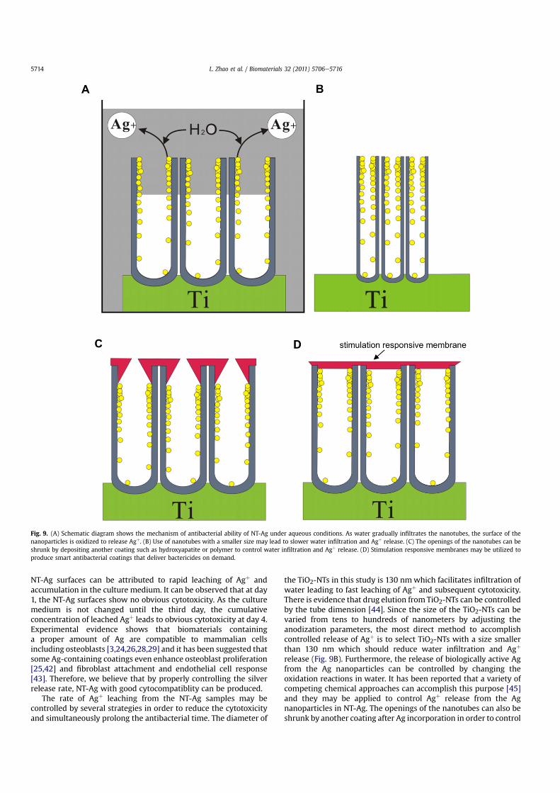
Although longer term studies up to 6 months to perhaps 2 yearsshould be conducted in the next stage of clinical trials to demon-strate the viability, the effective period in which NT-Ag can resistbacteria adhesion is expected to be longer than the 30 daysexperimentally demonstrated here. It is worth mentioning that inour antibacterial assay, the samples are subject to intense bacteriaattack by immersing in 1 ml of the bacteria suspension containing105 CFU/ml and changing the bacteria suspension daily. Theseconditions are much harsher than the normal situation in vivo.Consequently, the samples that show excellent antibacterial abilityduring the first 30 days are expected to be good for a much longerperiod of time under normal conditions. Even though Humblotet al. [39] reported that their antibacterial peptide Magainin Icovalently bound coatings exhibited activity up to six months, theircoatings were actually only subjected to a total of four bacteriaattacks (initial assay and assays after one week, one month, and sixmonths) and were incubated for only 3 h each time. Hence, theseverity of their bacterial challenge is far less than that in thepresent study. Besides, the Magainin I covalently bound coatingshave an inhibition rate of 60e70% and show obvious decline in thefourth assay. Therefore, our NT-Ag samples produce stronger andmore long-lasting antibacterial effects than the Magainin I immo-bilized coatings. Release of biologically active Ag from the NT-Ag isrelated to the aqueous reaction in which the Ag nanoparticles formAgþ [41]. As water infiltrates the nanotubes, the surface of thenanoparticles is oxidized to release Agþ (Fig. 9A). As the NT-Ag isloaded with Ag along the entire depth of NTs and infiltration ofbody fluids into the nanotubes is slow, slow Agþ release andconsequently long-term bacteria resistance are expected.
Ag is generally regarded to be cytocompatible at low concen-trations, but high doses of Ag can induce cytotoxicity leading tosafety concerns [28,29]. In this study, the NT-Ag samples areultrasonically cleaned to remove loosely adhered Ag nanoparticlesand adsorbed Agþ to avoid excessive Agþ release in the early stage.Nonetheless, the NT-Ag surfaces still show some cytotoxicity asdemonstrated by the higher level of LDH in the culture mediumcompared to the control after 4 days of culture, especially NT-Ag2.0which has the largest concentration of Ag. The cytotoxicity of theNT-Ag surfaces is also verified by the DNA content assay, intracel-lular total protein content, and early differentiation marker ALPactivity, which are also depressed by Ag incorporation with theextent depending on the Ag concentration. The cytotoxicity of the
Fig. 8. (A) Intracellular total protein synthesis and (B) ALP activity of osteoblasts on thespecimens after incubation for 7 days. The data are expressed as means � standarddeviations (n ¼ 3). One-way ANOVA followed by SNK post hoc test is utilized todetermine the level of significance. *p < 0.05 and **p < 0.01.
L. Zhao et al. / Biomaterials 32 (2011) 5706e5716 5713
NT-Ag surfaces can be attributed to rapid leaching of Agþ andaccumulation in the culture medium. It can be observed that at day1, the NT-Ag surfaces show no obvious cytotoxicity. As the culturemedium is not changed until the third day, the cumulativeconcentration of leached Agþ leads to obvious cytotoxicity at day 4.Experimental evidence shows that biomaterials containinga proper amount of Ag are compatible to mammalian cellsincluding osteoblasts [3,24,26,28,29] and it has been suggested thatsome Ag-containing coatings even enhance osteoblast proliferation[25,42] and fibroblast attachment and endothelial cell response[43]. Therefore, we believe that by properly controlling the silverrelease rate, NT-Ag with good cytocompatiblity can be produced.
The rate of Agþ leaching from the NT-Ag samples may becontrolled by several strategies in order to reduce the cytotoxicityand simultaneously prolong the antibacterial time. The diameter of
the TiO2-NTs in this study is 130 nmwhich facilitates infiltration ofwater leading to fast leaching of Agþ and subsequent cytotoxicity.There is evidence that drug elution from TiO2-NTs can be controlledby the tube dimension [44]. Since the size of the TiO2-NTs can bevaried from tens to hundreds of nanometers by adjusting theanodization parameters, the most direct method to accomplishcontrolled release of Agþ is to select TiO2-NTs with a size smallerthan 130 nm which should reduce water infiltration and Agþ
release (Fig. 9B). Furthermore, the release of biologically active Agfrom the Ag nanoparticles can be controlled by changing theoxidation reactions in water. It has been reported that a variety ofcompeting chemical approaches can accomplish this purpose [45]and they may be applied to control Agþ release from the Agnanoparticles in NT-Ag. The openings of the nanotubes can also beshrunk by another coating after Ag incorporation in order to control
Fig. 9. (A) Schematic diagram shows the mechanism of antibacterial ability of NT-Ag under aqueous conditions. As water gradually infiltrates the nanotubes, the surface of thenanoparticles is oxidized to release Agþ. (B) Use of nanotubes with a smaller size may lead to slower water infiltration and Agþ release. (C) The openings of the nanotubes can beshrunk by depositing another coating such as hydroxyapatite or polymer to control water infiltration and Agþ release. (D) Stimulation responsive membranes may be utilized toproduce smart antibacterial coatings that deliver bactericides on demand.
L. Zhao et al. / Biomaterials 32 (2011) 5706e57165714
the Agþ leaching rate (Fig. 9C). A simple and direct method isdeposition of hydroxyapatite in simulated body fluids. The nano-tubular structure can induce hydroxyapatite deposition [13] and bychoosing the suitable simulated body fluid and controlling theimmersion time, the tube openings can be moderately narrowed[46]. The openings of the nanotubes can be reduced significantly oreven completely closed by a thin and chemically reactive polymerproduced by plasma polymerization. This provides the basis forfurther surface functionalization by attaching desirable molecules[47]. Smart coatings which deliver bactericidal agents only whenbacteria invasion occurs are ideal, because a longer antibacterialperiod can be achieved and side effects caused by long-termbactericidal agent delivery can be circumvented simultaneously.There are many stimulation responsive biomaterials such as pH-sensitive, temperature-sensitive and electric-signal-sensitivehydrogels [48] which may be applied to the openings of thenanotubes loaded with Ag as an “on-off” switch to control Agþ
release on demand, but it is a big challenge to do so properly andreliably (Fig. 9D). Drug release triggered by bacterial toxin fromgold nanoparticle-stabilized liposomes has recently been reported[49] demonstrating hopeful realization of long-term smart anti-bacterial coatings encompassing the NT-Ag structure.
Besides controlling Ag leaching, the technique described in thispaper can be improved to incorporate a larger amount of Ag intothe TiO2-NTs. Parameters such as the AgNO3 concentration, addi-tives, and immersion time can be further optimized and ultrasonictreatment may be utilized to facilitate AgNO3 infiltration andincrease the amount of incorporated Ag. Besides, nanotubes witha bigger size can be used and it may be of interest to try other Agloading methods. Macak et al. have reported that TiO2-NTs can befilled with copper by a self-doping and electro-deposition tech-nique [50] which may be extended to load TiO2-NTs with Ag. Weare performing some of these experiments in our laboratory andadditional findings will be reported in due course.
5. Conclusion
In summary, Ag nanoparticles are incorporated into TiO2-NTs onTi implants using a simple procedure involving AgNO3 immersionand UV irradiation. The Ag nanoparticles adhere strongly to theinner walls of the nanotubes along the entire length, and the sizeand amount of Ag nanoparticles can be regulated by adjusting theAgNO3 concentration and immersion time. The NT-Ag possessesthe ability to kill all the planktonic bacteria in the culture mediumduring the first several days and the capability to prevent bacterialadhesion is maintained for 30 days, with the exception of NT-Ag0.5which has the smallest Ag concentration. Since the conditions usedin our experiments are more severe than those encountered nor-mally in vivo, the materials are expected to be effective for a muchlonger time in the normal situation. This relatively long-term effectbodes well for prevention of initial and intermediate-stage infec-tion after operation and perhaps even late infection. Although theNT-Ag samples show some cytotoxicity, it can be reduced bycontrolling the Ag release rate and the properties can be furthertailored to accomplish both long-term antibacterial ability and bio-integration. The materials are thus very attractive for biomedicalimplants due to prevention of implant associated infection andpromotion of osseointegration.
Acknowledgments
This work was jointly supported by National High TechnologyResearch and Development Program of China No. 2009AA02Z416,National Natural Science Foundation of China No. 50902104 and81070862, Hubei Province Natural Science Foundation
No.2010CDB03402, City University of Hong Kong Strategic ResearchGrant (SRG) No. 7008009, and Hong Kong Research Grants Council(RGC) General Research Funds (GRF) No. CityU 112510.
References
[1] Zhao L, Chu PK, Zhang Y, Wu Z. Antibacterial coatings on titanium implants.J Biomed Mater Res B Appl Biomater 2009;91:470e80.
[2] Darouiche RO. Treatment of infections associated with surgical implants.N Engl J Med 2004;350:1422e9.
[3] Hardes J, Ahrens H, Gebert C, Streitbuerger A, Buerger H, Erren M, et al. Lack oftoxicological side-effects in silver-coated megaprostheses in humans.Biomaterials 2007;28:2869e75.
[4] Schmalzried TP, Amstutz HC, Au MK, Dorey FJ. Etiology of deep sepsis in totalhip arthrosplasty. The significance of hematogenous and recurrent infections.Clin Orthop Rel Res 1992;280:200e7.
[5] Green SA, Ripley MJ. Chronic osteomyelitis in pin tracks. J Bone Jt Surg Am1984;66:1092e8.
[6] Monteiro DR, Gorup LF, Takamiya AS, Ruvollo-Filho AC, de Camargo ER,Barbosa DB. The growing importance of materials that prevent microbialadhesion: antimicrobial effect of medical devices containing silver. Int JAntimicrob Agents 2009;34:103e10.
[7] Mah TF, O’Toole GA. Mechanisms of biofilm resistance to antimicrobial agents.Trends Microbiol 2001;9:34e9.
[8] Webster TJ, Ejiofor JU. Increased osteoblast adhesion on nanophase metals: Ti,Ti6Al4V, and CoCrMo. Biomaterials 2004;25:4731e9.
[9] Hajicharalambous CS, Lichter J, Hix WT, Swierczewska M, Rubner MF,Rajagopalan P. Nano- and sub-micron porous polyelectrolyte multilayerassemblies: biomimetic surfaces for human corneal epithelial cells. Biomate-rials 2009;30:4029e36.
[10] Mendonca G, Mendonca DB, Aragao FJ, Cooper LF. Advancing dental implantsurface technologyefrom micron- to nanotopography. Biomaterials 2008;29:3822e35.
[11] Zhao L, Mei S, Chu PK, Zhang Y, Wu Z. The influence of hierarchical hybridmicro/nano-textured titanium surface with titania nanotubes on osteoblastfunctions. Biomaterials 2010;31:5072e82.
[12] Zhao L, Mei S, Wang W, Chu PK, Wu Z, Zhang Y. The role of sterilization in thecytocompatibility of titania nanotubes. Biomaterials 2010;31:2055e63.
[13] Oh SH, Finones RR, Daraio C, Chen LH, Jin S. Growth of nano-scale hydroxy-apatite using chemically treated titanium oxide nanotubes. Biomaterials2005;26:4938e43.
[14] Oh S, Daraio C, Chen LH, Pisanic TR, Finones RR, Jin S. Significantly acceleratedosteoblast cell growth on aligned TiO2 nanotubes. J Biomed Mater Res Part A2006;78:97e103.
[15] Brammer KS, Oh S, Cobb CJ, Bjursten LM, van der Heyde H, Jin S. Improvedbone-forming functionality on diameter-controlled TiO2 nanotube surface.Acta Biomater 2009;5:3215e23.
[16] Das K, Bose S, Bandyopadhyay A. TiO2 nanotubes on Ti: influence of nanoscalemorphology on bone cell-materials interaction. J Biomed Mater Res A 2009;90:225e37.
[17] Crawford GA, Chawla N, Das K, Bose S, Bandyopadhyay A. Microstructure anddeformation behavior of biocompatible TiO2 nanotubes on titanium substrate.Acta Biomater 2007;3:359e67.
[18] Popat KC, Eltgroth M, Latempa TJ, Grimes CA, Desai TA. Decreased Staphylo-coccus epidermis adhesion and increased osteoblast functionality onantibiotic-loaded titania nanotubes. Biomaterials 2007;28:4880e8.
[19] Eaninwene 2nd G, Yao C, Webster TJ. Enhanced osteoblast adhesion to drug-coated anodized nanotubular titanium surfaces. Int J Nanomedicine 2008;3:257e64.
[20] Campoccia D, Montanaro L, Arciola CR. The significance of infection related toorthopedic devices and issues of antibiotic resistance. Biomaterials 2006;27:2331e9.
[21] Arciola CR, Campoccia D, Gamberini S, Donati ME, Pirini V, Visai L, et al.Antibiotic resistance in exopolysaccharide-forming Staphylococcus epi-dermidis clinical isolates from orthopaedic implant infections. Biomaterials2005;26:6530e5.
[22] Arciola CR, Baldassarri L, Campoccia D, Creti R, Pirini V, Huebner J, et al. Strongbiofilm production, antibiotic multi-resistance and high gelE expression inepidemic clones of Enterococcus faecalis from orthopaedic implant infections.Biomaterials 2008;29:580e6.
[23] Hendriks JG, van Horn JR, van der Mei HC, Busscher HJ. Backgrounds ofantibiotic-loaded bone cement and prosthesis-related infection. Biomaterials2004;25:545e56.
[24] Alt V, Bechert T, Steinrucke P, Wagener M, Seidel P, Dingeldein E, et al. Anin vitro assessment of the antibacterial properties and cytotoxicity of nano-particulate silver bone cement. Biomaterials 2004;25:4383e91.
[25] Bosetti M, Masse A, Tobin E, Cannas M. Silver coated materials for externalfixation devices: in vitro biocompatibility and genotoxicity. Biomaterials2002;23:887e92.
[26] Chen W, Liu Y, Courtney HS, Bettenga M, Agrawal CM, Bumgardner JD, et al. Invitro anti-bacterial and biological properties of magnetron co-sputteredsilver-containing hydroxyapatite coating. Biomaterials 2006;27:5512e7.
L. Zhao et al. / Biomaterials 32 (2011) 5706e5716 5715
[27] Necula BS, Fratila-Apachitei LE, Zaat SA, Apachitei I, Duszczyk J. In vitroantibacterial activity of porous TiO2-Ag composite layers against methicillin-resistant Staphylococcus aureus. Acta Biomater 2009;5:3573e80.
[28] Agarwal A, Weis TL, Schurr MJ, Faith NG, Czuprynski CJ, McAnulty JF, et al.Surfaces modified with nanometer-thick silver-impregnated polymeric filmsthat kill bacteria but support growth of mammalian cells. Biomaterials 2010;31:680e90.
[29] Ramstedt M, Ekstrand-Hammarstrom B, Shchukarev AV, Bucht A, Osterlund L,Welch M, et al. Bacterial and mammalian cell response to poly(3-sulfopropylmethacrylate) brushes loaded with silver halide salts. Biomaterials 2009;30:1524e31.
[30] Zhao L, Wei Y, Li J, Han Y, Ye R, Zhang Y. Initial osteoblast functions on Ti-5Zr-3Sn-5Mo-15Nb titanium alloy surfaces modified by microarc oxidation.J Biomed Mater Res A 2010;92:432e40.
[31] Cui L, Liu B, Liu G, Zhang W, Cen L, Sun J, et al. Repair of cranial bone defectswith adipose derived stem cells and coral scaffold in a canine model.Biomaterials 2007;28:5477e86.
[32] Jin M, Zhang X, Nishimoto S, Liu Z, Tryk DA, Emeline AV, et al. Light-stimulatedcomposition conversion in TiO2-based nanofibers. J Phys Chem C 2007;111:658e65.
[33] Chua PH, Neoh KG, Kang ET, Wang W. Surface functionalization of titaniumwith hyaluronic acid/chitosan polyelectrolyte multilayers and RGD forpromoting osteoblast functions and inhibiting bacterial adhesion. Biomate-rials 2008;29:1412e21.
[34] Zhang F, Zhang Z, Zhu X, Kang ET, Neoh KG. Silk-functionalized titaniumsurfaces for enhancing osteoblast functions and reducing bacterial adhesion.Biomaterials 2008;29:4751e9.
[35] Harris LG, Tosatti S, Wieland M, Textor M, Richards RG. Staphylococcus aureusadhesion to titanium oxide surfaces coated with non-functionalized andpeptide-functionalized poly(L-lysine)-grafted-poly(ethylene glycol) copoly-mers. Biomaterials 2004;25:4135e48.
[36] Paramasivam I, Macak JM, Ghicov A, Schmuki P. Enhanced photochromism ofAg loaded self-organized TiO2 nanotube layers. Chem Phys Lett 2007;445:233e7.
[37] Antoci Jr V, Adams CS, Parvizi J, Davidson HM, Composto RJ, Freeman TA, et al.The inhibition of Staphylococcus epidermidis biofilm formation by
vancomycin-modified titanium alloy and implications for the treatment ofperiprosthetic infection. Biomaterials 2008;29:4684e90.
[38] Costa F, Carvalho IF, Montelaro RC, Gomes P, Martins MC. Covalent immobi-lization of antimicrobial peptides (AMPs) onto biomaterial surfaces. ActaBiomater 2011;7:1431e40.
[39] Humblot V, Yala JF, Thebault P, Boukerma K, Hequet A, Berjeaud JM, et al. Theantibacterial activity of Magainin I immobilized onto mixed thiols Self-Assembled Monolayers. Biomaterials 2009;30:3503e12.
[40] Percival SL, Bowler PG, Russell D. Bacterial resistance to silver in wound care.J Hosp Infect 2005;60:1e7.
[41] Kumar R, Howdle S, Munstedt H. Polyamide/silver antimicrobials: effect offiller types on the silver ion release. J Biomed Mater Res B Appl Biomater2005;75:311e9.
[42] VerneE,DiNunzioS,BosettiM,AppendinoP,BrovaroneCV,MainaG,etal. Surfacecharacterization of silver-doped bioactive glass. Biomaterials 2005;26:5111e9.
[43] Hsu SH, Tseng HJ, Lin YC. The biocompatibility and antibacterial properties ofwaterborne polyurethane-silver nanocomposites. Biomaterials 2010;31:6796e808.
[44] Peng L, Mendelsohn AD, LaTempa TJ, Yoriya S, Grimes CA, Desai TA. Long-termsmall molecule and protein elution from TiO2 nanotubes. Nano Lett 2009;9:1932e6.
[45] Liu J, Sonshine DA, Shervani S, Hurt RH. Controlled release of biologicallyactive silver from nanosilver surfaces. ACS Nano 2010;4:6903e13.
[46] Dey T, Roy P, Fabry B, Schmuki P. Anodic mesoporous TiO2 layer on Ti forenhanced formation of biomimetic hydroxyapatite. Acta Biomater 2011;7:1873e9.
[47] Vasilev K, Poh Z, Kant K, Chan J, Michelmore A, Losic D. Tailoring the surfacefunctionalities of titania nanotube arrays. Biomaterials 2010;31:532e40.
[48] Qiu Y, Park K. Environment-sensitive hydrogels for drug delivery. Adv DrugDeliv Rev 2001;53:321e39.
[49] Pornpattananangkul D, Zhang L, Olson S, Aryal S, Obonyo M, Vecchio K, et al.Bacterial toxin-triggered drug release from gold nanoparticle-stabilizedliposomes for the treatment of bacterial infection. J Am Chem Soc 2011;133:4132e9.
[50] Macak JM, Gong BG, Hueppe M, Schmuki P. Filling of TiO2 nanotubes by self-doping and Electrodeposition. Adv Mater 2007;19:3027e31.
L. Zhao et al. / Biomaterials 32 (2011) 5706e57165716