Embed Size (px)
Citation preview
Antioxidative and free radical scavenging effectsof ecdysteroids from Serratula strangulata
Yu-Jun Cai, Jin-Qiu Dai, Jian-Guo Fang, Lan-Ping Ma, Li-Fen Hou, Li Yang, andZhong-Li Liu
Abstract: The antioxidative and free radical scavenging effects of four ecdysteroids, 20-hydroxyecdysone (E1), 25-deoxy-11,20-dihydroxyecdysone (E2), 24-(2-hydroxyethyl)-20-hydroxyecdysone (E3), and 20-hydroxyecdysone-20,22-monoacetonide (E4), isolated from the Chinese herb Serratula strangulata have been investigated in vitro. Theseecdysteroids could protect human erythrocytes against oxidative hemolysis induced by a water-soluble azo initiator 2,2�-azobis(2-amidinopropane hydrochloride) (AAPH). They could also inhibit the peroxidation of rat liver microsomes in-duced by hydroxyl radicals, as monitored by the formation of thiobarbituric acid reactive substances (TBARS), and pre-vent radical-induced decrease of membrane fluidity as determined by fluorescence polarization. They reacted withgalvinoxyl radicals in homogeneous solution, and the pseudo-first-order rate constants were determined spectrophoto-metrically by following the disappearance of galvinoxyl radicals. Compounds E1 and (or) E3 were the most active inboth antioxidative and radical-scavenging reactions.
Key words: ecdysteroid, Serratula strangulata, free radical, erythrocyte, lipid peroxidation, antioxidant.
Résumé : On a examiné in vitro les effets antioxydatifs et antiradicalaires de quatre ecdystéroïdes, 20-hydroxyecdysone(E1), 25-désoxy-11,20-dihydroxyecdysone (E2), 24-(2-hydroxyéthyl)-20-hydroxyecdysone (E3) et 20-hydroxyecdysone-20,22-monoacétonide (E4), isolés de la plante chinoise Serratula strangulata. Ces ecdystéroïdes pourraient protéger lesérythrocytes humains contre l’hémolyse oxydative induite par l’initiateur azo hydrosoluble, chlorhydrate de 2,2�-azobis(2-amidinopropane) (AAPH). Ils pourraient aussi inhiber la peroxydation induite par les radicaux hydroxyl desmicrosomes hépatiques de rats, tel qu’indiqué par la formation de substances réagissant avec l’acide thiobarbiturique(TBARS), et prévenir la diminution induite par les radicaux de la fluidité membranaire, tel que déterminé en utilisantla polarisation de fluorescence. Ils ont réagi avec le radical galvinoxyl dans une solution homogène, et les constantesde pseudo-premier ordre ont été déterminées par spectrophotométrie en suivant la disparition du radical. Les composésE1 et (ou) E3 sont les plus actifs dans les réactions antioxydatives et antiradicalaires.
Mots clés : ecdystéroïde, Serratula strangulata, radical libre, érythrocyte, peroxydation lipidique, antioxydant.
[Traduit par la Rédaction] Cai et al. 1194
Introduction
Ecdysteroids were initially recognized as steroidal hor-mones controlling moulting and metamorphosis in insects,and it was realized recently that these steroids are present inall stages of insect development, regulating many biochemi-cal and physiological processes (Dinan 2001). Since the firstisolation of ecdysteroids from plant sources (Nakanishi et al.1966), phytoecdysteroids have been found in over 100 ter-restrial plant families, including ferns, gymnosperms, andangiosperms. Many of them have been reported to possessinteresting pharmacological effects, such as control of diabetes(Najmutdinova and Saatov 1999), anticarcinogenesis(Takasaki et al. 1999), and being used in gene replacement
therapy to correct inborn errors and used as gene switches(Luers et al. 2000; Albance et al. 2000). However, an anti-oxidative effect of ecdysteroids has not yet been reported.
It is now commonly recognized that reactive oxygen spe-cies are involved in a variety of physiological and pathologi-cal processes, including cellular signal transduction, cellproliferation and differentiation, and apoptosis, as well asischemia–reperfusion injury, inflammation, and many degen-erative diseases (Halliwell and Gutteridge 1999; Sen andPacker 1996; Kroemer et al. 1995; Abuja and Albertini2001). In particular, lipid peroxidation and DNA damagemediated by reactive oxygen species are associated with avariety of chronic health problems, such as cancer, ageing,and atherosclerosis (Marnett 2000; Shigenaga et al. 1994;Bland 1995). Plant- and food-derived antioxidants, such asgreen-tea polyphenols and resveratrol, have been reported tobe beneficial in protecting against these diseases (Ahmadand Mukhtar 1999; Jankun et al. 1997; Jang et al. 1997);hence, antioxidant therapy has become an attractive thera-peutic strategy (Rice-Evans et al. 1993). As a part of our on-going research program on antioxidants (Zhou et al. 2000a,b;Chen et al. 2001; Liu et al. 2000), we report herein theantioxidative and free radical scavenging effects of four
Can. J. Physiol. Pharmacol. 80: 1187–1194 (2002) DOI: 10.1139/Y02-152 © 2002 NRC Canada
1187
Received 16 July 2002. Published on the NRC ResearchPress Web site at http://cjpp.nrc.ca on 18 December 2002.
Y.-J. Cai, J.-Q. Dai, J.-G. Fang, L.-P. Ma, L.-F. Hou,L. Yang, and Z.-L. Liu.1 National Laboratory of AppliedOrganic Chemistry, Lanzhou University, Lanzhou, Gansu730000, China.
1Corresponding author (e-mail: [email protected]).
0
5
25
75
95
100
0
5
25
75
95
100
0
5
25
75
95
100
0
5
25
75
95
100
phytoecdysteroids, 20-hydroxyecdysone (E1), 25-deoxy-11,20-dihydroxyecdysone (E2), 24-(2-hydroxyethyl)-20-hydroxyecdysone (E3), and 20-hydroxyecdysone-20,22-monoacetonide (E4) isolated from Serratula strangulata(Dai et al. 2001). Generally, antioxidants are moleculesbearing active hydroxyl groups, such as vitamins E and C(Liu 1995) and tea polyphenols (Liu et al. 2000). Therefore,it is interesting to find that these ecdysteroids, which do nothave active hydroxyl groups, are also active antioxidants andfree-radical scavengers.
Materials and methods
MaterialsEcdysteroids E1–E4 were isolated by repeated column
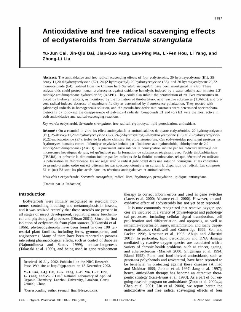
chromatography and preparative thin-layer chromatographyof an alcoholic extract of the whole S. strangulata plant.Their structures were identified by spectroscopic methodsand two-dimensional NMR as 20-hydroxyecdysone (E1),25-deoxy-11,20-dihydroxyecdysone (E2), 24-(2-hydroxy-ethyl)-20-hydroxyecdysone (E3), and 20-hydroxyecdysone-20,22-monoacetonide (E4), as reported previously (Fig. 1)(Dai et al. 2001). (–)-Epigallocatechin gallate (EGCG) wasisolated from green-tea leaves by extraction with methanol,water, and ethyl acetate, consecutively, and chromatographicseparation on a Sephadex LH-20 column, with reference toprocedures reported previously (Nonaka et al. 1983). Thestructures and purity of the compounds were confirmed by[1H]- and [13C]-NMR spectra and HPLC, as reported previ-ously (Fig. 1) (Jia et al. 1998).
Thiobarbituric acid (TBA), butylated hydroxytoluene(BHT), reduced L-glutathione (GSH), and 1,6-diphenyl-1,3,5-hexatriene (DPH) were purchased from Sigma (St.Louis, Mo.); 2,2�-azobis(2-amidinopropane hydrochloride)(AAPH) and galvinoxyl were from Aldrich (Milwaukee,
Wisc.); (±)-�-tocopherol was from Merck (Darmstadt,Germany). All other chemicals were of the highest qualityavailable.
Assay for hemolysis of human erythrocytesHuman erythrocytes were separated from heparinized
blood that was drawn from a healthy donor. The blood wascentrifuged at 1000 × g for 10 min to separate the erythro-cytes from plasma, and the erythrocytes were washed threetimes with phosphate-buffered saline (PBS) (pH 7.4). Duringthe last washing, the cells were centrifuged at 1000 × g for10 min to obtain a consistently packed cell preparation. A5% suspension of human erythrocytes in PBS waspreincubated with antioxidants at 37°C for 5 min, to whichAAPH was added to initiate hemolysis. The ecdysteroidsand �-tocopherol were dissolved in dimethyl sulfoxide(DMSO) before the experiment, and the volume of DMSOsolution added to the erythrocyte suspension was less than0.1% v/v of the reaction mixture. GSH and EGCG were dis-solved in PBS and added directly to the reaction mixture.The reaction mixture was gently shaken at 37°C. Aliquots ofthe reaction mixture was taken out at specific intervals anddiluted with 10 volumes of 0.15 M NaCl. The absorbance(A1) of the supernatant at 540 nm was measured. Similarly,the reaction mixture was treated with distilled water to yieldcomplete hemolysis, and the absorbance (A2) at 540 nm ofthe supernatants after centrifugation at 1000 × g for 10 minwas measured. The percentage hemolysis was calculatedfrom the ratio of the measurements as follows: (A1/A2) ×100 (Miki et al. 1987). The lag phase of the oxidativehemolysis was calculated as described by Palozza et al.(1992) by drawing a straight line through the linear portionof the propagation phase until it intercepts the abscissa. Ev-ery experiment was repeated three times and the values rep-resent means ± SE of the three experiments.
© 2002 NRC Canada
1188 Can. J. Physiol. Pharmacol. Vol. 80, 2002
Fig. 1. Molecular structures of the ecdysteroids, galvinoxyl, and epigallocatechin gallate (EGCG).
0
5
25
75
95
100
0
5
25
75
95
100
0
5
25
75
95
100
0
5
25
75
95
100
Preparation of rat liver microsomesFemale Wister rats weighing 250 ± 20 g were starved
overnight before cervical dislocation. Liver microsomeswere prepared by tissue homogenization with ice-cold STEbuffer (0.25 M sucrose – 0.01 M Tris (pH 7.4) with 1 mMEDTA) in a motor-driven glass homogenizer (Greenwald1985). Microsomal vesicles were isolated by removal of thenuclear faction at 8000 × g for 10 min and removal of mito-chondrial fraction at 18 000 × g for 10 min. The microsomalfraction was sedimented in a Hitachi 55P-72 ultracentrifuge(Hitachi High Technologies, Tokyo, Japan) at 105 000 × gfor 60 min and washed twice with 0.15 M KCl at 105 000 ×g for 30 min. The membranes, suspended in 0.1 M potas-sium phosphate buffer (pH 7.5), were stored in a deepfreezer maintained at –20°C. Microsomal protein was deter-mined by the method of Lowry et al. (1951).
Microsomal peroxidation as measured by TBA-reactivesubstance formation
The formation of TBA-reactive substances (TBARS) wasused to monitor lipid peroxidation (Buege and Aust 1978).Rat liver microsomes were incubated at 37°C in 0.1 M po-tassium phosphate buffer (pH 7.5) and made up to a finalprotein concentration of 1.0 mg·mL–1. The peroxidation wasinitiated by 50 �M FeSO4 and 200 �M cysteine. After30 min, 2 mL of TCA–TBA–HCl reagent (15% w/v trichloro-acetic acid, 0.37% thiobarbituric acid, and 0.25 N HCl)was added to the reaction mixture, together with 0.02% w/vBHT. This amount of BHT completely prevents the forma-tion of any nonspecific TBARS (Palozza et al. 1992). Thesolution was heated for 15 min in a boiling water bath. Aftercooling, the precipitate was removed by centrifugation. Thelevel of TBARS in the supernatant was determined at532 nm by use of an extinction coefficient of 1.56 ×10⁵ M–1·cm–1 (Buege and Aust,1978). Every experiment wasrepeated three times and the values represent means ± SE ofthe three experiments.
Microsomal peroxidation as measured by membranefluidity
The membrane fluidity of rat liver microsomes was mea-sured by fluorescence depolarization using DPH as the lipidprobe (Shintzek and Barrenholz 1974). A small volume ofDPH solution (2 mM) in tetrahydrofuran was injected withrapid stirring into 1000 volumes of PBS (pH 7.4) at roomtemperature. The suspension was stirred for 2 h. In a typicalexperiment, a mixture containing 1.5 mL PBS, 200 �gmicrosomal protein, 1.5 mL of 2 �M DPH, 100 �MFeSO4, and 750 �M cysteine was incubated at 25°C for30 min. Fluorescence polarization was measured in a Hitachi850 spectrofluorimeter (Hitachi High Technologies)equipped with a polarizer, using an excitation wavelength of362 nm and emission wavelength of 430 nm (Dave et al.1981). The degree of polarization (P) was calculated by theformulas P = (I0,0 – GI0,90)/(I0,0 + GI0,90) and G =I90,0/I90,90, where I0,0 is the fluorescence intensity of theemitted light when the excitation and emission lightpolarizers are both vertical, I0,90 is the fluorescence intensitywhen the excitation polarizer is vertical and the emission po-larizer is horizontal; I90,90 is the fluorescence intensity whenthe the excitation and emission light polarizers are both hori-
zontal; I90,0 is the fluorescence intensity when the excitationpolarizer is horizontal and the emission polarizer is vertical;and G is a correction factor. A smaller value of P demon-strates a greater lipid membrane fluidity.
The microviscosity of membrane fluidity, �, of mem-branes was calculated by the formula � = 2P/(0.46 – P).
Reaction with galvinoxyl radicalThe reaction kinetics of galvinoxyl (5 �M) with
ecdysteroids (10–100 �M) in ethanol solution were moni-tored spectrophotometrically at 429 nm (Tsychiya et al.1985) with a Hitachi model 557 UV spectrometer (HitachiHigh Technologies) at 37°C.
Statistical analysisValues are means ± SE. For most experiments, means
were compared using Student’s t test to evaluate statisticaldifferences.
Results and discussion
Inhibition of AAPH-induced erythrocyte hemolysis byecdysteroids
Erythrocyte membranes are rich in polyunsaturated fattyacids, which are very susceptible to oxidative stress medi-ated by free radicals. AAPH is a water-soluble azo com-pound that could decompose at physiological temperaturesto generate alkyl radicals, which can attack membrane lipidsfrom outside of erythrocytes and eventually cause oxidativemembrane damage and hemolysis (Miki et al. 1987; Ma etal. 2000a,b). Since AAPH is water soluble and the rate offree-radical generation from AAPH can be easily controlledand measured, it has been extensively used as a free-radicalinitiator for biological and related studies (Liu et al. 2000;Zhou et al. 2000a,b) and the hemolysis induced by AAPHprovides a good approach for studying membrane damageinduced by free radicals (Miki et al. 1987; Kuang et al.1994). Human erythrocytes were stable and little hemolysisoccurred within 4 h when incubated in PBS under air at37°C in the absence of AAPH. Addition of AAPH inducedfast hemolysis after a short inhibition period, demonstratinglipid peroxidation and the presence of endogenous antioxi-dants in human erythrocyte membranes, such as �-tocopheroland ubiquinol-10 (Miki et al. 1987), which can trap the initi-ating and (or) propagating radicals to inhibit hemolysis. Ithas been proven that the inhibition period is correlated in aconcentration-dependent manner with the concentration ofAAPH (Ma et al. 2000).
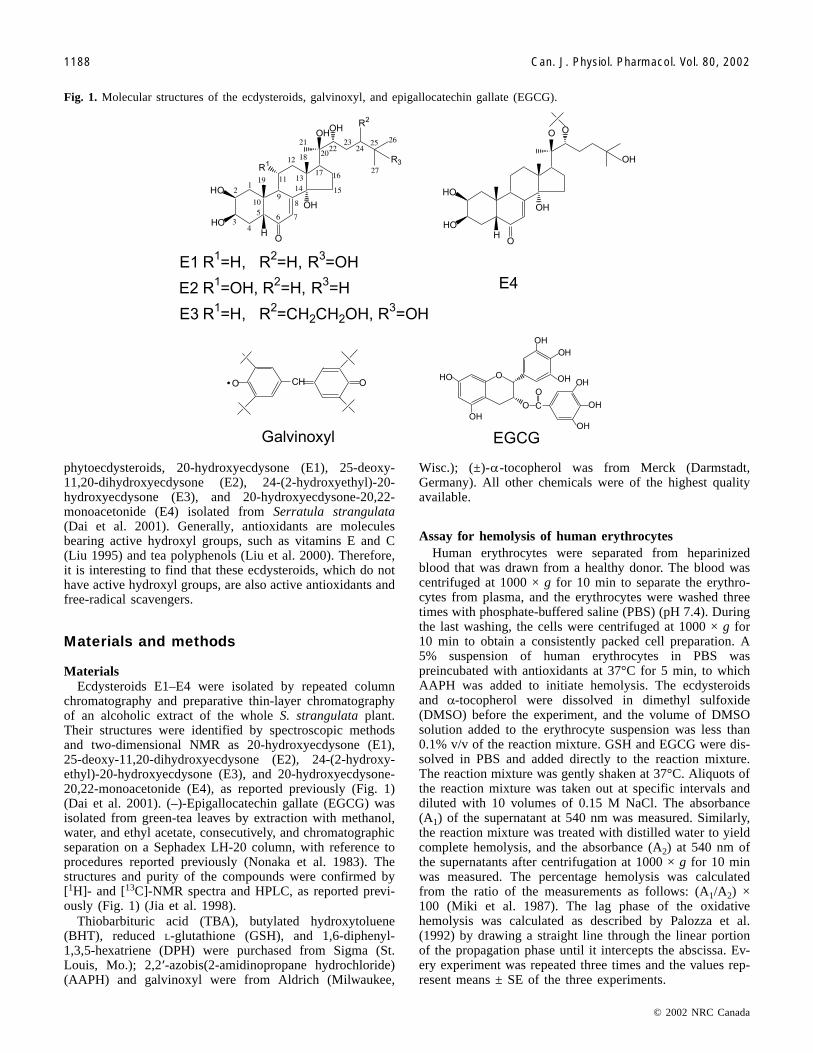
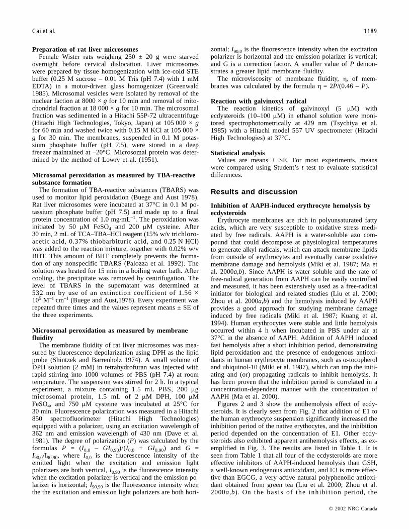
Figures 2 and 3 show the antihemolysis effect of ecdy-steroids. It is clearly seen from Fig. 2 that addition of E1 tothe human erythrocyte suspension significantly increased theinhibition period of the native erythocytes, and the inhibitionperiod depended on the concentration of E1. Other ecdy-steroids also exhibited apparent antihemolysis effects, as ex-emplified in Fig. 3. The results are listed in Table 1. It isseen from Table 1 that all four of the ecdysteroids are moreeffective inhibitors of AAPH-induced hemolysis than GSH,a well-known endogenous antioxidant, and E3 is more effec-tive than EGCG, a very active natural polyphenolic antioxi-dant obtained from green tea (Liu et al. 2000; Zhou et al.2000a,b). On the basis of the inhibi t ion period, the
© 2002 NRC Canada
Cai et al. 1189
0
5
25
75
95
100
0
5
25
75
95
100
0
5
25
75
95
100
0
5
25
75
95
100
inhibitory activity against AAPH-induced erythrocytehemolysis follows the sequence of E3 > EGCG � E1 > E2 >E4 > GSH.
Inhibition of AAPH-induced erythrocyte hemolysis bythe ecdysteroids and exogenous �-tocopherol
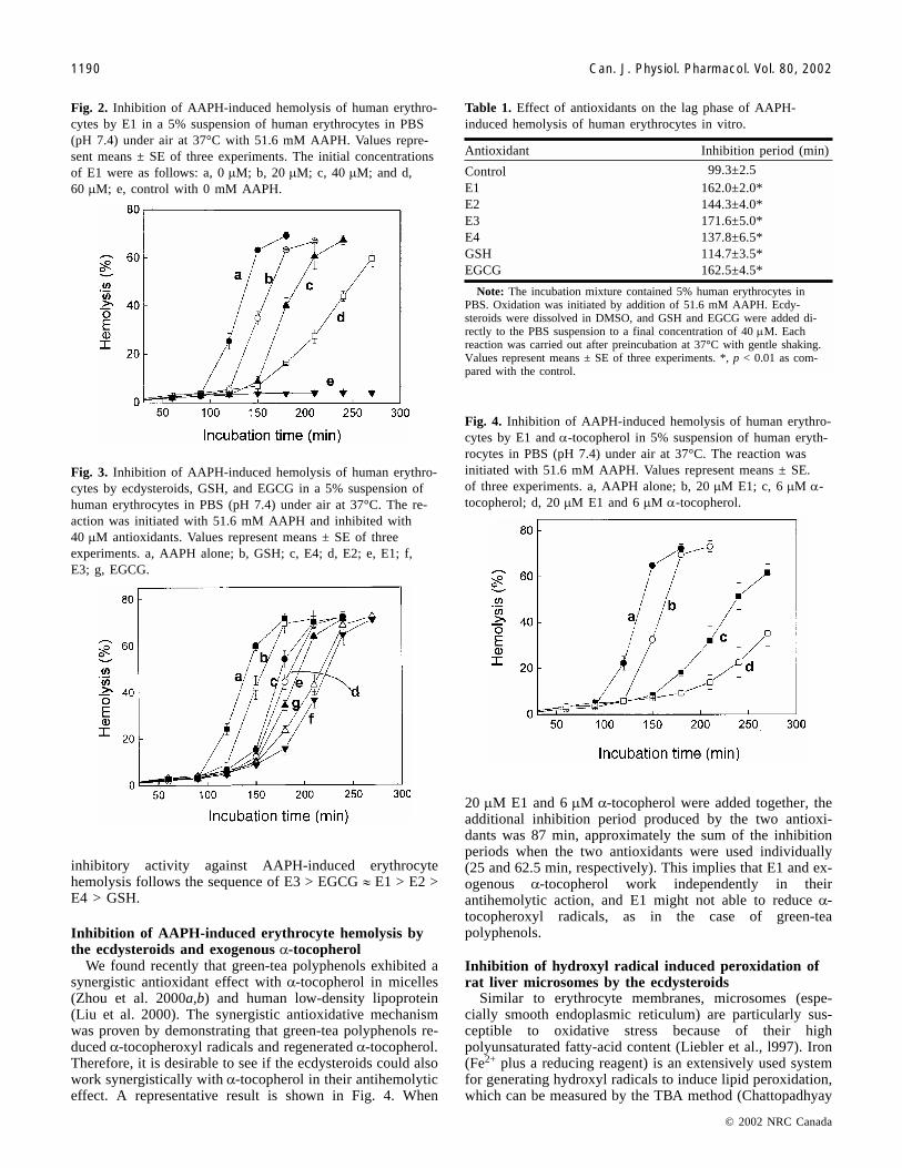
We found recently that green-tea polyphenols exhibited asynergistic antioxidant effect with �-tocopherol in micelles(Zhou et al. 2000a,b) and human low-density lipoprotein(Liu et al. 2000). The synergistic antioxidative mechanismwas proven by demonstrating that green-tea polyphenols re-duced �-tocopheroxyl radicals and regenerated �-tocopherol.Therefore, it is desirable to see if the ecdysteroids could alsowork synergistically with �-tocopherol in their antihemolyticeffect. A representative result is shown in Fig. 4. When
20 �M E1 and 6 �M �-tocopherol were added together, theadditional inhibition period produced by the two antioxi-dants was 87 min, approximately the sum of the inhibitionperiods when the two antioxidants were used individually(25 and 62.5 min, respectively). This implies that E1 and ex-ogenous �-tocopherol work independently in theirantihemolytic action, and E1 might not able to reduce �-tocopheroxyl radicals, as in the case of green-teapolyphenols.
Inhibition of hydroxyl radical induced peroxidation ofrat liver microsomes by the ecdysteroids
Similar to erythrocyte membranes, microsomes (espe-cially smooth endoplasmic reticulum) are particularly sus-ceptible to oxidative stress because of their highpolyunsaturated fatty-acid content (Liebler et al., l997). Iron(Fe2+ plus a reducing reagent) is an extensively used systemfor generating hydroxyl radicals to induce lipid peroxidation,which can be measured by the TBA method (Chattopadhyay
© 2002 NRC Canada
1190 Can. J. Physiol. Pharmacol. Vol. 80, 2002
Fig. 2. Inhibition of AAPH-induced hemolysis of human erythro-cytes by E1 in a 5% suspension of human erythrocytes in PBS(pH 7.4) under air at 37°C with 51.6 mM AAPH. Values repre-sent means ± SE of three experiments. The initial concentrationsof E1 were as follows: a, 0 �M; b, 20 �M; c, 40 �M; and d,60 �M; e, control with 0 mM AAPH.
Fig. 3. Inhibition of AAPH-induced hemolysis of human erythro-cytes by ecdysteroids, GSH, and EGCG in a 5% suspension ofhuman erythrocytes in PBS (pH 7.4) under air at 37°C. The re-action was initiated with 51.6 mM AAPH and inhibited with40 �M antioxidants. Values represent means ± SE of threeexperiments. a, AAPH alone; b, GSH; c, E4; d, E2; e, E1; f,E3; g, EGCG.
Antioxidant Inhibition period (min)
Control 99.3±2.5E1 162.0±2.0*E2 144.3±4.0*E3 171.6±5.0*E4 137.8±6.5*GSH 114.7±3.5*EGCG 162.5±4.5*
Note: The incubation mixture contained 5% human erythrocytes inPBS. Oxidation was initiated by addition of 51.6 mM AAPH. Ecdy-steroids were dissolved in DMSO, and GSH and EGCG were added di-rectly to the PBS suspension to a final concentration of 40 �M. Eachreaction was carried out after preincubation at 37°C with gentle shaking.Values represent means ± SE of three experiments. *, p < 0.01 as com-pared with the control.
Table 1. Effect of antioxidants on the lag phase of AAPH-induced hemolysis of human erythrocytes in vitro.
Fig. 4. Inhibition of AAPH-induced hemolysis of human erythro-cytes by E1 and �-tocopherol in 5% suspension of human eryth-rocytes in PBS (pH 7.4) under air at 37°C. The reaction wasinitiated with 51.6 mM AAPH. Values represent means ± SE.of three experiments. a, AAPH alone; b, 20 �M E1; c, 6 �M �-tocopherol; d, 20 �M E1 and 6 �M �-tocopherol.
0
5
25
75
95
100
0
5
25
75
95
100
0
5
25
75
95
100
0
5
25
75
95
100
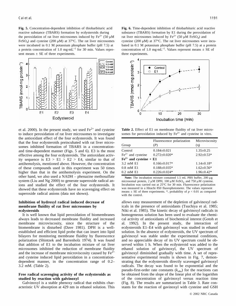
et al. 2000). In the present study, we used Fe2+ and cysteineto induce peroxidation of rat liver microsomes to investigatethe antioxidant effect of the four ecdysteroids. It was foundthat the four ecdysteroids preincubated with rat liver micro-somes inhibited formation of TBARS in a concentration-and time-dependent manner (Figs. 5 and 6). E3 is the mosteffective among the four ecdysteroids. The antioxidant activ-ity sequence is E3 > E1 > E2 > E4, similar to that ofantihemolysis, mentioned above. However, the concentrationof these compounds used in this experiment was 50 timeshigher than that in the antihemolysis experiment. On theother hand, we also used a NADH – phenazine methosulfatesystem (Liu and Ng 2000) to generate superoxide radical an-ions and studied the effect of the four ecdysteroids. Itshowed that these ecdysteroids have no scavenging effect onsuperoxide radical anions (data not shown).
Inhibition of hydroxyl radical induced decrease ofmembrane fluidity of rat liver microsomes byecdysteroids
It is well known that lipid peroxidation of biomembranesalways leads to decreased membrane fluidity and increasedmembrane microviscosity, since the structure of thebiomembrane is disturbed (Dave 1981). DPH is a well-established and efficient lipid probe that can insert into lipidbilayers for monitoring membrane fluidity by fluorescencepolarization (Shintzek and Barrenholz 1974). It was foundthat addition of E1 to the incubation mixture of rat livermicrosomes inhibited the reduction of membrane fluidityand the increase of membrane microviscosity caused by Fe2+
and cysteine induced lipid peroxidation in a concentration-dependent manner, in the concentration range of 0.2–3.2 mM. (Table 2).
Free radical scavenging activity of the ecdysteroids asstudied by reaction with galvinoxyl
Galvinoxyl is a stable phenoxy radical that exhibits char-acteristic UV absorption at 429 nm in ethanol solution. This
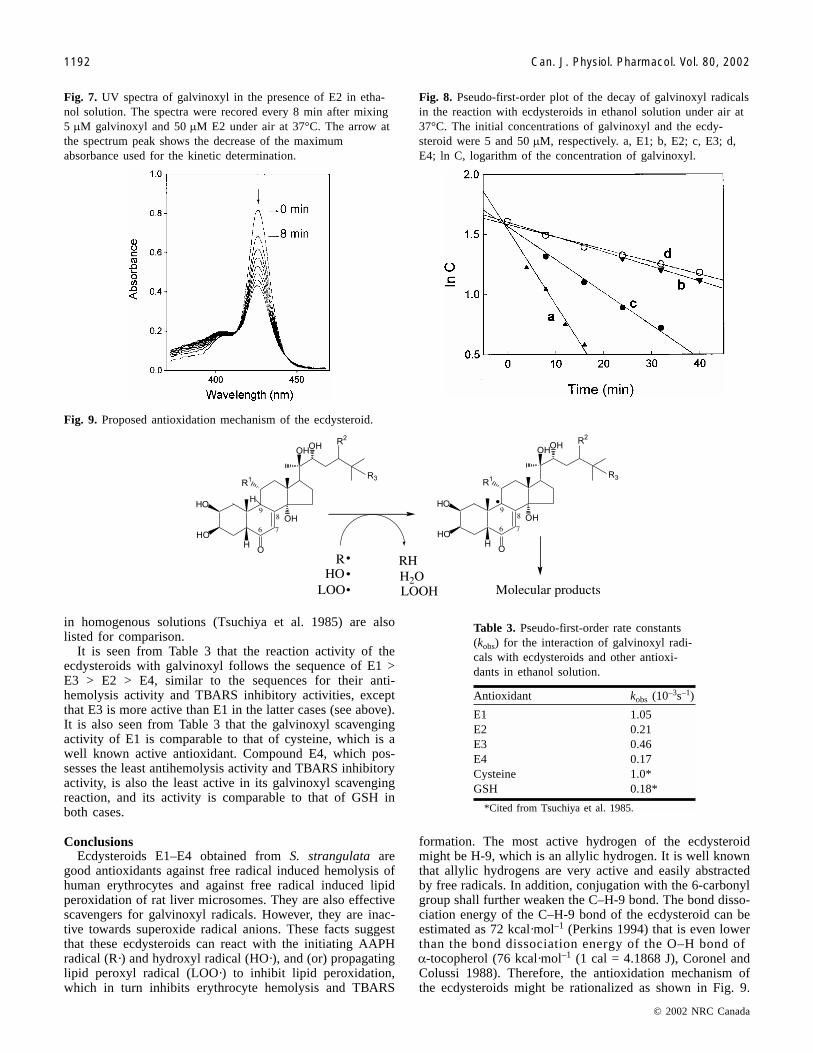
allows easy measurement of the depletion of galvinoxyl rad-icals in the presence of antioxidants (Tsuchiya et al. 1985;Niki et al. 1985). The kinetic decay of galvinoxyl radicals inhomogeneous solution has been used to evaluate the chemi-cal activity of antioxidants of biochemical interest (Gotoh etal. 1992). In the present study, the interaction ofecdysteroids E1–E4 with galvinoxyl was studied in ethanolsolution. In the absence of ecdysteroids, the UV spectrum ofgalvinoxyl was stable under the experimental conditions,and no appreciable decay of its UV spectrum could be ob-served within 1 h. When the ecdysteroid was added to theethanol solution of galvinoxyl, the UV spectrum ofgalvinoxyl diminished gradually with time. A set of repre-sentative experimental results is shown in Fig. 7, demon-strating that the ecdysteroids directly scavenged galvinoxylradicals. The decay was found to be exponential and thepseudo-first-order rate constants (kobs) for the reactions canbe obtained from the slope of the linear plot of the logarithmof the concentration of galvinoxyl versus reaction time(Fig. 8). The results are summarized in Table 3. Rate con-stants for the reaction of gavinoxyl with cysteine and GSH
© 2002 NRC Canada
Cai et al. 1191
Fig. 5. Concentration-dependent inhibition of thiobarbituric acidreactive substance (TBARS) formation by ecdysteroids duringthe peroxidation of rat liver microsomes induced by Fe2+ (50 �MFeSO4) and cysteine (200 �M) at 37°C. The rat liver microsomeswere incubated in 0.1 M potassium phosphate buffer (pH 7.5) ata protein concentration of 1.0 mg·mL–1 for 30 min. Values repre-sent means ± SE of three experiments.
Fig. 6. Time-dependent inhibition of thiobarbituric acid reactivesubstance (TBARS) formation by E1 during the peroxidation ofrat liver microsomes induced by Fe2+ (50 �M FeSO4) andcysteine (200 �M) at 37°C. The rat liver microsomes were incu-bated in 0.1 M potassium phosphate buffer (pH 7.5) at a proteinconcentration of 1.0 mg·mL–1. Values represent means ± SE ofthree experiments.
GroupFluorescence polarization(P)
Microviscosity(�)
Control 0.184±0.021 1.35±0.25Fe2+ and cysteine 0.272±0.020* 2.92±0.53*Fe2+ and cysteine + E13.2 mM E1 0.166±0.017* 1.14±0.18*0.8 mM E1 0.188±0.035* 1.62±0.56*0.2 mM E1 0.226±0.024* 1.96±0.42*
Note: The incubation mixture contained 1.5 mL PBS buffer, 200 �gmicrosomal protein, 2 �M DPH, 100 �M FeSO4, and 750 �M cysteine.Incubation was carried out at 25°C for 30 min. Fluorescence polarizationwas measured in a Hitachi 850 fluorophotometer. The values representmeans ± SE of three experiments. *, probability of p < 0.01 as comparedwith the control.
Table 2. Effect of E1 on membrane fluidity of rat liver micro-somes for peroxidation induced by Fe2+ and cysteine in vitro.
0
5
25
75
95
100
0
5
25
75
95
100
0
5
25
75
95
100
0
5
25
75
95
100
in homogenous solutions (Tsuchiya et al. 1985) are alsolisted for comparison.
It is seen from Table 3 that the reaction activity of theecdysteroids with galvinoxyl follows the sequence of E1 >E3 > E2 > E4, similar to the sequences for their anti-hemolysis activity and TBARS inhibitory activities, exceptthat E3 is more active than E1 in the latter cases (see above).It is also seen from Table 3 that the galvinoxyl scavengingactivity of E1 is comparable to that of cysteine, which is awell known active antioxidant. Compound E4, which pos-sesses the least antihemolysis activity and TBARS inhibitoryactivity, is also the least active in its galvinoxyl scavengingreaction, and its activity is comparable to that of GSH inboth cases.
ConclusionsEcdysteroids E1–E4 obtained from S. strangulata are
good antioxidants against free radical induced hemolysis ofhuman erythrocytes and against free radical induced lipidperoxidation of rat liver microsomes. They are also effectivescavengers for galvinoxyl radicals. However, they are inac-tive towards superoxide radical anions. These facts suggestthat these ecdysteroids can react with the initiating AAPHradical (R·) and hydroxyl radical (HO·), and (or) propagatinglipid peroxyl radical (LOO·) to inhibit lipid peroxidation,which in turn inhibits erythrocyte hemolysis and TBARS
formation. The most active hydrogen of the ecdysteroidmight be H-9, which is an allylic hydrogen. It is well knownthat allylic hydrogens are very active and easily abstractedby free radicals. In addition, conjugation with the 6-carbonylgroup shall further weaken the C–H-9 bond. The bond disso-ciation energy of the C–H-9 bond of the ecdysteroid can beestimated as 72 kcal·mol–1 (Perkins 1994) that is even lowerthan the bond dissociation energy of the O–H bond of�-tocopherol (76 kcal·mol–1 (1 cal = 4.1868 J), Coronel andColussi 1988). Therefore, the antioxidation mechanism ofthe ecdysteroids might be rationalized as shown in Fig. 9.
© 2002 NRC Canada
1192 Can. J. Physiol. Pharmacol. Vol. 80, 2002
Fig. 7. UV spectra of galvinoxyl in the presence of E2 in etha-nol solution. The spectra were recored every 8 min after mixing5 �M galvinoxyl and 50 �M E2 under air at 37°C. The arrow atthe spectrum peak shows the decrease of the maximumabsorbance used for the kinetic determination.
Fig. 8. Pseudo-first-order plot of the decay of galvinoxyl radicalsin the reaction with ecdysteroids in ethanol solution under air at37°C. The initial concentrations of galvinoxyl and the ecdy-steroid were 5 and 50 �M, respectively. a, E1; b, E2; c, E3; d,E4; ln C, logarithm of the concentration of galvinoxyl.
Antioxidant kobs (10–3s–1)
E1 1.05E2 0.21E3 0.46E4 0.17Cysteine 1.0*GSH 0.18*
*Cited from Tsuchiya et al. 1985.
Table 3. Pseudo-first-order rate constants(kobs) for the interaction of galvinoxyl radi-cals with ecdysteroids and other antioxi-dants in ethanol solution.
Fig. 9. Proposed antioxidation mechanism of the ecdysteroid.
0
5
25
75
95
100
0
5
25
75
95
100
0
5
25
75
95
100
0
5
25
75
95
100
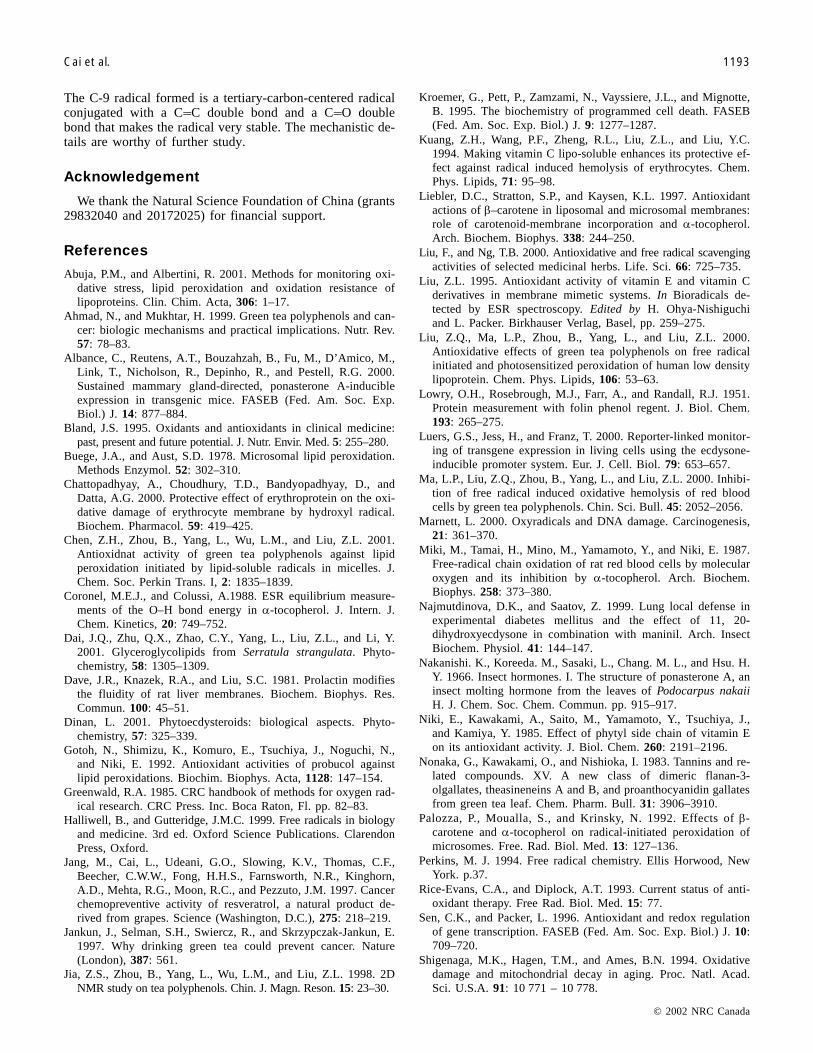
The C-9 radical formed is a tertiary-carbon-centered radicalconjugated with a C�C double bond and a C�O doublebond that makes the radical very stable. The mechanistic de-tails are worthy of further study.
Acknowledgement
We thank the Natural Science Foundation of China (grants29832040 and 20172025) for financial support.
References
Abuja, P.M., and Albertini, R. 2001. Methods for monitoring oxi-dative stress, lipid peroxidation and oxidation resistance oflipoproteins. Clin. Chim. Acta, 306: 1–17.
Ahmad, N., and Mukhtar, H. 1999. Green tea polyphenols and can-cer: biologic mechanisms and practical implications. Nutr. Rev.57: 78–83.
Albance, C., Reutens, A.T., Bouzahzah, B., Fu, M., D’Amico, M.,Link, T., Nicholson, R., Depinho, R., and Pestell, R.G. 2000.Sustained mammary gland-directed, ponasterone A-inducibleexpression in transgenic mice. FASEB (Fed. Am. Soc. Exp.Biol.) J. 14: 877–884.
Bland, J.S. 1995. Oxidants and antioxidants in clinical medicine:past, present and future potential. J. Nutr. Envir. Med. 5: 255–280.
Buege, J.A., and Aust, S.D. 1978. Microsomal lipid peroxidation.Methods Enzymol. 52: 302–310.
Chattopadhyay, A., Choudhury, T.D., Bandyopadhyay, D., andDatta, A.G. 2000. Protective effect of erythroprotein on the oxi-dative damage of erythrocyte membrane by hydroxyl radical.Biochem. Pharmacol. 59: 419–425.
Chen, Z.H., Zhou, B., Yang, L., Wu, L.M., and Liu, Z.L. 2001.Antioxidnat activity of green tea polyphenols against lipidperoxidation initiated by lipid-soluble radicals in micelles. J.Chem. Soc. Perkin Trans. I, 2: 1835–1839.
Coronel, M.E.J., and Colussi, A.1988. ESR equilibrium measure-ments of the O–H bond energy in �-tocopherol. J. Intern. J.Chem. Kinetics, 20: 749–752.
Dai, J.Q., Zhu, Q.X., Zhao, C.Y., Yang, L., Liu, Z.L., and Li, Y.2001. Glyceroglycolipids from Serratula strangulata. Phyto-chemistry, 58: 1305–1309.
Dave, J.R., Knazek, R.A., and Liu, S.C. 1981. Prolactin modifiesthe fluidity of rat liver membranes. Biochem. Biophys. Res.Commun. 100: 45–51.
Dinan, L. 2001. Phytoecdysteroids: biological aspects. Phyto-chemistry, 57: 325–339.
Gotoh, N., Shimizu, K., Komuro, E., Tsuchiya, J., Noguchi, N.,and Niki, E. 1992. Antioxidant activities of probucol againstlipid peroxidations. Biochim. Biophys. Acta, 1128: 147–154.
Greenwald, R.A. 1985. CRC handbook of methods for oxygen rad-ical research. CRC Press. Inc. Boca Raton, Fl. pp. 82–83.
Halliwell, B., and Gutteridge, J.M.C. 1999. Free radicals in biologyand medicine. 3rd ed. Oxford Science Publications. ClarendonPress, Oxford.
Jang, M., Cai, L., Udeani, G.O., Slowing, K.V., Thomas, C.F.,Beecher, C.W.W., Fong, H.H.S., Farnsworth, N.R., Kinghorn,A.D., Mehta, R.G., Moon, R.C., and Pezzuto, J.M. 1997. Cancerchemopreventive activity of resveratrol, a natural product de-rived from grapes. Science (Washington, D.C.), 275: 218–219.
Jankun, J., Selman, S.H., Swiercz, R., and Skrzypczak-Jankun, E.1997. Why drinking green tea could prevent cancer. Nature(London), 387: 561.
Jia, Z.S., Zhou, B., Yang, L., Wu, L.M., and Liu, Z.L. 1998. 2DNMR study on tea polyphenols. Chin. J. Magn. Reson. 15: 23–30.
Kroemer, G., Pett, P., Zamzami, N., Vayssiere, J.L., and Mignotte,B. 1995. The biochemistry of programmed cell death. FASEB(Fed. Am. Soc. Exp. Biol.) J. 9: 1277–1287.
Kuang, Z.H., Wang, P.F., Zheng, R.L., Liu, Z.L., and Liu, Y.C.1994. Making vitamin C lipo-soluble enhances its protective ef-fect against radical induced hemolysis of erythrocytes. Chem.Phys. Lipids, 71: 95–98.
Liebler, D.C., Stratton, S.P., and Kaysen, K.L. 1997. Antioxidantactions of �–carotene in liposomal and microsomal membranes:role of carotenoid-membrane incorporation and �-tocopherol.Arch. Biochem. Biophys. 338: 244–250.
Liu, F., and Ng, T.B. 2000. Antioxidative and free radical scavengingactivities of selected medicinal herbs. Life. Sci. 66: 725–735.
Liu, Z.L. 1995. Antioxidant activity of vitamin E and vitamin Cderivatives in membrane mimetic systems. In Bioradicals de-tected by ESR spectroscopy. Edited by H. Ohya-Nishiguchiand L. Packer. Birkhauser Verlag, Basel, pp. 259–275.
Liu, Z.Q., Ma, L.P., Zhou, B., Yang, L., and Liu, Z.L. 2000.Antioxidative effects of green tea polyphenols on free radicalinitiated and photosensitized peroxidation of human low densitylipoprotein. Chem. Phys. Lipids, 106: 53–63.
Lowry, O.H., Rosebrough, M.J., Farr, A., and Randall, R.J. 1951.Protein measurement with folin phenol regent. J. Biol. Chem.193: 265–275.
Luers, G.S., Jess, H., and Franz, T. 2000. Reporter-linked monitor-ing of transgene expression in living cells using the ecdysone-inducible promoter system. Eur. J. Cell. Biol. 79: 653–657.
Ma, L.P., Liu, Z.Q., Zhou, B., Yang, L., and Liu, Z.L. 2000. Inhibi-tion of free radical induced oxidative hemolysis of red bloodcells by green tea polyphenols. Chin. Sci. Bull. 45: 2052–2056.
Marnett, L. 2000. Oxyradicals and DNA damage. Carcinogenesis,21: 361–370.
Miki, M., Tamai, H., Mino, M., Yamamoto, Y., and Niki, E. 1987.Free-radical chain oxidation of rat red blood cells by molecularoxygen and its inhibition by �-tocopherol. Arch. Biochem.Biophys. 258: 373–380.
Najmutdinova, D.K., and Saatov, Z. 1999. Lung local defense inexperimental diabetes mellitus and the effect of 11, 20-dihydroxyecdysone in combination with maninil. Arch. InsectBiochem. Physiol. 41: 144–147.
Nakanishi. K., Koreeda. M., Sasaki, L., Chang. M. L., and Hsu. H.Y. 1966. Insect hormones. I. The structure of ponasterone A, aninsect molting hormone from the leaves of Podocarpus nakaiiH. J. Chem. Soc. Chem. Commun. pp. 915–917.
Niki, E., Kawakami, A., Saito, M., Yamamoto, Y., Tsuchiya, J.,and Kamiya, Y. 1985. Effect of phytyl side chain of vitamin Eon its antioxidant activity. J. Biol. Chem. 260: 2191–2196.
Nonaka, G., Kawakami, O., and Nishioka, I. 1983. Tannins and re-lated compounds. XV. A new class of dimeric flanan-3-olgallates, theasineneins A and B, and proanthocyanidin gallatesfrom green tea leaf. Chem. Pharm. Bull. 31: 3906–3910.
Palozza, P., Moualla, S., and Krinsky, N. 1992. Effects of �-carotene and �-tocopherol on radical-initiated peroxidation ofmicrosomes. Free. Rad. Biol. Med. 13: 127–136.
Perkins, M. J. 1994. Free radical chemistry. Ellis Horwood, NewYork. p.37.
Rice-Evans, C.A., and Diplock, A.T. 1993. Current status of anti-oxidant therapy. Free Rad. Biol. Med. 15: 77.
Sen, C.K., and Packer, L. 1996. Antioxidant and redox regulationof gene transcription. FASEB (Fed. Am. Soc. Exp. Biol.) J. 10:709–720.
Shigenaga, M.K., Hagen, T.M., and Ames, B.N. 1994. Oxidativedamage and mitochondrial decay in aging. Proc. Natl. Acad.Sci. U.S.A. 91: 10 771 – 10 778.
© 2002 NRC Canada
Cai et al. 1193
0
5
25
75
95
100
0
5
25
75
95
100
0
5
25
75
95
100
0
5
25
75
95
100
© 2002 NRC Canada
1194 Can. J. Physiol. Pharmacol. Vol. 80, 2002
Shintzek, M., and Barrenholz, Y. 1974. Dynamics of the hydrocar-bon layer in liposomes of lecithin and sphingomyelin containingdicetylphosphate. J. Biol. Chem. 249: 2652–2657.
Takasaki, M., Tokuda, H., Nishino, H., and Konoshima, T. 1999.Cancer chemopreventative agents (antitumor-promoters) fromAjuga decumbens. J. Nat. Prod. 62: 972–975.
Tsuchiya, J., Yamada, T., Nike, E., and Kamiya, Y. 1985. Interac-tion of galvinoxyl radical with ascorbic acid, cysteine, andglutathione in homogeneous solutions and in aqueous disper-sions. Bull. Chem. Soc. Jpn. 58: 326–330.
Zhou, B., Chen, Z., Jia, Z., Jia, Y., Zeng, L., Wu, L., Yang, L., andLiu, Z.L. 2000a. Kinetic EPR studies on bio-antioxidants. Appl.Magn. Reson. 18: 397–406.
Zhou, B., Jia, Z.S., Chen, Z.H., Yang, L., Wu, L.M., and Liu, Z.L.2000b. Synergistic antioxidant effect of green tea polyphenolswith �-tocopherol on free radical initiated peroxidation of lino-leic acid in micelles. J. Chem. Soc. Perkin Trans. I, 2: 785–791.
0
5
25
75
95
100
0
5
25
75
95
100
0
5
25
75
95
100
0
5
25
75
95
100