Embed Size (px)
Citation preview
DISSERTATION ON
“ROLE OF DEVASCULARISATION IN SURGICALLY MANAGED CASES OF JUVENILE NASOPHARYNGEAL
ANGIOFIBROMA”
Submitted in partial fulfillment of the requirements for
M.S. DEGREE BRANCH -IV OTORHINOLARYNGOLOGY
of
THE TAMILNADU DR. M.G.R. MEDICAL UNIVERISTY,
UPGRADED INSTITUTE OF OTORHINOLARYNGOLOGY
MADRAS MEDICAL COLLEGE
CHENNAI – 600 003
MARCH – 2009
CERTIFICATE
This is to certify that this dissertation entitled “ ROLE OF
DEVASCULARISATION IN SURGICALLY MANAGED CASES OF
JUVENILE NASOPHARYNGEAL ANGIOFIBROMA” submitted by Dr.
S.SHANMUGA ASHOK, appearing for M.S. E.N.T.. Branch IV Degree
examination in March 2009 is a bonafide record of work done by him under my
direct guidance and supervision in partial fulfillment of regulations of the Tamil
Nadu Dr. M.G.R. Medical University, Chennai. I forward this to the Tamil Nadu
Dr.M.G.R. Medical University, Chennai, Tamil Nadu, India.
DIRECTOR & PROFESSOR,
Upgraded Institute of Otorhinolaryngology,
Madras Medical College,
Government General Hospital,
Chennai – 600 003.
CERTIFICATE
This is to certify that this dissertation “ROLE OF
DEVASCULARISATION IN SURGICALLY MANAGED CASES OF
JUVENILE NASOPHARYNGEAL ANGIOFIBROMA” submitted by
Dr.S.SHANMUGA ASHOK, appearing for M.S. E.N.T.. Branch IV Degree
examination in March 2009 is a bonafide record of work done by him under my
direct guidance and supervision in partial fulfillment of regulations of the Tamil
Nadu Dr. M.G.R. Medical University, Chennai. I forward this to the Tamil Nadu
Dr.M.G.R. Medical University, Chennai, Tamil Nadu, India.
DEAN,
Madras Medical College,
Government General Hospital,
Chennai – 600 003.
DECLARATION
I solemnly declare that the dissertation “ ROLE OF
DEVASCULARISATION IN SURGICALLY MANAGED CASES OF
JUVENILE NASOPHARYNGEAL ANGIOFIBROMA” is done by me at the
Madras Medical College and Government General Hospital, Chennai during 2006-
2008 under the guidance and supervision of Prof. S. KULASEKARAN,
M.S., D.L.O.
This dissertation is submitted to The Tamilnadu Dr. M.G.R Medical
University, towards partial fulfillment of regulation for the award of M.S.
DEGREE IN OTORHINOLARYNGOLOGY (BRANCH–IV).
DR.S.SHANMUGA ASHOK
M.S. E.N.T. post graduate,
Place: Upgraded Institute of Otorhinolaryngology ,
Date: Madras Medical College
ACKNOWLEDGEMENT
I am immensely grateful to Prof. S. Kulasekaran, M.S. D.L.O., The
Director, Upgraded Institute of Otorhinolaryngology, for his valuable guidance,
suggestions, encouragement and help in conducting this study.
I am greatly indebted to Prof. K. Balakumar M.S., D.L.O., Professor,
Upgraded Institute of Otorhinolaryngology, who encouraged and helped me
throughout this study.
I express my sincere gratitude to Ex-Director and Professor Late
Dr. A. K. Sukumaran M.S., D.L.O., for his valuable support in conducting the
study.
I would like to express my sincere gratitude to Prof.T.P.KALANITI,
M.D., The DEAN, Madras Medical College, for having permitted me to use the
hospital material in this study.
I express my sincere thanks to all the Assistant Professors, for their
thoughtful guidance throughout the work.
I thank the Secretary and Chairman of Institutional Ethical
Committee, Government General Hospital and Madras Medical College, Chennai.
I thank all my colleagues and friends for their constant encouragement
and valuable criticism.
Last but not least, I express my gratitude for the generosity shown by all
the patients who participated in the study.
I am extremely thankful to my family members for their continuous
support. Above all I thank God Almighty for His immense blessings.
CONTENTS AIM - 3 REVIEW OF LITERATURE - 7 SURGICAL ANATOMY - 8 MATERIALS AND METHODS - 29 SURGICAL PROCEDURES - 31 PREOPERATIVE DEVASCULARISATION - 47 OBSERVATION - 51 DISCUSSION - 53 CONCLUSION - 59 BIBILOGRAPHY - 61 PROFORMA - 65 MASTER CHART - 69
1
INTRODUCTION
Juvenile nasopharyngeal angiofibroma (JNA) is a benign, locally
aggressive, highly vascular tumour often occurring at the superior margin of the
sphenopalatine foramen occurring exclusively in adolescent males.
JNA is uncommon and accounts for approximately 0.5% of all head and
neck neoplasms.
The median age at diagnosis is 15 years. Presenting symptoms commonly
include nasal obstruction and epistaxis. Approximately 20% of patients have
evidence of skull base invasion at the time of diagnosis. JNAs may be hormonally
dependant. Hagen et al studied androgen receptor binding in cultured tumour
fibroblasts from 3 patients with JNA and demonstrated that cell proliferation
increased with testosterone and decreased with antiandrogens.
Spontaneous regression is unlikely but has been observed with increasing
age. The main stay of treatment of JNA is surgical excision. Preoperative role of
devascularizaion is discussed.
2
This study has been conducted in the Upgraded institute of
Otorhinolaryngology, Madras medical college, Chennai-03 during the period 2006
to 2009. The following is a discussion of the devascularization of surgically
managed advance cases of juvenile nasal angiofibroma.
3
AIMS OF THE STUDY
1. To study the age distribution, clinical presentations of JNA.
2. To Study and apply various Radiological investigations for assessment of the
exact extent and vascular supply of locally advanced cases of JNA.
3. To choose appropriate preoperative devascularization methods in surgically
managed advanced cases of JNA
4. To assess the effectiveness and complications of devascularizations and to
explore ways to minimize complications and optimize the outcome of treatment.
4
REVIEW OF LITERATURE
The history of JNA dates back to Hippocrates, who described removal
of a ‘hard nasal polyp’ using a midline nasal splitting incision. Harma presented a
comprehensive of the subject in 1958 and noted that piecemeal removal of the
tumour through the external nares was done till 1834. Chelius(1834) advanced the
understanding by reporting that less bleeding was encountered when the tumour
was removed along with its roots(Vascular pedicle).In 1906, Chaveau coined the
term juvenile nasopharyngeal angiofibroma.
THEORIES OF ORIGIN OF JNA
Ringertz(1938)
The tumour arose from the periosteum or the nasopharyngeal vault.
Som and Neffson(1940)
Inequalities in the growth of the bones forming the skull base resulted in
hypertrophy of underlying periosteum in response to hormonal influence.
5
Bensch and Ewing (1941)
The tumour probably arose from the embryonic fibrocartilage between basi-
occiput and basi-spenoid.
Brunner (1942)
Originated from Pharyngobasilar and Buccopharyngeal fascia.
Osborn (1959)
These were either hamartomas or residues of fetile erectile tissues which
were subject to hormonal influences.
Girgis and Fahmy (1973)
Postulated the tumour to be a paraganglioma. Existence of
paragangliomatous tissues around the terminal part of internal maxillary artery in
the pterygopalatine fossa of stillborn infants.
Angiofibromas may arise from vestiges of atrophied stapedial
artery,although it is not possible to validate such assertion.
Hughes
Angiofibroma arises from remnants of craniopharyngeal duct.
6
Wills
Angiofibroma is the result of inflammatory immune response.
Stenberg
Angiofibroma is a type of hemangioma.
Martin (1948)
Use of testosterone caused atropy of the tumour.
Schiff (1959)
Combination of oestrogen and testosterone caused atrophy of tumour.
Muller (1999)
Transforming growth Factor Beta-1 may play a role in the stromal cell
proliferation and angiogenesis associated with JNA.
Hwang and Patterson
Presence of Androgen receptors in Angiofibromas explains the unique
clinicopathologic features of these tumours.
7
Reham-Schimd (1998)
Expression of CD-34 antigen in nasopharyngeal angiofibroma indicates the
increased proliferative potential of endothelium of small vessel component of
angiofibroma.
Lloyd G Howard 1999
Recurrence of angiofibroma was due to the residual mass in the
pterygopalatine fossa and invasion of sphenoid bone.
This can be differentiated radiologically by persistence of widening of distance
between root of pterygoid and posterior wall of maxilla in cases of recurrences
from pterygopalatine fossa.
Recurrence from the mass invading sphenoid shows pushing of root of
pterygoid anteriorly towards maxilla and reducing the distance between root of
pterygoid and posterior wall of maxilla.
Susan C. Abraham,Elizabeth A. Montgomery,
Francis M. Giardiello, and Tsung-Teh Wu (2001)
Frequent occurrences of JNA in cases of Familial Adenomatous
Polyposis(FAP) due to frequent beta-catenin mutations in JNA.
8
SURGICAL ANATOMY
Site of origin
Described by Neal.
Periosteum of the posterosuperior aspect of sphenopalatine
foramen.
At the junction between sphenoidal process of the palatine
bone and the pterygoid process of the sphenoid bone.
Mode of spread
- Spreads submucosally along preformed anatomical pathways
through foramina and fissures.
Anterior
- Spreads to the nasal septum, lateral nasal wall.
- Involves the ethmoid sinus, erodes the posterior wall of the maxillary sinus and
involves it.
9
Posterior
- Involves the anterior wall of sphenoid sinus and spreads into the sinus.
- Erodes the pterygoid plates.
Lateral
-Pterygopalatine fossa and may present in the cheek.
-Extends further laterally into the infratemporal fossa.
Inferior
-Nasopharynx and postnasal space.
Superior
-To orbit via the inferior orbital fissure and then superior orbital fissure and middle
cranial fossa.
Intracranial extension
-seen in 10 % of the cases
-mostly extradural
-The tumor reaches the cranial vault through three paths.
Two lateral pathways(lateral to carotid and cavernous sinus)
1. Through superior orbital fissure
2. Directly through the greater wing of sphenoid from
pterygopalatine fossa and Infratemporal fossa.
10
Medial pathway(medial to the carotid and cavernous sinus)
-through the sphenoid sinus and sella turcica making surgical excision
difficult.
-Intradural involvement is rare,when present is a contraindication for surgery.
PTERYGOPALATINE FOSSA
Boundaries:
Anterior : Superior aspect of posterior surface of maxilla.
Posterior : Root of pterygoid process and adjoining part of
Anterior surface of the greater wing of sphenoid.
Medial : Upper part of perpendicular plate of palatine bone.
Orbital and sphenoidal process of the palatine bone also
take part.
Lateral : Opens into infratemporal fossa,through the pterygo-
Maxillary fissure.
Superior : Undersurface of body of sphenoid.
Inferior : Closed by the pyramidal process of palatine in the
angle between the maxilla and the pterygoid process.
11
Communications:
Anterior : With the orbit through the medial end of the inferior
orbital fissure.
Posterior : Middle cranial fossa through the foramen rotundum.
: Foramen lacerum through the pterygoid canal.
: Pharynx ,through the palatovaginal canal.
Lateral : Infratemporal fossa through the pterygomaxillary
fissure.
Medially : With the nose through the sphenopalatine foramen.
Inferiorly : With the oral cavity through the greater and lesser
Palatine canals.
Contents:
- Third part of maxillary artery and its branches.
- Maxillary nerve and its two branches zygomatic and
posterior alveolar.
- Pterygopalatine ganglion and its numerous branches
Containing fibres of the maxillary nerve mixed with
autonomic fibres.
12
SPHENOPALATINE FORAMEN
Boundaries:
Superiorly : Body of the sphenoid
Anteriorly : By orbital process of palatine bone.
Posteriorly : Sphenoidal process of palatine bone.
Inferiorly : Perpendicular plate of palatine bone.
Oval in shape. Located at the posterior end of the superior meatus(85%) or the
middle meatus(5%) or is bridged by the basilar lamina of the middle turbinate
(10%).
Sphenopalatine vessels emerges out from pterygopalatine fossa, through the
foramen into the nasal cavity.
INFRATEMPORAL FOSSA
Roof:
Medially by the Infratemporal surface of the greater wing of the sphenoid and
by a small part of the temporal bone.
Laterally:
Roof is incomplete where the infratemporal fossa communicates with the
temporal fossa through the gap deep to the zygomatic arch.
13
Floor: The floor is open
Medial wall:
Formed by the lateral pterygoid plate and the pyramidal process of the
palatine bone.
Lateral wall:
Formed by the ramus of the mandible.
Anterior wall:
Formed by the infratemporal surface of the maxilla and by the medial
surface of the zygomatic bone.
Anterior and medial walls are separated in the upper parts by the
pterygomaxillary fissure through which the infratemporal fossa communicates with
the pterygopalatine fossa.
Upper end of the pterygomaxillary fissure is continuous with the anterior
part of the inferior orbital fissure through which the infratemporal fossa
communicates with the orbit.
Posterior wall: -It is open.
Contents of the infratemporal fossa:
1. Muscles - Medial and lateral pterygoid,temporalis.
2. Vessels - Maxillary artery and its five branches.
3. Nerves - Mandibular division of fifth cranial nerve.
14
TEMPORAL FOSSA
Boundaries:
Above - Temporal line of frontal bone.
Below - Upper border of zygomatic arch laterally and by
infratemporal crest of greater wing of sphenoid
bone medially.
Anterior wall- Zygomatic bone and parts of sphenoid and
frontal bones.
Floor - ‘H’ shaped suture where four bones (frontal,
Parietal,sphenoid,temporal) join.
MIDDLE CRANIAL FOSSA
It is shaped like a butterfly,narrow and shallow in the middle and deep
on each side.
Boundaries:
Anterior :
-Posterior border of lesser wing of the sphenoid.
-Anterior clinoid process and anterior margin of Chiasmaticus.
15
Posterior :
-Superior border of petrous temporal bone.
Floor :
- Dorsum sellae of sphenoid.
Lateral:
-Greater wing of sphenoid.
-Antero-inferior angle of parietal bone.
-Squamous temporal bone.
Medially:
- Body of sphenoid
CONTENTS:
Median area :
Sulcus chiasmatis - leads to optic canal.
Optic canal - Bounded laterally by lesser wing of sphenoid.
- Infront and behind by two roots of lesser wing.
- Medially by body of the sphenoid.
16
Sella turcica - Contains tuberculum sellae in front
- Hypophyseal fossa in the middle
- Dorsum sellae behind
Lateral area :
Lodges the temporal lobe of the brain.
SUPERIOR ORBITAL FISSURE:
Boundaries
-above by the lesser wing of sphenoid
-below by the greater wing of sphenoid
-medially by body of sphenoid
Foramina in greater wing of the sphenoid:
Foramen rotundum:
- leads anteriorly to the pterygopalatine fossa.
- lies posteroinferior to medial end of superior orbital fissure.
- Transmits maxillary nerve.
17
Foramen ovale :
- lies posterolateral to foramen rotundum and leads inferior to
the infratemporal fossa.
- Transmits mandibular nerve, lesser petrosal nerve, accessory
Meningeal artery,emissary vein connecting the cavernous
sinus with pterygoid plexus of veins.
Foramen spinosum:
- Lies posterolateral to foramen ovale
- Leads inferiorly to infratemporal fossa
- Transmits middle meningeal artery,meningeal branch of
mandibular nerve,posterior trunk of middle meningeal vein.
Emissary sphenoidal foramen(foramen of Vesalius):
-Transmits emissary vein connecting cavernous sinus with the
pterygoid plexus of veins.
Groove for the middle meningeal vessels:
-Leads forwards from foramen spinosum.
Foramen Lacerum:
- Lies posteromedial to foramen ovale,lower part of canal filled with
cartilage.
18
-No significant structure passes through it except for meningeal
branch of the ascending pharyngeal artery and an emissary vein
from the cavernous sinus.
-Upper part of the canal is traversed by the internal carotid artery.
- in upper part of the foramen,the greater petrosal nerve unites with
the deep petrosal nerve to form the nerve of pterygoid canal,which
leaves the canal by entering the pterygoid canal in the anterior wall of
foramen lacerum.
ORBIT
Inferior orbital fissure:
Transmits,
1. Maxillary nerve
2. Zygomatic nerve
3. Orbital branches of pterygopalatine ganglion
4. Infraorbital vessels
5. Communication between inferior ophthalmic veins
and the pterygoid plexus of veins.
19
Superior orbital fissure :
Lateral part
-lacrimal nerve
-frontal nerve
-trochlear nerve
-superior ophthalmic vein
-meningeal branch of lacrimal artery
Middle part
-Upper and lower divisions of the oculomotor nerve
-Nasociliary nerve
-Oculomotor nerve
-Abducent nerve
Medial part
-Inferior ophthalmic vein
-Symapathetic nerves from plexus around the internal
carotid artery.
20
CAVERNOUS SINUS
-Large venous sinus located in the middle cranial fossa,on
either side of the body of sphenoid bone.Its interior is divided
into number of spaces by trabeculae.
- Floor of sinus is formed by endosteal dura .
- Lateral wall,roof and medial wall is formed by meningeal dura
- Anteriorly it extends upto medial end of the superior orbital
fissure.
-Posteriorly upto apex of petrous temporal bone.
Structures in the lateral wall of cavernous sinus:
Occulomotor nerve
Trochlear nerve
Ophthalmic nerve
Maxillary nerve
Structures passing through the centre of the sinus:
Internal carotid artery
Abducent nerve
Structures in the sinus are separated from blood by
Endothelial lining.
21
Incoming channels:
From orbit:
- Superior ophthalmic vein.
- Branch of inferior ophthalmic vein.
- Central vein of retina.
From brain:
- Superficial middle cerebral vein.
- Inferior cerebral veins from temporal lobe.
From meninges:
- Sphenopareietal sinus
- Frontal trunk of meningeal vein.
Draining channels :
- Transverse sinus through the superior petrosal sinus.
- Internal jugular vein through the inferior petrosal sinus and
through a plexus around internal carotid artery.
- Pterygoid plexus through emissary veins passing through foramen
ovale,foramen lacerum,emissary sphenoidal foramen.
- Facial vein through superior ophthalmic vein
22
- Right and left cavernous sinus communicate with each other via
the anterior and posterior intercavernous sinuses and through the
basilar plexus of veins.
- All these communications are valveless communications and blood
can flow through them in either direction.
INTERNAL MAXILLARY ARTERY
Larger terminal branch of external carotid artery given off behind the neck of
mandible .
Divided into three parts (by the lateral pterygoid muscle)
First part(Mandibular part) - runs along the lower border of lateral
pterygoid muscle.
Second part(Pterygoid part) - runs either superficial or deep to the lower
head of lateral pterygoid muscle.
Third part(Pterygopalatine part) – passes between two heads of the
lateral pterygoid and through the
pterygomaxillary fissure to enter the
pterygopalatine fossa and lies in front
of the pterygopalatine ganglion.
23
Branches:
First part:
1. Deep auricular
2. Anterior tympanic
3. Middle meningeal
4. Accessory meningeal
5. Inferior alveolar
Second part:
1. Deep temporal
2. Pterygoid
3. Masseter
4. Buccal
Third part:
1. Posterior superior alveolar
2. Infraorbital
3. Greater palatine
4. Pharyngeal branch
5. Artery of pterygoid canal
6. Sphenopalatine artery
24
EXTERNAL CAROTID ARTERY
- Branch of common carotid artery
- Lies anterior to internal carotid artery
- Begins in the carotid triangle at the upper border of thyroid
cartilage.
- Terminates behind the neck of mandible into maxillary and
superficial temporal artery.
Branches
Anterior
Superior thyroid, lingual, Facial.
Posterior
Occipital, posterior auricular.
Medial
Ascending pharyngeal .
Lateral
Maxillary, Superficial temporal.
25
PATHOLOGY OF JNA
Gross appearance
Firm,rubbery lobulated swellings ,nodularity increases with
age.Nasopharyngeal portion which is covered by mucosa
appears pink and extrapharyngeal portion appears white or
grey.
On section
Tumour has reticulated,whorly or spongy appearance and lacks
a true capsule.Edge however is sharply demarcated and easily
distinguishable from surrounding structures(Pseudo capsule).
Microscopy
- composed of vascular and stromal components
- Vascular component predominates in early lesions
- Stromal component predominates in long standing cases.
- Vascular pattern is composed of large thin walled sinusoidal
vessels lined by flattened epithelium, unsupported by a muscular
coat and the closer the vessels to the surface the smaller they
become.
- In long standing cases , there is tendency towards gradual
compression of sinusoids so that the lining endothelial cells are
26
pushed against each other like cords ,while in others intravascular
thrombosis occurs.
- The stroma is composed of interlacing bundles of collagen in
which stromal cells are seemed to radiate outwards from the
vessels and in which localized areas of myxomatous degeneration
may be observed.
Immunochemistry
Androgen receptor has been detected in the stromal cells of
JNA. Antiandrogen hormonal therapy has been used preoperatively to
suppress JNA growth and enhance the resectability of the tumor and
minimize bleeding during surgery.
Familial adenomatous polyposis (FAP) results from germline
mutations in the adenomatous polyposis coli (APC) gene that
subsequently alters the [beta]-catenin signaling pathway. In addition
to a high tendency for the development of colorectal neoplasms,
patients with FAP develop JNA 25 times more frequently than an age-
matched population. Recently, activating [beta]-catenin mutation
without the APC gene mutation has been reported in sporadic
JNA.Expression of various growth factors, such as transforming
27
growth factor [beta]1 (TGF-[beta]1) and insulin-like growth factor II
(IGF-II), has also been detected in JNA.
DIFFERENTIAL DIAGNOSIS:
1. Antrochoanal polyp
2. Nasopharyngeal carcinoma
3. Craniopharyngioma
4. Chordoma
5. Chondroblastoma
6. Rhabdomyosarcoma
BIOPSY
- Since the diagnosis is easily done and biopsy is dangerous, routine
pre-op biopsy is not done.
- However Biopsy can be done in operating room under general
anaesthesia in following circumstances(Batsakis)
1. Female patients
2. Ulcerative lesions
3. Suspected rhabdomyosarcoma
28
RADIOLOGICAL INVESTIGATIONS
Computerised tomography helps in accurate preoperative assessment of the
extent of the disease and aids in surgical planning and also in postoperative
surveillance.
MRI scan is a very helpful tool in assessing the intracranial extension of
JNA and also MR angiography aids in identifying the feeding vessel in a non
invasive manner.
The tumour typically shows a “vascular blush” in the capillary phase which
is typical of JNA.
HOLMAN MILLER SIGN
Forward bowing of the posterior wall of maxilla due to mass in the
pterygopalatine fossa noted in the CT scan axial and saggital view.
HANDOUSA SIGN
Maxillo-mandibular space widening due to extension of the mass from
infratemporal fossa into the cheek.
29
Materials and methods
The present study consists of a series of ‘32’histopathologically proven
cases of JNA. Some of the cases had been operated already for the disease and
presented with recurrence.
This study was done at the Upgraded institute of otorhinolaryngology,
Madras medical college,Chennai-03 during the period of June 2006 to oct 2008
Detailed history of each patient was taken. History of previous surgery and
thorough clinical examination was done.
Diagnostic nasal endoscopy was done in all cases.
Following provisional diagnosis , all the cases were subjected to CT
scanning with contrast. Selected cases also underwent MRI,MRA.
Angiography was done in cases with intracranial extension where blood
supply is shared between in internal and external carotid arterial systems is
expected and in cases planned for pre-op embolisation.
A Proforma was prepared for the study to record the history, clinical
examination and investigations.
30
Grading of the tumour was done using Radowski staging system and
appropriate surgical approach was employed.
Cases were studied for effectveness of devsaclarization.
Inclusion criteria
- Only advanced cases(Stage II a and above) were included in the
study.
- Only cases histopathologically proved postoperatively were
included in the study.
Exclusion criteria
- Cases with small tumours(Stage I ) were excluded from the study.
- Cases with histopathology other than JNA or doubtful HPE were
excluded.
Study period and centre
- June 2006 to Oct 2008
- Upgraded institute of Otorhinolaryngology,MMC
31
Postoperative follow up
- Diagnostic nasal endoscopy in all patients every month for 6
months and once every 3 months thereafter.
- CT scan was done in patients suspected to have recurrence.
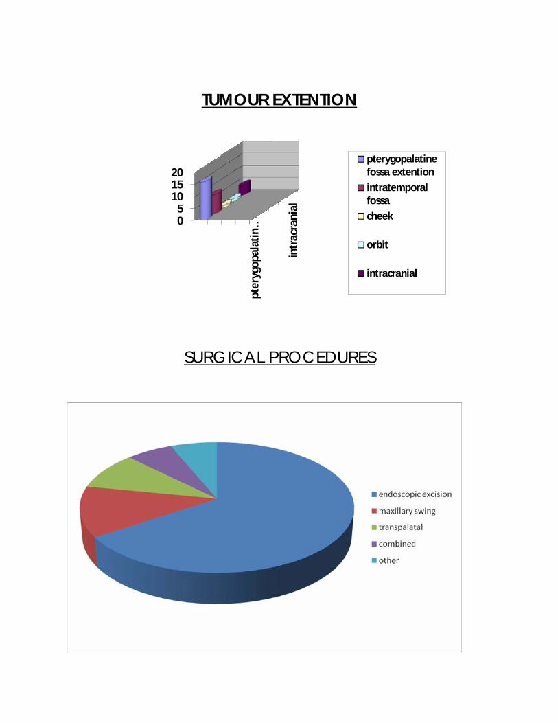
SURGICAL PROCEDURES
In this study , Endoscopic transnasal approach was used in 23 cases.
Maxillary swing procedure was done in 4 cases.
Transpalatine approach, previously the commonest approach employed was
used only in three cases.
Combined transantral and transpalatine excision was done in one case.
Combined Transpalatine and endoscopic excision was done in one case.
Transantral excision was done in one case.
Lateral rhinotomy was employed in one case.
All the cases were done under Hypotensive anaesthesia (Halothane, Propofol,
Nitroglycerine drip)
32
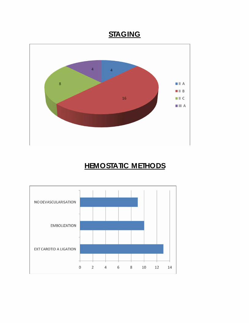
External carotid artery ligation was done in 13 cases.
Embolisation was done in 10 cases.
No devascularisation was done in 9 cases.
Amount of Blood transfused :-
Minimum – no transfusion.
Maximum –3 units.
Recurrent tumour was seen in 8 cases. Recurrent tumour was commonly seen in
the pterygopalatine fossa.
Postoperative post nasal packing was done in all cases and were removed on
second postoperative day in the operating room.
All cases were postoperatively treated with Inj.Cefotaxim 1gm i.v B.D,
Inj.Metronidazole 500 mg i.v B.D for 7 to 10 days.
Postoperative follow up was done every month for six months and every 6 months
thereafter. It consists of diagnostic nasal endoscopy and CT if any suspicion of
recurrence.
10 cases were lost to follow up after 5 months.
33
SURGICAL PROCEDURES AND PRE
OPERATIVE DEVASCULARIZATION DONE IN
OUR STUDY
In this study Endoscopic Transnasal approach was the commonest
approach employed. External carotid artery ligation was done 13 cases.
Embolisation was done for 10 cases.
Performed under general anaesthesia. Throat packing was done in all
cases.
Endoscopic transnasal approach :
Position of the patient- supine with 15˚ head end elevation.
Nose is gently packed with cottonoids soaked in 4% xylocaine and 1 in
10,000 adrenaline solution, 15 minutes prior to the surgery.
The nasal cavity is first visualized with 0 degree Karl storz endoscope.
Local infiltration is given over the root of middle turbinate, uncinate process, nasal
septum,posterior end of middle turbinate using 1in 100,000 adrenaline and 1%
xylocaine mixture.
34
The procedure starts with uncinectomy,partial middle turbinectomy and a
wide middle meatal antrostomy.Complete ethmoidectomy and sphenoidotomy
were done.
The posterior wall of maxillary sinus,often pushed forwards and thinned
out in cases of JNA is removed and the Pterygopalatine fossa is exposed.
The attachments of the mass are easy to delineate due to the excellent
direct visualization offered by endoscope. Sphenopalatine artery is cauterized.The
attachments are systematically released using monopolar cautery.
The extensions into the pterygopalatine fossa and infratemporal fossa are
removed using 45 deg and 70 deg endoscopes and angled instruments.
After releasing from its attachments the mass is pushed posteriorly into
the nasopharynx.
Doyens mouth gag is applied and the softpalate is retracted using suction
catheters passed through the opposite nostril.
The mass is now delivered in toto through the oral cavity.Any remnants
can be visualized,cauterized and removed under direct vision through the nose.
Postnasal packing is done using either 14 size Foleys catheter or
conventional postnasal packs. Anterior nasal packing is done if anterior oozing is
still present. Wide spectrum intravenous antibiotics are used for 3 – 4 days.
35
Post nasal packs are removed on postop day 2 in the operating room
,keeping things ready for a repack. The patient is discharged on 4 th or 5 th postop
day.
MAXILLARY SWING APPROACH
This is relatively new procedure described by wei and associates for
approach to nasopharyngeal tumours. We have used this approach quite often in
our institute for excision of extensive stage II c /III lesions where endoscopic
approach cannot be used.
Wide spectrum antibiotic coverage is started on the day of surgery.The
procedure is done under general anaesthesia. Temporary tarsoraphhy is done. After
an initial extrernal carotid artery ligation on the side of the lesion, a Weber-
Ferguson- Longmire Facial incision is made. The incision extends 0.5 cm below
the lower lid margin and is extended laterally till the preauricular region. The
incision curves around the medial canthus and extends along the nasomaxillary
groove and wind around the nasolabial sulcus to reach the philtrum. Now a lip
splitting incision is made and the incision is extended along the gingivobuccal
36
sulcus turned around the last molar tooth and comes forward between the two hard
palates in the midline.
Flaps are raised and contrary to maxillectomy the anterior maxillary wall is
left attached to the masseter flap. Osteotomies are made just below the infra orbital
rim,through the floor of the ipsilateral nasal cavity, and through the posterior wall
of the maxillary sinus. The pterygoid plates are then separated from the maxillary
tuberosity with a osteotome.
The maxilla, which is left pedicled to the masseter and cheek flap can now
be swung out of the way to gain wide exposure of the nasopharynx and
parapharyngeal space.
Now the entire tumour with all its extension can be seen. The mass is
separated from its attachments by blunt finger dissection. The mass is deliverd in
toto. Hemostasis is secured by placing hot saline pads in the cavity.
The maxilla is repositioned and fixed by wiring along the lines of
osteotomies. The skin is sutured with atraumatic 3-0 ethilon sutures. Ryles tube is
inserted. Oral feeds are started from second post op day. Sutures are removed on
the sixth post op day. Patient is discharged after 8 to 10 days.
37
TRANSPALATINE APPROACH
Anaesthesia – GA with orotracheal intubation, tracheostomy.
Position - Supine ,Rose position.
Boyle Davis mouth gag is applied .Care is taken to see that the blade of the
instrument does not occlude the anaesthetic tube or damage the posterior
pharyngeal wall by pressure.
The mucosa over the palate is infiltrated with 2 % xylocaine and 1:100000
adrenaline 5 minutes before making the incision. A Curved incision , bowed
forwards is made with a 15 size bard parker blade between the two maxillary
tuberosities,keeping internal to the greater palatine foramen.
Adequate exposure to the postnasal space can be obtained through an
insision that extends just in front of the posterior margin of the hard palate.
Incision is made through the mucosa and periosteum down to the bone.
Inorder to simplify subsequent suturing, it is easy to start by elevating for a short
distance, the edge of the mucoperiosteal flap on the anterior side of the incision.
The posterior flap is then separated completely from the undersurface of the hard
palate.
38
A small right angled dissector is used to separate the flap from the posterior
margin of the bone. But a knife may be needed to free the attachment to the
posterior spine of the hard palate.
Mucosa on the upper surface of the palate is divided transversely and the
postnasal space is examined.
Near the pterygo mandibular raphe and the pterygoid hamulus , tensor palati
muscle fans out from the tendon passing round the hamulus and if necessary it is
divided just medial to the hamulus. Soft palate is retracted by means of two rubber
catheters placed in the retracted site. Care is taken not to jeopardize the greater
palatine blood supply to the soft palate. By retracting the entire tumour is
visualized. The mass is then freed from its attachments by blunt dissection.
Then by using a periosteal elevator, mass is mobilised from sphenopalatine
foramen region. The mass is then held with a babcock forceps and the entire mass
is removed in toto by side by side movement along with continuous lift by
perisoteal elevator.
Cavity is packed with hot saline pad. After achieving hemostasis, the flaps
are sutured in the midline. Vicryl is used and the sutures are allowed to absorb by
themselves.
Ryles tube is inserted and oral feeds are started by second post op day. The
patient is discharged 8 to 10 days after the surgergery.
39
TRANS ANTRAL APPROACH
A temporary tarsoraphhy is done . Weber – Ferguson incision is made about
2-3 mm below the lid margin along the lower lid. This allows the skin to be
separated from the underlying orbicularis oculi and minimize the postoperative
edema.
Incisison curves inferiorly just lateral to the medial canthus and extends
along the nasomaxillary groove and the curves around the nasolabial fold to reach
the columella. Then a lip splitting incision is made and the incision is carried along
the gingivo buccal sulcus upto the maxillary tuberosity.
And the palatine of the incision is not done. The anterior wall of the maxilla
removed by osteotomy. Posterior wall of the maxilla is removed. Maxillary artery
is identified and ligated.
The mass can be visualized and released from its attachments and removed
in one piece. Hemostasis secured with hot saline packs. Wound closed in layers.
Cavity is packed with antibiotic smeared gauze and postnasal packing done.
40
LATERAL RHINOTOMY (MOURE’ s)
The upper part of the incision need to be no higher than the point midway
between the medial canthus and dorsum of the nose. To avoid an unsightly
depressed scar it should run just medial to the nasomaxillary groove but follow the
ala to finish within the ipsilateral nasal cavity.
All layers are divided including the nasal mucosa and retraction of the alar
region away from the incision is done.Periosteum and skin is elevated over the
nasal bone and the fronto nasal process of maxilla ,so as to allow full visualization
of the mass.
Medial maxillectomy is done and the mass is removed in toto after
separating from the attachments. Anterior and posterior nasal packing is done.
Wound is closed in layers.
41
COMBINED TRANSPALATINE AND
TRANS ANTRAL APPROACH
This approach was done in one of the cases in our study. It was done since
the tumour was extensive. But this approach does not result in any better exposure
than medial maxillectomy and resulted in more morbidity and prolonged hospital
stay.
COMBINED TRANSPALATINE AND
ENDOSCOPIC APPROACH
Used in one of the cases in our study, the procedure was started as an
endoscopic approach and after releasing the lateral attachments, transpalatine
approach was used to release attachments to the nasopharynx.
42
OTHER APPROACHES
SARDANA ‘S APPROACH
Sublabial incision is combined with transpalatine approach to facilitate
removal of lateral extensions of the mass. Sublabial incision is made from the
midline, extending upto the maxillary tuberosity.
Surgeon ‘s index finger is inserted into pterygoid space and blunt dissection
is used to free the tumour from its lateral attachments. It can then be delivered into
the nasopharynx.
FACIAL TRANSLOCATION
This procedure is done by the ENT surgeon jointly with the neuro surgeon.
A extended Weber- Ferguson incision is made and a inferiorly based flap is raised.
Cranio facial skeleton is exposed. Osteotomies are performed to free the
anterior face of the maxilla, the malar eminence,zygomatic arch,inferior and lateral
orbital rims and the orbital floor. Orbito-maxillary complex is removed and
preserved for later replacement.
An osteotomy is performed at the base of the coronoid
process and temporalis muscle is transposed inferiorly. A Fronto temporal
43
osteotomy is then performed and the foramen ovale , spinosum and rotundum as
well as the superior orbital fissure are identified.
Surgical field can extend from the contralateral Eustachian tube to the
ipsilateral geniculate ganglion and from the superior orbital fissure and cavernous
sinus area to the level of the hard palate. It included the nasopharynx ,clivus,
sphenoid,cavernous sinus and infratemporal fossa.
After tumour removal dura is repaired and temporalis muscle is mobilized to
line the defect. Orbito maxillary complex is then replaced and fixed. skin is sutured
in layers.
MIDFACIAL DEGLOVING ( Conley and Price )
Sublabial approach to the nasal and nasopharyngeal cavities in which the
midface is essentially degloved from the anterior maxillary facial skeleton by
extension of a sublabial incision bilaterally. This exposure provides generous
access to sinuses and nasopharyngeal cavity.
The maxillary antrum as well as the pterygomaxillary spaces are readily
accessible.
44
TRANSMANDIBULAR APPROACH (Kermen)
The incision begins vertically in front of the ear and carried down the neck
anterior to the sternomastoid muscle. Dissection is started in the neck by exposing
the carotid artery bifurcation at which level the external carotid is ligated.
Lower pole of parotid is dissected free and the vessels entering the substance
from below are transected. At this level one severs the insertion of masseter muscle
into the mandible. Incision is curved down to the periosteum at the angle of
mandible so as to expose its entire lateral aspect. Periosteum is stripped upwards
from the lateral aspect of the ascending ramus of the mandible.
Transection of the mandible is done with gigli saw at a point about 1 cm
below the coronoid and condyloid processes. Seperation and retraction of
mandibular fragments expose tubomuscular wall of the nasopharynx which is
incised on its lateral wall, so that its lumen is entered.
Tumour is exposed and dissected out. Nasopharynx is closed. Mandible is
approximated with stainless steel wires.
45
DENKERS MODIFICATION
Tumour in maxillary antrum and anterior part of the nose may removed by
sublabial incision as well as by performing a caldwel-luc antrostomy using
Denker’s modification of removing the lateral wall of the nose.
TRANSHYOID APPROACH
This is suitable for the tumour localized to nasopharynx without extension
into the surrounding structure. The disadvantage is that usually requires temporary
tracheostomy for two to three days.
TRIPLE APPROACH (HIRANANDANI)
Transpalatal and lateral rhinotomy approach is combined with caldwel
incision. Complete exposure of pterygo palatine fossa is possible by removing
posterior wall of maxillary antrum after opening the antrum through caldwel –Luc
incision.
Once the pterygo palatine fossa is exposed, tumour can be easily palpated
bimanually with one finger in fossa an another in the nasopharynx. The whole
tumour is removed in toto under direct vision.
46
MODIFIED BARBOSA TECHNIQUE
Weber Ferguson incision extending from the eye to the tragus is made.
Three bony cuts are made , two at the zygoma and one at the coronoid process to
reach the infratemoral fossa.
FISCH INFRATEMPORAL FOSSA APPROACH
TYPE- C
For extensive cases of JNA with intracranial extension. The morbidity of the
procedure includes conductive hearing loss on the involved side and anaesthesia
over the distribution of V2 and V3.
Endoscopic and KTP laser-assisted surgery
This is a recent modality of surgery where the endoscopic approach is
combined with KTP laser assisted dissection to minimize bleeding and improve
exposure and precision or tumour removal. Needs further evaluation before
accepting as a standard modality of treatment. sixth post op day. Patient is
discharged after 8 to 10 days.
47
PREOPERATIVE DEVASCULARISATION
External carotid artery ligation
Neck is extended by placing a sand bag under the shoulder and the head is
partially rotated to the other side. Curved incision following one of the skin folds
of the neck is made centered over the bifurcation of the common carotid artery at
the upper border of the thyroid cartilage.
Incision is carried through the platysma muscle to the deep cervical fascia.
The great auricular nerve should be preserved. Deep cervical fascia is divided
along its attachment to the sternomastoid muscle and infrahyoid muscles are
exposed by blunt dissection.
Common Facial vein is ligated and divided. Carotid sheath is opened to
expose the common carotid artery and jugular vein with the vagus nerve lying
posteriorly. Bifurcation of common carotid artery is at the level of the upper border
of the thyroid cartilage.
48
Posterior belly of the digastric muscle and the stylohyoid muscle should be
identified where they cross lateral to the internal and external carotid arteries and
retracted to expose the carotid bulb. Hypoglossal nerve should be carefully
identified where it crosses lateral to both the arteries.
External carotid artery is identified by finding one of its distal branches.
Variations in the origin and arrangement of all the branches of the external carotid
must be expected. External carotid artery is ligated with silk sutures. Deep cervical
fascia is approximated to the sternomastoid muscle. Skin including the platysma is
closed with interrupted silk sutures.
CAROTID ANGIOGRAM AND EMBOLISATION
Informed high risk consent is taken from the patient and his attendants. Groin is
shaved .Nil oral for an appropriate period prior to procedure to avoid aspiration
during a possible reaction to the contrast medium. Premedication with inj.
Pentazocine(fortwin) 30 mg im/iv is given.
49
The procedure is done within 48- 72 hours prior to the surgery. After
cleansing the area with a suitable preparation , 5 to 10 ml of 1- 2% xylocaine is
infiltrated around the femoral artery.
Percutaneous needle puncture of an artery (femoral) is done. Inner stylet is
removed when the needle is in the lumen of the artery and is replaced with guide
wire of appropriate diameter. The needle is removed and a catheter or introducer is
inserted over the wire.
Then the guide wire is withdrawn. Then injection of contrast through the
catheter is done(IOHEXOL). Maxillary artery and feeding vessel of the tumour is
identified by the tumour blush. Embolisation of the feeding vessel is done with gel
foam beads.
After the procedure is over , the catheter is withdrawn and tight bandage is
applied over the area. Wound site is checked at regular intervals. Patient should
remain on bed rest overnight following the procedure.
50
COMPLICATIONS
1. Hemorrhage
2. vascular thrombosis
3. vascular stenosis
4. pseudo aneurysm formation
5. local sepsis
6. allergy to contrast
7. air embolism
8. vasovagal reaction
9. loss of guide wire
10. Cerebro vascular accident with paralysis- 1 case
11. Death due to embolisation of ICA -1case
51
OBSERVATION
In this study of 32 locally advanced cases of Juvenile angiofibroma, the clinical
presentation was as follows
Mass in nasal cavity, nasopharynx, Pterygopalatine fossa – 16 cases
Mass extending to infratemporal fossa - 8 cases
Extension to the cheek - 1 case
Extension to orbit - 1 case
Intracranial extension - 4 cases
Clinical Presentation
1. Nasal obstruction – all cases
2. Epistaxis - 30 cases
3. Cheek involvement - 1 case
4. Proptosis - 1 case
52
One of our patient presented only with history of nasal obstruction and had no
history of epistaxis. The diagnosis was done based on clinical appearance of the
tumour and CT findings.
Treatment Modality
Surgery - 32 cases
1. Endoscopic excision -21 cases
2. Maxillary swing - 4 cases
3. Transpalatal excision - 3 cases
4. Combined approach - 2 cases
5. Other approaches - 2 cases
STAGING OF DISEASE
Stage II A – 4 cases (12.5%)
Stage II B – 16 cases (50%)
Stage II C – 8 cases (25%)
Stage III A – 3 cases (9.4%)
Stage III B – 1cases (3.1%)
53
DISCUSSION
In our case study , 32 cases of Juvenile nasopharyngeal angiofibroma were
managed by surgery. Hormonal therapy & radiotherapy were not tried in any of
our cases. In all the cases, history was evaluated carefully.
Of the 32 cases 18 cases were primary cases and 14 cases were recurrent
cases. The average age of presentation of primary cases was 15 years. The
youngest was 9 yrs and the oldest was 29 yrs old. The average age of presentation
of recurrent cases was 19 years. The youngest was 9 yrs and the oldest was 31 yrs.
Nasal obstruction and epistaxis were the commonest symptoms. Anosmia,
Rhinolalia, cheek swelling and proptosis were the other symptoms. The average
delay in diagnosis was 4 months.
Preoperative Computerised tomographic scanning in coronal and axial views
were done in all cases. Magnetic resonance imaging and MR angiography were
done in 7 cases. The extent of the tumour was assessed accurately in all cases.
Staging of the disease with Radowsky staging system based on CT and MRI
findings was done. Only stage II a and above cases were included in the study.
Carotid angiography with embolisation was done in 10 cases. The internal
maxillary artery was the commonest feeding vessel. We had one post embolisation
death and one case had quadriparesis following the procedure, which recovered
with conservative management.
54
Other hemostatic methods used. External carotid artery ligation was done in
13 cases. It is a safe and effective procedure and decreases the vascularity of the
tumour considerably.
Embolisation helps in decreasing the vascularity of the mass and also shrinks
the mass. It was done in 10 cases . We had two embolisation related
complications. Embolisation results in shrinkage of tumour leading to incomplete
resection.In our study embolisation done in 5 primary cases and 5 recurrent cases.
In this 10 cases endoscpic approach used 3 cases and external approach 5
cases.2 cases no surgical procedure done because of the embolisation related
complication.
The complication were death and cerebrovascular accident.these
complications were mainly seen in recurrent cases.
During embolisation minor complications noted all cases which includes
post embolisation fever and local pain.Both subsided after administration of
Tab.Paracetamol.Transient Bradycardia observed one case.It resolved after
administration of Atropine.
Major complication observed 2 cases.
1.Death
2.Cerebrovascular accident.
55
This complication was due to three factors
1. Selection of inappropriate embolic material.
2. Reflux of emboli.(caused by vascular spasm,injecting too many
particle too rapidly).
3. Failure to recognize potentially dangerous external-internal carotid
vascular anastamosis.(due to incomplete angiographic delineation or inadequate
anatomical analysis).
But in our study these complication seen in recurrent cases. In recurrent
cases multiple collateral formation were common. It is very difficult to delineate
the feeding vessel. Embolisation has its own hazards. Embolisation done 72 hours
prior to surgery otherwise multiple collateral formation occurs. Other method
,external carotid artery ligation done just prior to definitive procedure, that is on
the table. It is safe and simple procedure. Hence we prefer external carotid artery
ligation. Devascularisation were done on 20 cases. Of these 23 cases surgical
procedure done on 21 cases. No blood transfusion -7 cases. One unit was
transfused fourteen cases.9 cases no devascularisation was done. In these 9 cases ,
4 cases required minimum 4 units of blood transfusion. 5 cases required 3 units. In
devascularised tumors better delineation of the tumor was possible. Complete
removal of the tumor was easily done. Especially in endoscopic approach these
devascularisation procedures were verymuch useful. After devascularisation the
56
surgical field was clear, better delineation of the tumor, decreased surgical
duration, less need of postoperative blood transfusion and anesthesia related
complications were minimal. The major complication were seen in recurrent cases
especially after embolisation. These complications were due to collaterals
formation and embolic material that entered in internal carotid artery. Recurrence
rate was more in embolised cases. These were due to shrinkage of tumor after
embolisation, and incomplete removal. These recurrence rate was minimal after
external carotid artery ligation. Embolisation done for 10 cases. Maximum blood
transfusion in embolised cases were 1 units. Recurrence were 3 cases. Of these
embolisation related major complication were 2 cases. Recurrence was less after
externalcarotid ligation. Of these 13 cases recurrence were 1 cases. No
devascularisation was done in 9 cases. recurrence was 4 cases. Blood loss was
more and maximum transfusion recquired were 3 units.
Maxillary swing approach and removal was done in 4 cases. Two cases were
stage II C and two cases were stage III A. Two cases were embolised and two
cases were external carotid artery ligation done. Transnasal endoscopic surgery
done 23 cases. Of these 16 cases were devascularised. Externalcarotid artery
ligation done for 10 cases. Embolisation was done for 3 cases.
57
Transpalatine surgery, previously the most common surgical route for JNA
excision was done only in 2 cases. All cases were Stage II A. Embolisation done
for 1 case. External carotid artery ligation done for 1 case.
Combined Transpalatine and endoscopic excision was done in one case
(Stage II B).
Combined transantral and transpalatine approach was done in one case.
(Stage II B).
Lateral rhinotomy was done in one case (Stage II B)
The feeding vessel in all cases were internal maxillary artery..
Mass was sent for Histopathological examination in all cases for confirmation of
diagnosis. All the cases was treated with intravenous antibiotics ( Cefotaxim and
Metronidazole).
58
There were eight cases of recurrence. All the cases were taken up for
surgery. Cause of the recurrence was due to persistence of mass in the
pterygopalatine fossa.
Of these eight cases of recurrence, five cases underwent endoscopic excision
and two cases underwent maxillary swing procedure.One case underwent
transpalatine approach.
59
CONCLUSION
In this study , 32 cases were analysed. Nasal obstruction and epistaxis were
the common symptoms noticed in almost all cases.
CT scan was done in all cases . MRI and MRA was done in 7 cases. These
investigations were used to accurately assess the extent of the tumour,vascular
supply of the tumor and plan appropriate surgical procedure. Staging based on
Radowski ‘ s system, a widely accepted and comprehensive staging system was
employed. Intracranial spread was noted in three cases.
Carotid angiogram with embolistion was done in ten cases. The typical
capillary phase tumour blush was seen in all cases. The internal maxillary artery
was the feeding vessel in all cases. One patient died following the procedure and
another patient had quadriparesis.
External carotid artery ligation in our opinion is a safe and effective method
for decreasing blood loss during surgery. Combined with hypotensive anaesthesia
provides a very good blood less field, essential for complete tumour removal
60
the efficacy of a particular devascularisation procedure evaluated based on local
control and complications. External carotid artery ligation is the best procedure in
preoperative devascularisation of JNA excision. The advantages of this approach
are listed below,
1. Done on the same day of surgery.
2. No need of modern instruments
3. safe procedure
4. Decreased blood loss
5. Can be repeated any number of times in cases of recurrences with no
extra morbidity.
6. Decreased operating time
7. Better delineation of tumor because no shrinkage of the mass.
8. Recurrence rate is less because complete removal is possible.
The complications encountered in this study were,
1.Death
2.Cerebrovascular accident
3.Bleeding
4.Recurrence
61
Death and cerebrovascular accident mainly in embolised ,recurrent
cases.Embolisation better avoided in recurrent cases.No major complication
encounterd in external carotid artery ligation,both primary aswellas recurrent
cases.In our study external carotid artery ligation was the best way to devascularise
the surgically managed advance cases of juvenile nasopharyngeal angiofibroma.
62
Bibiliography
Martin H, Ehrlich HE, Abels JC. Juvenile nasopharyngeal
angiofibroma. Ann Surg 1948;127:513-536.
Bremer JW, Neel HB III, De Santo LW, et al. Angiofibroma: Treatment
trends in 150 patients during 40 years. Laryngoscope 1986; 96: 1321-1329.
Cansiz H, Guvenc MG, Sekecioglu N. Surgical approaches to juvenile
nasopharyngeal angiofibroma. J Craniomaxillofac Surg. 2006 Jan;34(1):3-8.
Epub 2005 Dec 15.
Cummings BJ, Blend R, Keane T, et al. Primary radiation therapy for
juvenile nasopharyngeal angiofibroma. Laryngoscope 1984; 94: 1599-1605.
Douglas R, Wormald PJ. Endoscopic surgery for juvenile nasopharyngeal
angiofibroma: where are the limits? Curr Opin Otolaryngol Head Neck
Surg. 2006 Feb;14(1):1-5.
Enepekides DJ. Recent advances in the treatment of juvenile angiofibroma.
Curr Opin Otolaryngol Head Neck Surg. 2004 Dec;12(6):495-499.
Hardillo JA, Vander Velden LA, Knegt PP. Denker operation is an effective
surgical approach in managing juvenile nasopharyngeal angiofibroma. Ann
Otol Rhinol Laryngol. 2004 Dec;113(12):946-950.
63
Herman F, Lot G, Chapot R, et al. Long term follow up of juvenile
nasopharyngeal angiofibromas: Analysis of recurrences. Laryngoscope
1999; 109: 140-147.
Hosseini SM, Borghei P, Borghei SH, Ashtiani MT, Shirkhoda A.
Angiofibroma: an outcome review of conventional surgical approaches. Eur
Arch Otorhinolaryngol. 2005 Oct;262(10):807-812. Epub 2005 Mar 1.
Andrews JC, Fisch U, Valavanis A, Aeppli U, Makek MS. The
surgical management of extensive nasopharyngeal angiofibromas
with the infratemporal fossa approach. Laryngoscope 1989;99:
429-437.
Roger G, Tran Ba Huy P, Froelich P, et al. Exclusively endoscopic
removal of juvenile nasopharyngeal angiofibroma: trends and limits. Arch
Otolaryngol Head Neck Surg 2002;128:928-935.
Hagen R, Romalo G, Schwab B, Hoppe F, Schweikert HU. Juvenile
nasopharyngeal fibroma: androgen receptors and their significance for tumor
growth. Laryngoscope 1994;104:1125-1129.
Deschler DG, Kaplan MJ, Boles R. Treatment of large juvenile
nasopharyngeal angiofibroma. Otolaryngol Head Neck Surg
1992;106:278-284

64
Radkowski D, McGill T, Healy GB, Ohlms L, Jones DT. Angiofibroma.
Changes in staging and treatment. Arch Otolaryngol Head Neck Surg
1996;122:122-129
Frequent b-Catenin Mutations in Juvenile
Nasopharyngeal Angiofibromas
Susan C. Abraham, Elizabeth A. Montgomery,
Francis M. Giardiello, and Tsung-Teh Wu
American Journal of Pathology, Vol. 158, No. 3, March 2001
Copyright © American Society for Investigative Pathology
TUMOUR EXTENTION
pter
ygop
alat
in…
intra
cran
ial
05
101520
pterygopalatine fossa extentionintratemporal fossacheek
orbit
intracranial
SURGICAL PROCEDURES
STAGING
HEMOSTATIC METHODS