Embed Size (px)
Citation preview
133Case Report
Aortic Root Pseudoaneurysm Following Surgery for Aortic Valve Endocarditis
Kuei-Ton Tsai, MD; Nye-Jen Cheng1, MD; Jaw-Ji Chu2, MD; Pyng Jing Lin2, MD
Prosthetic aortic valve replacement for aortic valve endocarditis remains a primarypractice of most cardiac surgeons. Usually it cures endocarditis and restores cardiac func-tion. However, in advanced aortic valve endocarditis with complex annular destruction,complications following prosthetic aortic valve replacement do occur and present a formida-ble challenge for reoperation.
Herein, we describe a case of an adult man who was operated on initially for advancedaortic valve endocarditis with a large periannular abscess cavity and who developed conges-tive heart failure 3 months later. Furthermore, he was diagnosed with a giant pseudoa-neurysm around the aortic root without evidence of recurrent infection or aortic prostheticincompetence. During his reoperation, a cryopreserved aortic homograft as a root replace-ment that included reimplantation of bilateral coronary artery buttons was used to exteriorizethis pseudoaneurysm and reconstruct a left ventricular outflow tract. The postoperativecourse was unremarkable, and the patient, during a follow-up of 2 years, remained in NewYork Heart Association functional class I.
Aortic root pseudoaneurysm following prosthetic aortic valve replacement for infectiveendocarditis is rare in clinical practice and can cause rapid hemodynamic deteriorationwhich requires imminent reoperation. Homograft aortic root replacement has proven to be aversatile treatment option of this complex disease. (Chang Gung Med J 2002;25:133-8)
Key words: aortic valve endocarditis, aortic root pseudoaneurysm, homograft aortic rootreplacement.
From the the Division of Thoracic and Cardiovascular Surgery, Department of Surgery, Chang Gung Memorial Hospital,Kaohsiung; 1First Section of Cardiology, Department of Medicine; 2Division of Thoracic and Cardiovascular Surgery, Departmentof Surgery, Chang Gung Memorial Hospital, TaipeiReceived: Mar. 8, 2001; Accepted: Jul. 10, 2001Address for reprints: Dr. Kuei-Ton Tsai, Division of Thoracic and Cardiovascular Surgery, Department of Surgery, Chang GungMemorial Hospital. 123, Ta-Pei Road, Niaosung, Kaohsiung 83305, Taiwan, R.O.C. Tel.: 886-7-7317123 ext. 8003; Fax: 886-7-7318762; E-mail: [email protected]
Surgical management of complications followingprosthetic aortic valve replacement remains a for-
midable challenge with associated high morbidityand mortality.(1) Usually these complications are dueto persistent or recurrent endocarditis with prostheticincompetence.(2) However, aortic root pseudoa-neurysm formation in the absence of recurrent infec-tion following prosthetic aortic valve replacementfor infective endocarditis is rare. The presentation ofthis entity and its successful management with
homograft aortic root replacement are describedherein.
CASE REPORT
A 45-year-old man with a history of heavydrinking and alcoholic liver disease was operated oninitially for infective endocarditis due to persistentfever and congestive heart failure. Although the pre-operative blood culture was positive only for strepto-
Chang Gung Med J Vol. 25 No. 2February 2002
Kuei-Ton Tsai, et alAortic root pseudoaneurysm
134
coccus viridans, during the operation the aorticleaflets were revealed to be severely damaged byinfection with multiple vegetations on the noncoro-nary and left coronary cusps. In addition, a largeabscess cavity measuring 15¡ 10 mm in diameternear the membranous septum had been excavatedbelow the noncoronary annulus and extended alongthe aorto-mitral fibrous continuity with consequentdownward and lateral displacement of the mitralannulus. The infected leaflets were excised, and theabscess cavity received radical debridement andcurettage. A porcine aortic valve (23 mmCarpentier-Edward aortic bioprosthesis) was insertedobliquely along the upper margin of the debridedcavity to avoid distorting the underlying mitral valveintegrity and damaging the conduction system.Following the operation, the patient recovered withno major events and was discharged 4 weeks laterafter completion of an intravenously administeredantibiotic course.
Unfortunately, during the outpatient follow-up,he developed progressive dyspnea and chest tight-ness and was readmitted for further examination.Notably, no fever occurred during this period, andrepeated blood cultures were negative for microor-ganism growth. Transesophageal echocardiographyrevealed that, although there was adequate prosthetic
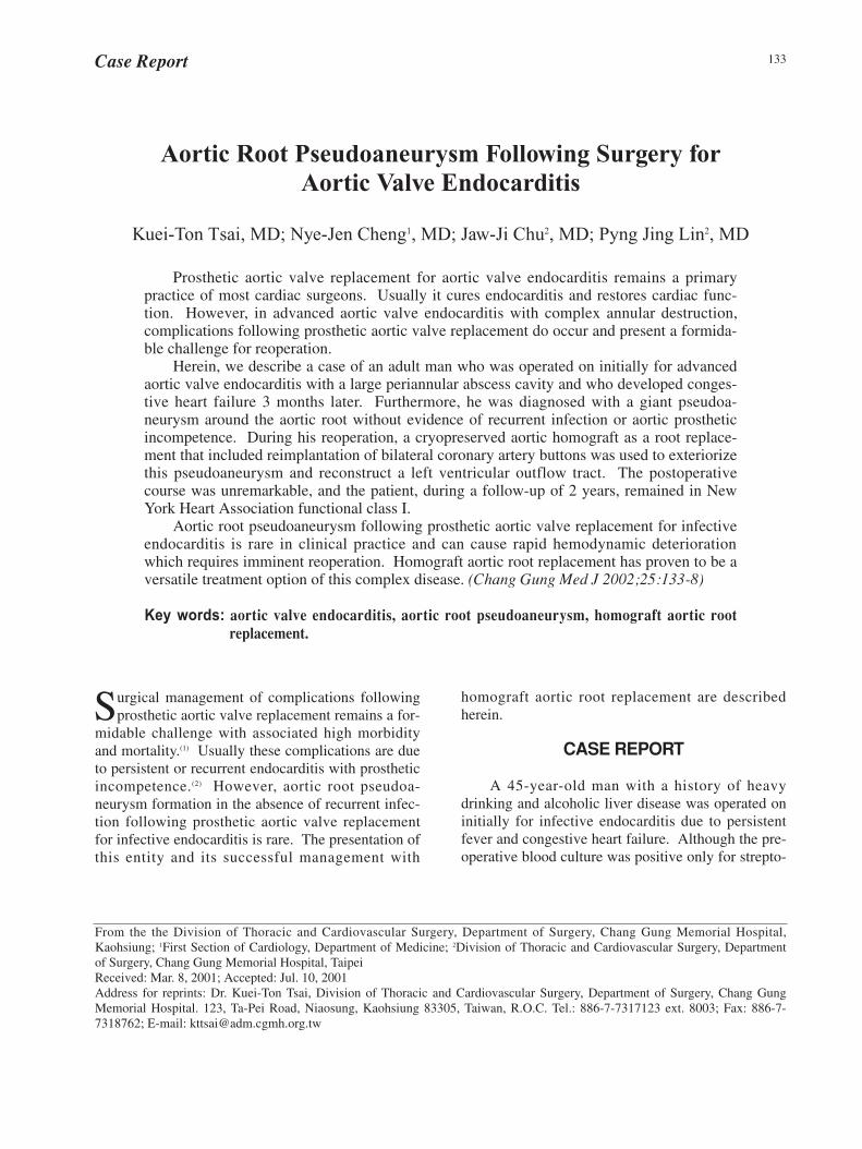
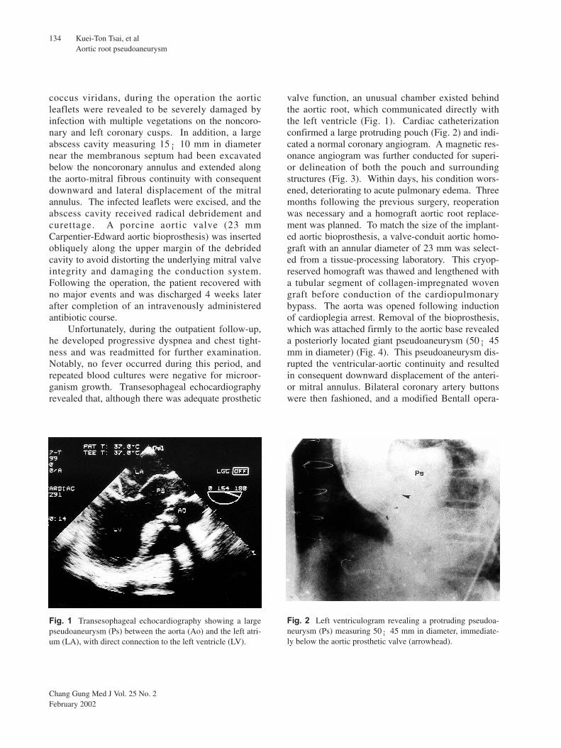
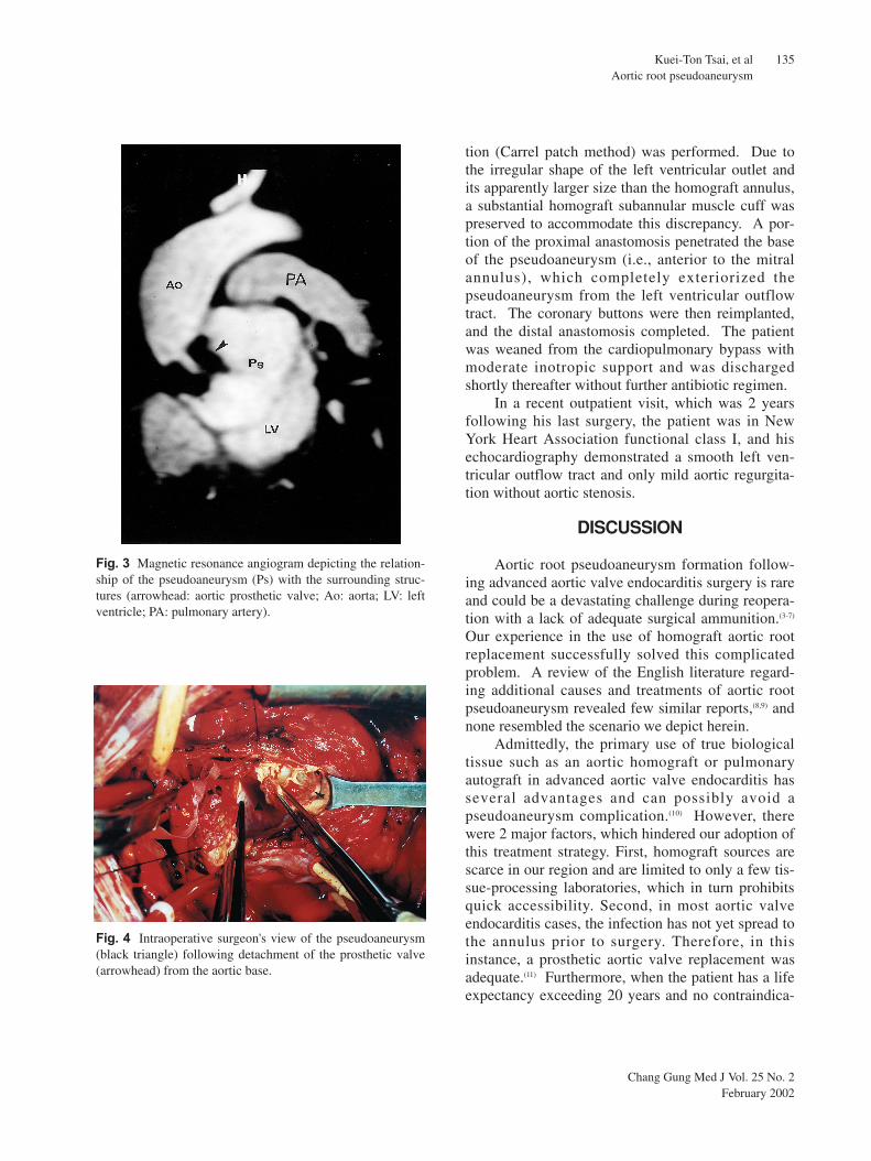
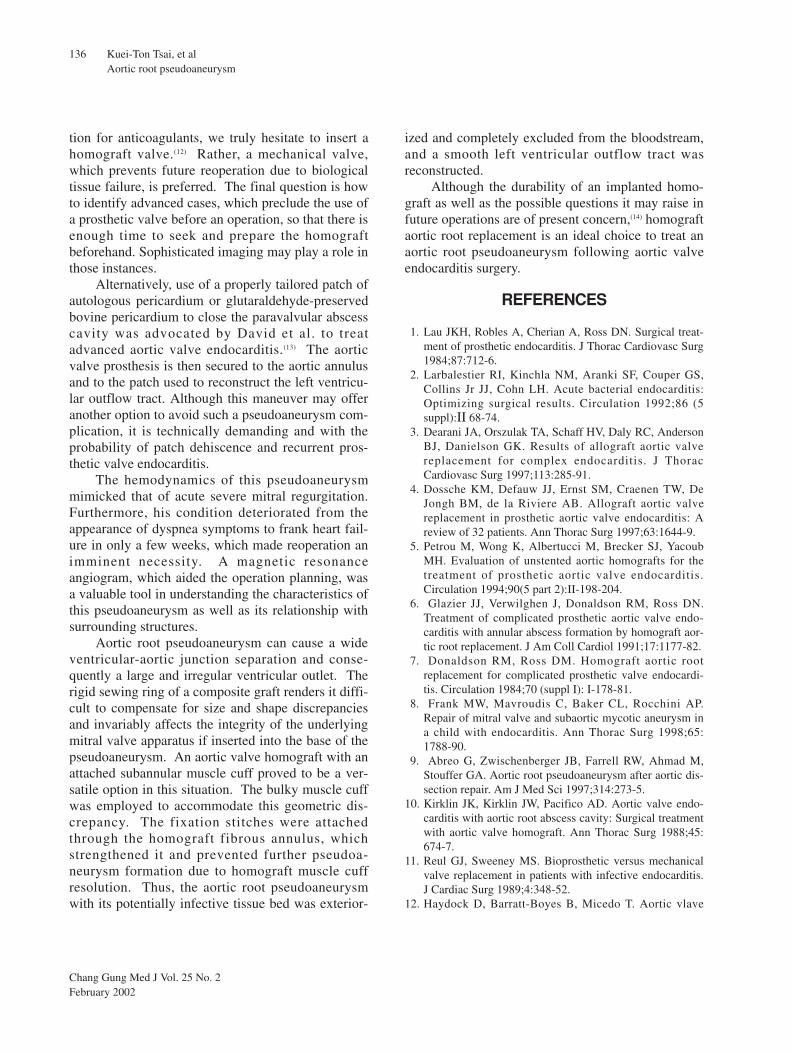
valve function, an unusual chamber existed behindthe aortic root, which communicated directly withthe left ventricle (Fig. 1). Cardiac catheterizationconfirmed a large protruding pouch (Fig. 2) and indi-cated a normal coronary angiogram. A magnetic res-onance angiogram was further conducted for superi-or delineation of both the pouch and surroundingstructures (Fig. 3). Within days, his condition wors-ened, deteriorating to acute pulmonary edema. Threemonths following the previous surgery, reoperationwas necessary and a homograft aortic root replace-ment was planned. To match the size of the implant-ed aortic bioprosthesis, a valve-conduit aortic homo-graft with an annular diameter of 23 mm was select-ed from a tissue-processing laboratory. This cryop-reserved homograft was thawed and lengthened witha tubular segment of collagen-impregnated wovengraft before conduction of the cardiopulmonarybypass. The aorta was opened following inductionof cardioplegia arrest. Removal of the bioprosthesis,which was attached firmly to the aortic base revealeda posteriorly located giant pseudoaneurysm (50¡ 45mm in diameter) (Fig. 4). This pseudoaneurysm dis-rupted the ventricular-aortic continuity and resultedin consequent downward displacement of the anteri-or mitral annulus. Bilateral coronary artery buttonswere then fashioned, and a modified Bentall opera-
Fig. 1 Transesophageal echocardiography showing a largepseudoaneurysm (Ps) between the aorta (Ao) and the left atri-um (LA), with direct connection to the left ventricle (LV).
Fig. 2 Left ventriculogram revealing a protruding pseudoa-neurysm (Ps) measuring 50¡ 45 mm in diameter, immediate-ly below the aortic prosthetic valve (arrowhead).
Chang Gung Med J Vol. 25 No. 2February 2002
Kuei-Ton Tsai, et alAortic root pseudoaneurysm
135
tion (Carrel patch method) was performed. Due tothe irregular shape of the left ventricular outlet andits apparently larger size than the homograft annulus,a substantial homograft subannular muscle cuff waspreserved to accommodate this discrepancy. A por-tion of the proximal anastomosis penetrated the baseof the pseudoaneurysm (i.e., anterior to the mitralannulus), which completely exteriorized thepseudoaneurysm from the left ventricular outflowtract. The coronary buttons were then reimplanted,and the distal anastomosis completed. The patientwas weaned from the cardiopulmonary bypass withmoderate inotropic support and was dischargedshortly thereafter without further antibiotic regimen.
In a recent outpatient visit, which was 2 yearsfollowing his last surgery, the patient was in NewYork Heart Association functional class I, and hisechocardiography demonstrated a smooth left ven-tricular outflow tract and only mild aortic regurgita-tion without aortic stenosis.
DISCUSSION
Aortic root pseudoaneurysm formation follow-ing advanced aortic valve endocarditis surgery is rareand could be a devastating challenge during reopera-tion with a lack of adequate surgical ammunition.(3-7)
Our experience in the use of homograft aortic rootreplacement successfully solved this complicatedproblem. A review of the English literature regard-ing additional causes and treatments of aortic rootpseudoaneurysm revealed few similar reports,(8,9) andnone resembled the scenario we depict herein.
Admittedly, the primary use of true biologicaltissue such as an aortic homograft or pulmonaryautograft in advanced aortic valve endocarditis hasseveral advantages and can possibly avoid apseudoaneurysm complication.(10) However, therewere 2 major factors, which hindered our adoption ofthis treatment strategy. First, homograft sources arescarce in our region and are limited to only a few tis-sue-processing laboratories, which in turn prohibitsquick accessibility. Second, in most aortic valveendocarditis cases, the infection has not yet spread tothe annulus prior to surgery. Therefore, in thisinstance, a prosthetic aortic valve replacement wasadequate.(11) Furthermore, when the patient has a lifeexpectancy exceeding 20 years and no contraindica-
Fig. 3 Magnetic resonance angiogram depicting the relation-ship of the pseudoaneurysm (Ps) with the surrounding struc-tures (arrowhead: aortic prosthetic valve; Ao: aorta; LV: leftventricle; PA: pulmonary artery).
Fig. 4 Intraoperative surgeon's view of the pseudoaneurysm(black triangle) following detachment of the prosthetic valve(arrowhead) from the aortic base.
Chang Gung Med J Vol. 25 No. 2February 2002
Kuei-Ton Tsai, et alAortic root pseudoaneurysm
136
tion for anticoagulants, we truly hesitate to insert ahomograft valve.(12) Rather, a mechanical valve,which prevents future reoperation due to biologicaltissue failure, is preferred. The final question is howto identify advanced cases, which preclude the use ofa prosthetic valve before an operation, so that there isenough time to seek and prepare the homograftbeforehand. Sophisticated imaging may play a role inthose instances.
Alternatively, use of a properly tailored patch ofautologous pericardium or glutaraldehyde-preservedbovine pericardium to close the paravalvular abscesscavity was advocated by David et al. to treatadvanced aortic valve endocarditis.(13) The aorticvalve prosthesis is then secured to the aortic annulusand to the patch used to reconstruct the left ventricu-lar outflow tract. Although this maneuver may offeranother option to avoid such a pseudoaneurysm com-plication, it is technically demanding and with theprobability of patch dehiscence and recurrent pros-thetic valve endocarditis.
The hemodynamics of this pseudoaneurysmmimicked that of acute severe mitral regurgitation.Furthermore, his condition deteriorated from theappearance of dyspnea symptoms to frank heart fail-ure in only a few weeks, which made reoperation animminent necessity. A magnetic resonanceangiogram, which aided the operation planning, wasa valuable tool in understanding the characteristics ofthis pseudoaneurysm as well as its relationship withsurrounding structures.
Aortic root pseudoaneurysm can cause a wideventricular-aortic junction separation and conse-quently a large and irregular ventricular outlet. Therigid sewing ring of a composite graft renders it diffi-cult to compensate for size and shape discrepanciesand invariably affects the integrity of the underlyingmitral valve apparatus if inserted into the base of thepseudoaneurysm. An aortic valve homograft with anattached subannular muscle cuff proved to be a ver-satile option in this situation. The bulky muscle cuffwas employed to accommodate this geometric dis-crepancy. The fixation stitches were attachedthrough the homograft fibrous annulus, whichstrengthened it and prevented further pseudoa-neurysm formation due to homograft muscle cuffresolution. Thus, the aortic root pseudoaneurysmwith its potentially infective tissue bed was exterior-
ized and completely excluded from the bloodstream,and a smooth left ventricular outflow tract wasreconstructed.
Although the durability of an implanted homo-graft as well as the possible questions it may raise infuture operations are of present concern,(14) homograftaortic root replacement is an ideal choice to treat anaortic root pseudoaneurysm following aortic valveendocarditis surgery.
REFERENCES
1. Lau JKH, Robles A, Cherian A, Ross DN. Surgical treat-ment of prosthetic endocarditis. J Thorac Cardiovasc Surg1984;87:712-6.
2. Larbalestier RI, Kinchla NM, Aranki SF, Couper GS,Collins Jr JJ, Cohn LH. Acute bacterial endocarditis:Optimizing surgical results. Circulation 1992;86 (5suppl):II 68-74.
3. Dearani JA, Orszulak TA, Schaff HV, Daly RC, AndersonBJ, Danielson GK. Results of allograft aortic valvereplacement for complex endocarditis. J ThoracCardiovasc Surg 1997;113:285-91.
4. Dossche KM, Defauw JJ, Ernst SM, Craenen TW, DeJongh BM, de la Riviere AB. Allograft aortic valvereplacement in prosthetic aortic valve endocarditis: Areview of 32 patients. Ann Thorac Surg 1997;63:1644-9.
5. Petrou M, Wong K, Albertucci M, Brecker SJ, YacoubMH. Evaluation of unstented aortic homografts for thetreatment of prosthetic aortic valve endocarditis.Circulation 1994;90(5 part 2):II-198-204.
6. Glazier JJ, Verwilghen J, Donaldson RM, Ross DN.Treatment of complicated prosthetic aortic valve endo-carditis with annular abscess formation by homograft aor-tic root replacement. J Am Coll Cardiol 1991;17:1177-82.
7. Donaldson RM, Ross DM. Homograft aortic rootreplacement for complicated prosthetic valve endocardi-tis. Circulation 1984;70 (suppl I): I-178-81.
8. Frank MW, Mavroudis C, Baker CL, Rocchini AP.Repair of mitral valve and subaortic mycotic aneurysm ina child with endocarditis. Ann Thorac Surg 1998;65:1788-90.
9. Abreo G, Zwischenberger JB, Farrell RW, Ahmad M,Stouffer GA. Aortic root pseudoaneurysm after aortic dis-section repair. Am J Med Sci 1997;314:273-5.
10. Kirklin JK, Kirklin JW, Pacifico AD. Aortic valve endo-carditis with aortic root abscess cavity: Surgical treatmentwith aortic valve homograft. Ann Thorac Surg 1988;45:674-7.
11. Reul GJ, Sweeney MS. Bioprosthetic versus mechanicalvalve replacement in patients with infective endocarditis.J Cardiac Surg 1989;4:348-52.
12. Haydock D, Barratt-Boyes B, Micedo T. Aortic vlave
Chang Gung Med J Vol. 25 No. 2February 2002
Kuei-Ton Tsai, et alAortic root pseudoaneurysm
137
replacement for aortic endocarditis in 108 patients: Acomparison of free hand allograft valves with mechanicalprosthesis and bioprosthesis. J Thorac Cardiovasc Surg1992;103:130-9.
13. David TE, Komeda M, Brofman PR. Surgical treatment of
aortic root abscess. Circulation 1989;80 (suppl I): 269-74.14. O'Brien MF, Stafford EG, Gardner MAH. Allograft aortic
valve replacement: Long term follow up. Ann ThoracSurg 1995;60:S65-70.
138
1 2 2
( 2002;25:133-8)
“ł'‹'´ | “¶fl|ˇ ¥~‹‡¡ fl ⁄ ⁄ ƒƒ”¥~‹¡F 1“ł'‹'´ | ¥x¥_|ˇ ⁄”‹‡¡ †⁄@⁄ ƒ⁄”‹¡F 2¥~‹‡¡ fl ⁄⁄ ƒƒ”¥~‹¤⁄⁄Ø·¡G¥ Œ 90ƒ~3⁄º1⁄Ø¡F–¤¥Z‚¡G¥ Œ 90ƒ~7⁄º10⁄Ø¡Cfl ¤œ' ƒL¥»‡B¡G‰†¶Q· ´ fiv¡A“ł' ‹ ' ´ | fl ⁄ ⁄ ƒƒ ” ¥~‹ ¡C “¶fl‰u‡ “Q¶m⁄j æ‚ 123‚„¡C Tel.: (07)7317123´ 8003;Fax: (07)7318763; E-mail: [email protected]