Embed Size (px)
Citation preview
LUND UNIVERSITY
PO Box 117221 00 Lund+46 46-222 00 00
Appendicitis in Children. Clinical, diagnostic and pathogenic factors
SALÖ, MARTIN
2016
Link to publication
Citation for published version (APA):SALÖ, MARTIN. (2016). Appendicitis in Children. Clinical, diagnostic and pathogenic factors. Lund: LundUniversity, Faculty of Medicine.
Creative Commons License:Unspecified
General rightsCopyright and moral rights for the publications made accessible in the public portal are retained by the authorsand/or other copyright owners and it is a condition of accessing publications that users recognise and abide by thelegal requirements associated with these rights.
• Users may download and print one copy of any publication from the public portal for the purpose of private studyor research. • You may not further distribute the material or use it for any profit-making activity or commercial gain • You may freely distribute the URL identifying the publication in the public portalTake down policyIf you believe that this document breaches copyright please contact us providing details, and we will removeaccess to the work immediately and investigate your claim.

1
Appendicitis in children

2
3
Appendicitis in children Clinical, diagnostic and pathogenic factors
Martin Salö
DOCTORAL DISSERTATION by due permission of the Faculty of Medicine, Lund University, Sweden.
To be defended at Segerfalksalen, Lund, 20160514, 10:00
Faculty opponent
Gabriel Sandblom
Department of Gastrointestinal Surgery, Karolinska University Hospital Stockholm, Sweden
4
Organization LUND UNIVERSITY Faculty of Medicine
Department of clinical sciences, Pediatrics, Lund
Lund University
S-221 85 Lund, Sweden
Document name: DOCTORAL DISSERTATION
Date of issue: 20160514
Author: Martin Salö, MD Sponsoring organization
Title and subtitle: Appendicitis in children – clinical, diagnostic and pathogenic factors
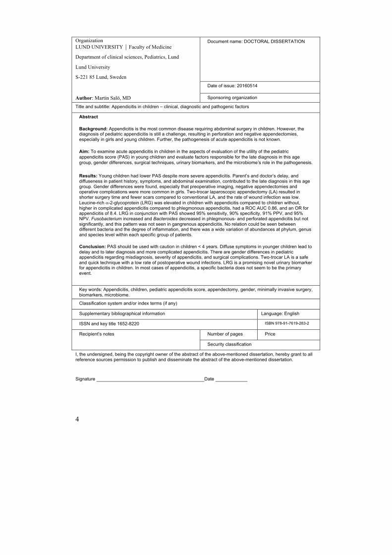
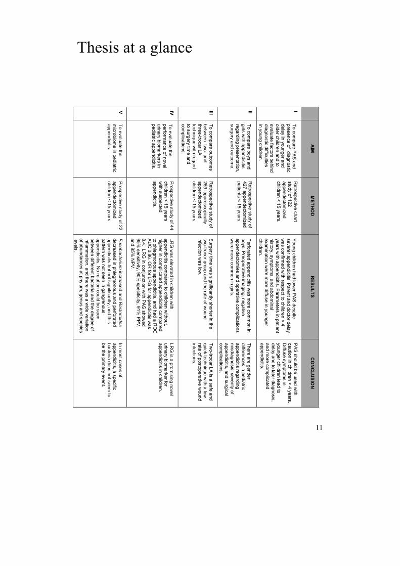
Abstract Background: Appendicitis is the most common disease requiring abdominal surgery in children. However, the diagnosis of pediatric appendicitis is still a challenge, resulting in perforation and negative appendectomies, especially in girls and young children. Further, the pathogenesis of acute appendicitis is not known. Aim: To examine acute appendicitis in children in the aspects of evaluation of the utility of the pediatric appendicitis score (PAS) in young children and evaluate factors responsible for the late diagnosis in this age group, gender differences, surgical techniques, urinary biomarkers, and the microbiome’s role in the pathogenesis. Results: Young children had lower PAS despite more severe appendicitis. Parent’s and doctor’s delay, and diffuseness in patient history, symptoms, and abdominal examination, contributed to the late diagnosis in this age group. Gender differences were found, especially that preoperative imaging, negative appendectomies and operative complications were more common in girls. Two-trocar laparoscopic appendectomy (LA) resulted in shorter surgery time and fewer scars compared to conventional LA, and the rate of wound infection was low. Leucine-rich α-2-glycoprotein (LRG) was elevated in children with appendicitis compared to children without, higher in complicated appendicitis compared to phlegmonous appendicitis, had a ROC AUC 0.86, and an OR for appendicitis of 8.4. LRG in conjunction with PAS showed 95% sensitivity, 90% specificity, 91% PPV, and 95% NPV. Fusobacterium increased and Bacteroides decreased in phlegmonous- and perforated appendicitis but not significantly, and this pattern was not seen in gangrenous appendicitis. No relation could be seen between different bacteria and the degree of inflammation, and there was a wide variation of abundances at phylum, genus and species level within each specific group of patients. Conclusion: PAS should be used with caution in children < 4 years. Diffuse symptoms in younger children lead to delay and to later diagnosis and more complicated appendicitis. There are gender differences in pediatric appendicitis regarding misdiagnosis, severity of appendicitis, and surgical complications. Two-trocar LA is a safe and quick technique with a low rate of postoperative wound infections. LRG is a promising novel urinary biomarker for appendicitis in children. In most cases of appendicitis, a specific bacteria does not seem to be the primary event.
Key words: Appendicitis, children, pediatric appendicitis score, appendectomy, gender, minimally invasive surgery, biomarkers, microbiome.
Classification system and/or index terms (if any)
Supplementary bibliographical information Language: English
ISSN and key title 1652-8220 ISBN 978-91-7619-283-2
Recipient’s notes Number of pages Price
Security classification
I, the undersigned, being the copyright owner of the abstract of the above-mentioned dissertation, hereby grant to all reference sources permission to publish and disseminate the abstract of the above-mentioned dissertation.
Signature Date

5
Appendicitis in children
Martin Salö
6
Cover photo and back page photo by: Sofia Salö
All photos in this book have been retrieved from Wikipedia Commons or from own library.
Copyright: Martin Salö
Faculty of Medicine | Department of Clinical Sciences, Pediatrics ISBN 978-91-7619-283-2 ISSN 1652-8220 Printed in Sweden by Media-Tryck, Lund University Lund 2016
7
Of all the ills within the abdomen which cause affliction to the sons of men There's none more often puts them in a fix than trouble in the worm-like appendix That caecal tail which sometimes tells a story or figures in a scene which may be gory That arch-deceiver, symbol of the devil which leads to every kind of septic evil That unexploded bomb which soon or late aperients may serve to detonate That worm which often turns to bad effect and makes us treat it with a great respect That foul assassin whose supreme delight choosing the place and knowing well the site To stab below the belt and on the right is causing that dread disease - appendicitis. -” Zeta” (Sir Zachary Cope). The acute abdomen in rhyme. 2nd ed. London, H.K. Lewis & Co. Ltd, 1949.
To Sofia, Mamma, Syrran, Ester
8
Contents
Contents ................................................................................................................... 8 Thesis at a glance ................................................................................................... 11 Papers included in the thesis .................................................................................. 12 Abbreviations ......................................................................................................... 13 Populärvetenskaplig sammanfattning .................................................................... 14
Blindtarmsinflammation hos barn – från orsak till operation 14 Introduction ............................................................................................................ 18
The appendix 18 History ................................................................................................ 18 Embryology ........................................................................................ 18 Anatomy .............................................................................................. 19 Histology ............................................................................................. 20 Function .............................................................................................. 20
Appendicitis 21 History of appendicitis and appendectomy ......................................... 21 Epidemiology ...................................................................................... 22 Pathogenesis – what causes appendicitis? .......................................... 22 Histopathology and severity of appendicitis ....................................... 24 Natural course of appendicitis ............................................................ 26 Young children and girls are at risk .................................................... 26
Diagnosing appendicitis 27 Clinical scoring systems ..................................................................... 28 Laboratory tests ................................................................................... 30 Radiology ............................................................................................ 31 Why we can’t completely rely on existing diagnostic aids ................ 33
Treatment of appendicitis 33 Surgical treatment ............................................................................... 34 Antibiotic treatment ............................................................................ 35
Aims ....................................................................................................................... 37 Paper I ................................................................................................. 37 Paper II ................................................................................................ 37
9
Paper III .............................................................................................. 37 Paper IV .............................................................................................. 37 Paper V ............................................................................................... 38
Settings and patients .............................................................................................. 39 Settings 39 Patients 39
Paper I ................................................................................................. 39 Paper II ................................................................................................ 39 Paper III .............................................................................................. 40 Paper IV .............................................................................................. 40 Paper V ............................................................................................... 41
Methods.................................................................................................................. 43 Study design 43
Papers I–III ......................................................................................... 43 Papers IV-V ........................................................................................ 44
Definitions 46 Routine management of appendicitis at the Department of Pediatric Surgery, Lund ..................................................................................... 46 Severity of appendicitis ...................................................................... 46 Histopathology .................................................................................... 47 Analysis of routine blood tests ............................................................ 47
Laboratory methods 47 Enzyme-Linked Immuno-Sorbant Assay (ELISA) ............................. 47 Microbiome analyses .......................................................................... 48
Statistical analyses 48 Ethics 49
Results .................................................................................................................... 50 Age (I) 50 Gender (II) 51 Pediatric appendicitis score (I, II, IV) 53 Two- vs. three-trocar laparoscopic appendectomy (III) 54 Urinary biomarkers (IV) 55 Microbiome (V) 57
Evaluation of the microbiome at phylum, genus and species level .... 57 Microbiome in relation to histopathology and sample site ................. 59 Diversity .............................................................................................. 61
Discussion .............................................................................................................. 62 Problems at young age 63
10
Girls 64 Two trocars 66 Urinary biomarkers 68 Bacteria or not, that is the question 70
Conclusions ............................................................................................................ 74 Paper I ................................................................................................. 74 Paper II ................................................................................................ 74 Paper III .............................................................................................. 74 Paper IV .............................................................................................. 74 Paper V ............................................................................................... 75
Future aspects ......................................................................................................... 76 Acknowledgements ................................................................................................ 77 References .............................................................................................................. 79 Papers I-V ............................................................................................................ 102
11
Thesis at a glance
V IV
III
II I
To evaluate the microbiom
e in pediatric appendicitis.
To evaluate the perform
ance of novel urinary biom
arkers in pediatric appendicitis. To com
pare outcomes
between tw
o- and three-trocar LA technique w
ith regard to surgery tim
e and com
plications.
To compare boys and
girls with appendicitis
regarding presentation, surgery and outcom
e. To com
pare PAS and presence of diagnostic delay in younger and older children and to evaluate factors behind diagnostic difficulties in young children.
AIM
Prospective study of 22 appendectom
ized children < 15 years.
Prospective study of 44 children < 15 years with suspected appendicitis.
Retrospective study of 259 laparoscopically appendectom
ized children < 15 years.
Retrospective study of 427 appendectom
ized patients < 15 years.
Retrospective chart study of 122 appendectom
ized children < 15 years.
METH
OD
Fusobacterium increased and B
acteroides decreased in phlegm
onous and perforated appendicitis but not significantly, and this pattern w
as not seen in gangrenous appendicitis. N
o relation could be seen betw
een different bacteria and the degree of inflam
mation, and there w
as a wide variation
of abundances at phylum, genus and species
levels.
LRG was elevated in children w
ith appendicitis com
pared to children without,
higher in complicated appendicitis com
pared to phlegm
onous appendicitis, and had a ROC
AUC 0.86. O
R for LR
G for appendicitis w
as 8.4. LR
G in conjunction w
ith PAS showed
95% sensitivity, 90%
specificity, 91% PPV,
and 95% NPV.
Surgery time w
as significantly shorter in the two-trocar group and the rate of w
ound infection w
as low.
Perforated appendicitis was m
ore common in
boys. Preoperative imaging, negative
appendectomies and operative com
plications were m
ore common in girls.
Young children had lower PAS despite
severer appendicitis. Parent and doctor delay was confirm
ed with respect to children < 4
years with appendicitis. Param
eters in patient history, sym
ptoms, and abdom
inal exam
ination were m
ore diffuse in younger children.
RESU
LTS
In most cases of
appendicitis, a specific bacteria does not seem
to be the prim
ary event.
LRG is a prom
ising novel urinary biom
arker for appendicitis in children.
Two-trocar LA is a safe and
quick technique with a low
rate of postoperative w
ound infections.
There are gender differences in pediatric appendicitis regarding misdiagnosis, severity of
appendicitis, and surgical com
plications, PAS should be used w
ith caution in children < 4 years. Diffuse sym
ptoms in
younger children lead to delay and to later diagnosis, and m
ore complicated
appendicitis.
CONCLUSIO
N
12
Papers included in the thesis
This thesis is based upon the following papers, referred to as Paper I-V:
I. Salö M, Friman G, Stenström P, Ohlsson B, Arnbjörnsson E. Appendicitis in Children: Evaluation of the Pediatric Appendicitis Score in Younger and Older Children. Surgery Research and Practice, vol. 2014, Article ID 438076, 6 pages, 2014. doi:10.1155/2014/438076.
II. Salö M, Ohlsson B, Arnbjörnsson E, Stenström P. Appendicitis in children from a gender perspective. Pediatric Surgery International. 2015 Sep;31(9):845-53.
III. Salö M, Järbur E, Hambraeus M, Ohlsson B, Stenström P, Arnbjörnsson E. Two-trocar appendectomy in children – description of technique and comparison with conventional laparoscopic appendectomy. Submitted.
IV. Salö M, Roth B, Stenström P, Arnbjörnsson E, Ohlsson B.
Urinary biomarkers in pediatric appendicitis. Submitted.
V. Salö M, Marungruang N, Roth B, Sundberg T, Stenström P,
Arnbjörnsson E, Fåk F, Ohlsson B. Evaluation of the microbiome in children’s appendicitis. Submitted.
13
Abbreviations
5-HIAA: 5-hydroxyindoleacetic acid
ANC: absolute neutrophile count
AUC: area under the curve
CI: confidence interval
CRP: C-reactive protein
CT: computed tomography
IL-6: interleukin 6
LA: laparoscopic appendectomy
LRG: leucine-rich alfa-2-glycoprotein
MRI: magnetic resonance imaging
NPV: negative predictive value
NSAID: non-steroidal inflammatory drugs
OA: open appendectomy
PAS: pediatric appendicitis score
PCR: polymerase chain reaction
PPV: positive predictive value
RLQ: right lower quadrant
rRNA: ribosomal ribonucleic acid
RLQ: Right lower quadrant
ROC: receiver operating characteristic
US: ultrasound
UTI: urinary tract infection
WBC: white blood cell

14
Populärvetenskaplig sammanfattning
Blindtarmsinflammation hos barn – från orsak till operation
De flesta känner någon som blivit opererad för blindtarmsinflammation (appendicit), vilket beror på att det är en vanlig sjukdom där nästan var 10:e person någon gång under livet drabbas. Många insjuknar när de är relativt unga, vanligast är det mellan cirka 10 – 30 års ålder, men sjukdomen kan också drabba spädbarn liksom mycket gamla människor. Trots att sjukdomen är vanlig vet man inte exakt varför blindtarmsinflammation uppstår. Det är klart att bakterier har en roll i sjukdomsförloppet men det är oklart om de startar själva processen eller kommer in senare i förloppet. Fler personer än dem som egentligen har blindtarmsinflammation, får sin blindtarm bortopererad. Detta gäller framförallt flickor/kvinnor, men till viss del även pojkar/män. Kirurgen tror alltså att det är blindtarmsinflammation men vid operationen eller vid den efterföljande mikroskopiska undersökningen så finner man att blindtarmen var frisk. Detta vittnar om de diagnostiska svårigheterna som föreligger vid misstänkt appendicit. Svårigheten att veta om det är blindtarmsinflammation som orsakar barnets magont kan förutom en onödig operation också leda till att diagnosen missas vilket kan leda till komplikationer för barnet. Avhandlingens syfte var att utvärdera flera aspekter av appendicit hos barn; bakteriers roll i sjukdomsutvecklingen, hur diagnostik och behandling påverkas av barnets ålder och kön, om ämnen (biomarkörer) kan förbättra diagnostiken, samt om modifiering av en befintlig operationsmetod kan vara till nytta för patienten. Barnen som studerades och ingår i avhandlingen är alla behandlade på Barn- och ungdomskirurgiska kliniken i Lund.
15
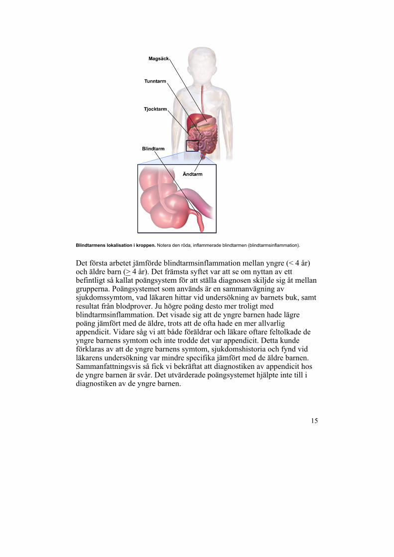
Blindtarmens lokalisation i kroppen. Notera den röda, inflammerade blindtarmen (blindtarmsinflammation).
Det första arbetet jämförde blindtarmsinflammation mellan yngre (< 4 år) och äldre barn (> 4 år). Det främsta syftet var att se om nyttan av ett befintligt så kallat poängsystem för att ställa diagnosen skiljde sig åt mellan grupperna. Poängsystemet som används är en sammanvägning av sjukdomssymtom, vad läkaren hittar vid undersökning av barnets buk, samt resultat från blodprover. Ju högre poäng desto mer troligt med blindtarmsinflammation. Det visade sig att de yngre barnen hade lägre poäng jämfört med de äldre, trots att de ofta hade en mer allvarlig appendicit. Vidare såg vi att både föräldrar och läkare oftare feltolkade de yngre barnens symtom och inte trodde det var appendicit. Detta kunde förklaras av att de yngre barnens symtom, sjukdomshistoria och fynd vid läkarens undersökning var mindre specifika jämfört med de äldre barnen. Sammanfattningsvis så fick vi bekräftat att diagnostiken av appendicit hos de yngre barnen är svår. Det utvärderade poängsystemet hjälpte inte till i diagnostiken av de yngre barnen.
16
I det andra arbetet jämfördes flickor och pojkar som opererats för blindtarmsinflammation. Syftet var att se om det fanns könsskillnader i insjuknande, diagnostik, operation och eftervård. Vi kunde se att flickor oftare feldiagnosticerades, trots att de oftare genomgick ultraljud av buken som ett led i diagnostiken. Vidare hade flickorna fler komplikationer vid operationen, dock inte när blindtarmen opererades ut med titthålsteknik. Pojkar hade oftare sprucken blindtarmsinflammation, detta till trots att det inte förlöpte längre tid från insjuknande till operation jämfört med flickorna. Sammanfattningsvis sågs flera könsskillnader vid blindtarmsinflammation hos barn varav det mest nedslående var den stora andelen flickor som feldiagnosticerades.
Vid blindtarmsinflammation är det idag vanligast att man försöker operera ut blindtarmen med hjälp av titthålsteknik. Detta innebär enkelt formulerat att man genom små hål i huden för in instrument i bukhålan som är uppblåst av gas för bättre insyn. I det tredje arbetet beskrev vi en modifierad titthålsteknik vid operation av blindtarmsinflammation hos barn och jämförde den med den konventionella titthålstekniken. Den modifierade tekniken visade sig vara snabbare jämfört med den vanliga och resulterade inte i fler komplikationer. Dock kan den modifierade tekniken troligtvis inte användas vid alla blindtarmsinflammationer, t.ex. om blindtarmen sitter fast mot bukväggen.
Biomarkörer är ämnen i kroppen som kan användas för att diagnosticera sjukdomar eller följa ett sjukdomsförlopp. I det fjärde arbetet var syftet att se om man med biomarkörer i urin kunde erhålla en säkrare diagnos vid blindtarmsinflammation. Urinprov togs på barn som sökte på barnakutmottagningen med misstänkt blindtarmsinflammation. Av de fyra markörerna som analyserades visade sig en vara lovande: LRG. Denna markör ökade mer i koncentration hos barn med blindtarmsinflammation jämfört med barnen med annan förklaring till buksmärtan. LRG var också mer ökad hos de barnen med allvarligare appendicit jämfört med de med en mer begränsad sjukdom. Vi utvärderade sedan hur säker diagnosen av appendicit hos de undersökta barnen var om man kombinerade LRG med det i första arbetet utvärderade poängsystemet. Om både poängsystemet och LRG hade värden som talade emot appendicit så hade endast 5% av barnen feldiagnosticerats. Sammanfattningsvis verkar LRG i urin vara en lovande biomarkör men fler och framförallt större studier behövs för att bekräfta dessa resultat.
17
I det femte och sista arbetet utvärderade vi bakteriers roll vid utveckling av blindtarmsinflammation hos barn. De flesta tidigare studier har använt metoder (odling) där man missar ca 90% av alla bakteriearter. Enstaka studier med nya metoder, där man genom att undersöka förekomsten av bakteriernas arvsmassa inte missar några bakterier, har indikerat att bakterier som i vanliga fall finns i munhålan verkar ligga bakom utvecklingen av appendicit. Vi kartlade den bakteriella arvsmassan i sjuka blindtarmar, och jämförde med den i friska blindtarmar som tagits ut från barn opererade för annan sjukdom i buken. Något överraskande fann vi ökad förekomst av munhålebakterier i de sjuka blindtarmarna, men statistiskt sett var det ingen skillnad mot de friska. Dessutom ökade inte förekomsten av munhålebakterierna i följd med allt mer allvarlig blindtarmsinflammation. Vidare såg vi att det fanns en stor variation i förekomsten av olika bakteriearter mellan varje patient, även om man jämförde barn med samma svårighetsgrad av appendicit. Vi drog slutsatsen att bakterier i de flesta fall förmodligen inte är primärt ansvariga för utvecklingen av blindtarmsinflammation.
Sammanfattningsvis är och förblir blindtarmsinflammation en vanlig men lurig sjukdom hos barn där vi inte vet orsaken till sjukdomen och har svårt att diagnosticera den hos framförallt yngre barn och flickor. Det är uppenbart att det behövs nya metoder att diagnosticera sjukdomen, där biomarkörer kan vara en väg att gå, framförallt hos de yngre barnen där befintliga poängsystem inte verkar vara till nytta. En gissning är att det kommer att krävas en kombination av diagnostiska metoder, t.ex. poängsystem och biomarkörer, för att nå en förbättrad diagnostisk säkerhet. Slutligen har denna avhandling visat att man genom modifiering av en befintlig operationsmetod kan göra nytta för barnet, förutsatt att diagnostiken från början varit rätt...
18
Introduction
The appendix
History
There are several findings of drawings of what is thought to be appendix and appendicitis in early history, going back to the ancient Egyptians and further on to Hippocrates (1,2). The first specific documentation is from 1492 when Leonardo da Vinci sketched the appendix (3) and in 1521 it was described in words by Berengario da Carpi (3). In 1543, Andreas Vesalius, a professor in anatomy, both illustrated and described the appendix, but naming it caecum (blind pouch) (1). Appendix vermiformis was finally formulated in 1530 by the Vidius Vidius (4).
Embryology
The major part of the intestines, including the cecum and appendix, develops from the embryonic midgut. Around the 6th week of gestation, a cecal bud develops from the antimesenteric border of the caudal part of midgut loop (5). Because the lower part does not grow equally fast, the appendix develops and can (histologically) be seen at the 8th week (5,6). By week 12, the cecum and appendix lie in the right upper quadrant, as the 270o degree rotation of the gut is completed. As the proximal colon elongates, the appendix and cecum are displaced down to the right side of the abdomen where the appendix can assume different, probably random, positions (5,6). Because of the unequal growth of the cecum, the appendix becomes displaced medially and upward (7). Lymphatic tissue including lymph nodes can be seen at 4–5 months of gestation (5). Abnormalities, such a agenesis or duplications, are very rare (8).
19
Anatomy
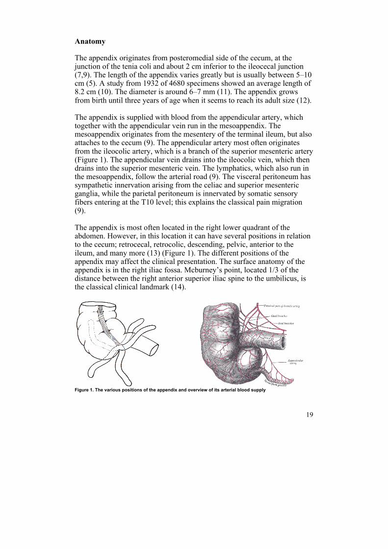
The appendix originates from posteromedial side of the cecum, at the junction of the tenia coli and about 2 cm inferior to the ileocecal junction (7,9). The length of the appendix varies greatly but is usually between 5–10 cm (5). A study from 1932 of 4680 specimens showed an average length of 8.2 cm (10). The diameter is around 6–7 mm (11). The appendix grows from birth until three years of age when it seems to reach its adult size (12). The appendix is supplied with blood from the appendicular artery, which together with the appendicular vein run in the mesoappendix. The mesoappendix originates from the mesentery of the terminal ileum, but also attaches to the cecum (9). The appendicular artery most often originates from the ileocolic artery, which is a branch of the superior mesenteric artery (Figure 1). The appendicular vein drains into the ileocolic vein, which then drains into the superior mesenteric vein. The lymphatics, which also run in the mesoappendix, follow the arterial road (9). The visceral peritoneum has sympathetic innervation arising from the celiac and superior mesenteric ganglia, while the parietal peritoneum is innervated by somatic sensory fibers entering at the T10 level; this explains the classical pain migration (9). The appendix is most often located in the right lower quadrant of the abdomen. However, in this location it can have several positions in relation to the cecum; retrocecal, retrocolic, descending, pelvic, anterior to the ileum, and many more (13) (Figure 1). The different positions of the appendix may affect the clinical presentation. The surface anatomy of the appendix is in the right iliac fossa. Mcburney’s point, located 1/3 of the distance between the right anterior superior iliac spine to the umbilicus, is the classical clinical landmark (14).
Figure 1. The various positions of the appendix and overview of its arterial blood supply
20
Histology
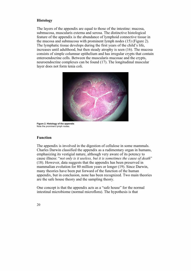
The layers of the appendix are equal to those of the intestine: mucosa, submucosa, muscularis externa and serosa. The distinctive histological feature of the appendix is the abundance of lymphoid connective tissue in the mucosa and submucosa with prominent lymph nodes (15) (Figure 2). The lymphatic tissue develops during the first years of the child’s life, increases until adulthood, but then steady atrophy is seen (16). The mucosa consists of simple columnar epithelium and has irregular crypts that contain enteroendocrine cells. Between the muscularis mucosae and the crypts, neuroendocrine complexes can be found (17). The longitudinal muscular layer does not form tenia coli.
Figure 2. Histology of the appendix Note the prominent lymph nodes.
Function
The appendix is involved in the digestion of cellulose in some mammals. Charles Darwin classified the appendix as a rudimentary organ in humans, emphasizing its vestigial nature, although very aware of its potency to cause illness: “not only is it useless, but it is sometimes the cause of death” (18). However, data suggests that the appendix has been preserved in mammalian evolution for 80 million years or longer (19). Since Darwin, many theories have been put forward of the function of the human appendix, but in conclusion, none has been recognized. Two main theories are the safe house theory and the sampling theory. One concept is that the appendix acts as a “safe house” for the normal intestinal microbiome (normal microflora). The hypothesis is that
21
microflora is guarded by the appendix mucosa which contains a biofilm with secretory IgA. After an infection in the colon, the protected microflora in the appendix may be able to re-inoculate the colon (20). Another theory is that the appendix acts as a sentinel sampling organ. This theory is supported by the fact that the appendix is part of gut-associated lymphatic tissue (GALT), the significant increase in lymphatic follicles from birth to a peak in adulthood, and its production of immunoglobulins (15). This, together with the highly strategic position after small intestines and the ileocecal valve makes it a candidate for being responsible for sampling of antigens (21,22).
Appendicitis
History of appendicitis and appendectomy
The history of appendicitis is somewhat diffuse, often due to not being specifically separated from other acute diseases in the abdomen and because of the confusion between cecum and appendix (3,23). Probably, Jean Fernel, a French physician, mathematician and philosopher, presented the first true description of appendicitis in 1544 (3). But it was not until 1886 that the term appendicitis became commonly recognized when introduced by the Harvard professor, Reginald Fitz, who combined the Latin word, appendere, to hang upon, with the Greek suffix, -itis, relating to (3). Interestingly, in his article “Perforating inflammation of the vermiform appendix, with special reference to its early diagnosis and treatment”, Fitz noted that the disease may spontaneously resolve (3). The first appendectomy, that is, removal of the appendix and not just drainage, was performed in 1735 by Claudius Amyand (24). The patient was an 11-year-old boy with a congenital scrotal hernia in which the appendix had become incarcerated; the incision was made through the hernia (3). The patient recovered slowly but survived. The first abdominal appendectomy was performed in 1880 by the Scottish surgeon, Robert Lawson Tait (23). In 1884, the work of Charles McBurney was published regarding the now famous point and incision (23). Over 100 years after the first abdominal appendectomy, 1981, Kurt Semm performed the first laparoscopic appendectomy (25).
22
Epidemiology
Appendicitis is the most common disease requiring abdominal surgery in children over 2 years of age (26,27). The rate of appendicitis in children presenting with acute abdominal pain varies in different studies. In one prospective study of over 1000 children aged 2–12 years, approximately 1% had appendicitis (27). In a more recent study, with a retrospective review of over 9000 children, the rate was > 4% (28). The life-time risk of developing acute appendicitis has been estimated to 8.6% for boys and 6.7% for girls (26). However, the life-time risk for an appendectomy is 12% for boys and 23% for girls (26). The difference in incidence between men and women has been reported to decrease in the early 2000s (29,30). The lower incidence among women is speculated to be related to female sex hormones (31). Studies from Europe indicate that the incidence of appendicitis in children is decreasing (32), while the opposite was seen in a study from the US (33). The incidence is highest among 10–19 year olds, with around 15 cases per 10 000 children per year (33). In the same study, the incidence for children 0–9 years old was around 6 cases per 10 000 children per year (33). Appendicitis is relatively uncommon in children under five years of age (34,35). A study from Denmark reported an incidence of 2.2/10 000 and 1.8/10 000 for boys and girls under 4 years of age, respectively (32). In Sweden, the incidence of appendicitis in patients 0–18 years was around 13/10 000 children in 2013, and in 2014 over 1800 appendectomies were performed in children 0–14 years old (36).
Pathogenesis – what causes appendicitis?
There are case reports of appendicitis being caused by foreign bodies, such as seeds or magnets (37,38). There are also case reports of traumatic appendicitis (39). However, appendicitis caused by foreign bodies or trauma is of course rare and despite appendicitis being a common disease, there have been and still are several controversies about its etiology. The main theories that have been suggested are based on diet and hygiene, obstruction, immunological characteristics of the patient, and infection. However, it is still today, almost 300 years since the first appendectomy, safe to say that no one truly knows what causes appendicitis. One general hypothesis is that a combination between the inherent immunological reactions of the individual, together with a local event in the appendix, may lead to appendicitis.
23
That diet may have role in appendicitis was suggested because of the higher incidence in developing countries (40–42). Low fiber intake together with high intake of refined sugar leading to slow transit time in the bowels was thought to explain the geographic difference. The diet hypothesis has however been questioned in more recent epidemiological studies where a decreasing incidence in appendicitis was seen despite no change in fiber intake (43). The hygiene hypothesis was based on the belief that improvement in hygiene in industrialized countries leads to less infection during infancy and altered immunity reaction to viral infection later on in life, which in turn would cause appendicitis (44). In conclusion, neither the diet nor the hygiene hypothesis is today seen as valid and does not work when looking at the recent epidemiology of appendicitis (43,45). It has been speculated that different characteristics of the patient’s immune response could be correlated with the incidence of appendicitis. There is evidence that a T-helper (Th) 2 response protects against appendicitis. The hypothesis is that an individual can have a propensity for a certain immune reaction in response to an antigen, with either a Th1 or Th2 response. This is, for example. supported by epidemiological studies showing correlation between Crohn’s disease and perforated appendicitis (Th1), and an inverse correlation between ulcerative colitis and appendicitis (46–48). Further, a study by Rubér et al. showed a Th17-like cytokine response in gangrenous but not in phlegmonous appendicitis (49) The most common explanation for the development of appendicitis is an obstruction of the lumen. This theory is by many still accepted as the direct cause of the major cases of appendicitis. As an example, one of the most renowned textbooks in pediatric surgery states obstruction to be the main cause of pediatric appendicitis (50). The theory is that obstruction is followed by the subsequent accumulation of secretions, a rising intraluminal pressure, the impairment of lymphatic and venous drainage, a compromised mucosal barrier, and finally the overgrowth and invasion of microbes within the appendiceal wall (51–54). However, an obstruction due to a fecalith, anatomic location, lymphoid hyperplasia, foreign bodies, tumors, among other reasons, is found only in around a third of all cases (55–57). Additionally, Arnbjörnsson and Bengmark measured the perioperative intraluminal pressure and did not find it to be increased (55). Their conclusion was that obstruction is not an important etiology but may develop secondary to inflammation. In summary, it is clear that the theory of an obstruction of the lumen cannot explain the majority of appendicitis cases (58).
24
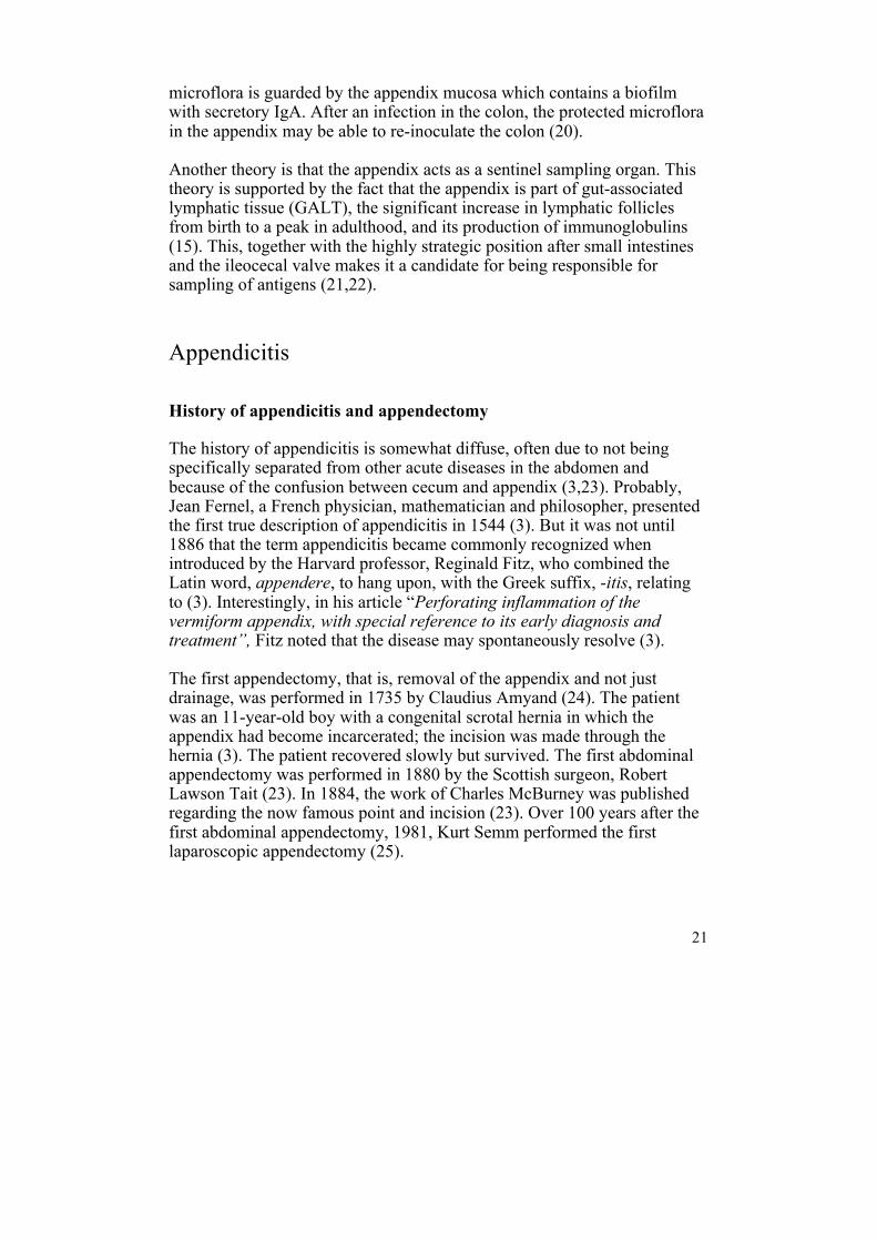
Infection has been proposed as a primary event causing appendicitis (59). This is based on studies reporting of appendicitis appearing in clusters (60) and of a seasonal variation of the incidence of acute appendicitis (61,62). From our own experience, there is a peak in children with appendicitis during the first school week after the summer holidays. Studies have, however, not found any correlation between viral infections and appendicitis (63). Bacteria has an obvious role in appendicitis but so far often seen as a secondary event, hence infection after the inflammation; and the bacteriology has been widely studied (64–66). Most past studies have used conventional culture techniques to evaluate the role of bacteria in acute appendicitis. These techniques are effective in evaluating solitary bacterial species, but lack the capability of characterizing the polymicrobial diversity present (59). With these conventional culturing methods, as much as 90–99% of microbes are missed (67). There are a few recent studies evaluating the whole microbiome in appendicitis (59,68–71) using rRNA-based fluorescence in situ hybridization (FISH) or 16S RNA sequencing (68,70,71). To summarize, these studies have found a significant increase in bacteria normally part of the oral flora in the inflamed appendices, especially Fusobacterium.
Figure 3. Perforated appendicitis The patient presented with a typical history and was taken to the operating theater for a laparoscopic appendectomy.
Histopathology and severity of appendicitis
The diagnosis of appendicitis in children is often confirmed by the intraoperative picture of an inflamed appendix. In equivocal cases, histopathology can confirm or rule out the diagnosis (which of course
25
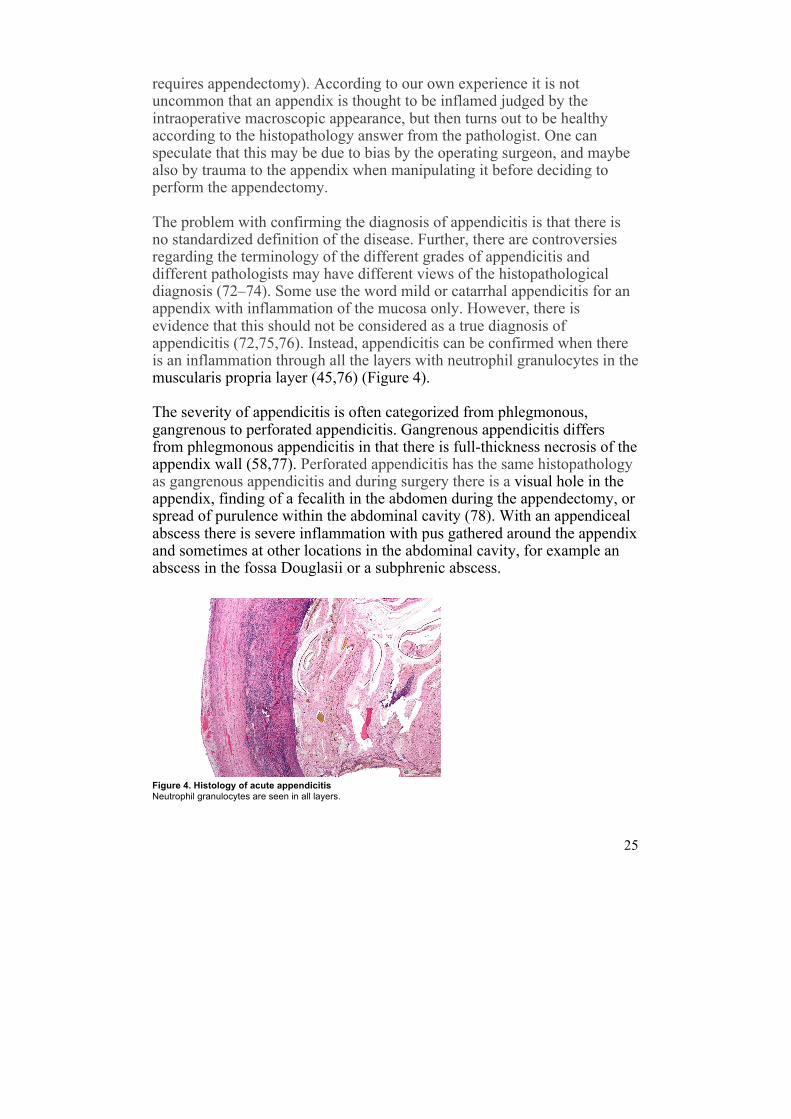
requires appendectomy). According to our own experience it is not uncommon that an appendix is thought to be inflamed judged by the intraoperative macroscopic appearance, but then turns out to be healthy according to the histopathology answer from the pathologist. One can speculate that this may be due to bias by the operating surgeon, and maybe also by trauma to the appendix when manipulating it before deciding to perform the appendectomy. The problem with confirming the diagnosis of appendicitis is that there is no standardized definition of the disease. Further, there are controversies regarding the terminology of the different grades of appendicitis and different pathologists may have different views of the histopathological diagnosis (72–74). Some use the word mild or catarrhal appendicitis for an appendix with inflammation of the mucosa only. However, there is evidence that this should not be considered as a true diagnosis of appendicitis (72,75,76). Instead, appendicitis can be confirmed when there is an inflammation through all the layers with neutrophil granulocytes in the muscularis propria layer (45,76) (Figure 4). The severity of appendicitis is often categorized from phlegmonous, gangrenous to perforated appendicitis. Gangrenous appendicitis differs from phlegmonous appendicitis in that there is full-thickness necrosis of the appendix wall (58,77). Perforated appendicitis has the same histopathology as gangrenous appendicitis and during surgery there is a visual hole in the appendix, finding of a fecalith in the abdomen during the appendectomy, or spread of purulence within the abdominal cavity (78). With an appendiceal abscess there is severe inflammation with pus gathered around the appendix and sometimes at other locations in the abdominal cavity, for example an abscess in the fossa Douglasii or a subphrenic abscess.
Figure 4. Histology of acute appendicitis Neutrophil granulocytes are seen in all layers.
26
Natural course of appendicitis
The main philosophy during the last century was that appendicitis always proceeded to gangrene and eventually perforation and abdominal sepsis. This of course led to the surgeons wanting to take the patients to the operating theater as soon as possible. Thus, in the past, the main view has been to never miss appendicitis, while accepting higher negative appendectomy rates (79). Today, there is increasing evidence that not all cases of appendicitis will progress to perforation. Already noted by Howie in 1964 (76), it seems that appendicitis may be self-limiting and spontaneous resolution may occur. Howie noted that being less aggressive in taking the patient to the operating theater led to fewer patients having to be appendectomized in the end. The same result was found by Andersson and colleagues 30 years later (80). They concluded that the rate of appendectomy does not influence the rate of perforated appendicitis, but the rate of non-perforated appendicitis. The same conclusion can be drawn from the studies by Morino et al. and Decadt et al. (81,82). In conclusion, it seems that less aggressive surgical management leads to fewer patients being diagnosed with appendicitis, fewer negative appendectomies, but not an increased number of perforations. Resolution of appendicitis has been described clinically and radiologically by several authors (83–86), and also shown histologically (87). There are studies showing that an increased time to appendectomy leads to a higher rate of perforations. In the light of the new philosophy, this can be explained by the fact that most perforations occur at an early stage often before arrival at the hospital and that self-limiting appendicitis is quite common (88). Hence, it seems hard to decrease the incidence of perforations (89,90) and appendectomy in the middle of the night can be questioned (90). In large epidemiological studies, it also seems that both the proportion of perforations and incidence of perforated appendicitis remain on the same levels (80,91,92). In conclusion, perforation rate is not as a measure of good diagnostic evaluation. Instead, the rate of negative appendectomies is a good measure (93,94).
Young children and girls are at risk
Appendicitis classically presents with vague periumbilical pain in the abdomen and maybe anorexia. This is followed by nausea and often
27
vomiting, pain migration to the RLQ and more intense pain, and development of fever. The process takes around 24–48 hours, and the child who despite the initial vague abdominal pain wanted to play, now prefers to lie still. Unfortunately, many children do not present in this classical manner. Instead, patient history and symptoms, and findings from clinical examination, are more diffuse, especially in the young children (35,95–98). The diffuse presentation confuses both caregivers and doctors and both parent’s and doctor’s delay have been suggested to contribute to the often late diagnosis in the youngest children (34,96,98,99). These diagnostic difficulties result in a higher rate of negative appendectomies, as well as a higher rate of perforation, increased morbidity, and longer hospital stay (34,35,95–98). This is further aggravated in the youngest children (95,97,100). In one study of over 63 000 children, every fourth child under 5 years of age was misdiagnosed (93). The perforation rate is also exceptionally high in the youngest children. In the study by Smink et al. of 33184 children, the overall perforation rate was 33% (101). The perforation rate was recently described to be correlated to age in children under five years of age (102). The rates were 86% (< 1 year), 74% (1–1.9 years), 60% (2–2.9 years), 64% (3–3.9 years), and 49% (4–4.9 years) (102). Similar rates have been reported from Denmark, with older children having a third of the rate seen in the younger children (32). Equal to young children, girls are in risk of misdiagnosis. A study by Smink et al. of 37109 children showed that the overall negative appendectomy rate was 9% (100). This percentage is in several studies significantly higher in girls (26,93,99,103). The life-time risk of appendectomy is 23% in girls compared to 12% in boys (26). The explanation for the misdiagnosis is often referred to girls having acute abdominal pain from ovarian pathology, such as ovarian torsion and salpingitis. However, both salpingitis and ovarian torsion are very uncommon in the premenarchal girl (104,105).
Diagnosing appendicitis
The doctor evaluating the child with acute abdominal pain has several aids for confirming or excluding the diagnosis of appendicitis. History and physical examination are important but sometimes not fully appreciated; instead the doctor relies on laboratory and imaging (93,106). From patient history and abdominal examination, together with blood tests, a clinical
28
prediction score can be used. Finally, imaging may aid the clinician, who, however, has to decide if the child benefits from the examination or not.
Clinical scoring systems
In general, clinical scores for appendicitis use information from patient history, abdominal examination, and certain blood tests, to give an estimate of how likely it is that the patient has a certain disease. There are several clinical scores that aim to confirm or exclude appendicitis (107–113). Of these, the Alvarado score (109), the appendicitis inflammatory response (AIR)-score (111), and the pediatric appendicitis score (PAS) (113) are probably the most well-known (Table 1). Together with PAS, the Lintula score is the only score developed specifically for children (108). The Alvarado score, described in 1986, was actually based on a retrospective study of 305 patients with a mean age of 25 (4–80) years (109). The score contains eight dichotomized parameters and the maximum score is 10 (Table 1). A score of 5–6 is indicative of possible appendicitis, a score of 7–8 implies probable appendicitis, and a score of 9–10 indicates very probable appendicitis. The AIR-score was described in 2008 in a prospective study of 545 patients with a mean age of 26 years, where the cohort was divided into two parts, one for the development of the score and the other for validation (111). The score contains eight parameters that are somewhat different from the ones in the Alvarado score, and some of the parameters are graded (Table 1). The score ranges from 0–12 and is divided into three degrees of probability: low (0–4 points), intermediate (5–8 points), and high (9–12). The Lintula score was described in 2005 and based on a cohort of children 4 – 15 years of age (108). The score was constructed in a cohort of 131 children, and the assessment of the score was conducted in 109 children. The score uses nine parameters and ranges from 0–32 points (Table 1). Two cut-offs at < 15 and > 21 points give three groups of probability: low, intermediate and high. PAS was the first true score for pediatric appendicitis, published in 2002 by Samuel, and based on a prospective study of 1170 patients between 4 – 15 years of age. The study uses eight variables and ranges from 0–10 points (Table 1). A child with a score > 6 has probable appendicitis. It was in the original study said to have a 100% sensitivity, 92% specificity, 96% PPV,
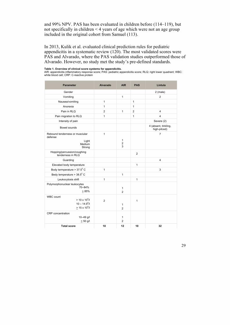
29
and 99% NPV. PAS has been evaluated in children before (114–119), but not specifically in children < 4 years of age which were not an age group included in the original cohort from Samuel (113).
In 2013, Kulik et al. evaluated clinical prediction rules for pediatric appendicitis in a systematic review (120). The most validated scores were PAS and Alvarado, where the PAS validation studies outperformed those of Alvarado. However, no study met the study’s pre-defined standards. Table 1. Overview of clinical score systems for appendicitis. AIR: appendicitis inflammatory response score;; PAS: pediatric appendicitis score;; RLQ: right lower quadrant;; WBC: white blood cell;; CRP: C-reactive protein
Parameter Alvarado AIR PAS Lintula
Gender 2 (male)
Vomiting 1 2
Nausea/vomiting 1 1
Anorexia 1 1
Pain in RLQ 2 1 2 4
Pain migration to RLQ 1 1 4
Intensity of pain Severe (2)
Bowel sounds 4 (absent, tinkling, high-pitced)
Rebound tenderness or musciular defense
Light Medium Strong
1 1 2 3
7
Hopping/percussion/coughing tenderness in RLQ 2
Guarding 4
Elevated body temperature 1
Body termperature > 37.50 C 1 3
Bedy temperature > 38.50 C 1
Leukocytosis shift 1 1
Polymorphonuclear leukocytes 70–84%
> 85%
1 2
WBC count > 10 x 109/l
10 – 14.99/l > 15 x 109/l
2
1 2
1
CRP concentration 10–49 g/l > 50 g/l
1 2
Total score 10 12 10 32
30
Laboratory tests
Routine laboratory tests normally used in the work-up in children with suspected appendicitis are white blood cell count (WBC), absolute neutrophil count (ANC), and C-reactive protein (CRP). These blood tests are used to reveal inflammation but are not specific for appendicitis and may be elevated in many of the possible differential diagnoses. Further, appendicitis in children has in several studies and cohorts been shown to occur with normal inflammatory biomarkers (121,122), which we have also seen in the cohort studied in this thesis. In a meta-analysis, WBC and CRP had a sensitivity and specificity of 62 and 75%, and 57 and 85%, respectively (123). In a prospective study of pediatric patients, 80% sensitivity and 79% specificity were seen when combining WBC and ANC (124). Another study, showed a 98% sensitivity when combining WBC and CRP but the specificity was low (125). In a JAMA meta-analysis, WBC, with different age-specific limits, had a likelihood-ratio (LR) of 3.4 for appendicitis (106). A WBC and ANC less than 8850/µL and 6750/µL, respectively, both had a likelihood ratio (LR) of 0.06 (106). CRP had greatly varying results as a predictor for appendicitis, a normal value seemed to reduce LR with 50% (106).
Novel biomarkers Because the traditional inflammatory markers (WBC, ANC, CRP) are not accurate enough, several new biomarkers have been evaluated. These consist of both already existing tests now evaluated for their role in pediatric appendicitis (e.g. Mean platelet volume, Bilirubin) and more novel biomarkers (126–136). The novel biomarkers have mainly been tested in serum but in a few studies also in urine. Urine analyses are preferable in children since they are easy to obtain and non-invasive. Only three studies have so far evaluated novel biomarkers in urine for pediatric appendicitis (130,131,133).
Urine 5-hydroxyindoleacetic acid (5-HIAA) was evaluated as a diagnostic marker by Ozel et al. (133) and was found to be significantly increased in appendicitis patients but had a low sensitivity and specificity. The marker does not seem promising in adult appendicitis either (137). Leucine-rich α-2-glycoprotein and calprotectin are biomarkers reflecting activation, chemotaxis, and neutrophil degranulation, and have been evaluated as biomarkers for pediatric appendicitis (130,132). LRG (both in serum and urine) have shown promising results (130,132), while calprotectin did not (132). LRG is a glycoprotein belonging to the leucine-rich repeat (LRR) family of proteins which is involved in signal transduction, cell adhesion, and protein-protein interactions (138). The exact mechanisms of the function of LRG are not known, but it has been described to be
31
elevated in bacterial diseases and is expressed by neutrophils undergoing differentiation in the liver and by high endothelial venules of the mesentery such as the mesoappendix (139,140). One speculation is that LRG, compared to the routine inflammatory markers, reflects a local inflammation, such as the one in appendicitis (139). In one of the studies, an immunoassay interference was described when analyzed with enzyme-linked immunosorbent assay (ELISA) (130). Further, none of the studies has adjusted for dehydration which one would expect to be important, especially when analyzing urine. Finally, no study has used a novel biomarker in conjunction with a clinical prediction score, compared to some of the traditional blood tests that are often incorporated in the scores.
Radiology
Imaging has gained an increased importance in the evaluation of the child with suspected appendicitis (141). The main purposes of imaging are to acquire an earlier diagnosis of appendicitis (or a differential diagnosis) and to reduce the rate of negative appendectomies and perforations (141). Abdominal radiography has little value unless a concomitant intestinal obstruction is suspected. Today, ultrasound (US) is the method of choice and preferred to computed tomography (CT), largely due to concerns over the risk of radiation exposure (142,143). In a meta-analysis, the sensitivity and specificity for diagnosing appendicitis in children were 88% and 94%, respectively, for US and 94% and 95%, respectively, for CT (144). However, despite the use of an inferior method of imaging, perforation rates and negative appendectomies do not increase (142). Finally, there is an increasing number of publications regarding MRI for suspected appendicitis in children. In a recent study of over 500 children, both sensitivity and specificity were 97% (145). Thus, MRI seems to have superior diagnostic accuracy compared to both CT and US, and lacks radiation exposure. Aspelund et al. compared ultrasound, followed by MRI in equivocal cases, with CT in children with suspected appendicitis (146). The radiation-free imaging pathway did not result in delay of administration of antibiotics or appendectomy, nor in increased negative appendectomy rate, perforation rate or length of hospital stay. However, for many centers, lack of availability is the major setback. Further, costs, and perhaps feasibility in a pediatric population are other concerns.
32
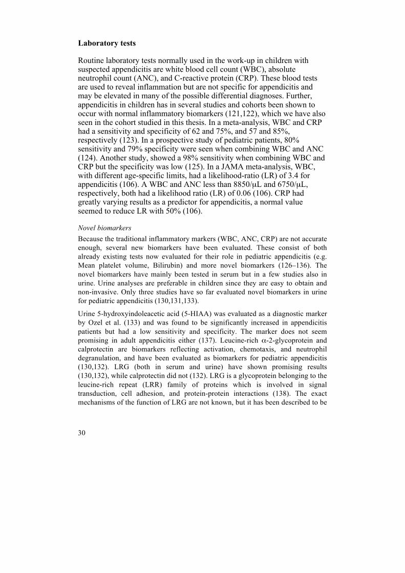
Figure 5. Ultrasound showing acute appendicitis
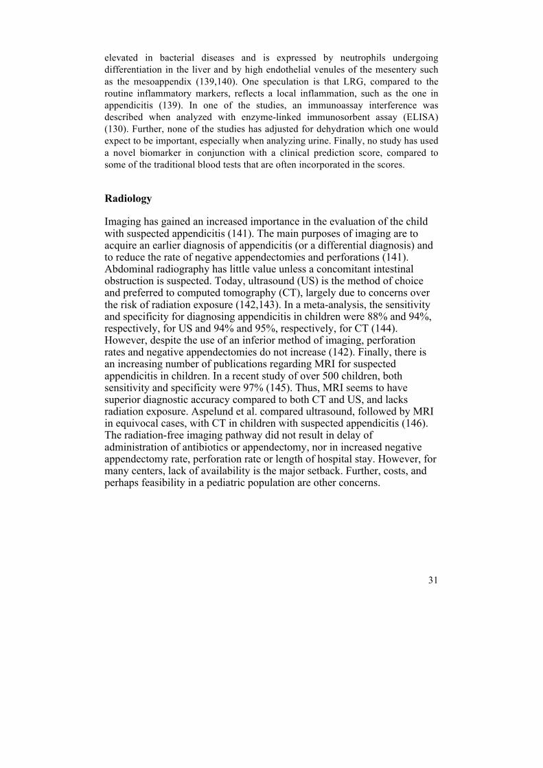
Figure 6. CT of child with an appendicolith (x) and perforated appendicitis with multiple abscesses (*)
33
Why we can’t completely rely on existing diagnostic aids
No clinical scoring system is perfect. One can assume that no present or future clinical scoring system can ever be completely trusted. Further, it is likely that the score works best in the “same” cohort it was developed from, and has lower predictive values in another study population (147). Further, in children, one could guess that the score performs differently in a three-year-old compared to a teenager. However, a good clinical scoring system for pediatric appendicitis can significantly aid the (especially young) surgeon, and also be an aid in the triage of children with acute abdominal pain (147). Routine blood tests revealing inflammation are often used and may aid the clinician in the management of the child with suspected appendicitis, but they neither confirm nor rule out appendicitis with sufficient accuracy. Several novel biomarkers have been evaluated in pediatric appendicitis, where a few seem promising, but the field of research is quite new and more studies are needed with improved analyzing techniques and clinical parameters used in conjunction with the biomarkers. Imaging is often relied upon and US is the most used method of imaging in children with suspected appendicitis. It has the advantage of being radiation-free, but it is operator-dependent and inferior to CT and MRI. MRI seems to be superior to both US and CT, but has limits in availability, costs and probably feasibility. Finally, a populations-based analysis in JAMA showed that despite the introduction of US and CT, the rate of perforation has not decreased over time (93).
Treatment of appendicitis
The treatment of appendicitis has for a long time equated with surgical removal of the appendix, appendectomy. In the 1980s, minimally invasive surgery was introduced for treatment of appendicitis, and has undergone dramatic developments since then (148). During the last decade, treatment with antibiotics has been introduced and evaluated.
34
Surgical treatment
Open appendectomy (OA) with a McBurney muscle splitting incision in the right iliac fossa has been the standard treatment since it was described in 1894 (149), and has practically not changed significantly since then. The technique is often safe but may be difficult if the appendix is not located in its normal position. In 1981, despite massive critique and skepticism from colleagues, Kurt Semm performed the first laparoscopic appendectomy (LA) (25). The first study of LA in children was presented in 1992 by Ure and colleagues (150). In their study of 43 children they concluded that LA was safe but not superior to OA regarding pain intensity or use of analgesics. In a Cochrane review from 2004 (151), laparoscopy and LA were recommended when applicable and available. LA was found to have significantly fewer wound infections, but twice as large risk of intra-abdominal abscess. However, only five of the studies included had a pediatric population. In 2006, Aziz et al. presented a meta-analysis of LA vs. OA in a pediatric population (152). Twenty-three studies were included with a total of 6477 children. LA had significantly fewer wound infections and ileus, and length of hospital stay was shortened, but OA had shorter operative time. The meta-analysis from Esposito et al. a few years later included over 120 000 patients between 0–18 years, and confirmed the shorter operative time for OA but only in the case of complicated appendicitis (153). LA had shorter hospital stay in all inflammation groups (153). The minimally invasive or minimally access surgery has continued to develop, and the traditional laparoscopic appendectomy with three trocars is now being challenged by techniques using only two (154) or one-port (single-port laparoscopic appendectomy) (155) or a single incision (156,157).

35
Figure 7. Two-trocar laparoscopic appendectomy The appendix is drawn out through the sheath, and an extra corporeal appendectomy is performed.
Antibiotic treatment
Antibiotic treatment instead of surgery for acute appendicitis has been described in several reports throughout history, but it is not until the last one or two decades that standardized and randomized trial have been performed, both in adults and children. In the first studies of the pediatric population, the cohort consisted of children with perforated appendicitis, where one study showed that antibiotic treatment followed by interval appendectomy had significantly more adverse events and more time away from normal activities than early appendectomy (158). The failure rate of non-operative treatment of perforated appendicitis in children is reported to be between 10–41% (159). In 2015, Svensson et al. (160) published the first RCT of non-operative treatment with antibiotics versus surgery for non-perforated appendicitis in children. Of the initial 24 patients who received antibiotic treatment, 22 had initial resolution of symptoms of which one later had recurrence during follow-up. Further, another six patients had appendectomy because of recurrent abdominal pain or request from parents. In conclusion, 62% of the children with antibiotic treatment did not have appendectomy during the follow-up of one year (160). In a study from Tanaka et al. 29% had recurrence after non-operative treatment of uncomplicated appendicitis, after a follow-up of 4.3 years (161). Finally, a very recent study with 24

36
patients and 50 controls, concluded that antibiotic treatment is feasible, cost-effective, safe, and also preferred by patients and parents (162).
37
Aims
The overall aim of this thesis was to evaluate several aspects of appendicitis in children, regarding pathogenesis, clinical factors, diagnostics, and surgical techniques.
Paper I
The primary aim was to evaluate the diagnostic performance of PAS in children operated on for suspected appendicitis comparing children < 4 years of age with children > 4 years of age. Secondary aims were to study if there was a diagnostic delay in diagnosing appendicitis in younger compared to older children, and to identify factors responsible for the possible late diagnosis in younger children.
Paper II
To compare girls and boys with appendicitis with regard to presentation, differences in perioperative care, and outcomes after appendectomy.
Paper III
To describe the technique of two-trocar LA and compare outcomes between two- and three-trocar techniques with regard to surgery time and complications, including the rate of postoperative wound infection.
Paper IV
To evaluate predictive values of LRG, calprotectin, IL-6, and Substance P in urine in children presenting with suspected appendicitis, and to use the most promising of these biomarkers in conjunction with PAS to see whether this could improve the accuracy of diagnosing appendicitis.

38
Paper V
The primary aim was to evaluate the microbiome in the normal appendix and in appendicitis specifically divided into the three clinically and histopathologically defined grades of inflammation (i.e. phlegmonous, gangrenous, and perforated appendicitis). Secondary aims were to examine whether there were any microbiome differences between proximal and distal appendices, and relate the microbiome with histopathological findings.
39
Settings and patients
Settings
The patients in papers I–V were all treated at the tertiary center of Pediatric Surgery at the Skåne University Hospital, Lund, Sweden. The center serves an area of 340 000 inhabitants with primary surgical care for children under 15 years of age, and an area of 1.3 million inhabitants with primary surgical care for children under three years of age. If there is suspicion of appendicitis, the patients are referred for a pediatric surgery consultation. The referral may be issued by either a pediatrician at the pediatric ER or directly from a general practitioner. The consultation is often carried out by a resident in pediatric surgery.
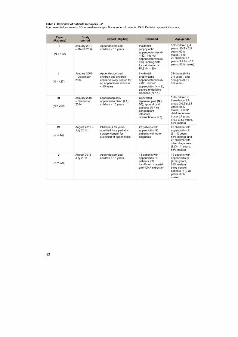
Patients
Paper I
The study included all children who underwent appendectomy, from January 2010 through March 2014. After excluding patients who had undergone an appendectomy during operations for other diseases (N = 32), patients with interval appendectomy (N = 6), and patients lacking data for calculation of PAS (N = 30), a total of 122 patients were included in the study. There were 102 children > 4 years of age with a mean age of 10.5 years (± 2.9) and 62% males, and 20 children < 4 years of age with a mean age of 2.6 (± 0.7) and 55% males.
Paper II
The study included all children who either underwent appendectomy or were conservatively treated for an appendiceal abscess, between January 2006 and December 2014. Excluded patients consisted of children who had
40
undergone an incidental prophylactic appendectomy during surgery for another diagnosis (N = 87), chronic appendicitis (N = 2), and children with severe underlying diseases making symptoms and length of hospital stay hard to interpret (N = 4). After exclusion, the study population consisted of 427 patients; 244 boys with mean age 9.8 years (± 3.4), and 183 girls with mean age 9.6 years (± 3.5).
Paper III
The study included all children who were operated on with laparoscopic appendectomy (LA), between January 2006 and December 2014. After exclusion of patients with converted LAs (N = 56), with appendiceal abscess (N = 6), or with concomitant intestinal obstruction (N = 3), a total of 259 children were left to study. Of these, 168 (65%) underwent surgery with the conventional three-trocar technique, and 91 (35%) were operated on with two-trocar laparoscopic appendectomy. The children in the three-trocar group had a mean age of 10.5 years (± 2.8) and 56% were males, compared to a mean age of 10.3 years (± 3.3) and 55% males in the two-trocar group.
Paper IV
During the study period between August 2013 and July 2014, a total of 160 children were admitted for a pediatric surgery consultation because of suspected appendicitis. Of these, 45 children had a final diagnosis of appendicitis of which 22 children were included with a median age of 11 years (6–14) years and 55% males. Gender, age, and degree of inflammation did not significantly differ between the 22 children included and the 23 children not included. Of the 115 children with other final diagnoses, 22 children were included with a median age of 9 (3–14) years and 68% males (Figure 8).
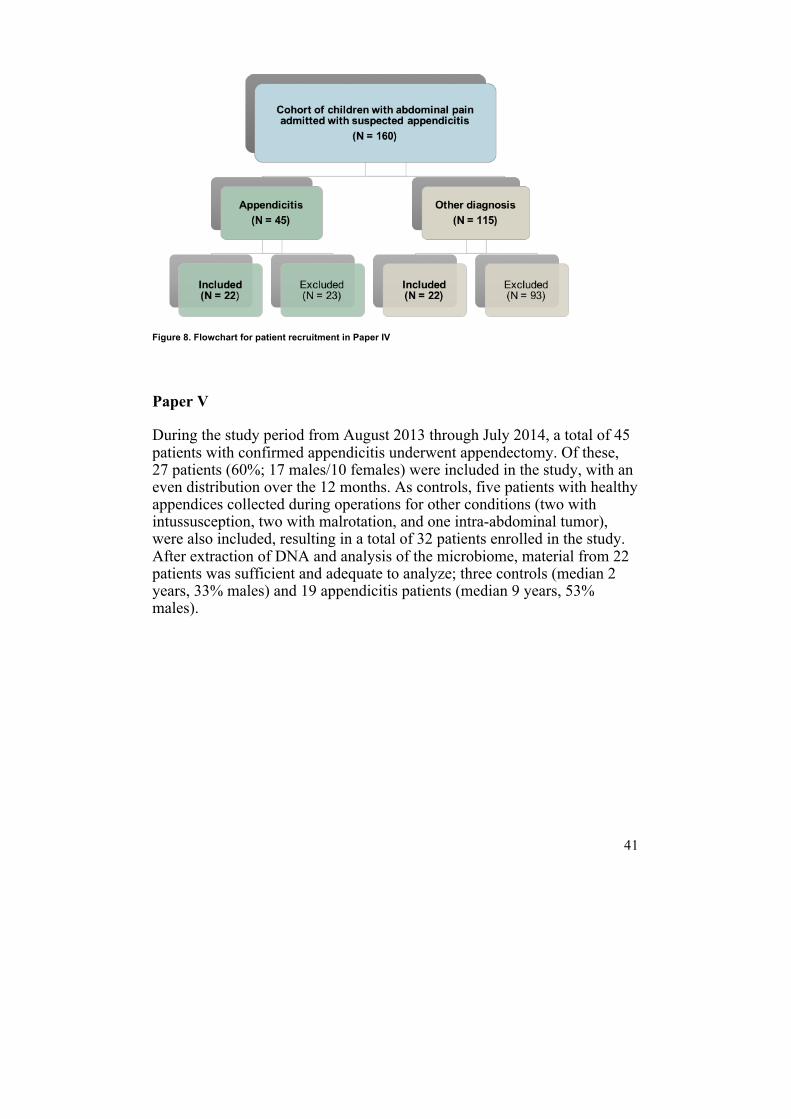
41
Figure 8. Flowchart for patient recruitment in Paper IV
Paper V
During the study period from August 2013 through July 2014, a total of 45 patients with confirmed appendicitis underwent appendectomy. Of these, 27 patients (60%; 17 males/10 females) were included in the study, with an even distribution over the 12 months. As controls, five patients with healthy appendices collected during operations for other conditions (two with intussusception, two with malrotation, and one intra-abdominal tumor), were also included, resulting in a total of 32 patients enrolled in the study. After extraction of DNA and analysis of the microbiome, material from 22 patients was sufficient and adequate to analyze; three controls (median 2 years, 33% males) and 19 appendicitis patients (median 9 years, 53% males).
42
Table 2. Overview of patients in Papers I–V Age presented as mean + SD, or median (range);; N = number of patients;; PAS: Pediatric appendicitis score;;
Paper (Patients)
Study period Cohort (eligible) Excluded Age/gender
I
(N = 122)
January 2010 – March 2014
Appendectomized children < 15 years
Incidental prophylactic appendectomies (N = 32), interval appendectomies (N = 6), lacking data for calculation of PAS (N = 30)
102 children > 4 years (10.5 ± 2.9 years, 62% males), and 20 children < 4 years of 2.6 (± 0.7 years, 55% males)
II
(N = 427)
January 2006 – December 2014
Appendectomized children and children conservatively treated for an appendiceal abscess < 15 years
Incidental prophylactic appendectomies (N = 87), chronic appendicitis (N = 2), severe underlying diseases (N = 4)
244 boys (9.8 ± 3.4 years), and 183 girls (9.6 ± 3.5 years).
III
(N = 259)
January 2006 – December 2014
Laparoscopically appendectomized (LA) children < 15 years
Converted laparoscopies (N = 56), appendiceal abscess (N = 6), concomitant intestinal obstruction (N = 3)
168 children in three-trocar LA group (10.5 ± 2.8 years, 56% males), and 91 children in two-trocar LA group (10.3 ± 3.3 years, 55% males)
IV
(N = 44)
August 2013 – July 2014
Children < 15 years admitted for a pediatric surgery consult for suspicion of appendicitis
23 patients with appendicitis, 93 patients with other diagnosis
22 children with appendicitis (11 (6–14) years, 55% males), and 22 children with other diagnoses (9 (3–14) years, 68% males)
V
(N = 22)
August 2013 – July 2014
Appendectomized children < 15 years
18 patients with appendicitis, 10 patients with insufficient material after DNA extraction
19 patients with appendicitis (9 (2-14) years, 53% males), three control patients (2 (2-3) years, 33% males)
43
Methods
Study design
Papers I–III
Papers I – III were retrospective, institution-based studies. The database of medical and surgical records of all children admitted to the Department of Pediatric Surgery was used. Patients were searched for using international classification of diseases (ICD-10) diagnosis codes (K35.2, K35.3, K35.8, K36.9, K37.9) and procedure codes (JEA00, JEA01, JEA10). Parameters were drawn from the journals and registered.
Paper I The following information was extracted from the medical and surgical records and registered: age, sex, time from onset of symptoms to seeking care (parent’s delay), if the child was triaged as acute abdominal pain, how often the child was evaluated by a doctor and sent home without suspicion of appendicitis and without a rescheduled follow-up (doctor’s delay), which diagnosis was presumed in patients with doctor’s delay, presenting symptoms, notes from the abdominal examination, presence of leukocytosis and/or neutrophilia, type of radiology used, surgeon’s description of the severity of the appendicitis, results from the histopathological analysis, duration of hospital stay, and complications. PAS was calculated for each patient using the information from patient history, abdominal examination, and laboratory tests.
Paper II The following information was extracted from the medical and surgical records and registered: Age, weight, time since the appendectomy, symptoms, finding from the abdominal examination, results from routine blood tests (WBC, ANC, CRP), type of imaging, time to surgery, severity of the appendicitis, method of operation, operative time, operative and postoperative complications, postoperative pain medication, and duration of hospital stay. PAS was calculated
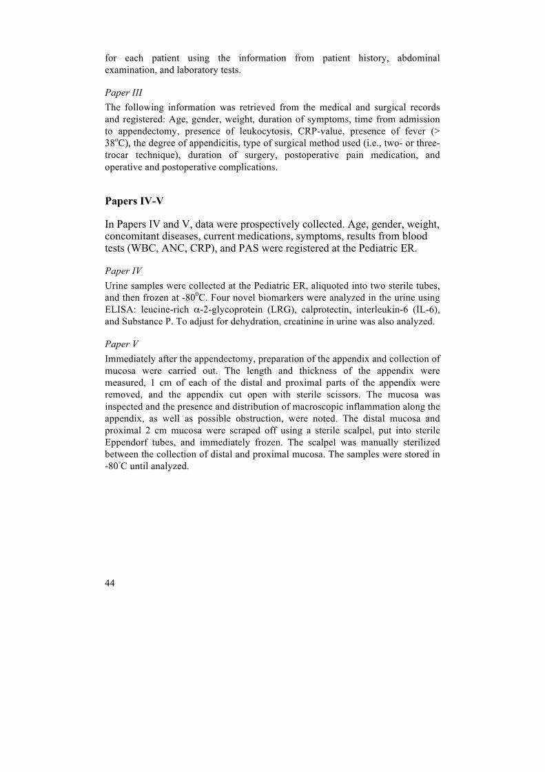
44
for each patient using the information from patient history, abdominal examination, and laboratory tests.
Paper III The following information was retrieved from the medical and surgical records and registered: Age, gender, weight, duration of symptoms, time from admission to appendectomy, presence of leukocytosis, CRP-value, presence of fever (> 38oC), the degree of appendicitis, type of surgical method used (i.e., two- or three-trocar technique), duration of surgery, postoperative pain medication, and operative and postoperative complications.
Papers IV-V
In Papers IV and V, data were prospectively collected. Age, gender, weight, concomitant diseases, current medications, symptoms, results from blood tests (WBC, ANC, CRP), and PAS were registered at the Pediatric ER.
Paper IV Urine samples were collected at the Pediatric ER, aliquoted into two sterile tubes, and then frozen at -800C. Four novel biomarkers were analyzed in the urine using ELISA: leucine-rich α-2-glycoprotein (LRG), calprotectin, interleukin-6 (IL-6), and Substance P. To adjust for dehydration, creatinine in urine was also analyzed.
Paper V Immediately after the appendectomy, preparation of the appendix and collection of mucosa were carried out. The length and thickness of the appendix were measured, 1 cm of each of the distal and proximal parts of the appendix were removed, and the appendix cut open with sterile scissors. The mucosa was inspected and the presence and distribution of macroscopic inflammation along the appendix, as well as possible obstruction, were noted. The distal mucosa and proximal 2 cm mucosa were scraped off using a sterile scalpel, put into sterile Eppendorf tubes, and immediately frozen. The scalpel was manually sterilized between the collection of distal and proximal mucosa. The samples were stored in -80C until analyzed.
45
Table 3. Overview of methods in Papers I-V PAS: Pediatric appendicitis score;; CRP: C-reactive protein;; LRG: leucine-rich α-2 glycoprotein;; IL-6: interleukin 6;; *Doctor’s delay: how often the child was evaluated by a doctor and sent home without suspicion of appendicitis and without a rescheduled follow-up
Paper I Paper II Paper III Paper IV Paper V
Study Retrospective, institution-based study Prospective study
Data were retrieved from the medical and surgical records and registered
registered at the pediatric ER
Parameters collected
Age, gender, weight, symptoms.
Duration of symptoms
If triaged as abdominal pain, doctor’s delay*, diagnosis when doctor’s delay.
Time to surgery, follow-up.
Time to surgery, follow-up.
Concomitant diseases, current medications,
Diagnosis Surgeon’s description and/or histopathological analysis. Surgeon’s description and histopathological analysis.
Blood tests
Presence of leukocytosis and/or neutrophilia
Presence of leukocytosis and/or neutrophilia, CRP-value
Presence of leukocytosis, CRP-value
Presence of leukocytosis and/or neutrophilia, proportion of neutrophils, CRP-value
Presence of leukocytosis and/or neutrophilia, CRP-value
PAS
PAS was calculated for each patient using the information from medical charts regarding patient history, abdominal examination, and laboratory tests
PAS was calculated for each patient after the work-up at the pediatric ER
Other samples
ELISA analysis of urine: LRG, calprotectin, Substance P, IL-6
Creatinine in urine.
Collection of proximal and distal appendix mucosa.
Imaging Proportion of patients undergoing preoperative imaging and type of imaging used.
Surgery Technique,
surgery time, complications
Technique, surgery time, complications
Fecalith, mucosa appearance
Postoperative Length of hospital stay, complications. Complications.
46
Definitions
Routine management of appendicitis at the Department of Pediatric Surgery, Lund
The management of the included children followed the current guidelines of the clinic, and was never changed due to the (prospective) studies. Children with suspected appendicitis are referred for a pediatric surgery consultation by the pediatrician at the pediatric ER or by the general practitioner. Appendicitis is diagnosed by means of patient history, physical examination, routine blood tests (WBC, ANC, CRP), and sometimes with the aid of ultrasound. Appendicitis is treated with appendectomy and never conservatively treated except in cases where an appendiceal abscess is diagnosed preoperatively. The appendectomy is performed either laparoscopically, with one, two, or three ports, or as an appendectomy with a traditional laparotomy in the RLQ. An attending surgeon performs or supervises the appendectomy. All children receive preoperative antibiotic prophylaxis with trimethoprim/sulfamethoxazole and metronidazole, with dosage according to age. In the case of a gangrenous or perforated appendicitis, intravenous antibiotics is continued for three and five days, respectively; additional treatment is often given orally after discharge from the hospital. There is no standardized protocol for postoperative pain management at the clinic but the treatment often consists of paracetamol, sometimes combined with NSAID, and if severe pain; morphine.
Severity of appendicitis
The classifications used for the description of the severity of the appendicitis in Papers I–V are phlegmonous, gangrenous, and perforated appendix, and appendiceal abscess. Gangrenous appendicitis was defined as an inflamed appendix with significant gray or black discoloration of the wall, and absence of the criteria for perforation (77). The definition of perforated appendicitis was a visual hole in the appendix, finding of a fecalith in the abdomen during the appendectomy, or spread of purulence within the abdominal cavity (78). Absence of macroscopic and/or microscopic inflammation rendered the diagnosis healthy appendix (negative appendectomy).
47
Histopathology
In the retrospective studies in Papers I–III the presence of appendicitis and the severity of the inflammation was determined by the intraoperative picture described by the surgeon, and in equivocal cases by histopathological analysis. In the prospective Papers IV–V all appendices were sent to the Department of Pathology for histopathological analysis.
Histopathological examination A specialist in pathology performed the histopathological examinations. The length and thickness of the appendix were measured, and the outer wall and lumen inspected with regard to obstruction, foreign bodies, purulence, and wall defects. As a routine, three sections of the appendix 3–5 mm in thickness were cut out: the base, the middle part, and the tip. If other parts of the appendix had a different gross appearance, sections from these parts were also cut out. The histopathological definition of appendicitis was the presence of infiltration of polymorphonuclear neutrophils in the muscularis propria layer (45). Gangrenous appendicitis was defined as full-thickness necrosis in any of the sections examined (77).
Analysis of routine blood tests
Analysis of WBC count, ANC, and CRP was performed at the Department of Clinical Chemistry according to standard protocols. Reference intervals for WBC were 6–16 x 109/L (3 months – 3 years), 5–15 x 109/L (3 – 6 years), and 5–13 x 109/L (7–15 years). Reference intervals for ANC were 1.6–6.5 x 10⁹/L (1 – 5 years), 2.4–6.5 x 10⁹/L (5 – 10 years), and 1.2–7 x 10⁹/L (10 – 15 years). Reference interval for CRP was < 3 mg/L. From the values of WBC and ANC, the proportion of neutrophils was calculated.
Laboratory methods
Enzyme-Linked Immuno-Sorbant Assay (ELISA)
ELISA was introduced in the 1970s, developed from radioimmunoassay (RIA) discovered by Yalow and Berson, who later received the Nobel Prize for this discovery (163). ELISA can be used to establish if, and in what amount, a certain protein is present in a sample. Simply put, the main
48
principle for the technique is as follows: an antibody specific for the protein (antigen) is added to the sample à a secondary antibody (tracer), specific for the primary antibody, is added together with a conjugated enzyme à the resulting reaction (often a color change) is measured with a spectrophotometry.
Microbiome analyses
In recent years, there has been an immense increase in the understanding of the human microbiome that resides in our gastrointestinal tract and in other parts of the body. Through the tremendous rise of research in the field, the great impact of the microbiome in health and disease has been elucidated (164,165). Since traditional cultures miss about 95% of microorganisms, a big step in the technological advances in microbiome research was the development of culture-independent analyses (166). The most common of these techniques is 16S rRNA sequencing. The technique uses the fact that the 16S rRNA gene is not found in eukaryotes and is specific for prokaryotes. After extraction and purification of nucleic acid from the microbiome sample, PCR is used to amplify the 16S rRNA gene. The retrieved sequences can then be compared with known bacterial sequences in a database.
Statistical analyses
The statistical methods used in Papers I–V are summarized in Table 4. In Papers I–IV, comparison of different parameters between two groups was carried out. In Paper IV, evaluation of novel urinary biomarkers was done by calculating predictive values and analysis of ROC curves. In Paper V, a comparison between controls and the three groups of appendiceal inflammation was performed. A power analysis was carried out in Papers I–IV. Significance was set to a p-value < 0.05 in all Papers. Statistical Package for the Social Sciences (IBM SPSS Statistics), version 22, was used for the statistical calculations.
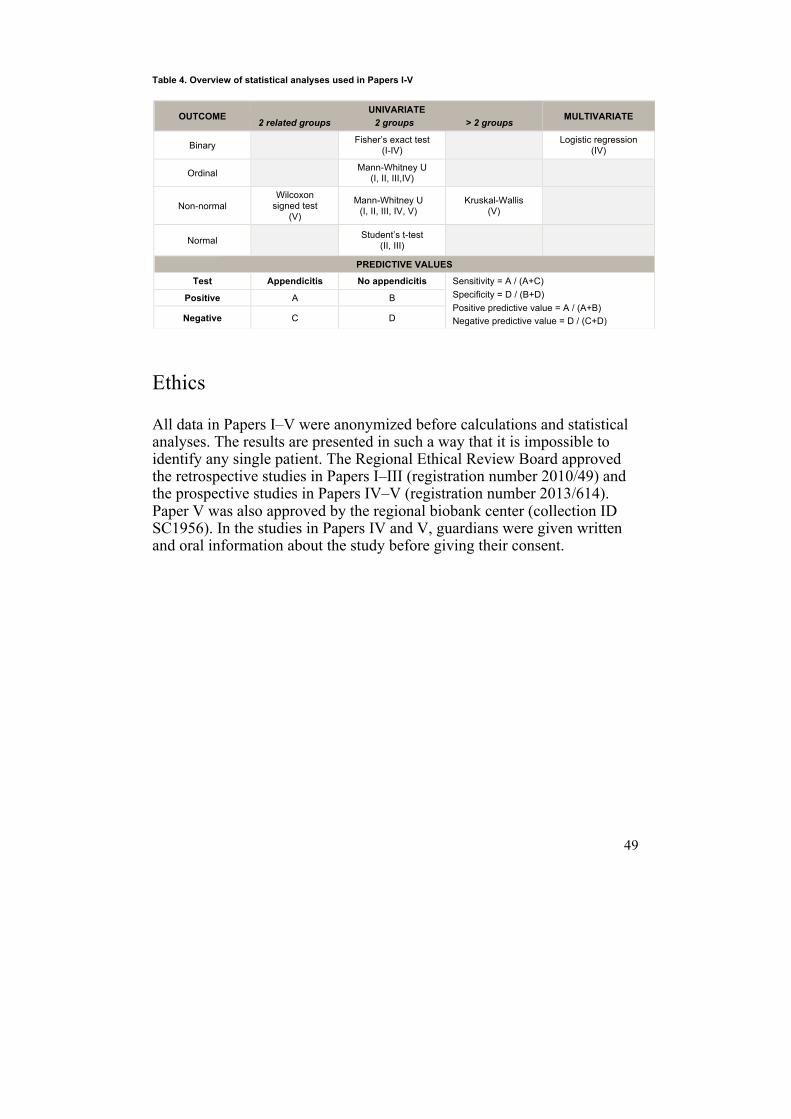
49
Table 4. Overview of statistical analyses used in Papers I-V
Ethics
All data in Papers I–V were anonymized before calculations and statistical analyses. The results are presented in such a way that it is impossible to identify any single patient. The Regional Ethical Review Board approved the retrospective studies in Papers I–III (registration number 2010/49) and the prospective studies in Papers IV–V (registration number 2013/614). Paper V was also approved by the regional biobank center (collection ID SC1956). In the studies in Papers IV and V, guardians were given written and oral information about the study before giving their consent.
OUTCOME UNIVARIATE
2 related groups 2 groups > 2 groups MULTIVARIATE
Binary Fisher’s exact test (I-IV) Logistic regression
(IV)
Ordinal Mann-Whitney U (I, II, III,IV)
Non-normal Wilcoxon signed test
(V)
Mann-Whitney U (I, II, III, IV, V)
Kruskal-Wallis (V)
Normal Student’s t-test (II, III)
PREDICTIVE VALUES
Test Appendicitis No appendicitis Sensitivity = A / (A+C) Specificity = D / (B+D) Positive predictive value = A / (A+B) Negative predictive value = D / (C+D)
Positive A B
Negative C D
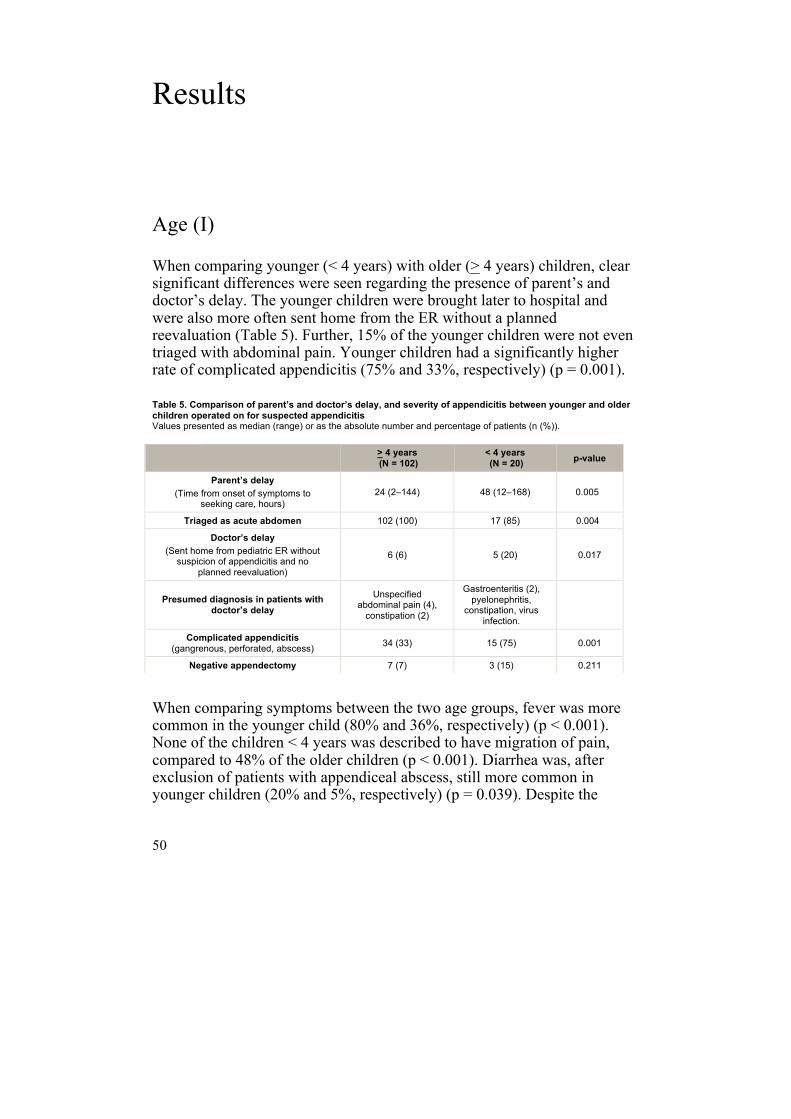
50
Results
Age (I)
When comparing younger (< 4 years) with older (> 4 years) children, clear significant differences were seen regarding the presence of parent’s and doctor’s delay. The younger children were brought later to hospital and were also more often sent home from the ER without a planned reevaluation (Table 5). Further, 15% of the younger children were not even triaged with abdominal pain. Younger children had a significantly higher rate of complicated appendicitis (75% and 33%, respectively) (p = 0.001).
Table 5. Comparison of parent’s and doctor’s delay, and severity of appendicitis between younger and older children operated on for suspected appendicitis Values presented as median (range) or as the absolute number and percentage of patients (n (%)).
When comparing symptoms between the two age groups, fever was more common in the younger child (80% and 36%, respectively) (p < 0.001). None of the children < 4 years was described to have migration of pain, compared to 48% of the older children (p < 0.001). Diarrhea was, after exclusion of patients with appendiceal abscess, still more common in younger children (20% and 5%, respectively) (p = 0.039). Despite the
> 4 years (N = 102)
< 4 years (N = 20) p-value
Parent’s delay (Time from onset of symptoms to
seeking care, hours) 24 (2–144) 48 (12–168) 0.005
Triaged as acute abdomen 102 (100) 17 (85) 0.004
Doctor’s delay (Sent home from pediatric ER without suspicion of appendicitis and no
planned reevaluation)
6 (6) 5 (20) 0.017
Presumed diagnosis in patients with doctor’s delay
Unspecified abdominal pain (4), constipation (2)
Gastroenteritis (2), pyelonephritis,
constipation, virus infection.
Complicated appendicitis (gangrenous, perforated, abscess) 34 (33) 15 (75) 0.001
Negative appendectomy 7 (7) 3 (15) 0.211
51
higher rate of complicated appendicitis, the children under 4 years were less likely to have tenderness in the RLQ (65% and 88%, respectively) (p = 0.016), and did not have a higher rate of peritonitis (50% and 52%, respectively). No differences between the two age groups were seen when comparing the presence of nausea/vomiting, anorexia, leukocytosis, neutrophilia, or symptoms of urinary tract infection.
Gender (II)
Girls and boys taken to the operating room due to suspicion of appendicitis were compared regarding symptoms, findings at the abdominal examination, and results from routine blood tests. No significant differences were found except that boys more often had local peritonitis in the RLQ (61% and 51%, respectively) (p = 0.042). Girls were more likely to have preoperative imaging (50% and 38%, respectively) (p = 0.021), but had a higher rate of negative appendectomy (Table 6). Despite no difference in time to operation, boys had a significantly higher rate of perforated appendicitis. Boys were also more likely to undergo open appendectomy (Table 6). There was a trend towards laparoscopic appendectomy taking longer time in girls than in boys (Table 6). No difference was found when comparing length of hospital stay between boys and girls. Neither were any differences found when comparing postoperative pain treatment with regard to the number of patients receiving morphine, amount of morphine administered, or the use of NSAIDs or paracetamol. Finally, boys and girls received equally long postoperative treatment with antibiotics in cases of complicated appendicitis. Girls had a significantly higher frequency of operative complications, and when sub-analyzed with regard to the operative modality, the significance was observed in open but not in laparoscopic appendectomy (Table 7). When comparing postoperative complications, no difference was seen between the genders.
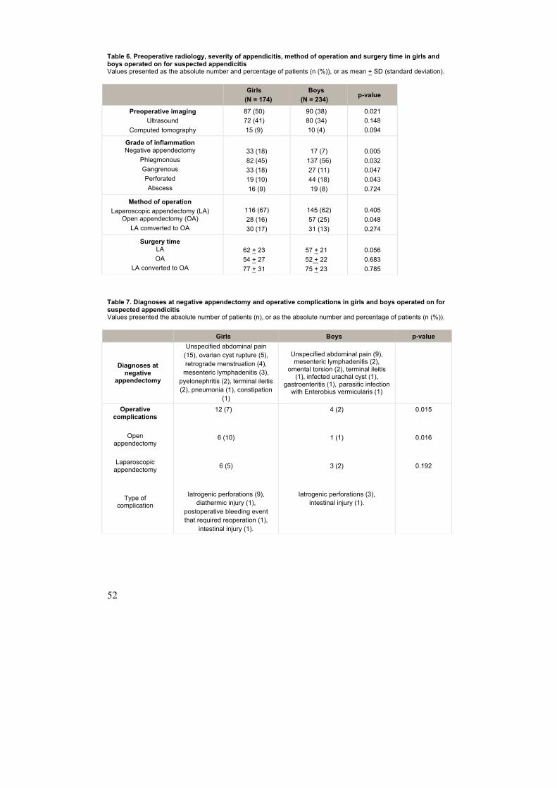
52
Table 6. Preoperative radiology, severity of appendicitis, method of operation and surgery time in girls and boys operated on for suspected appendicitis Values presented as the absolute number and percentage of patients (n (%)), or as mean + SD (standard deviation).
Table 7. Diagnoses at negative appendectomy and operative complications in girls and boys operated on for suspected appendicitis Values presented the absolute number of patients (n), or as the absolute number and percentage of patients (n (%)).
Girls
(N = 174) Boys
(N = 234) p-value
Preoperative imaging Ultrasound
Computed tomography
87 (50) 72 (41) 15 (9)
90 (38) 80 (34) 10 (4)
0.021 0.148 0.094
Grade of inflammation Negative appendectomy
Phlegmonous Gangrenous Perforated Abscess
33 (18) 82 (45) 33 (18) 19 (10) 16 (9)
17 (7) 137 (56) 27 (11) 44 (18) 19 (8)
0.005 0.032 0.047 0.043 0.724
Method of operation Laparoscopic appendectomy (LA)
Open appendectomy (OA) LA comverted to OA
116 (67) 28 (16) 30 (17)
145 (62) 57 (25) 31 (13)
0.405 0.048 0.274
Surgery time LA OA
LA converted to OA
62 + 23 54 + 27 77 + 31
57 + 21 52 + 22 75 + 23
0.056 0.683 0.785
Girls Boys p-value
Diagnoses at negative
appendectomy
Unspecified abdominal pain (15), ovarian cyst rupture (5), retrograde menstruation (4), mesenteric lymphadenitis (3), pyelonephritis (2), terminal ileitis (2), pneumonia (1), constipation
(1)
Unspecified abdominal pain (9), mesenteric lymphadenitis (2),
omental torsion (2), terminal ileitis (1), infected urachal cyst (1),
gastroenteritis (1), parasitic infection with Enterobius vermicularis (1)
Operative complications
Open
appendectomy
Laparoscopic appendectomy
Type of complication
12 (7)
6 (10)
6 (5)
Iatrogenic perforations (9), diathermic injury (1),
postoperative bleeding event that required reoperation (1),
intestinal injury (1).
4 (2)
1 (1)
3 (2)
Iatrogenic perforations (3), intestinal injury (1).
0.015
0.016
0.192
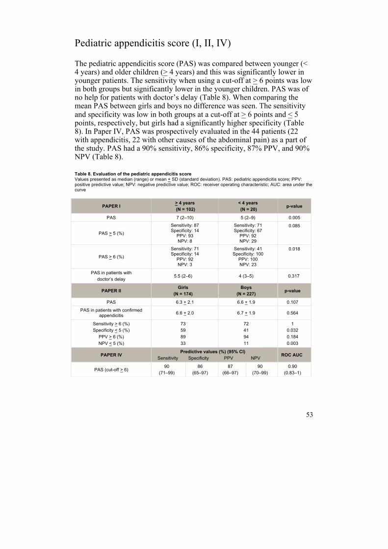
53
Pediatric appendicitis score (I, II, IV)
The pediatric appendicitis score (PAS) was compared between younger (< 4 years) and older children (> 4 years) and this was significantly lower in younger patients. The sensitivity when using a cut-off at > 6 points was low in both groups but significantly lower in the younger children. PAS was of no help for patients with doctor’s delay (Table 8). When comparing the mean PAS between girls and boys no difference was seen. The sensitivity and specificity was low in both groups at a cut-off at > 6 points and < 5 points, respectively, but girls had a significantly higher specificity (Table 8). In Paper IV, PAS was prospectively evaluated in the 44 patients (22 with appendicitis, 22 with other causes of the abdominal pain) as a part of the study. PAS had a 90% sensitivity, 86% specificity, 87% PPV, and 90% NPV (Table 8).
Table 8. Evaluation of the pediatric appendicitis score Values presented as median (range) or mean + SD (standard deviation). PAS: pediatric appendicitis score;; PPV: positive predictive value;; NPV: negative predictive value;; ROC: receiver operating characteristic;; AUC: area under the curve
PAPER I > 4 years (N = 102)
< 4 years (N = 20) p-value
PAS 7 (2–10) 5 (2–9) 0.005
PAS > 5 (%)
Sensitivity: 87 Specificity: 14 PPV: 93 NPV: 8
Sensitivity: 71 Specificity: 67 PPV: 92 NPV: 29
0.085
PAS > 6 (%)
Sensitivity: 71 Specificity: 14 PPV: 92 NPV: 3
Sensitivity: 41 Specificity: 100 PPV: 100 NPV: 23
0.018
PAS in patients with doctor’s delay
5.5 (2–6) 4 (3–5) 0.317
PAPER II Girls
(N = 174) Boys
(N = 227) p-value
PAS 6.3 + 2.1 6.6 + 1.9 0.107
PAS in patients with confirmed appendicitis 6.6 + 2.0 6.7 + 1.9 0.564
Sensitivity > 6 (%) Specificity < 5 (%) PPV > 6 (%) NPV < 5 (%)
73 59 89 33
72 41 94 11
1 0.032 0.184 0.003
PAPER IV Predictive values (%) (95% CI)
Sensitivity Specificity PPV NPV ROC AUC
PAS (cut-off > 6) 90
(71–99) 86
(65–97) 87
(66–97) 90
(70–99) 0.90
(0.83–1)
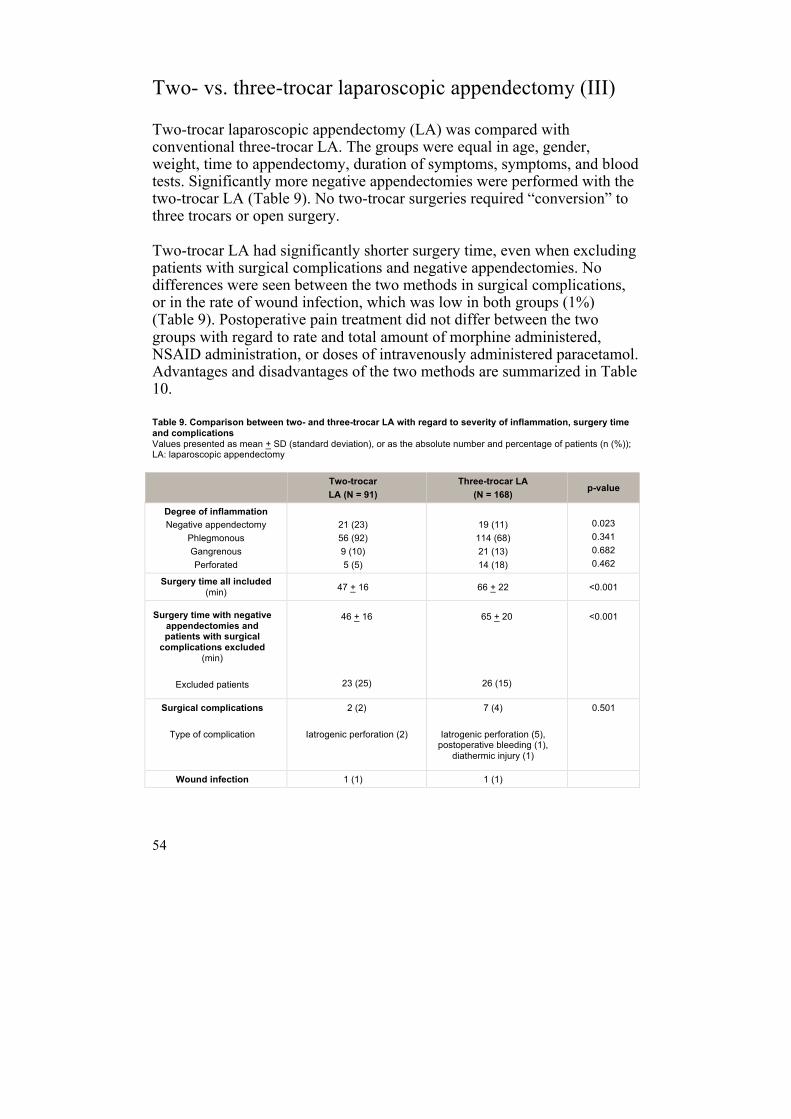
54
Two- vs. three-trocar laparoscopic appendectomy (III)
Two-trocar laparoscopic appendectomy (LA) was compared with conventional three-trocar LA. The groups were equal in age, gender, weight, time to appendectomy, duration of symptoms, symptoms, and blood tests. Significantly more negative appendectomies were performed with the two-trocar LA (Table 9). No two-trocar surgeries required “conversion” to three trocars or open surgery. Two-trocar LA had significantly shorter surgery time, even when excluding patients with surgical complications and negative appendectomies. No differences were seen between the two methods in surgical complications, or in the rate of wound infection, which was low in both groups (1%) (Table 9). Postoperative pain treatment did not differ between the two groups with regard to rate and total amount of morphine administered, NSAID administration, or doses of intravenously administered paracetamol. Advantages and disadvantages of the two methods are summarized in Table 10. Table 9. Comparison between two- and three-trocar LA with regard to severity of inflammation, surgery time and complications Values presented as mean + SD (standard deviation), or as the absolute number and percentage of patients (n (%));; LA: laparoscopic appendectomy
Two-trocar LA (N = 91)
Three-trocar LA (N = 168)
p-value
Degree of inflammation Negative appendectomy
Phlegmonous Gangrenous Perforated
21 (23) 56 (92) 9 (10) 5 (5)
19 (11) 114 (68) 21 (13) 14 (18)
0.023 0.341 0.682 0.462
Surgery time all included (min) 47 + 16 66 + 22 <0.001
Surgery time with negative appendectomies and patients with surgical complications excluded
(min)
Excluded patients
46 + 16
23 (25)
65 + 20
26 (15)
<0.001
Surgical complications
Type of complication
2 (2)
Iatrogenic perforation (2)
7 (4) Iatrogenic perforation (5), postoperative bleeding (1),
diathermic injury (1)
0.501
Wound infection 1 (1) 1 (1)
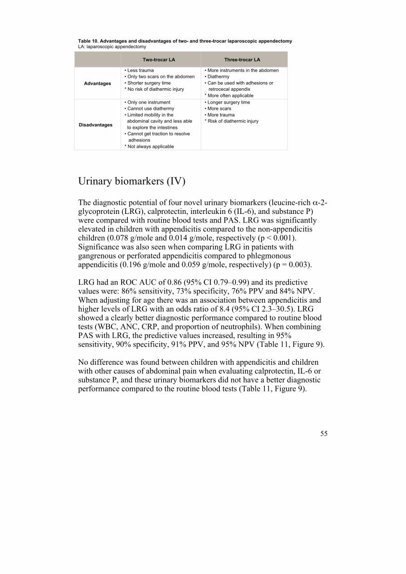
55
Table 10. Advantages and disadvantages of two- and three-trocar laparoscopic appendectomy LA: laparoscopic appendectomy
Urinary biomarkers (IV)
The diagnostic potential of four novel urinary biomarkers (leucine-rich α-2-glycoprotein (LRG), calprotectin, interleukin 6 (IL-6), and substance P) were compared with routine blood tests and PAS. LRG was significantly elevated in children with appendicitis compared to the non-appendicitis children (0.078 g/mole and 0.014 g/mole, respectively (p < 0.001). Significance was also seen when comparing LRG in patients with gangrenous or perforated appendicitis compared to phlegmonous appendicitis (0.196 g/mole and 0.059 g/mole, respectively) (p = 0.003). LRG had an ROC AUC of 0.86 (95% CI 0.79–0.99) and its predictive values were: 86% sensitivity, 73% specificity, 76% PPV and 84% NPV. When adjusting for age there was an association between appendicitis and higher levels of LRG with an odds ratio of 8.4 (95% CI 2.3–30.5). LRG showed a clearly better diagnostic performance compared to routine blood tests (WBC, ANC, CRP, and proportion of neutrophils). When combining PAS with LRG, the predictive values increased, resulting in 95% sensitivity, 90% specificity, 91% PPV, and 95% NPV (Table 11, Figure 9). No difference was found between children with appendicitis and children with other causes of abdominal pain when evaluating calprotectin, IL-6 or substance P, and these urinary biomarkers did not have a better diagnostic performance compared to the routine blood tests (Table 11, Figure 9).
Two-trocar LA Three-trocar LA
Advantages
• Less trauma • Only two scars on the abdomen • Shorter surgery time * No risk of diathermic injury
• More instruments in the abdomen • Diathermy • Can be used with adhesions or retrocecal appendix * More often applicable
Disadvantages
• Only one instrument • Cannot use diathermy • Limited mobility in the abdominal cavity and less able to explore the intestines • Cannot get traction to resolve adhesions * Not always applicable
• Longer surgery time • More scars • More trauma * Risk of diathermic injury
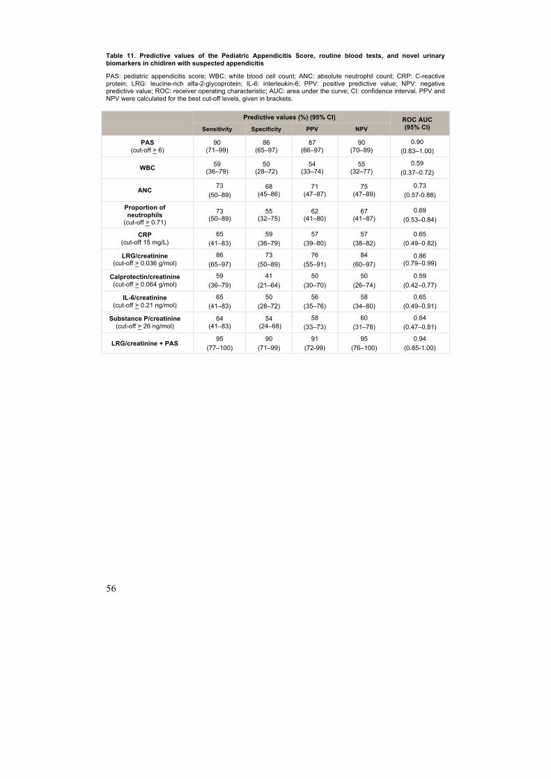
56
Table 11. Predictive values of the Pediatric Appendicitis Score, routine blood tests, and novel urinary biomarkers in chidlren with suspected appendicitis
PAS: pediatric appendicitis score;; WBC: white blood cell count;; ANC: absolute neutrophil count;; CRP: C-reactive protein;; LRG: leucine-rich alfa-2-glycoprotein;; IL-6: interleukin-6;; PPV: positive predictive value;; NPV: negative predictive value;; ROC: receiver operating characteristic;; AUC: area under the curve;; CI: confidence interval. PPV and NPV were calculated for the best cut-off levels, given in brackets.
Predictive values (%) (95% CI) ROC AUC
(95% CI) Sensitivity Specificity PPV NPV
PAS (cut-off > 6)
90 (71–99)
86 (65–97)
87 (66–97)
90 (70–99)
0.90 (0.83–1.00)
WBC 59 (36–79)
50 (28–72)
54 (33–74)
55 (32–77)
0.59 (0.37–0.72)
ANC 73
(50–89) 68
(45–86) 71
(47–87) 75
(47–89) 0.73
(0.57-0.88)
Proportion of neutrophils (cut-off > 0.71)
73 (50–89)
55 (32–75)
62 (41–80)
67 (41–87)
0.69 (0.53–0.84)
CRP (cut-off 15 mg/L)
65 (41–83)
59 (36–79)
57 (39–80)
57 (38–82)
0.65 (0.49–0.82)
LRG/creatinine (cut-off > 0.036 g/mol)
86 (65–97)
73 (50–89)
76 (55–91)
84 (60–97)
0.86 (0.79–0.99)
Calprotectin/creatinine (cut-off > 0.064 g/mol)
59 (36–79)
41 (21–64)
50 (30–70)
50 (26–74)
0.59 (0.42–0.77)
IL-6/creatinine (cut-off > 0.21 ng/mol)
65 (41–83)
50 (28–72)
56 (35–76)
58 (34–80)
0.65 (0.49–0.91)
Substance P/creatinine (cut-off > 26 ng/mol)
64 (41–83)
54 (24–68)
58 (33–73)
60 (31–78)
0.64 (0.47–0.81)
LRG/creatinine + PAS 95
(77–100) 90
(71–99) 91
(72-99) 95
(76–100) 0.94
(0.85-1.00)
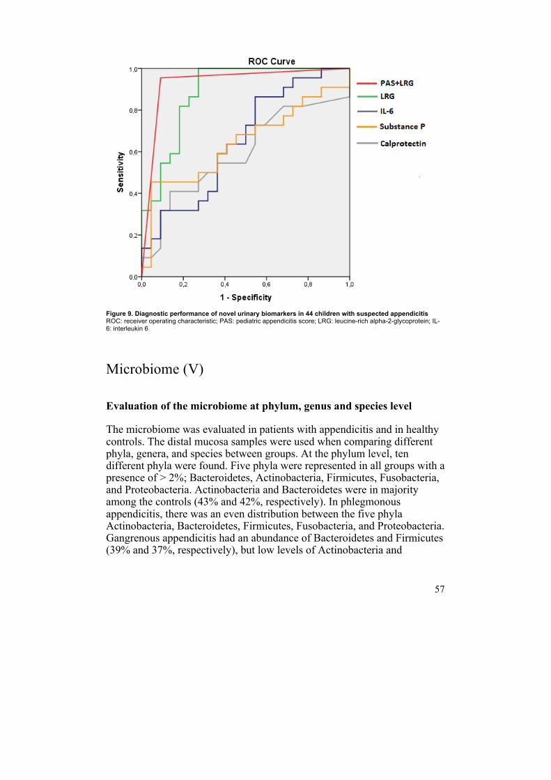
57
Figure 9. Diagnostic performance of novel urinary biomarkers in 44 children with suspected appendicitis ROC: receiver operating characteristic;; PAS: pediatric appendicitis score;; LRG: leucine-rich alpha-2-glycoprotein;; IL-6: interleukin 6
Microbiome (V)
Evaluation of the microbiome at phylum, genus and species level
The microbiome was evaluated in patients with appendicitis and in healthy controls. The distal mucosa samples were used when comparing different phyla, genera, and species between groups. At the phylum level, ten different phyla were found. Five phyla were represented in all groups with a presence of > 2%; Bacteroidetes, Actinobacteria, Firmicutes, Fusobacteria, and Proteobacteria. Actinobacteria and Bacteroidetes were in majority among the controls (43% and 42%, respectively). In phlegmonous appendicitis, there was an even distribution between the five phyla Actinobacteria, Bacteroidetes, Firmicutes, Fusobacteria, and Proteobacteria. Gangrenous appendicitis had an abundance of Bacteroidetes and Firmicutes (39% and 37%, respectively), but low levels of Actinobacteria and
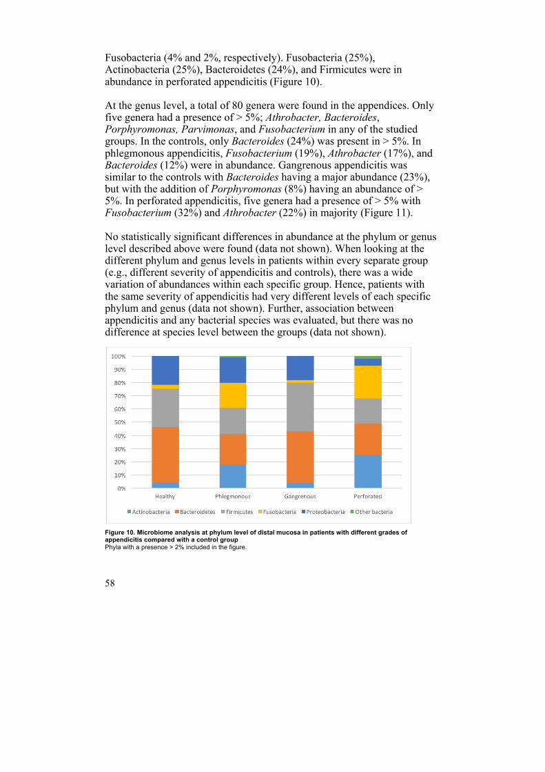
58
Fusobacteria (4% and 2%, respectively). Fusobacteria (25%), Actinobacteria (25%), Bacteroidetes (24%), and Firmicutes were in abundance in perforated appendicitis (Figure 10). At the genus level, a total of 80 genera were found in the appendices. Only five genera had a presence of > 5%; Athrobacter, Bacteroides, Porphyromonas, Parvimonas, and Fusobacterium in any of the studied groups. In the controls, only Bacteroides (24%) was present in > 5%. In phlegmonous appendicitis, Fusobacterium (19%), Athrobacter (17%), and Bacteroides (12%) were in abundance. Gangrenous appendicitis was similar to the controls with Bacteroides having a major abundance (23%), but with the addition of Porphyromonas (8%) having an abundance of > 5%. In perforated appendicitis, five genera had a presence of > 5% with Fusobacterium (32%) and Athrobacter (22%) in majority (Figure 11). No statistically significant differences in abundance at the phylum or genus level described above were found (data not shown). When looking at the different phylum and genus levels in patients within every separate group (e.g., different severity of appendicitis and controls), there was a wide variation of abundances within each specific group. Hence, patients with the same severity of appendicitis had very different levels of each specific phylum and genus (data not shown). Further, association between appendicitis and any bacterial species was evaluated, but there was no difference at species level between the groups (data not shown). Figure 10. Microbiome analysis at phylum level of distal mucosa in patients with different grades of appendicitis compared with a control group Phyla with a presence > 2% included in the figure.
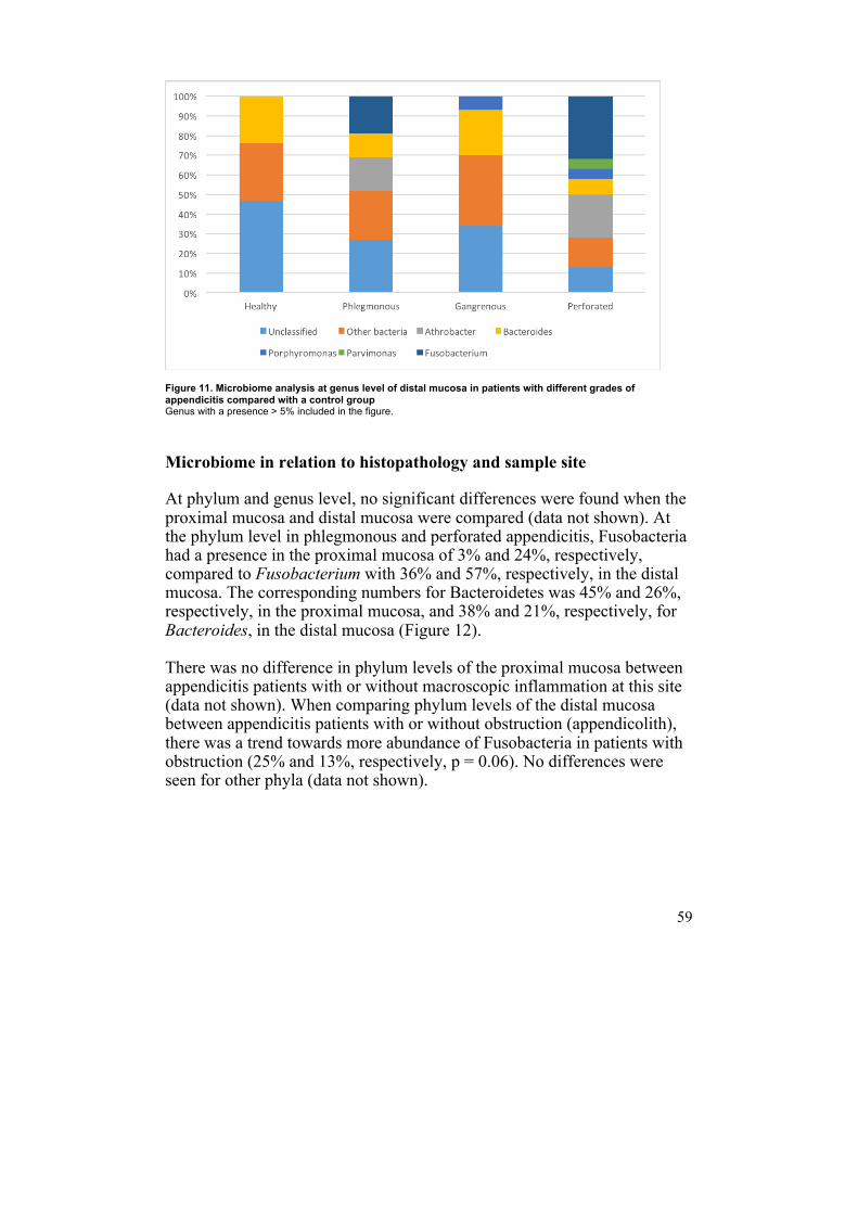
59
Figure 11. Microbiome analysis at genus level of distal mucosa in patients with different grades of appendicitis compared with a control group Genus with a presence > 5% included in the figure.
Microbiome in relation to histopathology and sample site
At phylum and genus level, no significant differences were found when the proximal mucosa and distal mucosa were compared (data not shown). At the phylum level in phlegmonous and perforated appendicitis, Fusobacteria had a presence in the proximal mucosa of 3% and 24%, respectively, compared to Fusobacterium with 36% and 57%, respectively, in the distal mucosa. The corresponding numbers for Bacteroidetes was 45% and 26%, respectively, in the proximal mucosa, and 38% and 21%, respectively, for Bacteroides, in the distal mucosa (Figure 12). There was no difference in phylum levels of the proximal mucosa between appendicitis patients with or without macroscopic inflammation at this site (data not shown). When comparing phylum levels of the distal mucosa between appendicitis patients with or without obstruction (appendicolith), there was a trend towards more abundance of Fusobacteria in patients with obstruction (25% and 13%, respectively, p = 0.06). No differences were seen for other phyla (data not shown).
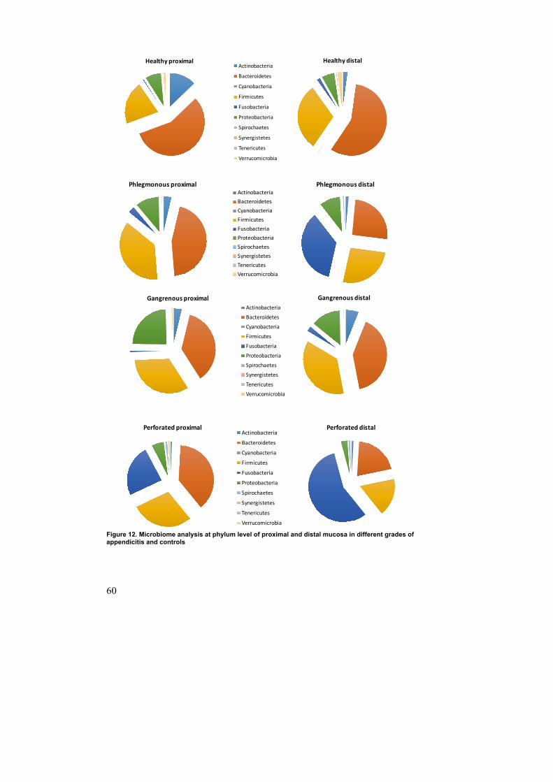
60
Figure 12. Microbiome analysis at phylum level of proximal and distal mucosa in different grades of appendicitis and controls
Healthy(proximalActinobacteria
Bacteroidetes
Cyanobacteria
Firmicutes
Fusobacteria
Proteobacteria
Spirochaetes
Synergistetes
Tenericutes
Verrucomicrobia
Healthy(distal
Phlegmonous+proximalActinobacteriaBacteroidetesCyanobacteriaFirmicutesFusobacteriaProteobacteriaSpirochaetesSynergistetesTenericutesVerrucomicrobia
Phlegmonous+distal
Gangrenous*proximalActinobacteriaBacteroidetesCyanobacteriaFirmicutesFusobacteriaProteobacteriaSpirochaetesSynergistetesTenericutesVerrucomicrobia
Gangrenous*distal
Perforated)proximalActinobacteria
Bacteroidetes
Cyanobacteria
Firmicutes
Fusobacteria
Proteobacteria
Spirochaetes
Synergistetes
Tenericutes
Verrucomicrobia
Perforated)distal
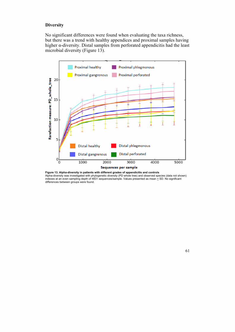
61
Diversity
No significant differences were found when evaluating the taxa richness, but there was a trend with healthy appendices and proximal samples having higher α-diversity. Distal samples from perforated appendicitis had the least microbial diversity (Figure 13). Figure 13. Alpha-diversity in patients with different grades of appendicitis and controls Alpha-diversity was investigated with phylogenetic diversity (PD whole tree) and observed species (data not shown) indexes at an even sampling depth of 4831 sequences/sample. Values presented as mean + SD. No significant differences between groups were found.
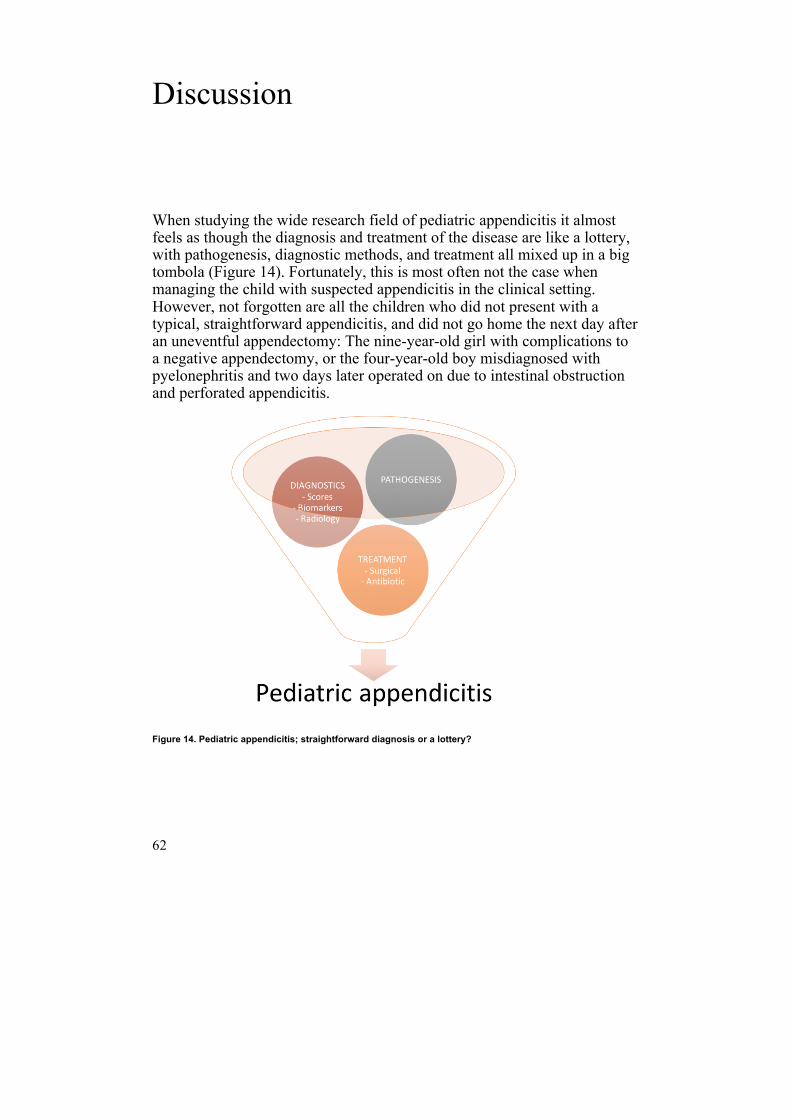
62
Discussion
When studying the wide research field of pediatric appendicitis it almost feels as though the diagnosis and treatment of the disease are like a lottery, with pathogenesis, diagnostic methods, and treatment all mixed up in a big tombola (Figure 14). Fortunately, this is most often not the case when managing the child with suspected appendicitis in the clinical setting. However, not forgotten are all the children who did not present with a typical, straightforward appendicitis, and did not go home the next day after an uneventful appendectomy: The nine-year-old girl with complications to a negative appendectomy, or the four-year-old boy misdiagnosed with pyelonephritis and two days later operated on due to intestinal obstruction and perforated appendicitis.
Figure 14. Pediatric appendicitis;; straightforward diagnosis or a lottery?
63
Problems at young age
Appendicitis in young children continues to be a clinical problem. There are several reasons for this. One: although appendicitis is the most common abdominal disease in children requiring acute surgery, we have to appreciate that its rate is only 1–4% of all children presenting at the pediatric ER with abdominal pain (27,28). Further, the incidence is considerably lower in younger than in older children (32,34,35), which may explain why the diagnosis is not directly suspected in the two-year-old child with fever and objectively abdominal pain. Hence, the doctor may not have that high suspicion of appendicitis before seeing the patient, which of course affects the clinical decisions and subsequent management and work-up (167). Two: especially seen in younger children with appendicitis, patient history and symptoms and findings from clinical examination are more diffuse (35,95–98). Together, this leads to misdiagnosis, high perforation rates, increased morbidity, and longer hospital stay (32,34,35,95–98,100–102). In Paper I, the misdiagnosis and morbidity mentioned above was confirmed. The younger children (< 4 years) had severer appendicitis and longer hospital stay. As seen in other studies (34,96,98,99), we saw a significant rate of both parent’s and doctor’s delay in the younger children. We also saw that 15% of the young children were not even triaged with “acute abdomen”, giving further evidence of the diffuseness of the symptoms. Finally, the rate of diarrhea was significantly higher among the young children, which of course may confuse the clinician. The main purpose of the study was to evaluate the pediatric appendicitis score (PAS) in younger children to see if this could be an aid for the clinician. The original study describing PAS did not include children < 4 years of age (113), and no study had evaluated the score between younger and older children before. Unfortunately, the results showed that PAS was not helpful; the younger children had a lower mean score, despite the severer appendicitis. The sensitivity of this test was low in both age groups, and this was despite the evaluation of children operated on for suspicion of appendicitis. The main disadvantages of the study were that it was retrospective, and that the cohort consisted of children who were appendectomized, instead of children with abdominal pain and suspicion of appendicitis. This of course, makes it hard to interpret the predictive values.
64
One speculation is that no present clinical prediction score really aids the diagnosis of appendicitis in the young children. Looking at the parameters in the different scores (108,109,111,113), one can see the problem: pain migration is of course difficult for the young child to describe, intensity of pain hard to evaluate in the young child, peritonitis less evident in the abdomen with less developed muscles, and nausea/vomiting and anorexia frequently seen in young children with extra abdominal disease. One might conclude that other diagnostic modalities are probably the right way to go when evaluating the young child with abdominal pain. Maybe we should be more liberal with ultrasound? Another possibility is the development of accurate biomarkers. It is probably hard to lower the perforation rate among young children to levels on a par with that of older children, since most perforations occur prehospitally (88). Hence, the main part of perforations could be speculated to be due to parent’s delay. However, perforated appendicitis is missed in hospital as well, and with improved diagnostics this doctor’s delay can be eliminated.
Girls
The incidence of appendicitis is lower in girls than in boys (26). Both girls and women have a higher risk of misdiagnosis with higher negative appendectomy rate compared to males (26,93,99,103). A striking difference of life-time risk of appendectomy was reported by Addiss et al. with 12% for boys and 23% for girls (26). In Paper II, we evaluated gender differences in pediatric appendicitis since the literature on this subject was scarce. The higher rate of negative appendectomy rate previously reported was confirmed in our cohort. We also saw that girls significantly had more preoperative imaging. Further, no differences in symptoms motivating earlier operative intervention were seen among girls. Instead, the specificity of PAS was higher in girls. Altogether, one might draw the conclusion that the girls are taken to the operating theater despite a clinically lower suspicion of appendicitis. One hypothesis is that surgeons are afraid of missing appendicitis in girls, possibly due to the misconception that perforated appendicitis may lead to infertility later in life (168). However, one would think that it is more equivocal cases that are misdiagnosed. Another speculation is that the girls
65
are operated on more liberally due to the possibility of ovarian pathology. However, this should not be an eligible reason since salpingitis and ovarian torsion are very uncommon in the premenarchal girl. Further, the sensitivity of ultrasound for ovarian torsion is equal to that for appendicitis (169,170). Hence, it is very unlikely that girls have an ovarian disease that requires surgical treatment. None of the 33 girls with negative appendectomies in our cohort had an ovarian disease that required operative treatment. As mentioned, preoperative imaging was used more frequently in girls, which also has been reported by others (171). In women, this only seems to delay time to surgery without reducing the frequency of negative appendectomies (172). Ultrasound for appendicitis has been reported to have lower sensitivity in girls (171). From our study, it does not seem that ultrasound resulted in fewer negative appendectomies, neither that the sensitivity for girls was lower. However, this was not given any specific attention. Girls had a higher proportion of gangrenous appendicitis and boys a higher rate of perforated appendicitis. However, if the negative appendectomies were excluded and rates compared only in patients with appendicitis, no differences were seen. A higher rate of perforation has been described before in men but not in boys (99,173). Further, girls had more operative complications when operated on with open appendectomy. Fortunately, these occurred in patients with gangrenous appendicitis (and not among negative appendectomies), and consisted of iatrogenic perforations. Perhaps, more “extensive” surgery to examine ovaries can explain this difference. In conclusion, the higher rate of negative appendectomies in girls continues to be reported in study after study. The reasons seem to be motivated by the wish to avoid infertility or by not missing ovarian pathology, of which both are misconceptions in most cases. Girls do not seem to present with symptoms differing from those of boys, and PAS seems equally accurate in both genders. Hence, there are no obvious “medical” reasons for the significantly higher rate of misdiagnosis in girls. From the literature, ultrasound does not seem to improve the diagnostic accuracy for appendicitis among girls (171). Perhaps, a different, maybe more restrictive, approach among surgeons is one way to solve the problem. This could even be motivated by the lower incidence of appendicitis among girls. Another possibility, like among the young children, is the development and evaluation of new biomarkers.
66
The study in Paper II was retrospective which, as always, is a disadvantage. However, awareness among the involved surgeons of a gender focus might have influenced the treatment and subsequently the results to become more gender equal. Finally, evaluating gender differences in pediatric surgery is of great importance, since it can have a major impact on the surgical care and, not least, on parental counseling. Unfortunately, there are today few studies that have specifically evaluated gender differences in pediatric surgery.
Two trocars
Since the first laparoscopic appendectomy, the minimally invasive surgery for appendicitis has continued to develop. From the conventional laparoscopic appendectomy using three trocars, techniques using two ports or only one, or a single incision, have been described, developed and evaluated. In Paper III, we evaluated two-trocar laparoscopic appendectomy (LA) and retrospectively compared it with the conventional three-trocar LA. The main purpose was to evaluate the operation time and its complications rate. There are to our knowledge five previous studies evaluating true two-trocar LA (154,174–177), often called laparoscopic assisted extracorporeal appendectomy or video-assisted extracorporeal appendectomy (Table 12). No study has, however, compared two- with three-trocar LA in children before. The main findings were that the two-trocar LA was quicker than the conventional LA, and that the wound infection rate was low (1%). The operation time was in comparison with what some reported (174,176), while others have shown significantly shorter duration (154,177). The rate of wound infection rate was in comparison with most other studies (154,176,177). One study reported a rate of 11% (174), and this was also the only one using a port placement in the right and left iliac fossa. Moreover, the type of trocars used may affect the wound infection rate.
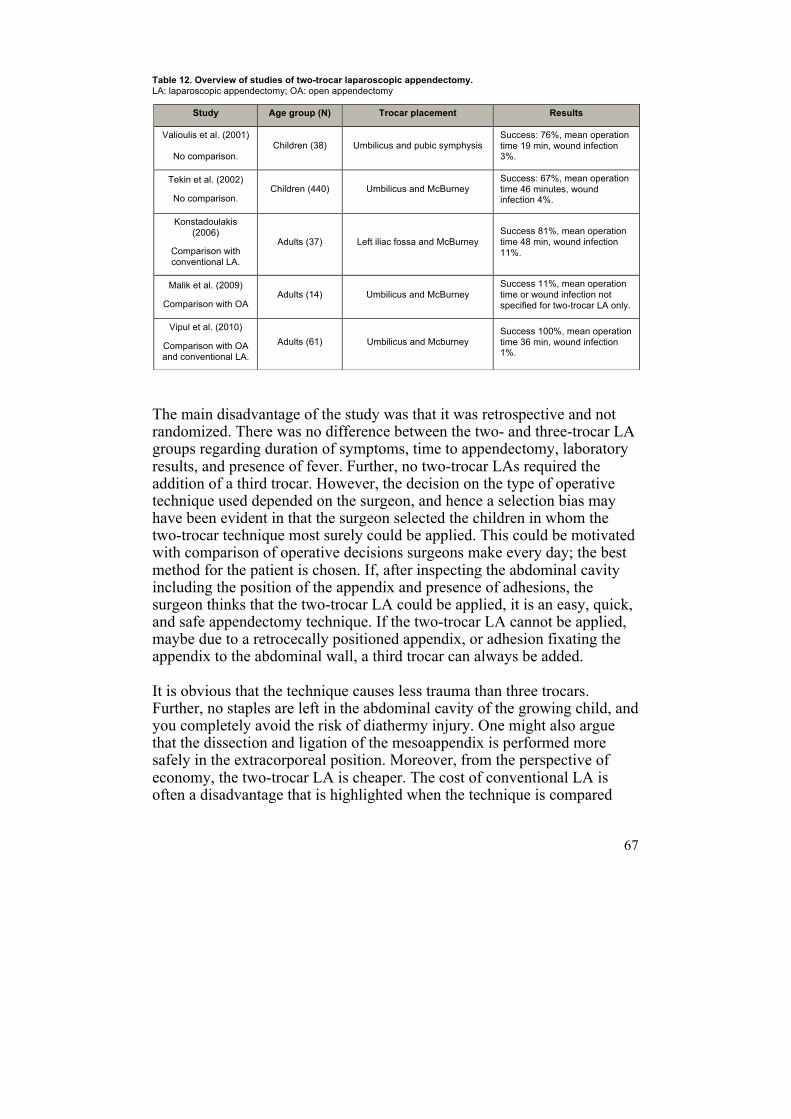
67
Table 12. Overview of studies of two-trocar laparoscopic appendectomy. LA: laparoscopic appendectomy;; OA: open appendectomy
The main disadvantage of the study was that it was retrospective and not randomized. There was no difference between the two- and three-trocar LA groups regarding duration of symptoms, time to appendectomy, laboratory results, and presence of fever. Further, no two-trocar LAs required the addition of a third trocar. However, the decision on the type of operative technique used depended on the surgeon, and hence a selection bias may have been evident in that the surgeon selected the children in whom the two-trocar technique most surely could be applied. This could be motivated with comparison of operative decisions surgeons make every day; the best method for the patient is chosen. If, after inspecting the abdominal cavity including the position of the appendix and presence of adhesions, the surgeon thinks that the two-trocar LA could be applied, it is an easy, quick, and safe appendectomy technique. If the two-trocar LA cannot be applied, maybe due to a retrocecally positioned appendix, or adhesion fixating the appendix to the abdominal wall, a third trocar can always be added. It is obvious that the technique causes less trauma than three trocars. Further, no staples are left in the abdominal cavity of the growing child, and you completely avoid the risk of diathermy injury. One might also argue that the dissection and ligation of the mesoappendix is performed more safely in the extracorporeal position. Moreover, from the perspective of economy, the two-trocar LA is cheaper. The cost of conventional LA is often a disadvantage that is highlighted when the technique is compared
Study Age group (N) Trocar placement Results
Valioulis et al. (2001)
No comparison. Children (38) Umbilicus and pubic symphysis
Success: 76%, mean operation time 19 min, wound infection 3%.
Tekin et al. (2002)
No comparison. Children (440) Umbilicus and McBurney
Success: 67%, mean operation time 46 minutes, wound infection 4%.
Konstadoulakis (2006)
Comparison with conventional LA.
Adults (37) Left iliac fossa and McBurney Success 81%, mean operation time 48 min, wound infection 11%.
Malik et al. (2009)
Comparison with OA Adults (14) Umbilicus and McBurney
Success 11%, mean operation time or wound infection not specified for two-trocar LA only.
Vipul et al. (2010)
Comparison with OA and conventional LA.
Adults (61) Umbilicus and Mcburney Success 100%, mean operation time 36 min, wound infection 1%.
68
with open appendectomy (178,179). Considering that appendectomy is a common operation, one would expect the two-trocar LA to reduce the costs substantially. There are numerous minimal access techniques described for appendectomy, the latest with one-port or a single incision. However, these techniques are technically more difficult and some require special equipment that is certainly not available in many hospitals. The two-trocar LA described by others, and in Paper III, uses regular instruments and is easy to learn, even for the resident. This makes it a useful technique besides being a clear benefit for the child.
Urinary biomarkers
So why are researchers trying to evaluate new biomarkers for appendicitis? The easy answer is that none of the present diagnostic tools is accurate enough. Clinical prediction scores have proven to not be sufficiently accurate (120). Ultrasound is operator dependent and not sensitive enough, CT should be used with precaution in children due to radiation (143), and MRI is superior to both US and CT, but has limits in availability, costs and probably feasibility. Finally, today available routine blood tests are neither sensitive or specific enough (123–125). In Paper IV, we evaluated four urinary biomarkers in children with suspected appendicitis; leucine-rich α-2-glycoprotein (LRG), calprotectin, IL-6, and Substance P. While no significant results were found for the last three, LRG had a better diagnostic performance than all the routine blood tests, and also seemed to correlate with the severity of the appendicitis. LRG has been evaluated in children with suspected appendicitis before. Kentsis et al. (130) found that LRG in urine was significantly elevated in patients with appendicitis compared to controls, and that LRG correlated with the severity of the appendicitis. However, the assay method was reported to play an important role in the outcome. When LRG was measured by ELISA, an immunoassay interference effect was described, and an AUC of only 0.80 was reached. When it was determined by mass spectrometry, the AUC reached impressive levels of 0.98–0.99 (130). Kharbanda et al. (132) found no difference between LRG in patients with phlegmonous appendicitis compared to patients without appendicitis, but a
69
significant difference between perforated appendicitis and non-perforated appendicitis. The ROC AUC for LRG was 0.63, and it showed 100% sensitivity, 23% specificity, and 100% NPV (132). In summary, our results with an AUC of 0.86 for LRG had a better performance compared to the two previous reports (130,132). This may be explained by a different ELISA compared to the other studies. Another possibility is that we adjusted for dehydration by measuring urine creatinine. The present study was the third one evaluating urinary biomarkers in children with suspected appendicitis. LRG is the most promising biomarker so far and should be further evaluated. There are studies showing that LRG, compared to the routine inflammatory markers, reflects a local inflammation, such as the one in appendicitis (139). For example, it has been shown that CRP and LRG, although both produced by the liver, seem to represent different physiological settings of inflammation (180). One might speculate that it is this type of inflammatory biomarker that in the future may prove to aid in the diagnosis of appendicitis; hence, not biomarkers that generally reflect inflammation, like CRP, IL-6, et cetera. Another consideration is that one way of finding a new biomarker is to look for substances in blood or urine that “leak” from the appendix when the disease starts. However, since the pathogenesis is not known, the “safest” way of seeking and evaluating novel biomarkers for appendicitis may still be to look for those reflecting (local) inflammation. Further, we combined LRG with PAS, and reached higher predictive values than with just LRG alone. Combining a biomarker with patient history and clinical examination is used in other fields of medicine, for example, in the management of deep vein thrombosis (181). This type of combination, with gathering information from patient history and examination before taking the test, uses the theory of pre-test probability. For pediatric appendicitis, the most useful biomarker is the one that can guide the surgeon in the decision of whether or not to take the child to the operating theater. Grading of the severity of the disease is less valuable. Upcoming studies should therefore have that focus. With a high negative predictive value, the surgeon can feel safe (regarding appendicitis) in not taking the child for a laparoscopy. In conclusion, LRG seems promising as a biomarker for pediatric appendicitis. A new accurate biomarker would be a simple and easy way to improve the management of children with suspected appendicitis. It is
70
important to point out that such a test should probably only be used in selected cases. Hence, patient history and abdominal examination will always be important. The test could, for example, also be used in primary care to aid in the decision of which patients should be referred. The strength of the study in Paper IV lies in being prospective; the same doctor performed the clinical evaluation of the patients, there was a homogenous cohort of only pediatric patients, and it was the first study trying to use a novel biomarker in conjunction with a clinical prediction score for pediatric appendicitis. The main weaknesses were the limited number of patients, and that the inclusion of patients was not random, so the diagnostic values may be biased.
Bacteria or not, that is the question
Several theories of the pathogenesis of appendicitis have been proposed of which the main are diet and hygiene, obstruction, immunological characteristics of the patient, and infection (40–49,51–54,59–62). Especially the concept of obstruction has been widely accepted despite several studies not supporting the theory (55–58). Bacteria has an obvious role in the development and consequences of appendicitis but so far often seen as a secondary event, hence infection after the inflammation, and the bacteriology have been widely studied (64–66). Most past studies have used conventional culture techniques to evaluate the role of bacteria in acute appendicitis. This technique is effective in evaluating solitary bacterial species, but lacks the capability of characterizing the polymicrobial diversity present (59). With these conventional culturing methods, as much as 90–99% of microbes are missed (67). There are a few recent studies evaluating the whole microbiome in appendicitis (59,68–71) using rRNA-based fluorescence in situ hybridization (FISH) or 16S RNA sequencing (68,70,71). To summarize, these studies have found a significant increase in bacteria normally part of the oral flora, in the inflamed appendices, especially Fusobacterium (Table 13). In Paper V, we evaluated the microbiome in children with appendicitis and in controls. An increase in the abundance of Fusobacterium and a decrease in Bacteroides was seen in phlegmonous and perforated appendicitis compared to controls, but no statistical significance was shown. Further, this pattern was not seen in gangrenous appendicitis, which more had a microbiome profile similar to the controls. Hence, no relationship between
71
different bacteria and severity of appendicitis was seen. Further, there was a wide variation of abundances at the phylum, genus, and species level within every specific group of patients. The study in Paper V could not confirm what previous studies have found with Fusobacterium being significantly increased in appendicitis compared to controls (59,68–71), or even a correlation between the grade of inflammation and the presence of Fusobacterium (59,68,71). However, the previous studies had no division of the appendicitis patients into the clinically and histopathologically defined groups of appendicitis. Hence, no specific group with gangrenous appendicitis was analyzed in these studies, and the conclusion with correlation of Fusobacterium to grade of inflammation was impossible to draw. No matter the significance of Fusobacterium, it is interesting with the finding of a bacteria that normally is part of the oropharyngeal flora. Though Fusobacterium seems to be a part of the normal appendiceal flora (68,70), it is also the most common oral anaerobe that gives rise to infection outside the oral cavity (182). Furthermore, reports exist on a possible negative correlation between inflammatory bowel disease (IBD) and appendectomy (47,183). Fusobacterium with its degree of invasive potential has also been shown to be associated with inflammatory bowel disease and the IBD status of the host (184). Alterations in the oral microbiome have also been linked to pediatric IBD (185). One may speculate that the presence of Fusobacterium may explain the link between appendectomy and IBD. In summary, with regard to the present and previous studies in the field (59,68,70,71), Fusobacteria may be part of the pathogenesis in some, but not the majority, of appendicitis cases.
The discussion about Fusobacterium could be “inversely extrapolated” to Bacteroides that was found to be abundant in healthy but also in gangrenous appendices. One study found Bacteroides to be inversely correlated to the degree of inflammation (70), but not others (68). However, as stated above, cited studies did not show a division of appendicitis patients into the three clinically and histopathologically defined groups of appendicitis, and hence, a specific group with gangrenous appendicitis was not analyzed. This may explain the difference in others results compared to our study, where Bacteroides was found distinctly increased in gangrenous compared to phlegmonous appendicitis.
72
Table 13. Overview of previous studies of non-culture dependent evaluation of appendicitis
One strength of the study in Paper V was the evaluation of different parts of the mucosa (proximal and distal), and relating the microbiome to the macroscopically seen inflammation. If you cut up the inflamed appendix it is evident that some display a general inflammation throughout the entire length, while in others the inflammation is more limited to a certain (often distal) part. From this observation, one would expect the results from the microbiome analysis to differ depending on from which site the sample was taken. Although differences were seen in the abundances of bacteria between proximal and distal mucosa, statistical significance was not reached, maybe due to the low number of patients studied. Thus, since there may be differences between proximal and distal mucosa, it is important to consider the sample site of the appendix when evaluating the microbiome. To summarize, there was a wide variation of abundances of bacteria within each specific group of appendicitis; this has also been described by others (70,71). One might think that this explains not only the lack of significances between the groups in our study, but also emphasizes the
Study Patients/ Controls Method Results
Swidsinski et al.
52 / 18 rRNA-based FISH
Invasion of Fusobacterium in the submucosa of the appendix. Fusobacterium not found in any controls and increased with the severity of inflammation.
Swidsinksi et al.
86 / X rRNA-based FISH
Guinane et al.
4 / 3 16sRNA sequencing
Highest amount of Fusobacterium found in appendicitis, but Fusobacterium also found in controls. Gemella, Parvimonas also abundantly increased in inflamed samples.
Zhong et al.
17 / 5 16sRNA sequencing
Increased abundance of Fusobacterium, Porphyromonas, Parvimonas, Gemella, and a reduced amount of Bacteroides in appendicitis compared to controls.
Jackson et al.
15 / 6 16sRNA sequencing
Fusobacter, Selonomonas, and Peptostreptococcus increased in normal appendices compared to normal rectal samples.
Peptostreptococcus, Bilophila, Bulleidia, Fusobacterium, Parvimonas, Mogibacterium, Aminobacterium, Proteus, Actinomycineae, Anaerovorax, Anaerofilum, and Porphyromonas increased in appendicitis compared to controls.
Bulleidia, Fusibacter, Prevotella, Porphyromonas, and Dialister increased in perforated appendicitis compared to non-perforated appendicitis. Bulleidia, Dialister, and Porphyromonas increased in rectal swabs of patients with appendicitis compared to controls.
73
hypothesis of whether the microbiome plays a primary etiological role in the pathogenesis of appendicitis. To conclude, despite the trends seen in our study and the significances found by others (59,68,70,71), it seems that in many cases of appendicitis, bacteria do not seem to be the primary event. The study in Paper V is, like most of the other studies evaluating the microbiome in pediatric appendicitis, small. Further studies are evidently needed, with larger cohorts, and with correlation to clinical parameters (laboratory tests, history of recent upper respiratory infection). Further, it could be of value to relate the microbiome findings with the histopathology report. Finally, the macroscopic appearance of the appendix lumen should be thoroughly evaluated, for example with regard to the presence of obstruction. Considering the ongoing discussion and evaluation of conservative treatment of appendicitis with antibiotics, it is of great importance to fully understand the role of the microbiome in appendicitis. The differences in outcomes previously reported in antibiotic use for appendicitis may be due to patients having a different microbial composition in the diseased appendix. It is a tempting thought that bacteriological findings may, in future, influence the choice of treatment with antibiotics or an operative intervention.
74
Conclusions
Paper I
PAS seems to be a scoring system for pediatric appendicitis, especially in younger children, and was of no help in the children with parent’s delay. Parent’s and doctor’s delay were contributing factors in the delayed diagnosis of appendicitis in younger children, which may explain the higher rate of complicated appendicitis in this group. Parameters in patient history, symptoms, and abdominal examination are more diffuse in younger children.
Paper II
There are important gender differences in pediatric appendicitis. Girls seem to have a higher rate of negative appendectomies despite more preoperative imaging, and they experienced more operative complications despite lower perforation rate. Boys have a higher perforation rate despite equal time to appendectomy.
Paper III
Two-trocar laparoscopic appendectomy seems to be a safe and quick technique with a low rate of postoperative wound infections, and could be a good and safe complement to the conventional three-trocar technique.
Paper IV
In children with suspected appendicitis, urine LRG is a promising biomarker for differentiating between patients with and without appendicitis, and for evaluating the severity of the disease. If LRG is used in conjunction with the pediatric appendicitis score, high predictive values seem reachable.

75
Paper V
The pattern of microbiome differed between the different inflammation groups, but also within the groups. No statistically significant differences could be found in the microbiome between groups or clinical conditions, and no correlation between a specific bacterium and grade of inflammation was found. In most cases of appendicitis, a specific bacterium does not seem to be the primary event.
76
Future aspects
Finally, what may be the next steps in the research and management of pediatric appendicitis? • Lowering the rate of misdiagnosis in young children with appendicitis.
Perhaps through specific age-adjusted clinical prediction scores, but more likely through development of imaging and new biomarkers.
• Lowering the rate of negative appendectomies in girls, perhaps through new biomarkers.
• Larger, prospective, preferably multi-center studies with comparison of clinical prediction scores.
• Continuous development and evaluation of minimally invasive techniques for appendectomy. Focus should be on rather simple methods with equipment available in every operation theater.
• Evaluation of new biomarkers to be used in conjunction with clinical prediction scores. Emphasis should be on reducing the numbers of negative appendectomies, and the biomarker should preferably aid in equivocal cases. Perforation rates are probably harder to reduce with a novel biomarker.
• The pathogenesis of appendicitis, with focus on the role of bacteria but also of the immunological events in the inflammation process. A more defined picture of the pathogenesis could facilitate the evaluation of different treatments.
Last but not least, until a clearer picture of the pathogenesis is presented, and better diagnostic accuracy with development of radiology and biomarkers is reached, we should not forget that patient history and abdominal examination are still the fundamental pillars of the clinician’s diagnostic arsenal.
77
Acknowledgements
Jag vill rikta ett stort tack till:
Mina fantastiska handledare Einar Arnbjörnsson, Bodil Ohlsson och Pernilla Stenström. Einar: För att du blandar en otrolig erfarenhet och kompetens med en ungdomlig entusiasm och energi. Allt känns lätt när man jobbar med dig oavsett om man opererar eller skriver manus. Slutligen måste du inneha världsrekord i att svara snabbast på mail.
Bodil: För din oerhörda erfarenhet som du delar med dig av, för din noggrannhet, för din förmåga att upprätta samarbeten och lösa problem. Men framförallt för att du lärt mig vad riktig forskning är.
Pernilla: För att du varvar värme, glädje, och nära till skratt med din oerhörda kompetens och kunnighet. Du kommer alltid med superba förslag till ändringar som förbättrar manuset på djupet. Jag hoppas att jag någon gång kommer i närheten av din kompetens som barnkirurg.
Mina medförfattare, framförallt Bodil Roth, för att du är ”värd din vikt i guld”, oerhört lätt att samarbeta med och för att de sista arbetena fortfarande inte varit klara utan din hjälp och kunnighet! Även stort tack till Frida Fåk, Nittaya Marungruang och Tiia-Marie Sundberg för all hjälp med de där bakterierna…
Sofia, fru och bästa vän, men också forskare. För att du stöttar mig i allt jag gör samtidigt som du försöker påminna mig om vad som är viktigt i livet. För att du är du och världens bästa mamma till Ester. Även stort tack för ditt tålamod vid kvällsundervisning i Excel (https://www.youtube.com/watch?v=N5mLK4V5P30).
Personalen på ”65:an: Tack för att ni gör arbetsdagen full av glädje och jag är imponerad av det omhändertagande och den omvårdnad barnen (och föräldrarna) får. Även stort tack för alla fikastunder på nätterna, de bidrar inte enbart till min vikt utan också till att man är skärpt på jouren! Ett speciellt tack till “Billis” för framtagning av patientlistor.
Magnus Anderberg, kollega och handledare, för roliga jourer och samtal, söndagsfika med forskningsprat som inte handlar om appendix, och för att du lyckades med bedriften att skaffa mig (och dig) en forskningsdator. Alla andra kollegor: Tobbe, Gunnar, Christina CK, Anna, Morten, Lars, Kristine, Hans,

78
Christina G, Erik, Ann, Mette, Maria, David, Louise, m.fl. för att ni är så fantastiskt duktiga och inspirerande och gör Barnkirurgen till bästa arbetsplatsen.
Mamma: För att du föreslog att jag skulle läsa medicin och för ditt tjat om att skolan alltid borde gå före ishockeyn.
Paulina: För systerlig uppfostran och läxförhör under min uppväxt ;)
Ester: Tittuuut!
Personalen på barnoperation, SUS Lund. För hjälpen med insamling av blindtarmarna och för ert varma omhändertagande när jag för första gången klev in på barnoperation.
Vänner: För gött häng med poker, bubbelbad och mat. Framförallt tack till Erik A för din gåva (jag hade på mig dem vid disputationen).
Region Skåne (Regionalt forskningsstöd) och Svensk Gastroenterologisk Förening (Mag-TarmFonden) för ekonomiskt bidrag.
79
References
1. Collins D. Historic phases of appendicitis. Ann Surg. 1931;94(179-96).
2. Cope Z. A history of the acute abdomen. London: Oxford University Press; 1965.
3. Hamill JK, Liley A, Hill AG. Historical aspects of appendicitis in children. ANZ J Surg. 2014;84(5):307–10.
4. Ansaloni L, Catena F PA. What Is the Function of the HumanVermiform Appendix? Evolution-Based Surgery: A New Perspective in the Darwinian Year 2009. Eur Surg Res. 2009;43:67–71.
5. Schumpelick V, Dreuw B, Ophoff K, Prescher a. Appendix and cecum. Embryology, anatomy, and surgical applications. Surg Clin North Am. 2000;80(1):295–318.
6. Schoenwolf GC, Bleyl SB, Brauer PR, Francis-West PH. Larsen’s human embryology. In: Larsen’s human embryology. 2014. p. 119–27.
7. Skandalakis JE, colborn GL, Weidman TA, Foster RS, Kingsworth AN, Skandalakis LJ, Skandalakis PN MP. Appendix, Surgical anatomy. Chapter 17. Philadelphia: Lippincott William & Wilkins; 2007. 843 - 860 p.
8. DC C. A study of 50,000 specimens of the human vermiform appendix.
80
Surg Gynecol Obs. 1955;101:437–45.
9. Standring S, editor. Gray’s anatomy. 40th ed. New York: Elsevier New York; 2009.
10. Collins D. The length and position of the vermiform appendix: A study of 4,680 specimens. Ann Surg. 1932;96(1044).
11. Ives EP, Sung S, McCue P, Durrani H, Halpern EJ. Independent Predictors of Acute Appendicitis on CT with Pathologic Correlation. Acad Radiol. 2008;15(8):996–1003.
12. Searle AR, Ismail KA, Macgregor D, Hutson JM. Changes in the length and diameter of the normal appendix throughout childhood. J Pediatr Surg. 2013;48(7):1535–9.
13. Wakeley C. The Position of the Vermiform Appendix as Ascertained by an Analysis of 10,000 Cases. J Anat. 1933;67:277–83.
14. Grover CA, Sternbach G. Charles McBurney: McBurney’s point. J Emerg Med. 2012;42(5):578–81.
15. Spencer J, Finn T, Isaacson PG. Gut associated lymphoid tissue: a morphological and immunocytochemical study of the human appendix. Gut. 1985;26(7):672–9.
16. Dasso JF, Obiakor H, Bach H, Anderson AO, Mage RG. A morphological and immunohistological study of the human and rabbit appendix for comparison with the avian bursa. Developmental and Comparative Immunology. 2000. p. 797–814.
81
17. Papadaki L, Rode J, Dhillon AP, Dische FE. Fine structure of a neuroendocrine complex in the mucosa of the appendix. Gastroenterology. 1983;84(3):490–7.
18. Darwin C. The descent of man and selection in relation to sex, in Charles Darwin, The origin of species and The descent of man (combined volume). J Anat Physiol. 1871;5(Pt 2):363–72.
19. Smith HF, Fisher RE, Everett ML, Thomas AD, Randal Bollinger R, Parker W. Comparative anatomy and phylogenetic distribution of the mammalian cecal appendix. J Evol Biol. 2009;22(10):1984–99.
20. Randal Bollinger R, Everett M Lou, Palestrant D, Love SD, Lin SS, Parker W. Human secretory immunoglobulin A may contribute to biofilm formation in the gut. Immunology. 2003;109(4):580–7.
21. Gebbers JO, Laissue JA. Bacterial translocation in the normal human appendix parallels the development of the local immune system. In: Annals of the New York Academy of Sciences. 2004. p. 337–43.
22. Bazar KA, Lee PY, Joon Yun A. An “eye” in the gut: The appendix as a sentinel sensory organ of the immune intelligence network. Medical Hypotheses. 2004. p. 752–8.
23. Thorén L. Appendicitens historia under 400 år 1521–1921. Nordisk Medicinhistorisk Årsbok; 1989.
24. Milanchi S, Allins AD. Amyand’s hernia: History, imaging, and management. Hernia. 2008;12(3):321–2.
25. Litynski GS. Kurt Semm and the fight against skepticism: endoscopic hemostasis, laparoscopic appendectomy, and Semm’s impact on the
82
“laparoscopic revolution.” JSLS. 1998;2(3):309–13.
26. Addiss DG, Shaffer N, Fowler BS, Tauxe R V. The epidemiology of appendicitis and appendectomy in the United States. Am J Epidemiol. 1990;132(5):910–25.
27. Scholer SJ, Pituch K, Orr DP, Dittus RS. Clinical outcomes of children with acute abdominal pain. Pediatrics. 1996;98(4 Pt 1):680–5.
28. Caperell K, Pitetti R, Cross KP. Race and acute abdominal pain in a pediatric emergency department. Pediatrics. 2013;131(6):1098–106.
29. Hui TT, Major KM, Avital I, Hiatt JR, Margulies DR. Outcome of elderly patients with appendicitis: effect of computed tomography and laparoscopy. Arch Surg. 2002;137(9):995–8; discussion 999–1000.
30. Lee SL, Walsh A. ., Ho HS, Schwesinger WH, Grosfeld JL, Kuhn J, et al. Computed tomography and ultrasonography do not improve and may delay the diagnosis and treatment of acute appendicitis. Archives of Surgery. 2001. p. 556–62.
31. Andersson RE, Lambe M. Incidence of appendicitis during pregnancy. Int J Epidemiol. 2001;30(6):1281–5.
32. Andersen S, Paerregaard A, Larsen K. Changes in the epidemiology of acute appendicitis and appendectomy in Danish children 1996-2004. Eur J Pediatr Surg. 2009;19(5):286–9.
33. Buckius MT, McGrath B, Monk J, Grim R, Bell T, Ahuja V. Changing epidemiology of acute appendicitis in the United States: Study period 1993-2008. J Surg Res. 2012;175(2):185–90.
83
34. Alloo J, Gerstle T, Shilyansky J, Ein SH. Appendicitis in children less than 3 years of age: A 28-year review. Pediatric Surgery International. 2004. p. 777–9.
35. Marzuillo P, Germani C, Krauss BS, Barbi E. Appendicitis in children less than five years old: A challenge for the general practitioner. World J Clin Pediatr. 2015;4(2):19–24.
36. Socialstyrelsens statistikdatabas. [Internet]. Available from: www.socialstyrelsen.se/statistik
37. Robinson AJ, Bingham J, Thompson RLE. Magnet induced perforated appendicitis and ileo-caecal fistula formation. Ulster Medical Journal. 2009. p. 4–6.
38. Kim JH, Lee DS, Kim KM. Acute appendicitis caused by foreign body ingestion. Ann Surg Treat Res. 2015;89(3):158–61.
39. Paschos KA, Boulas K, Liapis A, Georgiou E, Vrakas X. Traumatic appendicitis in minor blunt abdominal injury. EMA - Emerg Med Australas. 2012;24(3):343–6.
40. Larner AJ. The aetiology of appendicitis. Br J Hosp Med. 1988;39(6):540–2.
41. Brender JD, Weiss NS, Koepsell TD, Marcuse EK. Fiber intake and childhood appendicitis. Am J Public Health. 1985;75(4):399–400.
42. Morris J, Barker DJ, Nelson M. Diet, infection, and acute appendicitis in Britain and Ireland. J Epidemiol Community Health. 1987;41(1):44–9.
84
43. Walker a R, Segal I. What causes appendicitis? J Clin Gastroenterol. 1990;12(2):127–9.
44. Barker DJ. Acute appendicitis and dietary fibre: an alternative hypothesis. BMJ. 1985;290(6475):1125–7.
45. Andersson R. Appendicitis - epidemiology and diagnosis. Thesis. Linköping University, Sweden.; 1998.
46. Andersson RE, Olaison G, Tysk C, Ekbom A. Appendectomy is followed by increased risk of Crohn’s disease. Gastroenterology. 2003;124(1):40–6.
47. Andersson RE, Olaison G, Tysk C, Ekbom A. Appendectomy and protection against ulcerative colitis. N Engl J Med. 2001;344(11):808–14.
48. Frisch M, Pedersen B V, Andersson RE. Appendicitis, mesenteric lymphadenitis, and subsequent risk of ulcerative colitis: cohort studies in Sweden and Denmark. BMJ. 2009;338:b716.
49. Ruber M, Andersson M, Petersson BF, Olaison G, Andersson RE, Ekerfelt C. Systemic Th17-like cytokine pattern in gangrenous appendicitis but not in phlegmonous appendicitis. Surgery. 2010;147(3):366–72.
50. Holcomb III GW, Murphy JP, Ostlie DJ. Ashcraft’s pediatric surgery. 6th ed. Toronto: Elsevier; 2014.
51. Bundy DG, Byerley JS, Liles EA, Perrin EM, Katznelson J, Rice HE. Does this child have appendicitis? JAMA. 2007;298(4):438–51.
52. Walker ASI. What causes appendicitis? J Clin Gastroenterol. 1990;Apr(12(2)):127–9.
85
53. Humes DJ, Simpson J. Acute appendicitis. BMJ. 2006;333(7567):530–4.
54. Singh JP, Mariadason JG. Role of the faecolith in modern-day appendicitis. Ann R Coll Surg Engl. 2013;95(1):48–51.
55. Arnbjörnsson E, Bengmark S. Role of obstruction in the pathogenesis of acute appendicitis. Am J Surg. 1984;147:390–2.
56. Jones BA, Demetriades D, Segal I, Burkitt DP. The prevalence of appendiceal fecaliths in patients with and without appendicitis. A comparative study from Canada and South Africa. Ann Surg. 1985;202(1):80–2.
57. Chang AR. An analysis of the pathology of 3003 appendices. Aust N Z J Surg. 1981;51(2):169–78.
58. Carr NJ. The pathology of acute appendicitis. Ann Diagn Pathol. 2000;4(1):46–58.
59. Swidsinski A, Dörffel Y, Loening-Baucke V, Theissig F, Rückert JC, Ismail M, et al. Acute appendicitis is characterised by local invasion with Fusobacterium nucleatum/necrophorum. Gut. 2011;60(1):34–40.
60. Andersson R, Hugander A, Thulin A, Nystrom PO, Olaison G. Clusters of acute appendicitis - further evidence for an infectious etiology. International Journal of Epidemiology. 1995. p. 829–33.
61. Deng Y, Chang DC, Zhang Y, Webb J, Gabre-Kidan A, Abdullah F. Seasonal and day of the week variations of perforated appendicitis in US children. Pediatr Surg Int. 2010;26(7):691–6.
86
62. Zangbar B, Rhee P, Pandit V, Hsu C-H, Khalil M, Okeefe T, et al. Seasonal Variation in Emergency General Surgery. Ann Surg. 2015;
63. Alder AC, Fomby TB, Woodward W a, Haley RW, Sarosi G, Livingston EH. Association of viral infection and appendicitis. Arch Surg. 2010;145(1):63–71.
64. Leigh D a, Simmons K, Norman E. Bacterial flora of the appendix fossa in appendicitis and postoperative wound infection. J Clin Pathol. 1974;27:997–1000.
65. Roberts JP. Quantitative bacterial flora of acute appendicitis. Arch Dis Child. 1988;63(5):536–40.
66. Lamps LW. Beyond acute inflammation: a review of appendicitis and infections of the appendix. Diagnostic Histopathol. 2008;14(2):68–77.
67. Hugenholtz P, Goebel BM, Pace NR. Impact of culture-independent studies on the emerging phylogenetic view of bacterial diversity (Journal of Bacteriology (1998) 180:18 (4765-4774)). Journal of Bacteriology. 1998. p. 6793.
68. Jackson HT, Mongodin EF, Davenport KP, Fraser CM, Sandler AD, Zeichner SL. Culture-independent evaluation of the appendix and rectum microbiomes in children with and without appendicitis. PLoS One. 2014;9(4):e95414.
69. Swidsinski A, Loening-Baucke V, Biche-ool S, Guo Y, Dörffel Y, Tertychnyy A, et al. Mucosal invasion by fusobacteria is a common feature of acute appendicitis in Germany, Russia, and China. Saudi J Gastroenterol. 2012;18(1):55.
87
70. Zhong D, Brower-Sinning R, Firek B, Morowitz MJ. Acute appendicitis in children is associated with an abundance of bacteria from the phylum Fusobacteria. J Pediatr Surg. 2014;49(3):441–6.
71. Guinane CM, Tadrous A, Fouhy F, Ryan CA, Dempsey EM, Murphy B, et al. Microbial composition of human appendices from patients following appendectomy. MBio. 2013;4(1):e00366–12 – .
72. Campbell JS, Fournier P DT. When is the appendix normal? A study of acute inflammations of the appendix apparent only upon histologic examination. Can Med Assoc J. 1961;85:1155–7.
73. Andersson REB. Meta-analysis of the clinical and laboratory diagnosis of appendicitis. British Journal of Surgery. 2004. p. 28–37.
74. Riber C, Tønnesen H, Aru a, Bjerregaard B. Observer variation in the assessment of the histopathologic diagnosis of acute appendicitis. Scand J Gastroenterol. 1999;34(1):46–9.
75. Pieper R, Kager L, Näsman P. Clinical significance of mucosal inflammation of the vermiform appendix. Ann Surg. 1983;197(3):368–74.
76. Howie J. Too Few Appendicectomies? Lancet. 1964;1(7345):1240–2.
77. Emil S, Gaied F, Lo A, Laberge JM, Puligandla P, Shaw K, et al. Gangrenous appendicitis in children: A prospective evaluation of definition, bacteriology, histopathology, and outcomes. J Surg Res. 2012;177(1):123–6.
78. Peter SDS, Sharp SW, Holcomb GW, Ostlie DJ. An evidence-based definition for perforated appendicitis derived from a prospective
88
randomized trial. J Pediatr Surg. 2008;43(12):2242–5.
79. Todd A. Ponsky, Zhihuan J. Huang, Kory Kittle, Martin R. Eichelberger, James C. Gilbert; Fredrick Brody KDN. Hospital- and Patient-Level Characteristics and the Risk of Appendiceal Rupture and Negative Appendectomy in Children. JAMA. 2004;16(1977-1982).
80. Andersson R, Hugander A, Thulin A, Nyström PO, Olaison G. Indications for operation in suspected appendicitis and incidence of perforation. BMJ. 1994;308(6921):107–10.
81. Morino M, Pellegrino L, Castagna E, Farinella E, Mao P. Acute nonspecific abdominal pain: A randomized, controlled trial comparing early laparoscopy versus clinical observation. Ann Surg. 2006;244(6):881–6; discussion 886–8.
82. Decadt B, Sussman L, Lewis MP, Secker A, Cohen L, Rogers C, et al. Randomized clinical trial of early laparoscopy in the management of acute non-specific abdominal pain. Br J Surg. 1999;86(11):1383–6.
83. Cobben LP, de Van Otterloo AM, Puylaert JB. Spontaneously resolving appendicitis: frequency and natural history in 60 patients. Radiology. 2000;215(2):349–52.
84. Kirshenbaum M, Mishra V, Kuo D, Kaplan G. Resolving appendicitis: Role of CT. Abdom Imaging. 2003;28(2):276–9.
85. Migraine S, Atri M, Bret PM, Lough JO, Hinchey JE. Spontaneously resolving acute appendicitis: clinical and sonographic documentation. Radiology. 1997;205(1):55–8.
86. Barber MD, Mclaren J, Rainey JB. Recurrent appendicitis. Br J Surg.
89
1997;84(1):110–2.
87. Ciani S, Chuaqui B. Histological features of resolving acute, non-complicated phlegmonous appendicitis. Pathol Res Pract. 2000;196(2):89–93.
88. Andersson RE. The natural history and traditional management of appendicitis revisited: Spontaneous resolution and predominance of prehospital perforations imply that a correct diagnosis is more important than an early diagnosis. World Journal of Surgery. 2007. p. 86–92.
89. Maroju NK, Smile SR, Sistla SC, Narasimhan R, Sahai A. Delay in surgery for acute appendicitis. ANZ J Surg. 2004;74(9):773–6.
90. Yardeni D, Hirschl RB, Drongowski RA, Teitelbaum DH, Geiger JD, Coran AG, et al. Delayed Versus Immediate Surgery in Acute Appendicitis: Do We Need to Operate during the Night? In: Journal of Pediatric Surgery. 2004. p. 464–9.
91. Anderson JE, Bickler SW, Chang DC, Talamini MA. Examining a common disease with unknown etiology: Trends in epidemiology and surgical management of appendicitis in California, 1995-2009. World J Surg. 2012;36(12):2787–94.
92. Livingston EH, Woodward WA, Sarosi GA, Haley RW. Disconnect between incidence of nonperforated and perforated appendicitis: implications for pathophysiology and management. Ann Surg. 2007;245(6):886–92.
93. Flum DR, Morris A, Koepsell T, Dellinger EP. Has misdiagnosis of appendicitis decreased over time? A population-based analysis. JAMA. 2001;286(14):1748–53.
90
94. Flum DR, Koepsell T. The clinical and economic correlates of misdiagnosed appendicitis: nationwide analysis. Arch Surg. 2002;137(7):799–804; discussion 804.
95. Mallick MS. Appendicitis in pre-school children: A continuing clinical challenge. A retrospective study. Int J Surg. 2008;6(5):371–3.
96. Nance ML, Adamson WT, Hedrick HL. Appendicitis in the young child: a continuing diagnostic challenge. Pediatr Emerg Care. 2000;16(3):160–2.
97. Lee SL, Stark R, Yaghoubian A, Shekherdimian S, Kaji A. Does age affect the outcomes and management of pediatric appendicitis? In: Journal of Pediatric Surgery. 2011. p. 2342–5.
98. Williams N, Kapila L. Acute appendicitis in the under-5 year old. J R Coll Surg Edinb. 1994;39(3):168–70.
99. Korner H, Sondenaa K, Soreide J, Andersen E, Nysted A, Lende T, et al. Incidence of acute nonperforated and perforated appendicitis: age-specific and sex-specific analysis. World J Surg. 1997;21(3):313–7.
100. Smink DS, Finkelstein J a, Kleinman K, Fishman SJ. The effect of hospital volume of pediatric appendectomies on the misdiagnosis of appendicitis in children. Pediatrics. 2004;113(1 Pt 1):18–23.
101. Smink DS, Fishman SJ, Kleinman K, Finkelstein J a. Effects of race, insurance status, and hospital volume on perforated appendicitis in children. Pediatrics. 2005;115(4):920–5.
102. Bansal S, Banever GT, Karrer FM, Partrick D a. Appendicitis in children less than 5 years old: influence of age on presentation and outcome. Am J Surg. 2012;204(6):1031–5; discussion 1035.
91
103. Cheong LHA, Emil S. Determinants of appendicitis outcomes in Canadian children. In: Journal of Pediatric Surgery. 2014. p. 777–81.
104. Breech LL, Hillard PJA. Adnexal torsion in pediatric and adolescent girls. Curr Opin Obstet Gynecol. 2005;17(5):483–9.
105. Bagolan P, Giorlandino C, Nahom A, Bilancioni E, Trucchi A, Gatti C, et al. The management of fetal ovarian cysts. J Pediatr Surg. 2002;37(1):25–30.
106. Bundy DG, Byerley JS, Liles EA, Perrin EM, Katznelson J, Rice HE. Does this child have appendicitis? JAMA. 2007;298(4):438–51.
107. Kharbanda AB, Taylor GA, Fishman SJ, Bachur RG. A clinical decision rule to identify children at low risk for appendicitis. Pediatrics. 2005;116:709–16.
108. Lintula H, Pesonen E, Kokki H, Vanamo K, Eskelinen M. A diagnostic score for children with suspected appendicitis. Langenbeck’s Arch Surg. 2005;390(2):164–70.
109. Alvarado A. A practical score for the early diagnosis of acute appendicitis. Ann Emerg Med. 1986;15(5):557–64.
110. Ohmann C, Franke C, Yang Q. Clinical benefit of a diagnostic score for appendicitis: results of a prospective interventional study. German Study Group of Acute Abdominal Pain. Archives of surgery. 1999.
111. Andersson M, Andersson RE. The appendicitis inflammatory response score: A tool for the diagnosis of acute appendicitis that outperforms the Alvarado score. World J Surg. 2008;32(8):1843–9.
92
112. Kalan M, Talbot D, Cunliffe WJ, Rich AJ. Evaluation of the modified Alvarado score in the diagnosis of acute appendicitis: A prospective study. Ann R Coll Surg Engl. 1994;76(6):418–9.
113. Samuel M. Pediatric appendicitis score. J Pediatr Surg. 2002;37(6):877–81.
114. Zúñiga RV, Arribas JLF, Montes SP, Fernandez MNC, Abad CG, Martin LG, et al. Application of Pediatric Appendicitis Score on the Emergency Department of a Secondary Level Hospital. Pediatric Emergency Care. 2012. p. 489–92.
115. Schneider C, Kharbanda A, Bachur R. Evaluating Appendicitis Scoring Systems Using a Prospective Pediatric Cohort. Ann Emerg Med. 2007;49(6).
116. Goulder F, Simpson T. Pediatric appendicitis score: A retrospective analysis. J Indian Assoc Pediatr Surg. 2008;13(4):125–7.
117. Goldman RD, Carter S, Stephens D, Antoon R, Mounstephen W, Langer JC. Prospective Validation of the Pediatric Appendicitis Score. J Pediatr. 2008;153(2):278–82.
118. Z. P, S. R, I. M, I. J. Prospective validation of Alvarado score and pediatric appendicitis score for the diagnosis of acute appendicitis in children. Pediatr Emerg Care. 2015;31(3):164–8.
119. Bhatt M, Joseph L, Ducharme FM, Dougherty G, McGillivray D. Prospective validation of the pediatric appendicitis score in a Canadian Pediatric Emergency Department. Acad Emerg Med. 2009;16(7):591–6.
120. Kulik DM, Uleryk EM, Maguire JL. Does this child have appendicitis? A
93
systematic review of clinical prediction rules for children with acute abdominal pain. J Clin Epidemiol. 2013;66(1):95–104.
121. Grönroos JM. Do normal leucocyte count and C-reactive protein value exclude acute appendicitis in children? Acta Paediatr. 2001;90(6):649–51.
122. Vaughan-Shaw PG, Rees JR, Bell E, Hamdan M, Platt T. Normal inflammatory markers in appendicitis: evidence from two independent cohort studies. JRSM Short Rep. 2011;2:43.
123. Yu C-W, Juan L-I, Wu M-H, Shen C-J, Wu J-Y, Lee C-C. Systematic review and meta-analysis of the diagnostic accuracy of procalcitonin, C-reactive protein and white blood cell count for suspected acute appendicitis. Br J Surg. 2013;100:322–9.
124. Wang LT, Prentiss K a, Simon JZ, Doody DP, Ryan DP. The use of white blood cell count and left shift in the diagnosis of appendicitis in children. Pediatr Emerg Care. 2007;23(2):69–76.
125. Stefanutti G, Ghirardo V, Gamba P. Inflammatory markers for acute appendicitis in children: are they helpful? J Pediatr Surg. 2007;42(5):773–6.
126. Bilici S, Sekmenli T, Göksu M, Melek M, Avci V. Mean platelet volume in diagnosis of acute appendicitis in children. Afr Health Sci. 2011;11(3):427–32.
127. Cayrol J, Miguez MC, Guerrero G, Tomatis C, Simal I, Marañón R. Diagnostic accuracy and prognostic utility of D Dimer in acute appendicitis in children. European Journal of Pediatrics. 2015;
128. Dalal I, Somekh E, Bilker-Reich A, Boaz M, Gorenstein A, Serour F.
94
Serum and peritoneal inflammatory mediators in children with suspected acute appendicitis. Arch Surg. 2005;140(2):169–73.
129. Huckins DS, Simon HK, Copeland K, Spiro DM, Gogain J, Wandell M. A novel biomarker panel to rule out acute appendicitis in pediatric patients with abdominal pain. Am J Emerg Med. 2013;31(9):1368–75.
130. Kentsis A, Ahmed S, Kurek K, Brennan E, Bradwin G, Steen H, et al. Detection and Diagnostic Value of Urine Leucine-Rich alpha-2-Glycoprotein in Children With Suspected Acute Appendicitis. Ann Emerg Med. 2012;60(1):78–83 e1.
131. Kharbanda AB, Cosme Y, Liu K, Spitalnik SL, Dayan PS. Discriminative accuracy of novel and traditional biomarkers in children with suspected appendicitis adjusted for duration of abdominal pain. Acad Emerg Med. 2011;18(6):567–74.
132. Kharbanda AB, Rai AJ, Cosme Y, Liu K, Dayan PS. Novel serum and urine markers for pediatric appendicitis. Acad Emerg Med. 2012;19(1):56–62.
133. S. K. Ozel, N. Ilhan, A. Kazez, S. Apak NI. Is urinary 5-HIAA determination a valuable method in diagnosis of acute appendicitis in children? J Indian Assoc Pediatr Surg. 2006;11(1):35–8.
134. Sack U, Biereder B, Elouahidi T, Bauer K, Keller T, Tröbs R-B. Diagnostic value of blood inflammatory markers for detection of acute appendicitis in children. BMC Surg. 2006;6:15.
135. Schellekens DHSM, Hulsewé KWE, van Acker B a C, van Bijnen A a, de Jaegere TMH, Sastrowijoto SH, et al. Evaluation of the diagnostic accuracy of plasma markers for early diagnosis in patients suspected for acute appendicitis. Acad Emerg Med. 2013;20(7):703–10.
95
136. Ozguner IF, Kizilgun M, Karaman A, Cavusoglu YH, Erdogan D, Karaman I, et al. Are neutrophil CD64 expression and interleukin-6 early useful markers for diagnosis of acute appendicitis? European Journal of Pediatric Surgery. 2014. p. 179–83.
137. Hernandez R, Jain A, Rosiere L, Henderson SO. A prospective clinical trial evaluating urinary 5-hydroxyindoleacetic acid levels in the diagnosis of acute appendicitis. Am J Emerg Med. 2008;26(3):282–6.
138. Buchanan SGSC, Gay NJ. Structural and functional diversity in the leucine-rich repeat family of proteins. Prog Biophys Mol Biol. 1996;65(1):1–44.
139. Kentsis A, Lin YY, Kurek K, Calicchio M, Wang YY, Monigatti F, et al. Discovery and validation of urine markers of acute pediatric appendicitis using high-accuracy mass spectrometry. Ann Emerg Med. 2010;55(1):62–70.e4.
140. O’Donnell LC, Druhan LJ, Avalos BR. Molecular characterization and expression analysis of leucine-rich alpha2-glycoprotein, a novel marker of granulocytic differentiation. J Leukoc Biol. 2002;72:478–85.
141. Sivit CJ. Imaging the child with right lower quadrant pain and suspected appendicitis: Current concepts. Pediatric Radiology. 2004. p. 447–53.
142. Bachur RG, Levy J a., Callahan MJ, Rangel SJ, Monuteaux MC. Effect of Reduction in the Use of Computed Tomography on Clinical Outcomes of Appendicitis. JAMA Pediatr. 2015;1–6.
143. Pearce MS, Salotti J a., Little MP, McHugh K, Lee C, Kim KP, et al. Radiation exposure from CT scans in childhood and subsequent risk of leukaemia and brain tumours: A retrospective cohort study. Lancet.
96
2012;380(9840):499–505.
144. Doria AS, Moineddin R, Kellenberger CJ, Epelman M, Beyene J, Schuh S, et al. US or CT for Diagnosis of Appendicitis in Children and Adults? A Meta-Analysis. Radiology. 2006;241(1):83–94.
145. Kulaylat AN, Moore MM, Engbrecht BW, Brian JM, Khaku A, Hollenbeak CS, et al. An implemented MRI program to eliminate radiation from the evaluation of pediatric appendicitis. Journal of Pediatric Surgery. 2014;
146. Aspelund G, Fingeret A, Gross E, Kessler D, Keung C, Thirumoorthi A, et al. Ultrasonography/MRI versus CT for diagnosing appendicitis. Pediatrics. 2014;133(4):586–93.
147. Andersson RE. The magic of an appendicitis score. World J Surg. 2015;39:110–1.
148. Switzer NJ, Gill RS, Karmali S. The evolution of the appendectomy: from open to laparoscopic to single incision. Scientifica (Cairo). 2012;2012:895469.
149. McBurney C. IV. The Incision Made in the Abdominal Wall in Cases of Appendicitis, with a Description of a New Method of Operating. NY Med J. 1889;50:676–84.
150. Ure BM, Spangenberger W, Hebebrand D, Eypasch EP, Troidl H. Laparoscopic surgery in children and adolescents with suspected appendicitis: results of medical technology assessment. Eur J Pediatr Surg Off J Austrian Assoc Pediatr Surgery. 1992;2(6):336–40.
151. Sauerland S, Lefering R, Neugebauer EAM. Laparoscopic versus open surgery for suspected appendicitis. Cochrane database Syst Rev.
97
2004;(4):CD001546.
152. Aziz O, Athanasiou T, Tekkis PP, Purkayastha S, Haddow J, Malinovski V, et al. Laparoscopic Versus Open Appendectomy in Children. Ann Surg. 2006;243(1):17–27.
153. Esposito C, Calvo AI, Castagnetti M, Alicchio F, Suarez C, Giurin I, et al. Open versus laparoscopic appendectomy in the pediatric population: a literature review and analysis of complications. J Laparoendosc Adv Surg Tech A. 2012;22(8):834–9.
154. Valioulis I, Hameury F, Dahmani L, Levard G. Laparoscope-assisted appendectomy in children: the two-trocar technique. Eur J Pediatr Surg Off J Austrian Assoc Pediatr Surg . [et al] = Zeitschrift fur Kinderchirurgie. 2001;11(6):391–4.
155. Kim HJ, Lee JI, Lee YS, Lee IK, Park JH, Lee SK, et al. Single-port transumbilical laparoscopic appendectomy: 43 consecutive cases. Surg Endosc Other Interv Tech. 2010;24(11):2765–9.
156. Gao J, Li P, Li Q, Tang D, Wang DR. Comparison between single-incision and conventional three-port laparoscopic appendectomy: A meta-analysis from eight RCTs. International Journal of Colorectal Disease. 2013. p. 1319–27.
157. St Peter SD, Adibe OO, Juang D, Sharp SW, Garey CL, Laituri CA, et al. Single incision versus standard 3-port laparoscopic appendectomy: a prospective randomized trial. Ann Surg. 2011;254(4):586–90.
158. Blakely ML, Williams R, Dassinger MS, Eubanks JW, Fischer P, Huang EY, et al. Early vs interval appendectomy for children with perforated appendicitis. Arch Surg. 2011;146(6):660–5.
98
159. Talishinskiy, Toghrul. Limberg, Jessica, Ginsburg, Howard, Kuenzler, Keith, Fisher, Jason, Tomita S. Factors associated with failure of nonoperative treatment of complicated appendicitis in children. J Ped Surg. 2016;http://dx.
160. Svensson JF, Patkova B, Almström M, Naji H, Hall NJ, Eaton S, et al. Nonoperative Treatment With Antibiotics Versus Surgery for Acute Nonperforated Appendicitis in Children. Ann Surg. 2015;261(1):67–71.
161. Tanaka Y, Uchida H, Kawashima H, Fujiogi M, Takazawa S, Deie K, et al. Long-term outcomes of operative versus nonoperative treatment for uncomplicated appendicitis. J Pediatr Surg. 2015;50(11):1893–7.
162. Hartwich J, Luks FI, Watson-Smith D, Kurkchubasche AG, Muratore CS, Wills HE, et al. Nonoperative treatment of acute appendicitis in children: A feasibility study. J Pediatr Surg. 2015;
163. Yalow RS, Berson SA. Assay of plasma insulin in human subjects by immunological methods. Nature. 1959;184 (Suppl:1648–9.
164. Peterson J, Garges S, Giovanni M, McInnes P, Wang L, Schloss JA, et al. The NIH Human Microbiome Project. Genome Res. 2009;19(12):2317–23.
165. Qin J, Ruiqiang Li1, 3 MA* JR, Burgdorf4 KS, Manichanh5 C, Nielsen4 T, et al. A human gut microbial gene catalogue established by metagenomic sequencing: Commentary. Nature. 2010;11(1):28.
166. Robinson CJ, Bohannan BJM, Young VB. From structure to function: the ecology of host-associated microbial communities. Microbiol Mol Biol Rev. 2010;74(3):453–76.
167. Bornstein BH, Christine Emler A. Rationality in medical decision making:
99
A review of the literature on doctors’ decision-making biases. Journal of Evaluation in Clinical Practice. 2001. p. 97–107.
168. Andersson R, Lambe M, Bergstrom R, Bergström R. Fertility patterns after appendicectomy: historical cohort study. BMJ. 1999;318(7189):963–7.
169. Servaes S, Zurakowski D, Laufer MR, Feins N, Chow JS. Sonographic findings of ovarian torsion in children. Pediatr Radiol. 2007;37(5):446–51.
170. Vijayaraghavan SB. Sonographic whirlpool sign in ovarian torsion. J Ultrasound Med. 2004;23(12):1643–9; quiz 1650–1.
171. Saito JM, Yan Y, Evashwick TW, Warner BW, Tarr PI. Use and accuracy of diagnostic imaging by hospital type in pediatric appendicitis. Pediatrics. 2013;131(1):e37–44.
172. Wilson J, Skourat R, Lai L, Babu E, Kelley C. Delay To Surgery In Acute Appendicitis: Contributing Factors And Associated Morbidity. Internet J Surg. 2006;13(1).
173. Augustin T, Cagir B, VanderMeer TJ. Characteristics of Perforated Appendicitis: Effect of Delay Is Confounded by Age and Gender. J Gastrointest Surg. 2011;15(7):1223–31.
174. Konstadoulakis MM, Gomatos IP, Antonakis PT, Manouras A, Albanopoulos K, Nikiteas N, et al. Two-trocar laparoscopic-assisted appendectomy versus conventional laparoscopic appendectomy in patients with acute appendicitis. J Laparoendosc Adv Surg Tech A. 2006;16(1):27–32.
175. Malik AM, Talpur AH, Laghari A a. Video-assisted laparoscopic extracorporeal appendectomy versus open appendectomy. J Laparoendosc
100
Adv Surg Tech A. 2009;19(3):355–9.
176. Tekin A, Kurtoglu HC. Video-assisted extracorporeal appendectomy. J Laparoendosc Adv Surg Tech A. 2002;12(1):57–60.
177. Yagnik VD, Rathod JB, Phatak AG. A retrospective study of two-port appendectomy and its comparison with open appendectomy and three-port appendectomy. Saudi J Gastroenterol. 2010;16(4):268–71.
178. Vernon AH, Georgeson KE, Harmon CM. Pediatric laparoscopic appendectomy for acute appendicitis: A cost analysis. Surgical Endoscopy and Other Interventional Techniques. 2004. p. 75–9.
179. Ignacio RC, Burke R, Spencer D, Bissell C, Dorsainvil C, Lucha PA. Laparoscopic vs open appendectomy: What is the real difference? Results of a prospective randomized double-blinded trial. Surg Endosc Other Interv Tech. 2004;18(2):334–7.
180. Weivoda S, Andersen JD, Skogen A, Schlievert PM, Fontana D, Schacker T, et al. ELISA for human serum leucine-rich alpha-2-glycoprotein-1 employing cytochrome c as the capturing ligand. J Immunol Methods. 2008;336:22–9.
181. Wells P, Anderson DR, Bormanis J, Guy F, Mitchell M, Gray L, et al. Value of assessment of pretest probability of deep-vein thrombosis in clinical management. Lancet. 1997;350(9094):1795–8.
182. Bolstad a. I, Jensen HB, Bakken V. Taxonomy, biology, and periodontal aspects of Fusobacterium nucleatum. Clin Microbiol Rev. 1996;9(1):55–71.
183. Radford-Smith GL, Edwards JE, Purdie DM, Pandeya N, Watson M,

101
Martin NG, et al. Protective role of appendicectomy on onset and severity of ulcerative colitis and Crohn’s disease. Gut. 2002;51(6):808–13.
184. Strauss J, Kaplan GG, Beck PL, Rioux K, Panaccione R, Devinney R, et al. Invasive potential of gut mucosa-derived fusobacterium nucleatum positively correlates with IBD status of the host. Inflamm Bowel Dis. 2011;17(9):1971–8.
185. Docktor MJ, Paster BJ, Abramowicz S, Ingram J, Wang YE, Correll M, et al. Alterations in diversity of the oral microbiome in pediatric inflammatory bowel disease. Inflamm Bowel Dis. 2012;18(5):935–42.

102
Papers I-V