Embed Size (px)
Citation preview
1A
pp
licatio
n N
ote
Application properties of materials used for porous membranes in cell culture inserts Robert Scott, Cindy Neeley, and Joseph Granchelli
Key WordsCell culture insert, porous membrane, cell attachment, fluorescence imaging, barrier assay
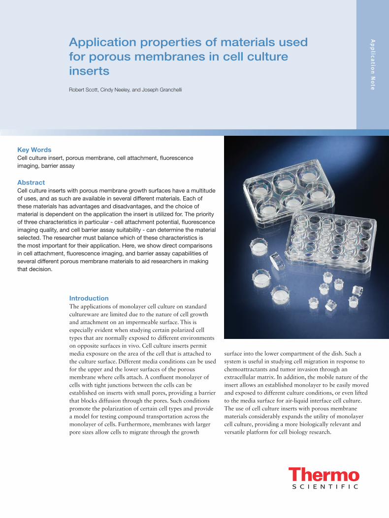
AbstractCell culture inserts with porous membrane growth surfaces have a multitude of uses, and as such are available in several different materials. Each of these materials has advantages and disadvantages, and the choice of material is dependent on the application the insert is utilized for. The priority of three characteristics in particular - cell attachment potential, fluorescence imaging quality, and cell barrier assay suitability - can determine the material selected. The researcher must balance which of these characteristics is the most important for their application. Here, we show direct comparisons in cell attachment, fluorescence imaging, and barrier assay capabilities of several different porous membrane materials to aid researchers in making that decision.
IntroductionThe applications of monolayer cell culture on standard cultureware are limited due to the nature of cell growth and attachment on an impermeable surface. This is especially evident when studying certain polarized cell types that are normally exposed to different environments on opposite surfaces in vivo. Cell culture inserts permit media exposure on the area of the cell that is attached to the culture surface. Different media conditions can be used for the upper and the lower surfaces of the porous membrane where cells attach. A confluent monolayer of cells with tight junctions between the cells can be established on inserts with small pores, providing a barrier that blocks diffusion through the pores. Such conditions promote the polarization of certain cell types and provide a model for testing compound transportation across the monolayer of cells. Furthermore, membranes with larger pore sizes allow cells to migrate through the growth
surface into the lower compartment of the dish. Such a system is useful in studying cell migration in response to chemoattractants and tumor invasion through an extracellular matrix. In addition, the mobile nature of the insert allows an established monolayer to be easily moved and exposed to different culture conditions, or even lifted to the media surface for air-liquid interface cell culture. The use of cell culture inserts with porous membrane materials considerably expands the utility of monolayer cell culture, providing a more biologically relevant and versatile platform for cell biology research.
2 With so many different applications for cell culture inserts, there are several types of materials that are commonly used to make the porous membrane in a variety of pore sizes. Different applications require different pore sizes, so the first step is to determine the pore size required for the experiment. The type of experiment will also determine the optimal membrane type being used, as materials such as polycarbonate (PC), polyethylene terephthalate (PET), and polytetrafluoroethylene (PTFE) each have strengths and weaknesses and are made for use under specific conditions. PC membranes, for example, are normally treated to promote cell attachment, and are made to have high pore density to allow more exchange through the membrane. Thus, PC inserts are best suited for transport studies and other applications where optimal cell growth is desired. PET membranes can be found with lower pore density, which allows greater transparency under microscopic conditions. Low pore density PET, therefore, is the membrane of choice when microscopic examination and/or photography is necessary. PTFE also is highly transparent for microscopic study and has low fluorescence background for immunofluorescent studies. However, the low binding properties of the PTFE material require coating with extracellular matrix proteins prior to seeding in order for the cells to properly attach.
Here we test each type of insert membrane material for a variety of applications. We examine the strengths and weaknesses of each material through experiments that allow for direct comparison between materials. These direct comparisons provide valuable information for researchers to balance their priorities in membrane material selection.
Results and Discussion:PC inserts have the best cell attachment property among the materials testedThe most important characteristic of any culture substrate is its ability to promote cell adhesion and growth. To determine the cell attachment properties of the insert
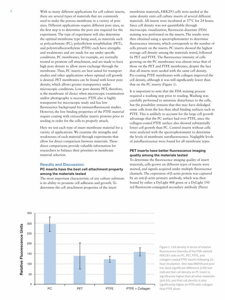
membrane materials, HEK293 cells were seeded at the same density onto cell culture inserts of several different materials. All inserts were incubated at 37°C for 24 hours. Since cell density was not quantifiable through microscopic visualization, fluorescein diacetate (FDA) staining was performed on the inserts. The results were then obtained using a spectrophotometer to determine fluorescence intensity, which corresponds to the number of cells present on the inserts. PC inserts showed the highest average cell density among the materials tested, followed by PET and PTFE. The fluorescence intensity of cells growing on the PC membranes was almost twice that of those on the PET and PTFE membranes, despite the fact that all inserts were seeded with the same cell density. Pre-coating PTFE membranes with collagen improved the cell density, although it was still significantly lower than that on the PC inserts (Figure 1).
It is important to note that the FDA staining process required a washing step prior to reading. Washing was carefully performed to minimize disturbance to the cells, but the possibility remains that this may have dislodged some cells from the less than ideal binding surfaces such as PTFE. This is unlikely to account for the large cell growth advantage that the PC surface had over PTFE, since the collagen-coated PTFE surface also showed substantially lower cell growth than PC. Control inserts without cells were analyzed with the spectrophotometer to determine the levels of membrane autofluorescence. Negligible levels of autofluorescence were found for all membrane types.
PET inserts have better fluorescence imaging quality among the materials testedTo determine the fluorescence imaging quality of insert materials, cells grown on different types of inserts were stained, and signals acquired under multiple fluorescence channels. The expression of β-actin protein was captured by an anti-β-actin primary antibody, which was then bound by either a DyLight 488 green or a DyLight 550 red fluorescent-conjugated secondary antibody (Pierce
Figure 1. Cell density in terms of relative fluorescence intensity of the FDA-stained HEK293 cells on PC, PET, PTFE, and collagen-coated PTFE inserts following 24-hour incubation. One-way ANOVA and post hoc least significant difference (LSD) test indicate that cell density on PC insert is significantly higher than all other materials (p<0.05), and that cell density is also significantly higher on PTFE with collagen than PTFE alone.PC PTFEPET PTFE + Collagen
350
250
200
150
100
50
0
300
Rel
ativ
e Fl
uore
scen
ce U
nits
3 Protein Biology). All cells were counterstained with the blue nuclear stain DAPI, which combined with the green and the red secondary antibodies covered a wide spectrum of light emission. This allows us to inspect the fluorescence imaging quality of a variety of insert materials for cellular analysis. Cells were photographed in grayscale using fluorescent filters matching the emission characteristics of FITC, Cy3, and DAPI; data were then pseudocolored and merged to create the final images (Figure 2).
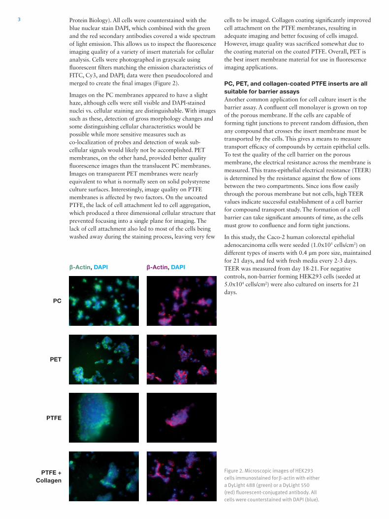
Images on the PC membranes appeared to have a slight haze, although cells were still visible and DAPI-stained nuclei vs. cellular staining are distinguishable. With images such as these, detection of gross morphology changes and some distinguishing cellular characteristics would be possible while more sensitive measures such as co-localization of probes and detection of weak sub-cellular signals would likely not be accomplished. PET membranes, on the other hand, provided better quality fluorescence images than the translucent PC membranes. Images on transparent PET membranes were nearly equivalent to what is normally seen on solid polystyrene culture surfaces. Interestingly, image quality on PTFE membranes is affected by two factors. On the uncoated PTFE, the lack of cell attachment led to cell aggregation, which produced a three dimensional cellular structure that prevented focusing into a single plane for imaging. The lack of cell attachment also led to most of the cells being washed away during the staining process, leaving very few
Figure 2. Microscopic images of HEK293 cells immunostained for β-actin with either a DyLight 488 (green) or a DyLight 550 (red) fluorescent-conjugated antibody. All cells were counterstained with DAPI (blue).
cells to be imaged. Collagen coating significantly improved cell attachment on the PTFE membranes, resulting in adequate imaging and better focusing of cells imaged. However, image quality was sacrificed somewhat due to the coating material on the coated PTFE. Overall, PET is the best insert membrane material for use in fluorescence imaging applications.
PC, PET, and collagen-coated PTFE inserts are all suitable for barrier assays Another common application for cell culture insert is the barrier assay. A confluent cell monolayer is grown on top of the porous membrane. If the cells are capable of forming tight junctions to prevent random diffusion, then any compound that crosses the insert membrane must be transported by the cells. This gives a means to measure transport efficacy of compounds by certain epithelial cells. To test the quality of the cell barrier on the porous membrane, the electrical resistance across the membrane is measured. This trans-epithelial electrical resistance (TEER) is determined by the resistance against the flow of ions between the two compartments. Since ions flow easily through the porous membrane but not cells, high TEER values indicate successful establishment of a cell barrier for compound transport study. The formation of a cell barrier can take significant amounts of time, as the cells must grow to confluence and form tight junctions.
In this study, the Caco-2 human colorectal epithelial adenocarcinoma cells were seeded (1.0x10⁵ cells/cm2) on different types of inserts with 0.4 µm pore size, maintained for 21 days, and fed with fresh media every 2-3 days. TEER was measured from day 18-21. For negative controls, non-barrier forming HEK293 cells (seeded at 5.0x10⁴ cells/cm2) were also cultured on inserts for 21 days.
β-Actin, DAPI
PC
PET
PTFE
PTFE +Collagen
β-Actin, DAPI
Ap
plica
tion
No
te
thermoscientific.com © 2013 Thermo Fisher Scientific Inc. All rights reserved. All (other) trademarks are the property of Thermo Fisher Scientific Inc. and its subsidiaries.
ANZ: Australia: 1300 735 292, New Zealand: 0800 933 966; Asia: China Toll-free: 800-810-5118 or 400-650-5118;India: +91 22 6716 2200, India Toll-free: 1 800 22 8374; Japan: +81-3-5826-1616; Other Asian countries: 65 68729717Europe: Austria: +43 1 801 40 0; Belgium: +32 2 482 30 30; Denmark: +45 4631 2000; France: +33 2 2803 2180; Germany: +49 6184 90 6000, Germany Toll-free: 0800 1-536 376; Italy: +39 02 95059 554; Netherlands: +31 76 571 4440; Nordic/Baltic countries: +358 9 329 10200; Russia/CIS: +7 (812) 703 42 15;Spain/Portugal: +34 93 223 09 18; Switzerland: +41 44 454 12 22; UK/Ireland: +44 870 609 9203North America: USA/Canada +1 585 586 8800; USA Toll-free: 800 625 4327South America: USA sales support: +1 585 899 7198 Countries not listed: +49 6184 90 6000 or +33 2 2803 2000
ANLSPCCINSERTMEM 0913
Conclusions:• PC insert membrane provides an excellent substrate for
cell attachment and growth, and may be advantageous in barrier assays as demonstrated by maintaining consistent TEER values over a longer period of time.
• PET membrane offsets its suboptimal cell growth capabilities with better image quality.
• PTFE membrane generates quality images but must be coated with extracellular matrix proteins for adequate cell attachment.
• PC, PET, and collagen-coated PTFE inserts with small pore size are all suitable for barrier assays with specific epithelial cell lines.
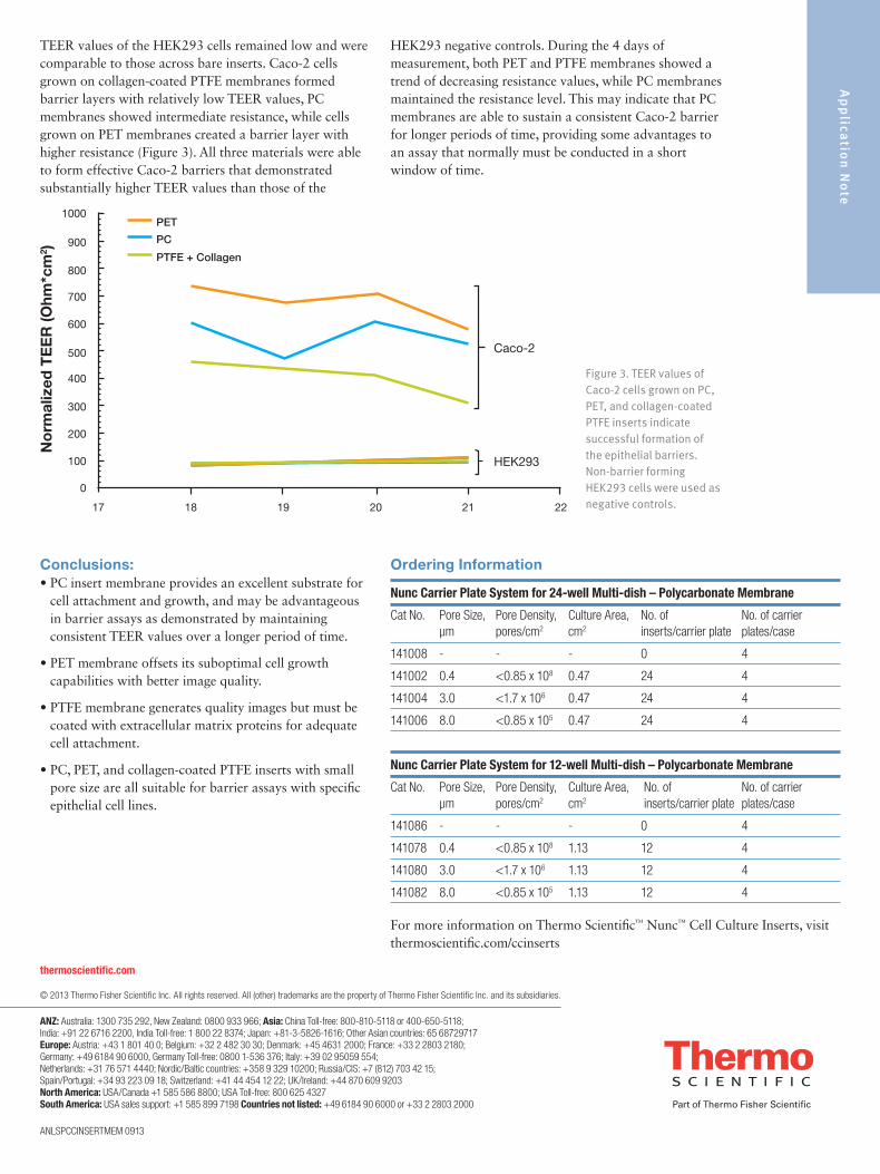
Figure 3. TEER values of Caco-2 cells grown on PC, PET, and collagen-coated PTFE inserts indicate successful formation of the epithelial barriers. Non-barrier forming HEK293 cells were used as negative controls.
Nunc Carrier Plate System for 24-well Multi-dish – Polycarbonate Membrane
Cat No. Pore Size, Pore Density, Culture Area, No. of No. of carrier μm pores/cm2 cm2 inserts/carrier plate plates/case
141008 - - - 0 4
141002 0.4 <0.85 x 10⁸ 0.47 24 4
141004 3.0 <1.7 x 10⁶ 0.47 24 4
141006 8.0 <0.85 x 10⁵ 0.47 24 4
Nunc Carrier Plate System for 12-well Multi-dish – Polycarbonate Membrane
Cat No. Pore Size, Pore Density, Culture Area, No. of No. of carrier μm pores/cm2 cm2 inserts/carrier plate plates/case
141086 - - - 0 4
141078 0.4 <0.85 x 10⁸ 1.13 12 4
141080 3.0 <1.7 x 10⁶ 1.13 12 4
141082 8.0 <0.85 x 10⁵ 1.13 12 4
Ordering Information
TEER values of the HEK293 cells remained low and were comparable to those across bare inserts. Caco-2 cells grown on collagen-coated PTFE membranes formed barrier layers with relatively low TEER values, PC membranes showed intermediate resistance, while cells grown on PET membranes created a barrier layer with higher resistance (Figure 3). All three materials were able to form effective Caco-2 barriers that demonstrated substantially higher TEER values than those of the
HEK293 negative controls. During the 4 days of measurement, both PET and PTFE membranes showed a trend of decreasing resistance values, while PC membranes maintained the resistance level. This may indicate that PC membranes are able to sustain a consistent Caco-2 barrier for longer periods of time, providing some advantages to an assay that normally must be conducted in a short window of time.
For more information on Thermo Scientific™ Nunc™ Cell Culture Inserts, visit thermoscientific.com/ccinserts
PTFE + Collagen
Caco-2
HEK293
No
rmal
ized
TE
ER
(Ohm
*cm
2 )
1000
900
800
700
600
500
400
300
200
100
17 18 19 20 21 22
0
PET
PC