Embed Size (px)
Citation preview
1
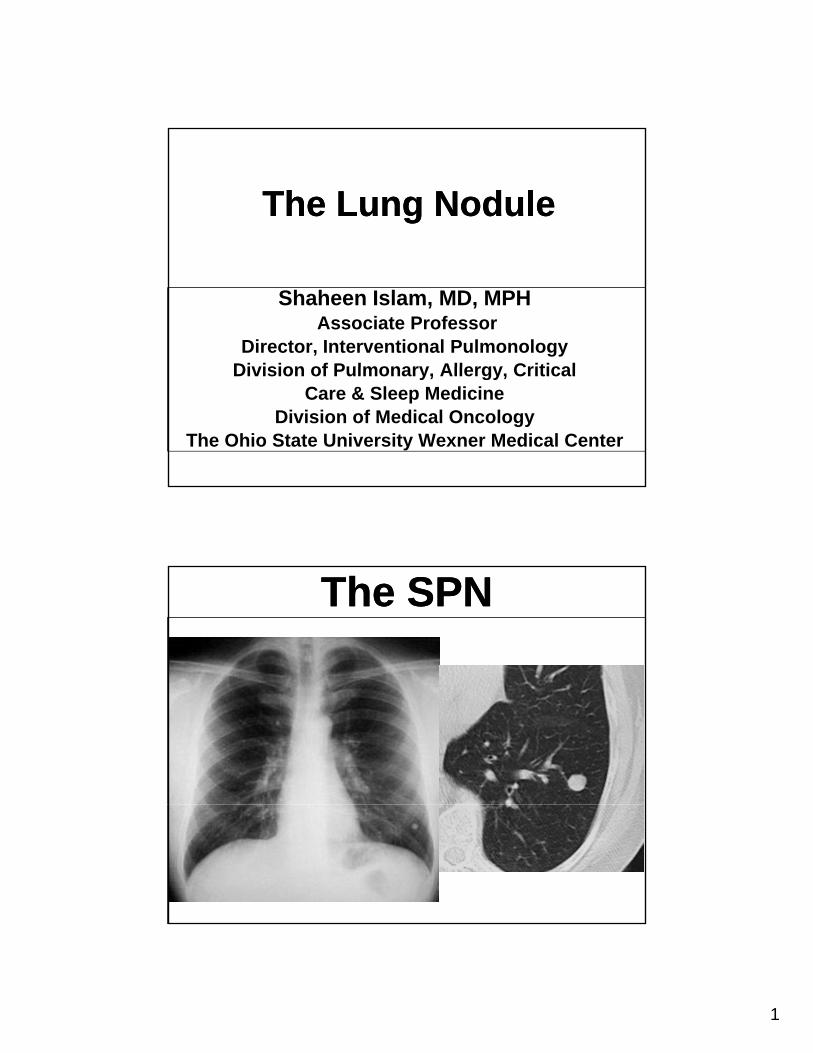
The Lung NoduleThe Lung Nodule
Shaheen Islam, MD, MPHAssociate Professor
Director, Interventional PulmonologyDivision of Pulmonary Allergy CriticalDivision of Pulmonary, Allergy, Critical
Care & Sleep MedicineDivision of Medical Oncology
The Ohio State University Wexner Medical Center
The SPNThe SPN
2
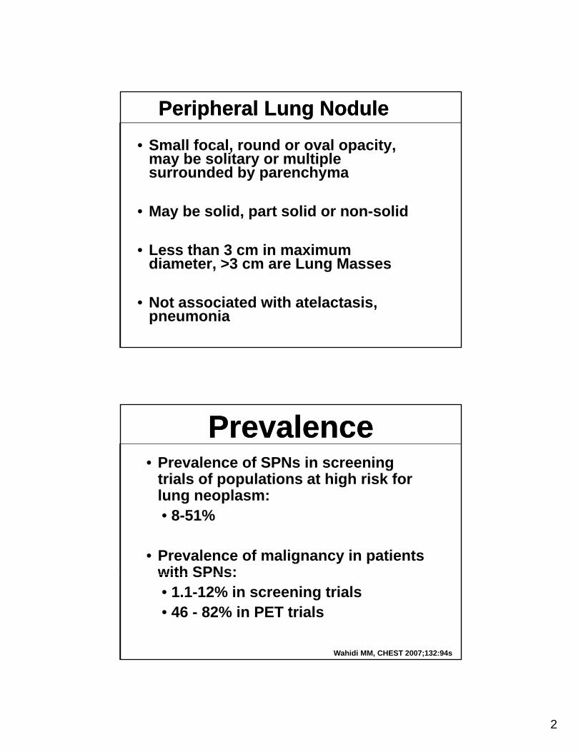
Peripheral Lung NodulePeripheral Lung Nodule
• Small focal, round or oval opacity, may be solitary or multiple surrounded by parenchymasurrounded by parenchyma
• May be solid, part solid or non-solid
• Less than 3 cm in maximum diameter >3 cm are Lung Massesdiameter, >3 cm are Lung Masses
• Not associated with atelactasis, pneumonia
PrevalencePrevalence• Prevalence of SPNs in screening
trials of populations at high risk for lung neoplasm:• 8-51%
• Prevalence of malignancy in patients with SPNs:with SPNs:• 1.1-12% in screening trials• 46 - 82% in PET trials
Wahidi MM, CHEST 2007;132:94s
3
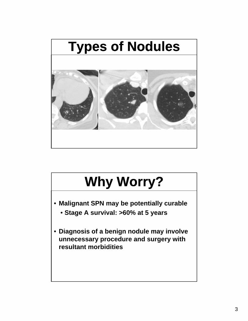
Types of NodulesTypes of Nodules
Why Worry?Why Worry?
• Malignant SPN may be potentially curable
• Stage A survival: >60% at 5 years
• Diagnosis of a benign nodule may involve unnecessary procedure and surgery with
lt t biditiresultant morbidities
4
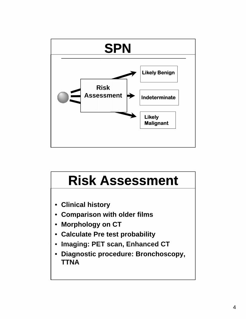
SPNSPN
Likely Benigny g
Indeterminate
Risk Assessment
Likely Malignant
Risk AssessmentRisk Assessment
• Clinical historyy
• Comparison with older films
• Morphology on CT
• Calculate Pre test probability
• Imaging: PET scan Enhanced CT• Imaging: PET scan, Enhanced CT
• Diagnostic procedure: Bronchoscopy, TTNA
5
SPNSPN
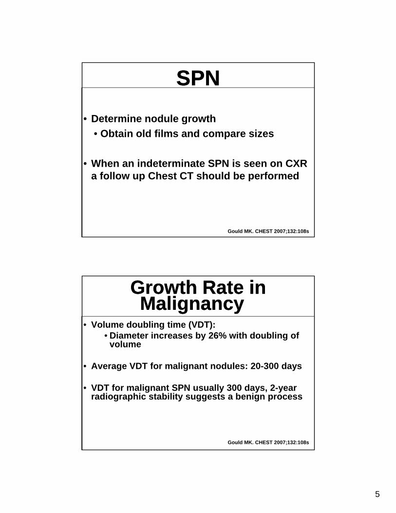
• Determine nodule growthDetermine nodule growth
• Obtain old films and compare sizes
• When an indeterminate SPN is seen on CXR a follow up Chest CT should be performeda follow up Chest CT should be performed
Gould MK. CHEST 2007;132:108s
Growth Rate in Malignancy
Growth Rate in Malignancy
• Volume doubling time (VDT): Diameter increases b 26% ith do bling of• Diameter increases by 26% with doubling of volume
• Average VDT for malignant nodules: 20-300 days
• VDT for malignant SPN usually 300 days, 2-year g y y , yradiographic stability suggests a benign process
Gould MK. CHEST 2007;132:108s
6
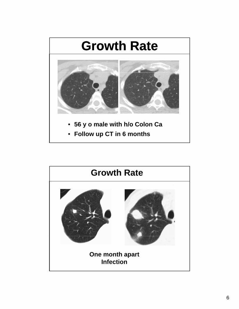
Growth RateGrowth Rate
• 56 y o male with h/o Colon Ca
• Follow up CT in 6 months
Growth Rate
One month apartInfection
7
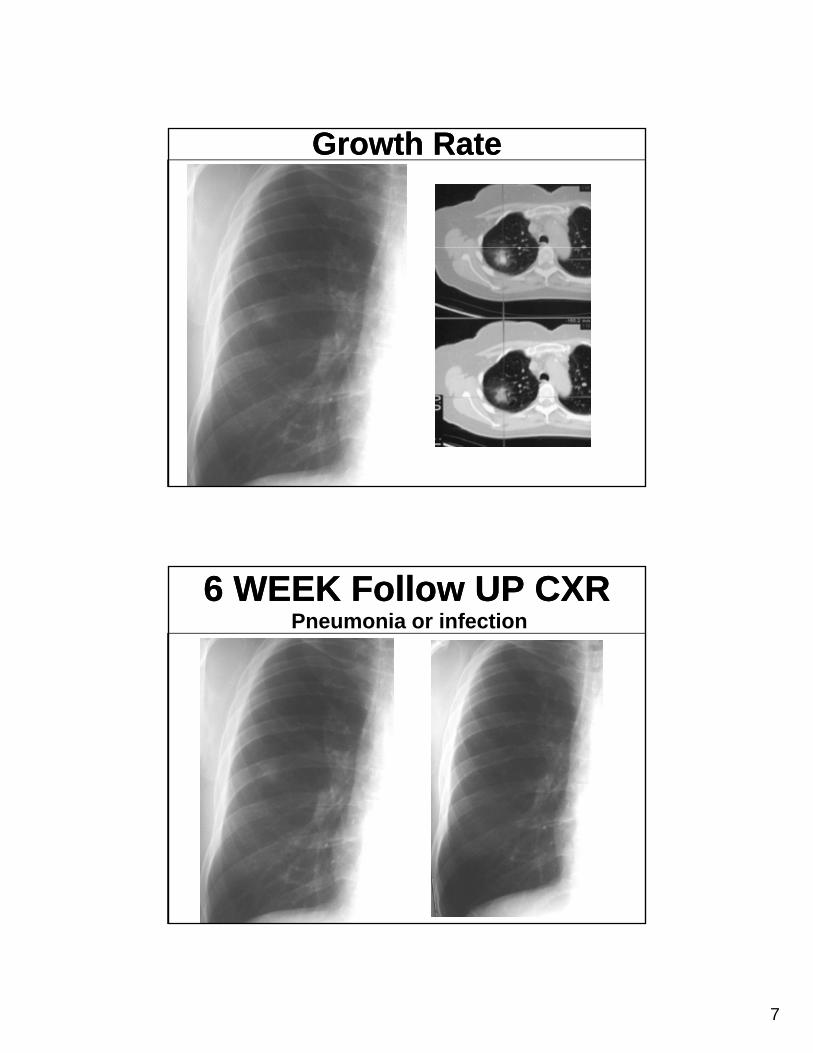
Growth RateGrowth Rate
6 WEEK Follow UP CXR6 WEEK Follow UP CXRPneumonia or infection
8
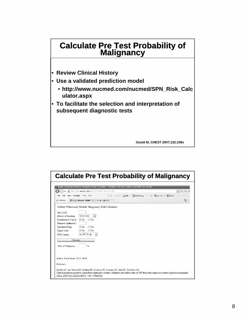
Calculate Pre Test Probability of Malignancy
Calculate Pre Test Probability of Malignancy
• Review Clinical Historyy
• Use a validated prediction model
• http://www.nucmed.com/nucmed/SPN_Risk_Calculator.aspx
• To facilitate the selection and interpretation of subsequent diagnostic testssubsequent diagnostic tests
Gould M, CHEST 2007;132:108s
Calculate Pre Test Probability of MalignancyCalculate Pre Test Probability of Malignancy
9
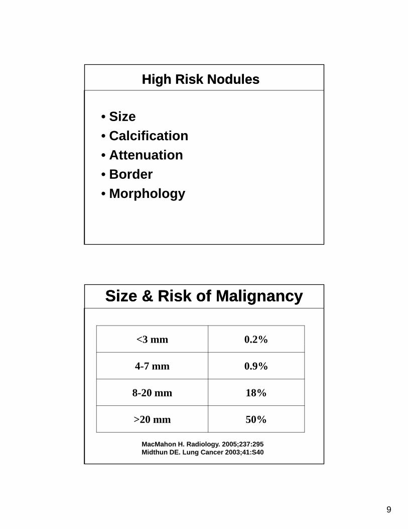
High Risk NodulesHigh Risk Nodules
• Size
• Calcification
• Attenuation
• Border
• Morphology
Size & Risk of MalignancySize & Risk of Malignancy
<3 mm 0.2%
4-7 mm 0.9%
8-20 mm 18%
>20 mm 50%
MacMahon H. Radiology. 2005;237:295Midthun DE. Lung Cancer 2003;41:S40
10
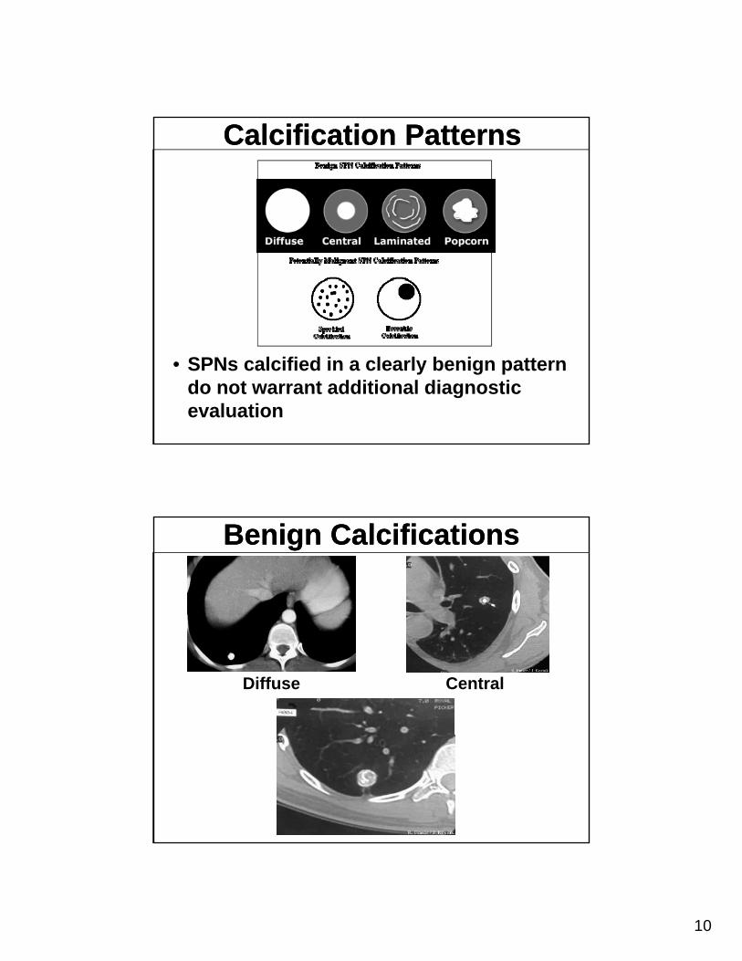
Calcification PatternsCalcification Patterns
• SPNs calcified in a clearly benign pattern do not warrant additional diagnostic evaluation
Benign CalcificationsBenign Calcifications
Diffuse Central
11
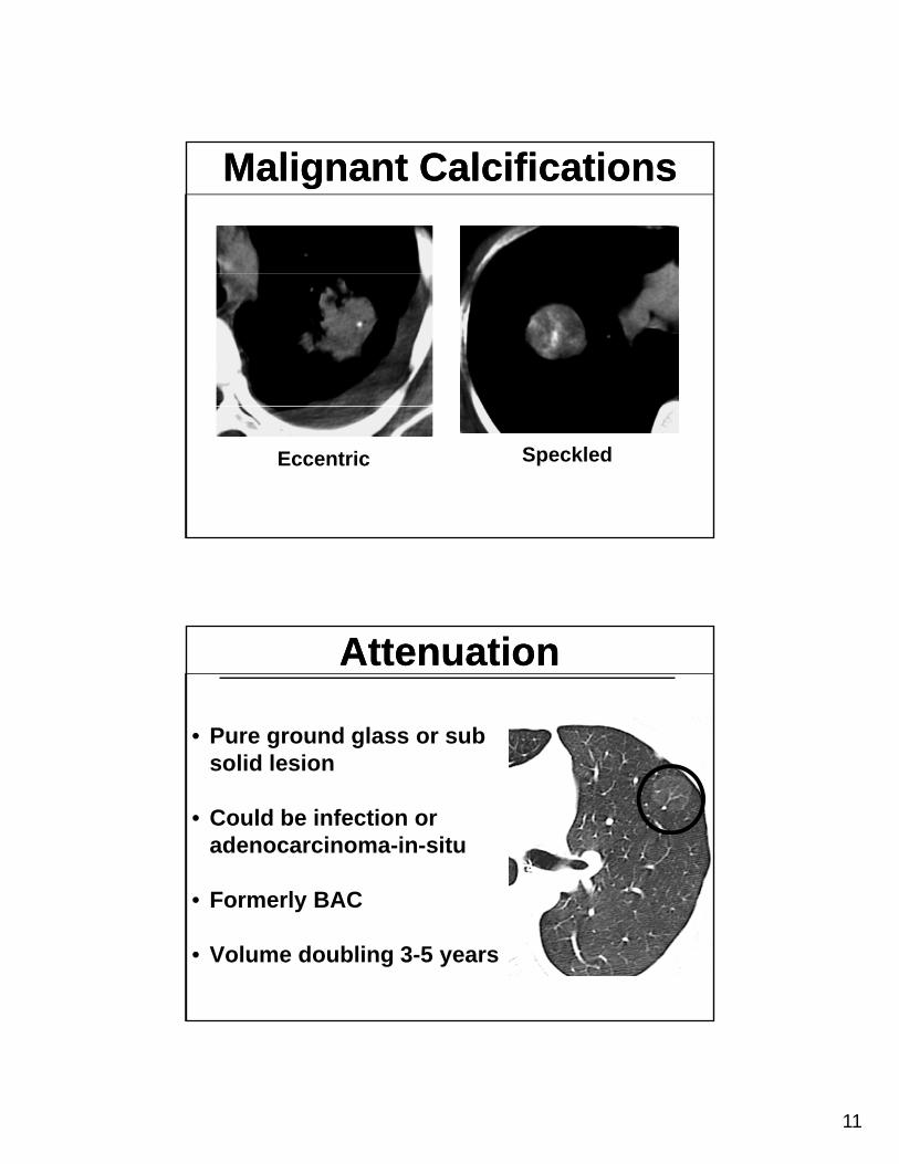
Malignant CalcificationsMalignant Calcifications
Eccentric Speckled
AttenuationAttenuation
• Pure ground glass or sub lid l isolid lesion
• Could be infection or adenocarcinoma-in-situ
• Formerly BAC
• Volume doubling 3-5 years
12
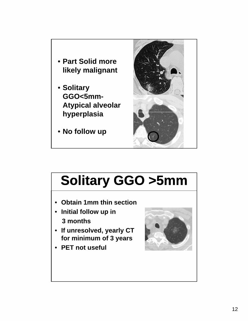
• Part Solid more likely malignant
• Solitary GGO<5mm-Atypical alveolar h l ihyperplasia
• No follow up
Solitary GGO >5mmSolitary GGO >5mm
• Obtain 1mm thin section
• Initial follow up in
3 months
• If unresolved, yearly CT for minimum of 3 years
PET t f l• PET not useful
13
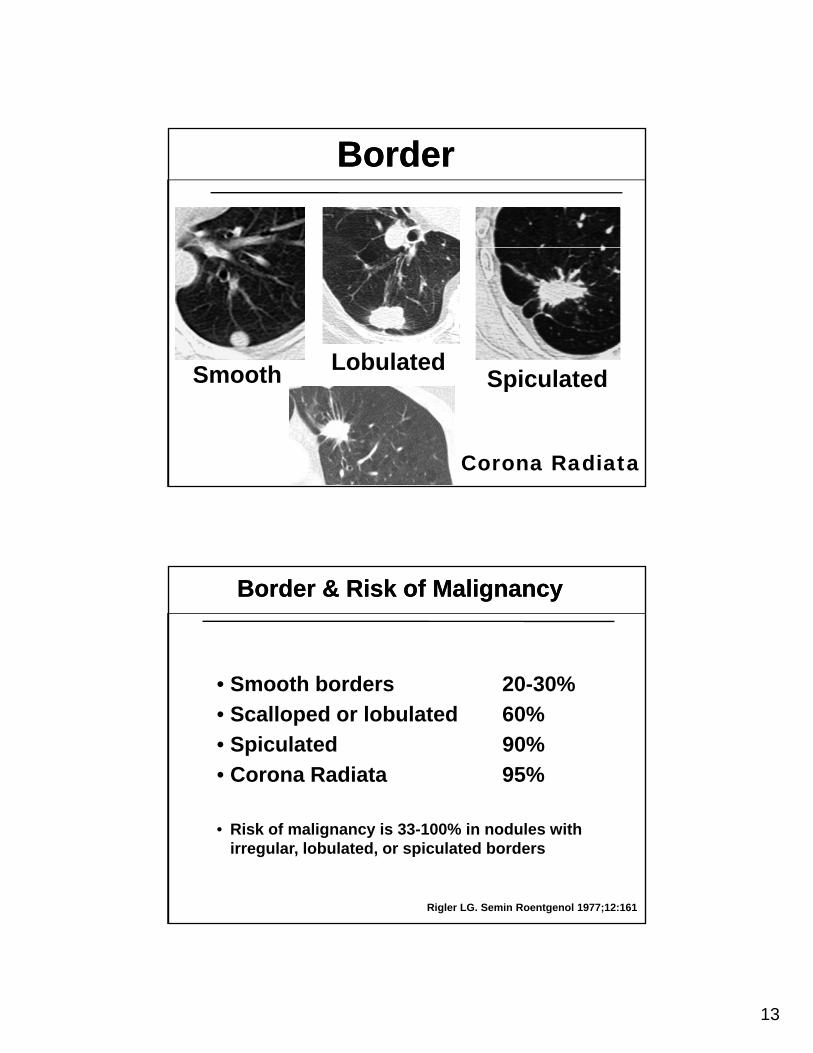
BorderBorder
S th LobulatedSmooth SpiculatedLobulated
Corona Radiata
• Smooth borders 20-30%
Border & Risk of MalignancyBorder & Risk of Malignancy
• Smooth borders 20-30%
• Scalloped or lobulated 60%
• Spiculated 90%
• Corona Radiata 95%
• Risk of malignancy is 33-100% in nodules with irregular, lobulated, or spiculated borders
Rigler LG. Semin Roentgenol 1977;12:161
14
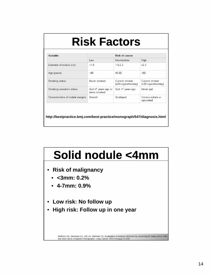
Risk FactorsRisk Factors
http://bestpractice.bmj.com/best-practice/monograph/547/diagnosis.html
Solid nodule <4mmSolid nodule <4mm• Risk of malignancy
• <3mm: 0 2%• <3mm: 0.2%
• 4-7mm: 0.9%
• Low risk: No follow up
• High risk: Follow up in one year• High risk: Follow up in one year
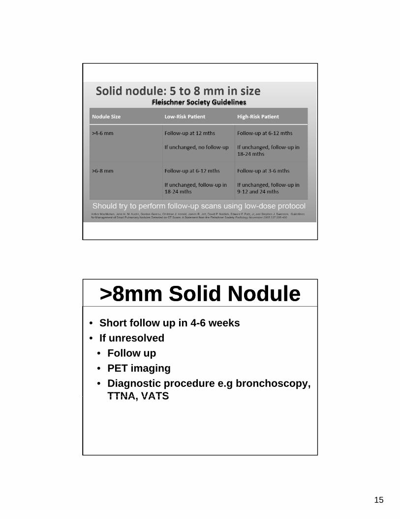
15
>8mm Solid Nodule>8mm Solid Nodule• Short follow up in 4-6 weeks
• If unresolved• If unresolved
• Follow up
• PET imaging
• Diagnostic procedure e.g bronchoscopy, TTNA VATSTTNA, VATS
16
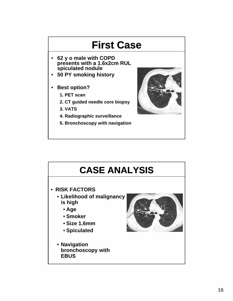
First CaseFirst Case• 62 y o male with COPD
presents with a 1.6x2cm RUL spiculated nodulep
• 50 PY smoking history
• Best option?1. PET scan
2. CT guided needle core biopsy
3. VATS
4. Radiographic surveillance
5. Bronchoscopy with navigation
CASE ANALYSISCASE ANALYSIS
• RISK FACTORS • Likelihood of malignancy• Likelihood of malignancy
is high• Age• Smoker• Size 1.6mm• Spiculated• Spiculated
• Navigation bronchoscopy with EBUS
17
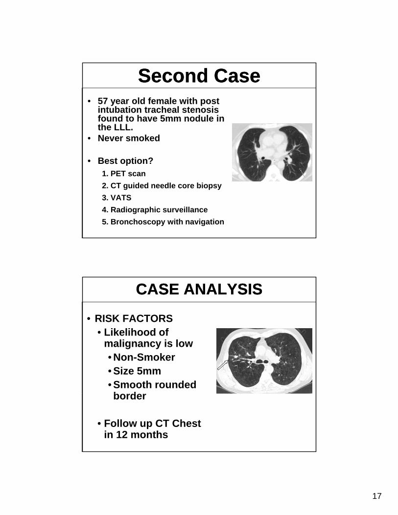
Second CaseSecond Case• 57 year old female with post
intubation tracheal stenosis found to have 5mm nodule infound to have 5mm nodule in the LLL.
• Never smoked
• Best option?1. PET scan
2. CT guided needle core biopsy
3. VATS
4. Radiographic surveillance
5. Bronchoscopy with navigation
CASE ANALYSISCASE ANALYSIS
• RISK FACTORS • Likelihood ofLikelihood of
malignancy is low• Non-Smoker• Size 5mm• Smooth rounded b dborder
• Follow up CT Chest in 12 months
18
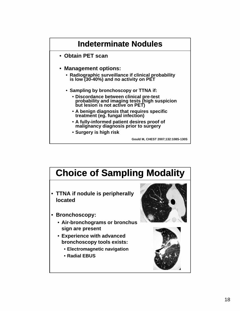
Indeterminate NodulesIndeterminate Nodules
• Obtain PET scan
• Management options:• Radiographic surveillance if clinical probability
is low (30-40%) and no activity on PET
• Sampling by bronchoscopy or TTNA if:• Discordance between clinical pre-test
probability and imaging tests (high suspicion but lesion is not active on PET)
• A benign diagnosis that requires specific treatment (eg. fungal infection)
• A fully-informed patient desires proof of malignancy diagnosis prior to surgery
• Surgery is high riskGould M, CHEST 2007;132:108S-130S
Choice of Sampling ModalityChoice of Sampling Modality
• TTNA if nodule is peripherally located
• Bronchoscopy:• Air-bronchograms or bronchus
sign are present
• Experience with advanced pbronchoscopy tools exists:• Electromagnetic navigation
• Radial EBUS
19
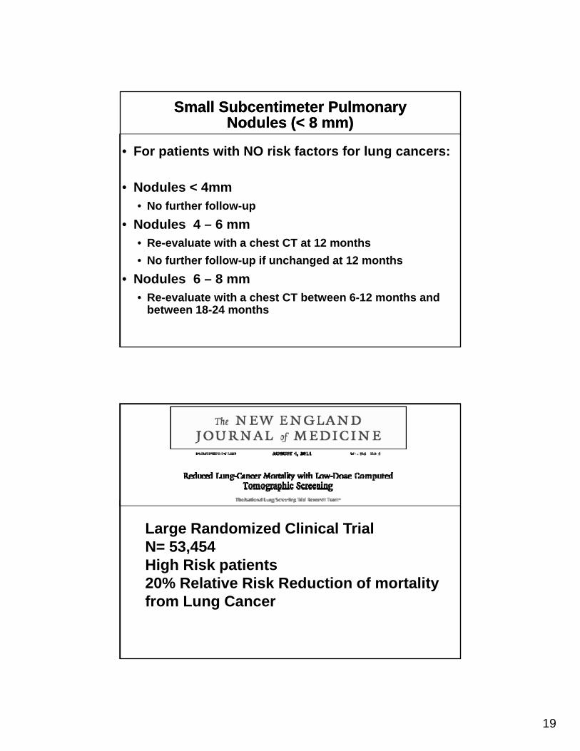
Small Subcentimeter Pulmonary Nodules (< 8 mm)
Small Subcentimeter Pulmonary Nodules (< 8 mm)
• For patients with NO risk factors for lung cancers:
• Nodules < 4mm• No further follow-up
• Nodules 4 – 6 mm• Re-evaluate with a chest CT at 12 months
• No further follow-up if unchanged at 12 monthsNo further follow-up if unchanged at 12 months
• Nodules 6 – 8 mm• Re-evaluate with a chest CT between 6-12 months and
between 18-24 months
Large Randomized Clinical TrialN= 53,454High Risk patients20% Relative Risk Reduction of mortality from Lung Cancer
20
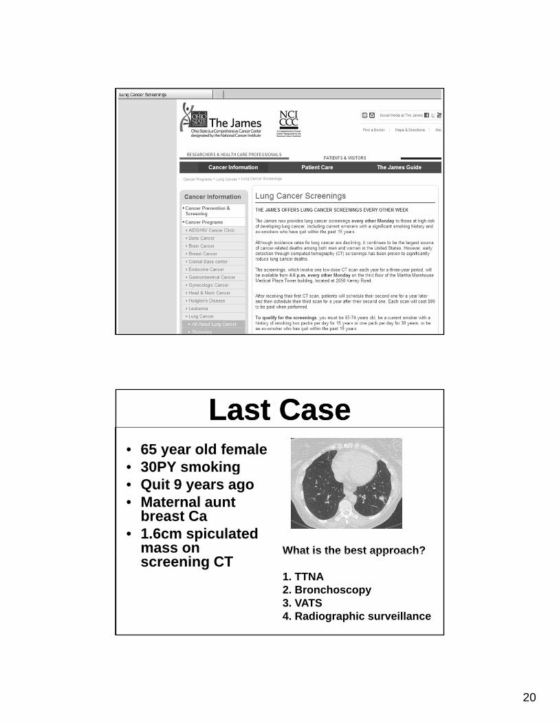
Last CaseLast Case• 65 year old female• 30PY smoking30PY smoking• Quit 9 years ago• Maternal aunt
breast Ca• 1.6cm spiculated
mass on What is the best approach?screening CT
What is the best approach?
1. TTNA2. Bronchoscopy3. VATS4. Radiographic surveillance
21
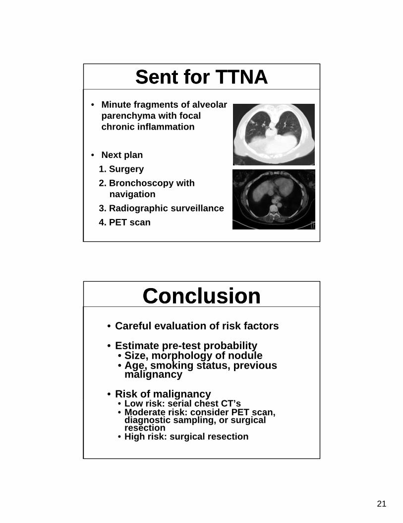
Sent for TTNASent for TTNA• Minute fragments of alveolar
parenchyma with focal chronic inflammation
• Next plan
1. Surgery
2 Bronchoscopy with2. Bronchoscopy with navigation
3. Radiographic surveillance
4. PET scan
ConclusionConclusion• Careful evaluation of risk factors
• Estimate pre-test probability• Size, morphology of nodule• Age, smoking status, previous
malignancy
• Risk of malignancy• Low risk: serial chest CT’s• Moderate risk: consider PET scan,
diagnostic sampling, or surgical resection
• High risk: surgical resection