Embed Size (px)
Citation preview
Journal of Neurology, Neurosurgery, and Psychiatry, 1974, 37, 1299-1305
Arachnodactyly, aminoaciduria, congenital cataracts,cerebellar ataxia, and delayed developmental
milestonesP. A. BHASKAR, K. JAGANNATHAN, AND K. VALMIKINATHAN
From the Institute of Neurology, Government General Hospital, Madras-3, India
SYNOPSIS Two male cousins are reported with arachnodactyly, selective aminoaciduria, congenitalcataracts, cerebellar ataxia, and delayed developmental milestones, and a distant female relative withsimilar abnormalities. The syndrome is thought to be previously undescribed, though it has resem-blances to Marinesco-Siogren and Marfan's syndromes.
A syndrome characterized by arachnodactyly,selective aminoaciduria, congenital cataracts,cerebellar ataxia, and delayed developmentalmilestones was observed in two young boys whowere first cousins. In certain respects the presenta-tion was similar to that of the Marinesco-Sjogren syndrome but with a few additionalclinical and biochemical features. In certainother respects it was suggestive of Marfan's ([ ] Csyndrome. To our knowledge, such a combina- , .Ition of clinical and biochemical features has not ibeen recordedhitherto.CaeI as2
CASE REPORTS
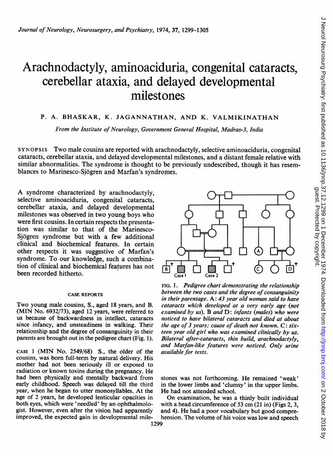
Two young male cousins, S., aged 18 years, and B.(MIN No. 6932/73), aged 12 years, were referred tous because of backwardness in intellect, cataractssince infancy, and unsteadiness in walking. Theirrelationship and the degree of consanguinity in theirparents are brought out in the pedigree chart (Fig. 1).
CASE 1 (MIN No. 2549/68) S., the older of thecousins, was born full-term by natural delivery. Hismother had not been seriously ill or exposed toradiation or known toxins during the pregnancy. Hehad been physically and mentally backward fromearly childhood. Speech was delayed till the thirdyear, when he began to utter monosyllables. At theage of 2 years, he developed lenticular opacities inboth eyes, which were 'needled' by an ophthalmolo-gist. However, even after the vision had apparentlyimproved, the expected gain in developmental mile-
FIG. 1. Pedigree chart demonstrating the relationshipbetween the two cases and the degree of consanguinityin their parentage. A: 43 year old woman said to havecataracts which developed at a very early age (notexamined by us). B and D: infants (males) who werenoticed to have bilateral cataracts and died at aboutthe age of3 years; cause of death not known. C: six-teen year old girl who was examined clinically by us.Bilateral after-cataracts, thin build, arachnodactyly,and Marfan-like features were noticed. Only urineavailable for tests.
1299
stones was not forthcoming. He remained 'weak'in the lower limbs and 'clumsy' in the upper limbs.He had not attended school.On examination, he was a thinly built individual
with a head circumference of 53 cm (21 in) (Figs 2, 3,and 4). He had a poor vocabulary but good compre-hension. The volume of his voice was low and speech
guest. Protected by copyright.
on 2 October 2018 by
http://jnnp.bmj.com
/J N
eurol Neurosurg P
sychiatry: first published as 10.1136/jnnp.37.12.1299 on 1 Decem
ber 1974. Dow
nloaded from

P. A. Bhaskar, K. Jagannathan, and K. Valmikinathan
FIG. 2. The first cousins. Left: case 1;right: case 2, showing the squint in bothand gynaecomastia in case 1.
..:
... ..~
FIG. 3. Cases 1 and 2. Lateral view showing dorsal FIG. 4. Cases I and 2. The thin build, Marfanoidkyphosis, flat chest, thin build, and the gynaecomastia features, and the normally developed externalgenitaliain case 1. are shown.
1300
.FTF,.vi
M--abi
guest. Protected by copyright.
on 2 October 2018 by
http://jnnp.bmj.com
/J N
eurol Neurosurg P
sychiatry: first published as 10.1136/jnnp.37.12.1299 on 1 Decem
ber 1974. Dow
nloaded from

Arachnodactyly, aminoaciduria, congenital cataracts, cerebellar ataxia, and mental retardation 1301
FIG. 5. Case 1. High arched palate.
syllables were split in a staccato way. Intelligencequotient tested on the Binet-Kamath scale gave amental age of 5 years. The visual acuity was poor(1/60) in both eyes. An 'after-cataract' had developedin the right eye. The pupils were normal in size andreaction and the appearance of the left ocular funduswas normal. A concomitant squint was evident onrelaxed forward gaze (Fig. 2). The intraocular tensionwas normal. The extraocular movements were fullwith coarse nystagmus on either horizontal gaze. Thelimbs were hypotonic with normal power in theupper limbs and MRC grade 4/5 power in bothlower limbs. Finger-nose-finger and heel-knee in-coordination were evident on both sides with mildtruncal ataxia and intense gait ataxia, necessitatingsupport while walking. The tendon jerks were quitebrisk with flexor plantar response bilaterally.Sensory appreciation and sphincter control wereintact.The other characteristic features noticed in this
young man were the thick bushy eyebrows and eye-lashes (Fig. 2), flat feet (Fig. 6), mild upper dorsalkyphosis (Fig. 3), sabre tibiae, arachnodactyly (Fig.7), high arched palate (Fig. 5), and bilateral gynaeco-
FIG. 6. Cases I and 2. Flat feet.
FIG. 7. Cases I and 2. Arachnodactyly.
guest. Protected by copyright.
on 2 October 2018 by
http://jnnp.bmj.com
/J N
eurol Neurosurg P
sychiatry: first published as 10.1136/jnnp.37.12.1299 on 1 Decem
ber 1974. Dow
nloaded from

P. A. Bhaskar, K. Jagannathan, and K. Valmikinathan
mastia (Fig. 3). His secondary sexual characters werewell developed (Fig. 4). His height measured 152 cm,span 163 cm, and crown to symphysis pubis 73 cm.Liver and spleen were not enlarged.
CASE 2 (MIN No. 6932/73) B., the younger of thetwo cousins, was aged 12 years at the time ofexamination. He was also born by natural deliveryafter a full-term gestation. Until the age of 4 years,he was not able to sit up without support. He de-veloped cataract at the age of 1l years. Two yearslater, an ophthalmologist 'needled' the cataracts andsome return of vision resulted. He was put to schoolonly at the age of 10 years but discontinued twoyears later as he was unable to grasp the lessonstaught. He continued to remain unsteady whilewalking and attempting to write.On examination, he appeared to be more cheerful
TABLE 1LABORATORY JNVESTIGATIONS
Case I Case 2
Blood and serumHaemoblobin (g/dl) 10-6 9 8Erythrocytes (million/mm3) 3-2 3-4Leucocytes (per mm3) 8,600 9,400Diff. count (P/L/E/M) 54/36/8/2 48/44/8/0ESR (mm/hr) 10 8Sugar (mg/dl) 78 70Urea (mg/dl) 18 20Cholesterol (mg/dl) 185 155Alkaline phosphatase (KA units) 12 7-6Acid phosphatase (KA units) 1 0 7Calcium (mg/dl) 9-4 10 2Phosphorus (mg/dl) 3-3 3-8Total proteins (g/dl) 6-3 6-9Albumin (g/dl) 3-2 3-7Alpha globulin (g/dl) 0-9 0-9Beta globulin (g/dl) 0-6 0 7Gamma globulin (g/dl) 1-6 1-6
Sodium (mEq/l) 140 139Chloride (mEq/l) 99 104Potassium (mEq/l) 3-1 3-6pH of arterial blood 7-34 7-22Serum creatinine (mg/dl) 0-80 0-74
Urine17-ketosteroids (mg per 24 hr) 8-4 5-6Calcium (mg per 24 hr) 112 Not donePhosphorus (mg per 24 hr) 943 Not doneProtein (mg per 24 hr) 98 88pH 5-78 5 90Creatinine (mg per 24 hr) 682 540
FIG. 8. Case 2. Dense after-cataract in the right eyeand capsular remnants of 'needling procedure' in theleft eye. The thick eyebrows and long eyelashes are
also seen.
and cooperative than his elder cousin; it was possibleto communicate with him by conversation. Speechwas normal. Intelligence as tested by the Binet-Kamath method placed his mental age at the 8 yearlevel. The visual acuity in the left eye was 6/18 andonly hand movements could be perceived by theright eye. A prominent convergent squint was seen(Fig. 2). Extraocular movements were full withcoarse nystagmus on looking to either side. In theright eye a dense after-cataract had developed whichhad occupied the entire pupillary aperture, while theleft eye contained the capsular remnants of the pre-vious needling procedure (Fig. 8).
All four limbs were hypotonic and exhibitedintention tremor. The tendon jerks were brisk withflexor plantar responses bilaterally. He was able to
walk without support but was unsteady, swayingfrom side to side. The gait was broad based andtandem walking was impossible. There was nosensory deficit or lack of sphincter control.On general examination, he was also of asthenic
build (Fig. 4) with a dorsal kyphosis (Fig. 3), flatchest (Fig. 3), arachnodactyly (Fig. 7), flat feet (Fig.6), high arched palate, thick eyebrows, and long eye-lashes (Fig. 8). He had sparse pubic hair and anormally developed phallus and testes (Fig. 4). Hisheight measured 142 cm, span 150 cm, and thecrown to symphysis pubis 65 cm. Liver and spleenwere not palpable.
LABORATORY INVESTIGATIONS The results of theroutine laboratory investigations are listed inTable 1.
Urine screening tests for inborn errors of metabol-ism (Benedict's test, cyanide-nitroprusside test,CTAB test, ferric chloride test, and DNPH test)were normal. However, two-dimensional chromato-graphic study of the urinary amino acids on paperusing butanol:acetic acid:water (40:7:5, v/v) andphenol: water (4: 1, v/v) systems showed evidence forlarge quantities of glycine, tyrosine, leucine, and
1302
....
guest. Protected by copyright.
on 2 October 2018 by
http://jnnp.bmj.com
/J N
eurol Neurosurg P
sychiatry: first published as 10.1136/jnnp.37.12.1299 on 1 Decem
ber 1974. Dow
nloaded from

Arachnodactyly, aminoaciduria, congenital cataracts, cerebellar ataxia, and mental retardation 1303
TABLE 2CONCENTRATION OF URINARY AMINO ACIDS
Case I Case 2 Case 3t Control$
Cystine 015 0-14 0-14 0-13Glycine 0-85 0 90 0-72 0-12Histidine 0-70 0-48 0-32 0-16Leucine 0 40 0-17 0 30 0-02Phenylalanine 0 04 005 0 04 0-06Proline 0 06 0-06 0 05 006Serine 0-05 0-06 0-05 004Threonine 0-12 0-11 011 0-11Tryptophane 0-02 0 03 0-03 0-02Tyrosine 0-83 0-67 0 74 0 03Valine 003 0-02 0 03 0-02
* Expressed as aminoacid/creatinine/24 hr.t Case 3 refers to the 16 year old relative of the other two casesmarked 'C' in the pedigree chart (Fig. 1).t Control: unrelated boy aged 12 years.
histidine in the urine of both cases. (The methodadopted for quantitation was that described byVarley, 1967). Analysis of the aminoacids in thepatients' sera was also conducted. The serum andurine of a 12 year old boy (control) was analysed forthe same aminoacids (Tables 2 and 3).
OTHER INVESTIGATIONS In both cases, radiographsof the skull and chest were normal, while those of thehand proved the presence of arachnodactyly(Sinclair et al., 1960). Metacarpal index in case 1
TABLE 3SERUM LEVELS OF AMINOACIDS EXCRETED IN EXCESS
IN URINE (mg/100 ml)
Case I Case 2 Control*
Glycine 2-1 1-4 1-8Histidine 1-4 1-7 1-4Leucine 2-4 2-1 2-5Tyrosine 1-2 1-8 1 3Creatinine 0-80 0-74 0-66
* Control: boy aged 12 years.
was 9 4, and in case 2 it was 9*5 (normal range: 5*4to 7-9). Buccal smear study in both of them provedto be chromatin negative with karyotype 22+XYwith no structural abnormalities of the chromo-somes. In both cases the electroencephalograph andelectrocardiogram were within normal limits.Electrophoretic patterns of serum lipoproteins werenormal.
Other family studies A distantly related 16 year oldgirl (case C in Fig. 1) also has bilateral after-cataracts, thin build, arachnodactyly, and Marfan-like features. Only urine was available for testing butshowed the same selective aminoaciduria as the pro-bands (Table 2). Unfortunately, the living sibling ofcase 1 is not available for study.
DISCUSSION
Both cases satisfied the criteria for the Marinesco-Sjogren syndrome (Marinesco et al., 1931;Sjogren, 1950)-namely, oligophrenia, cerebellarataxia, and congenital cataracts. Some of theother findings in our cases were also recorded byother authors: flat feet were observed byRichards (1950); brisk tendon jerks and equi-vocal plantar responses were recorded byDureux et al. (1958) and Sjogren (1950);scoliosis and kyphoscoliosis by Garland andMoorhouse (1953); and ocular squint by Alteret al. (1962). Many other clinical features addi-tional to the basic Marinesco-Sjogren syndromeare on record. These include prominent ears(Dogulu and Mutlu, 1957); pes cavus (Dureuxet al., 1958); genu valgum, deformed fingers, andshort stature (MacGillivray, 1957); anomalies ofsex organs (Freycon and Freycon, 1965); dentalanomalies, hypopituitarism, and retinitis pig-mentosa (Carrieri and Di Gennaro, 1965);myopathic features (Alter and Kennedy, 1968),etc.
Accepting our cases as basically examples ofthe Marinesco-Sjogren syndrome, there aresome additional observations: (1) arachnodactylywith high arched palate and other skeletalabnormalities reminiscent of Marfan's syndrome,and (2) presence of selective aminoaciduria(glycine, leucine, histidine, and tyrosine). Thesefindings could be considered coincidental, but, inview of their consistent presence in more thanone sib, appear less likely to be so.
In Marfan's syndrome, which our casesresembled morphologically, the presence ofspecific aminoacidurias, cerebellar features, andlenticular opacities have not been recorded.Again, the negative cyanide-nitroprusside urinetest excluded homocystinuria, which sometimespresents clinically as a Marfan-like syndrome(Dunn et al., 1966).
Aminoaciduria, cataracts, and oligophrenia
guest. Protected by copyright.
on 2 October 2018 by
http://jnnp.bmj.com
/J N
eurol Neurosurg P
sychiatry: first published as 10.1136/jnnp.37.12.1299 on 1 Decem
ber 1974. Dow
nloaded from

P. A. Bhaskar, K. Jagannathan, and K. Valmikinathan
constitute the oculocerebrorenal syndrome ofLowe (Lowe et al., 1952). Cerebellar dysfunc-tion, nystagmus, intention tremor (McCance et
al., 1960), and rachitic features have also beenrecorded in this clinical entity. However, theabsence of the following characteristic featuresof this syndrome does not favour the diagnosisof Lowe syndrome in our cases: (1) hypotoniaand areflexia (Chutorian and Rowland, 1966);(2) renal tubular acidosis (Sagel et al., 1970);(3) aminoaciduria, involving most of the com-
mon amino-acids (Schoen and Young, 1959).
The occurrence of aminoaciduria in cases ofthe Marinesco-Sjogren syndrome is unknown.However, involvement of the kidney has beenrecorded in literature. Crome et al. (1963)described a syndrome resembling a combinationof the Marinesco-Sjogren and Lowe's syndromespresenting with congenital cataracts, renaltubular necrosis, and encephalopathy in twosisters. Similarly D'Angelo and Ahlheid (1968)discovered deposits of a lipid substance in thelungs, kidney, liver, and spleen at necropsy in a
case of Marinesco-Sjogren syndrome. But selec-tive aminoaciduria was not described in either ofthese reports.
Histidine, leucine, and tyrosine share a com-
mon transport mechanism with other aminoacids like phenylalanine, valine, threonine, etc.
In the present study, the excretion of phenyl-alanine, threonine, serine, cystine, proline,valine, and tryptophane was well within normallimits, indicating a selective defect in the renaltubular transport mechanism. This defect seems
also to affect the transport of glycine, whichusually shares a common transport mechanismwith proline and hydroxyproline. The explana-tion for this selective transport defect resulting inthe excretion of this combination of aminoacidsremains obscure.
It is suggested that this syndrome, character-ized by arachnodactyly, selective aminoaciduria,congenital cataracts, cerebellar ataxia, anddelayed developmental milestones which was
observed in a few members of a family, is a
genetically determined disorder probably trans-mitted in the autosomal recessive manner. Thecondition could well be an instance of a hithertoundescribed disease with features common to
Marfan's and the Marinesco-Sjogren syndrome.
The authors are grateful to Professor B. Ramamurthi,Head of the Institute of Neurology and the Superin-tendent, Government General Hospital, for per-mitting them to publish an account of these patients.
REFERENCES
Alter, M., and Kennedy, W. (1968). Marinesco-Sjogrensyndrome. Hereditary cerebello-lental degeneration withmental retardation. Minnesota Medicine, 51, 901-906.
Alter, M., Talbert, 0. R., and Croffead, G. (1962). Cerebellarataxia, congenital cataracts, and retarded somatic andmental maturation. Report of cases of Marinesco-Sjogrensyndrome. Neurology (Minneap.), 12, 836-847.
Angelo, C. D', and Ahiheid, A. (1967). Sindrome di Mari-nesco-Sjogren. Due casi clinici con un reperto neuro-patologico. Lavoro Neuropsichiatrico, 40, 291-318.
Carrieri, G., and Di Gennaro, C. (1965). Sulla sindrome diMarinesco-Sjogren. Osservazione di due casi familiari.Giornale di Psichiatria e di Neuropatologia, 93, 667-681.
Chutorian, A., and Rowland, L. P. (1966). Lowe's syndrome.Neurology (Minneap.), 16, 115-122.
Crome, L., Duckett, S., and Franklin, A. W. (1963). Con-genital cataracts, renal tubular necrosis and encephalo-pathy in two sisters. Archives of Diseases in Childhood, 38,505-515.
Dogulu, S., and Mutlu, N. (1957). Iki kardeti konjenitalkatarakt, spinoserebellar ataksia ve oligofrenia. Deniz TipBulteni, 3, 1-4.
Dunn, H. G., Perry, T. L., and Dolman, C. L. (1966).Homocystinuria. A recently discovered cause of mentaldefect and cerebrovascular thrombosis. Neurology(Minneap.), 16, 407-420.
Dureux, J. B., Cordier, J., Ziza, P., and Tridon, P. (1958).H&r6do-ataxie, cataracte et oligophr6nie (syndrome deMarinesco-Sj6gren). Revue Neurologique, 98, 777-781.
Freycon, F., and Freycon, M. T. (1965). Le syndrome deMarinesco-Sj8gren. Pediatrie, 20, 101-103.
Garland, H., and Moorhouse, D. (1953). An extremely rare
recessive hereditary syndrome including cerebellar ataxia,oligophrenia, cataract, and other features. Journal ofNeurology, Neurosurgery, and Psychiatry, 16, 110-116.
Lowe, C. U., Terrey, M., and MacLachlan, E. A. (1952).Organic-aciduria, decreased renal ammonia production,hydrophthalmos, and mental retardation: a clinicalentity. American Journal of Diseases of Children, 83, 164-184.
McCance, R. A., Matheson, W. J., Gresham, G. A., andElkington, J. R. (1960). The cerebro-ocular-renal dystro-phies: a new variant. Archives of Disease in Childhood, 35,240-249.
MacGillivray, R. C. (1957). Oligophrenia, cerebellar ataxiaand cataract. The syndrome of Marinesco-Garland.American Journal of Mental Deficiency, 61, 719-724.
Marinesco, G., Draganesco, St, and Vasiliu, D. (1931).Nouvelle maladie familiale caract6ris6e par une cataracte
cong6nitale et un arret du d6veloppement somato-neuro-
psychique. L'Enciphale, 26, 97-109.Sagel, I., Ores, R. O., and Yuceoglu, A. M. (1970). Renal
function and morphology in a girl with oculocerebrorenalsyndrome. Journal of Pediatrics, 77, 124-127.
Santa, T., Araki, S., Terao, A., and Kuroiwa, Y. (1969).Marinesco-Sjogren syndrome: clinical study of two cases
1304
guest. Protected by copyright.
on 2 October 2018 by
http://jnnp.bmj.com
/J N
eurol Neurosurg P
sychiatry: first published as 10.1136/jnnp.37.12.1299 on 1 Decem
ber 1974. Dow
nloaded from

Arachnodactyly, aminoaciduria, congenital cataracts, cerebellar ataxia, and mental retardation 1305
in a family (Japanese). Brain and Nerve (Tokyo), 21, 821- (1960). The Marfan syndrome. Quarterly Journal of826. Medicine, 29, 19-46.
Schoen, E. J., and Young, G. (1959). "Lowe's syndrome". Sjogren, T. (1950). Hereditary congenital spinocerebellarAbnormalities in renal tubular function in combination ataxia accompanied by congenital cataract and oligo-with other congenital defects. American Journal of Medi- phrenia. Confinia Neurologica, 10, 293-308.cine, 27, 781-792. Varley, H. (1967). Practical Clinical Biochemistry, 4th edn,
Sinclair, R. J. G., Kitchin, A. H., and Turner, R. W. D. p. 223. Heinemann: London.
guest. Protected by copyright.
on 2 October 2018 by
http://jnnp.bmj.com
/J N
eurol Neurosurg P
sychiatry: first published as 10.1136/jnnp.37.12.1299 on 1 Decem
ber 1974. Dow
nloaded from