Embed Size (px)
Citation preview
r e v p o r t e s t o m a t o l m e d d e n t c i r m a x i l o f a c . 2 0 1 5;5 6(3):182–187
Revista Portuguesa de Estomatologia,Medicina Dentária e Cirurgia Maxilofacial
www.elsev ier .p t /spemd
Clinical case
Artrogryposis multiplex congenita associated withintraoral changes – Multidisciplinary approach
Helena Maltez Rodrigues ∗, Pedro Colaco Botelho, Paula Vaz, Pedro Mesquita,Maria João Ponces
Faculdade de Medicina Dentária da Universidade do Porto, Porto, Portugal
a r t i c l e i n f o
Article history:
Received 30 July 2014
Accepted 26 June 2015
Available online 10 August 2015
Keywords:
Tooth root
Short
Orthodontics
Arthrogryposis
a b s t r a c t
This article presents the clinical case of 21 years old female patient reporting history of
Arthrogryposis Multiplex Congenita (AMC). The extraoral examination disclosed clinical
AMC pathognomonic signs. The intraoral examination revealed slight compression of the
maxillary arch, conical upper lateral incisors, absence of 17 and 35, 53 persistence, upper
cuspids inclusion and agenesis of third molars. Additionally, there was a severe generalized
shortening of the tooth roots, with a general 1:1 root/crown proportion. The extraction of
23 was planned due to its maxillary position. Relatively to the 13, the orthodontical traction
with a microimplant was the option. Treatment planning established orthodontics to restore
esthetics and function followed by rehabilitation with implants. Finally, the esthetic com-
posite restorations of 12 and 22 were programmed, given the limited prognosis presented
by fixed prosthesis in the cases of root/crown low proportions.
© 2015 Sociedade Portuguesa de Estomatologia e Medicina Dentária. Published by
Elsevier España, S.L.U. This is an open access article under the CC BY-NC-ND license
(http://creativecommons.org/licenses/by-nc-nd/4.0/).
Artrogripose Múltipla Congénita associada a alteracões – intraoraisabordagem multidisciplinar
Palavras-chave:
Raiz dentária
Curta
r e s u m o
Este artigo apresenta um caso clínico de uma paciente de 21 anos, do sexo feminino,
que relatou história de Artrogripose Múltipla Congénita (AMC). O exame extraoral rev-
elou sinais clínicos patognomónicos de AMC. O exame intraoral revelou compressão da
OrtodontiaArtrogriposearcada maxilar, incisivos laterais superiores conoides, ausência do 17 e do 35, persistência
do 53, inclusão dos caninos superiores e agenesia dos terceiros molares. Adicionalmente,
verificou-se um encurtamento radicular severo generalizado, com a maioria das proporcões
raís/coroa 1:1. Foi planeada a extraccão do 23, dada a sua posicão na maxila. Relativamente
ao 13, a tracão ortodôntica com um microimplante foi a opcão eleita. Planeou-se o recurso
∗ Corresponding author.E-mail address: [email protected] (H.M. Rodrigues).
http://dx.doi.org/10.1016/j.rpemd.2015.06.0021646-2890/© 2015 Sociedade Portuguesa de Estomatologia e Medicina Dentária. Published by Elsevier España, S.L.U. This is an open accessarticle under the CC BY-NC-ND license (http://creativecommons.org/licenses/by-nc-nd/4.0/).
r e v p o r t e s t o m a t o l m e d d e n t c i r m a x i l o f a c . 2 0 1 5;5 6(3):182–187 183
à ortodontia para restabelecer estética e funcão, seguida de reabilitacão com implantes.
Finalmente, planearam-se restauracões estéticas a compósito nos dentes 12 e 22, dado o
limitado prognóstico oferecido pela prótese fixa em casos de baixas proporcões raíz/coroa.
© 2015 Sociedade Portuguesa de Estomatologia e Medicina Dentária. Publicado por
Elsevier España, S.L.U. Este é um artigo Open Access sob a licença de CC BY-NC-ND
I
Atirw
ad
msXibdntoi
msiIfiwat
akmrbta
(http://creativecommons.org/licenses/by-nc-nd/4.0/).
ntroduction
rthrogryposis Multiplex Congenita (AMC) is a rare congeni-al disorder,1 affecting 1 in 30002–4 to 1 in 12,0005 newborns. Itnvolves the presence of multiple non-progressive,1,3 symmet-ic joint contractures,1,3,4 sometimes associated with muscleeakness and fibrosis.1
This disease can present an isolated form or it can also bessociated with other congenital anomalies, as part of a syn-rome, with or without central nervous system involvement.6
This condition’s etiology is considered multifactorial anday be presented as a monogenic disease (autosomal reces-
ive transmission, autosomal dominant or associated with the chromosome), as a chromosomal disorder or as a congen-
tal malformation (involving various organs).7 AMC may alsoe associated with environmental factors such as infections,rugs administration, trauma, chronic diseases, oligohydram-ios or abnormal uterus structure (affecting the mother andhe developing fetus).7 These factors described in the etiologyf AMC are also common to approximately 7% of the congen-
tal abnormalities in general.3
In the recognition of early clinical signs of AMC, in the lastonths of pregnancy, decreased fetal movement (fetal akine-
ia) is considered a common denominator to all AMC affectedndividuals, conditioning a variety of minor fetal deformities.t is important to notice the absence of movement, essentialor joints and periarticular tissues development, leads to anncrease of connective tissue around the immobilized joint
ith rippling of the skin covering the joint, muscle atrophynd changes in the joint surface depending on the position ofhe immobilization.3,8
The involvement of the temporomandibularjoint (TMJ) is common AMC complication, conditioning the mandibularinetics.2,9 Other common features include the presence oficrognathia,2,8 slightly shortened limbs, intrauterine growth
estriction, pulmonary hypoplasia and short and/or immatureowel.8 Some cases of AMC were also found associated withhe presence of upper lateral conoid incisors,10 hypodontia11
nd delayed tooth eruption.2
This paper focused on the presentation of a clinical caseof AMC, emphasizing oral and craniofacial abnormalities andproposing a treatment approach.
Case report
A female patient, 21 years old, attended a dental appointmentto assess orthodontic treatment need, referring the closure ofexisting dental gaps in the upper anterior arch as a priority.During the anamnesis, the patient reported an AMC history,diagnosed since childhood.
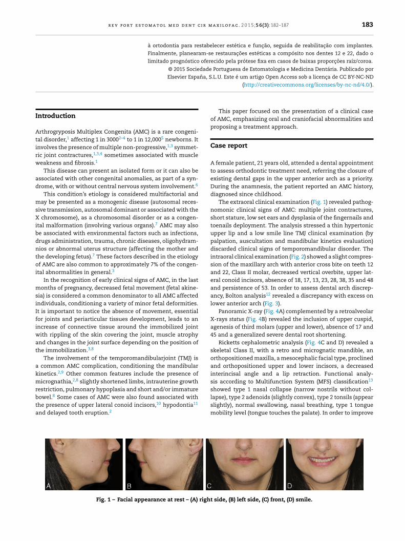
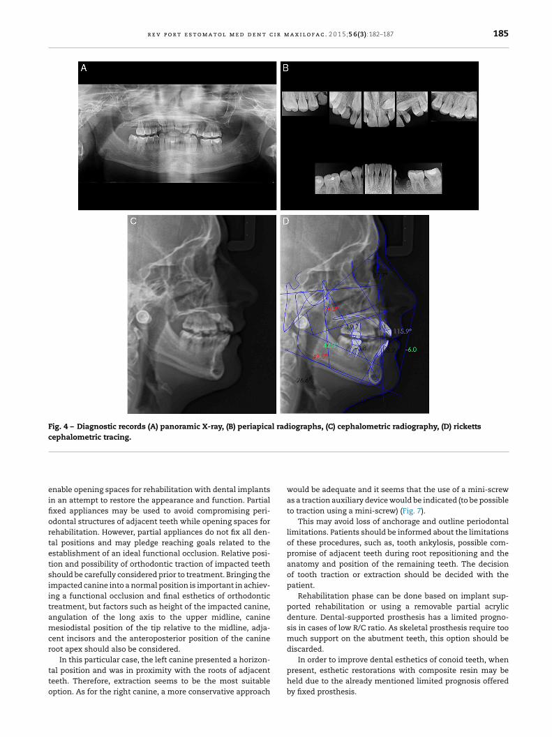
The extraoral clinical examination (Fig. 1) revealed pathog-nomonic clinical signs of AMC: multiple joint contractures,short stature, low set ears and dysplasia of the fingernails andtoenails deployment. The analysis stressed a thin hypertonicupper lip and a low smile line TMJ clinical examination (bypalpation, auscultation and mandibular kinetics evaluation)discarded clinical signs of temporomandibular disorder. Theintraoral clinical examination (Fig. 2) showed a slight compres-sion of the maxillary arch with anterior cross bite on teeth 12and 22, Class II molar, decreased vertical overbite, upper lat-eral conoid incisors, absence of 18, 17, 13, 23, 28, 38, 35 and 48and persistence of 53. In order to assess dental arch discrep-ancy, Bolton analysis12 revealed a discrepancy with excess onlower anterior arch (Fig. 3).
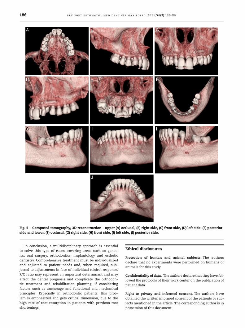
Panoramic X-ray (Fig. 4A) complemented by a retroalveolarX-rays status (Fig. 4B) revealed the inclusion of upper cuspid,agenesis of third molars (upper and lower), absence of 17 and45 and a generalized severe dental root shortening.
Ricketts cephalometric analysis (Fig. 4C and D) revealed askeletal Class II, with a retro and micrognatic mandible, anorthopositioned maxilla, a mesocephalic facial type, proclinedand orthopositioned upper and lower incisors, a decreasedinterincisal angle and a lip retraction. Functional analy-sis according to Multifunction System (MFS) classification13
showed type 1 nasal collapse (narrow nostrils without col-lapse), type 2 adenoids (slightly convex), type 2 tonsils (appearslightly), normal swallowing, nasal breathing, type 1 tonguemobility level (tongue touches the palate). In order to improve
Fig. 1 – Facial appearance at rest – (A) righ
t side, (B) left side, (C) front, (D) smile.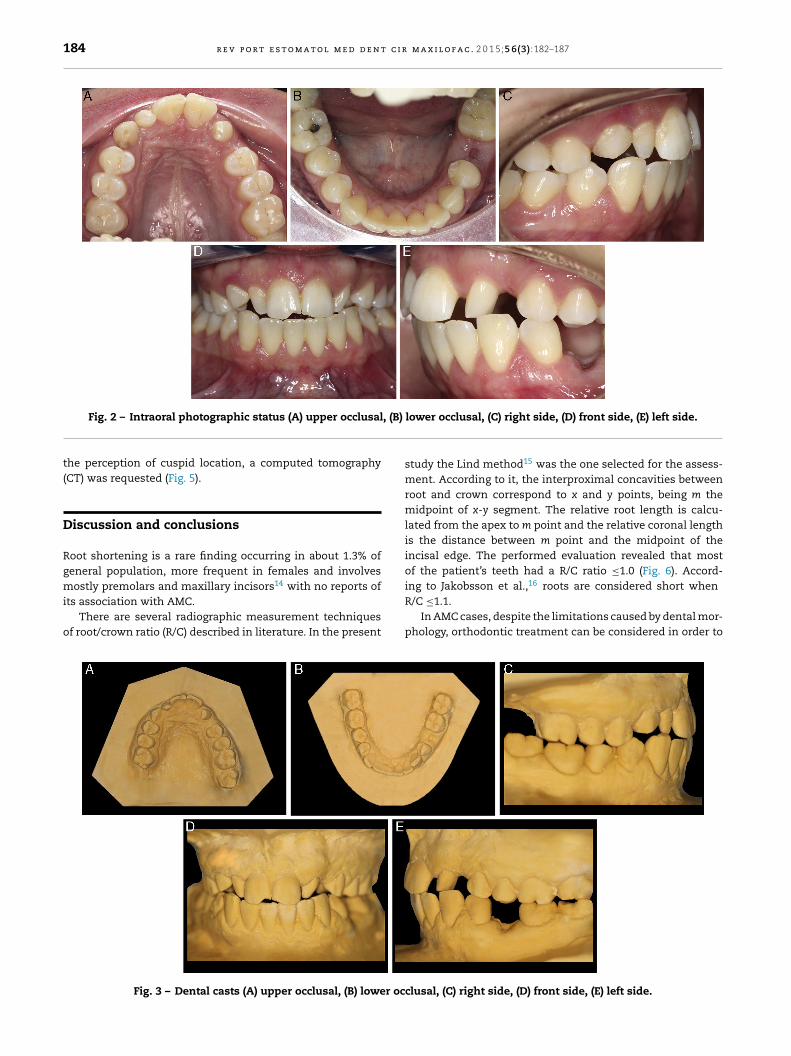
184 r e v p o r t e s t o m a t o l m e d d e n t c i r m a x i l o f a c . 2 0 1 5;5 6(3):182–187
, (B)
Fig. 2 – Intraoral photographic status (A) upper occlusalthe perception of cuspid location, a computed tomography(CT) was requested (Fig. 5).
Discussion and conclusions
Root shortening is a rare finding occurring in about 1.3% ofgeneral population, more frequent in females and involvesmostly premolars and maxillary incisors14 with no reports of
its association with AMC.There are several radiographic measurement techniquesof root/crown ratio (R/C) described in literature. In the present
Fig. 3 – Dental casts (A) upper occlusal, (B) lower oc
lower occlusal, (C) right side, (D) front side, (E) left side.
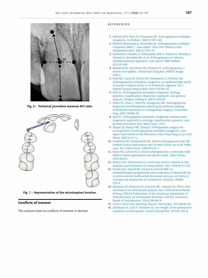
study the Lind method15 was the one selected for the assess-ment. According to it, the interproximal concavities betweenroot and crown correspond to x and y points, being m themidpoint of x-y segment. The relative root length is calcu-lated from the apex to m point and the relative coronal lengthis the distance between m point and the midpoint of theincisal edge. The performed evaluation revealed that mostof the patient’s teeth had a R/C ratio ≤1.0 (Fig. 6). Accord-ing to Jakobsson et al.,16 roots are considered short when
R/C ≤1.1.In AMC cases, despite the limitations caused by dental mor-phology, orthodontic treatment can be considered in order to
clusal, (C) right side, (D) front side, (E) left side.
r e v p o r t e s t o m a t o l m e d d e n t c i r m a x i l o f a c . 2 0 1 5;5 6(3):182–187 185
Fig. 4 – Diagnostic records (A) panoramic X-ray, (B) periapical radiographs, (C) cephalometric radiography, (D) rickettscephalometric tracing.
eifiortetsiitamcr
tto
nable opening spaces for rehabilitation with dental implantsn an attempt to restore the appearance and function. Partialxed appliances may be used to avoid compromising peri-dontal structures of adjacent teeth while opening spaces forehabilitation. However, partial appliances do not fix all den-al positions and may pledge reaching goals related to thestablishment of an ideal functional occlusion. Relative posi-ion and possibility of orthodontic traction of impacted teethhould be carefully considered prior to treatment. Bringing thempacted canine into a normal position is important in achiev-ng a functional occlusion and final esthetics of orthodonticreatment, but factors such as height of the impacted canine,ngulation of the long axis to the upper midline, canineesiodistal position of the tip relative to the midline, adja-
ent incisors and the anteroposterior position of the canineoot apex should also be considered.
In this particular case, the left canine presented a horizon-al position and was in proximity with the roots of adjacenteeth. Therefore, extraction seems to be the most suitableption. As for the right canine, a more conservative approach
would be adequate and it seems that the use of a mini-screwas a traction auxiliary device would be indicated (to be possibleto traction using a mini-screw) (Fig. 7).
This may avoid loss of anchorage and outline periodontallimitations. Patients should be informed about the limitationsof these procedures, such as, tooth ankylosis, possible com-promise of adjacent teeth during root repositioning and theanatomy and position of the remaining teeth. The decisionof tooth traction or extraction should be decided with thepatient.
Rehabilitation phase can be done based on implant sup-ported rehabilitation or using a removable partial acrylicdenture. Dental-supported prosthesis has a limited progno-sis in cases of low R/C ratio. As skeletal prosthesis require toomuch support on the abutment teeth, this option should bediscarded.
In order to improve dental esthetics of conoid teeth, whenpresent, esthetic restorations with composite resin may beheld due to the already mentioned limited prognosis offeredby fixed prosthesis.
186 r e v p o r t e s t o m a t o l m e d d e n t c i r m a x i l o f a c . 2 0 1 5;5 6(3):182–187
Fig. 5 – Computed tomography, 3D reconstruction – upper (A) occlusal, (B) right side, (C) front side, (D) left side, (E) posteriorft sid
side and lower, (F) occlusal, (G) right side, (H) front side, (I) leIn conclusion, a multidisciplinary approach is essentialto solve this type of cases, covering areas such as genet-ics, oral surgery, orthodontics, implantology and estheticdentistry. Comprehensive treatment must be individualizedand adjusted to patient needs and, when required, sub-jected to adjustments in face of individual clinical response.R/C ratio may represent an important determinant and mayaffect the dental prognosis and complicate the orthodon-tic treatment and rehabilitation planning, if consideringfactors such as anchorage and functional and mechanical
principles. Especially in orthodontic patients, this prob-lem is emphasized and gets critical dimension, due to thehigh rate of root resorption in patients with previous rootshortenings.e, (J) posterior side.
Ethical disclosures
Protection of human and animal subjects. The authorsdeclare that no experiments were performed on humans oranimals for this study.
Confidentiality of data. The authors declare that they have fol-lowed the protocols of their work center on the publication ofpatient data
Right to privacy and informed consent. The authors haveobtained the written informed consent of the patients or sub-jects mentioned in the article. The corresponding author is inpossession of this document.
r e v p o r t e s t o m a t o l m e d d e n t c i r m
Fig. 6 – Technical procedure measure R/C ratio.
C
T
r
1
1
1
1
1
Fig. 7 – Representation of the microimplant location.
onflicts of interest
he authors have no conflicts of interest to declare.
11
a x i l o f a c . 2 0 1 5;5 6(3):182–187 187
e f e r e n c e s
1. Alfonso IPO, Paez JO, Grossman JAI. Arthrogryposis multiplexcongenita. Int Pediatr. 2000;15:197–204.
2. Mielnik-Blaszczak M, Borowska M. Arthrogryposis multiplexcongenita (AMC) – case report. Ann Univ Mariae CurieSklodowska Med. 2002;57:437–41.
3. Binkiewicz-Glinska A, Sobierajska-Rek A, Bakula S, Wierzba J,Drewek K, Kowalski IM, et al. Arthrogryposis in infancy,multidisciplinary approach: case report. BMC Pediatr.2013;13:184.
4. Bamshad M, Van Heest AE, Pleasure D. Arthrogryposis: areview and update. J Bone Joint Surg Am. 2009;91 Suppl.4:40–6.
5. Hoff JM, Loane M, Gilhus NE, Rasmussen S, Daltveit AK.Arthrogryposis multiplexa congenita: an epidemiologic studyof nearly 9 million births in 24 EUROCAT registers. Eur JObstet Gynecol Reprod Biol. 2011;159:347–50.
6. Hall JG. Arthrogryposis multiplex congenita: etiology,genetics, classification, diagnostic approach, and generalaspects. J Pediatr Orthop B. 1997;6:159–66.
7. Alves PV, Zhao L, Patel PK, Bolognese AM. Arthrogryposis:diagnosis and therapeutic planning for patients seekingorthodontic treatment or orthognathic surgery. J CraniofacSurg. 2007;18:838–43.
8. Hall JG. Arthrogryposis (multiple congenital contractures):diagnostic approach to etiology, classification, genetics, andgeneral principles. Eur J Med Genet. 2014.
9. Kargel JS, Dimas VM, Chang P. Orthognathic surgery formanagement of arthrogryposis multiplex congenita: casereport and review of the literature. Can J Plast Surg (J Can ChirPlast). 2007;15:53–5.
0. Friedman BD, Heidenreich RA. Distal arthrogryposis type IIB:further clinical delineation and 54-year follow-up of an indexcase. Am J Med Genet. 1995;58:125–7.
1. Beals RK, LaFranchi S. Distal arthrogryposis: a new type withdistinct facial appearance and absent teeth. J Med Genet.2001;38:E22.
2. Bolton WA. Disharmony in tooth size and its relation to theanalysis and treatment of malocclusion. AJO. 1958;28:113–30.
3. Durán JCA, Ustrell JM, Echarri P, Arends MM. Laestímuloterapia programada como base para el desarrollo deun protocolo de reeducación funcional oral que nos lleva alconcepto de prevención en ortodoncia. Dentum. 2008;8:123–9.
4. Marques LS, Generoso R, Armond MC, Pazzini CA. Short-rootanomaly in an orthodontic patient. Am J Orthod DentofacialOrthop: Official Publication of the American Association ofOrthodontists, its Constituent Societies, and the American
Board of Orthodontics. 2010;138:346–8.5. Lind V. Short root anomaly. Scand J Dental Res. 1972;80:85–93.6. Jakobsson R, Lind V. Variation in root length of the permanent
maxillary central incisor. Scand J Dental Res. 1973;81:335–8.