Embed Size (px)
Citation preview
The Egyptian Journal of Radiology and Nuclear Medicine (2014) 45, 1017–1020
Egyptian Society of Radiology and Nuclear Medicine
The Egyptian Journal of Radiology andNuclearMedicine
www.elsevier.com/locate/ejrnmwww.sciencedirect.com
CASE REPORT
Ascending infection causing pyomyoma
in a young woman
* Corresponding author. Address: House No. 93/94, Pocket-2, Sector
22, Rohini, New Delhi 110086, India. Tel.: +91 9818427823.
E-mail address: [email protected] (A. Gupta).
Peer review under responsibility of Egyptian Society of Radiology and
Nuclear Medicine.
0378-603X � 2014 Production and hosting by Elsevier B.V. on behalf of Egyptian Society of Radiology and Nuclear Medicine.
http://dx.doi.org/10.1016/j.ejrnm.2014.05.014Open access under CC BY-NC-ND license.
Avantika Gupta *, Madhavi Mathur Gupta, Usha Manaktala
Department of Obstetrics & Gynaecology, Maulana Azad Medical College, New Delhi, India
Received 8 May 2014; accepted 18 May 2014
Available online 7 June 2014
KEYWORDS
Pyomyoma;
Escherichia coli;
CT scan;
Pus;
Laparotomy
Abstract Pyomyoma is a very rare complication of uterine fibroid, but is associated with high mor-
bidity and mortality. We report a case of pyomyoma in a young lady caused by an ascending infec-
tion. She presented with chronic fever. CT scan showed the presence of gas inside the pyomyoma.
An exploratory laparotomy was performed followed by drainage of pus. She responded well to the
treatment. Although rare, pyomyoma should be suspected in patients with leiomyoma, unexplained
fever, abdominal pain and no other apparent source of infection.� 2014 Production and hosting by Elsevier B.V. on behalf of Egyptian Society of Radiology and Nuclear
Medicine. Open access under CC BY-NC-ND license.
1. Introduction
Uterine leiomyomas are the most common uterine neoplasm.
The incidence of leiomyoma among women is generally citedas 20–25% but has been shown to be as high as 70–80% instudies using histological or sonographic examination (1).
Pyomyoma is a rare but potentially fatal complication of uter-ine leiomyoma. The diagnosis of pyomyoma is usually difficultbecause of its insidious presentation and lack of reportedimaging. The condition is usually fatal unless treated with sur-
gical intervention or appropriate antibiotics. Leiomyomas canget infected by bacterial seeding of necrotic foci, which hap-pens mostly in the postmenopausal women due to vascular
insufficiency or pregnancy due to haemorrhage and necrosis
(2–6). We report a case of a young woman who developedpyomyoma in an intramural fibroid caused by an ascending
infection of Escherichia coli.
2. Case report
A 35 year old nulliparous female was admitted with one monthhistory of intermittent low grade fever which was not associ-ated with chills or rigours. She had a history of multiple huge
fibroids six years ago for which she underwent myomectomyand had a recurrence of fibroid a year back. She had also beenundergoing multiple investigations for primary infertility. Her
menstrual history was unremarkable.On admission she had a temperature of 39.4 �C, pulse of
112 per minute, blood pressure of 120/82 mm of Hg and arespiratory rate of 24 per minute. General examination
revealed mild pallor. There was no lymphadenopathy. Uponabdominal examination, there was a mass corresponding to20 weeks of pregnant uterus, arising from pelvis which was
firm in consistency and nontender. The liver and spleen were
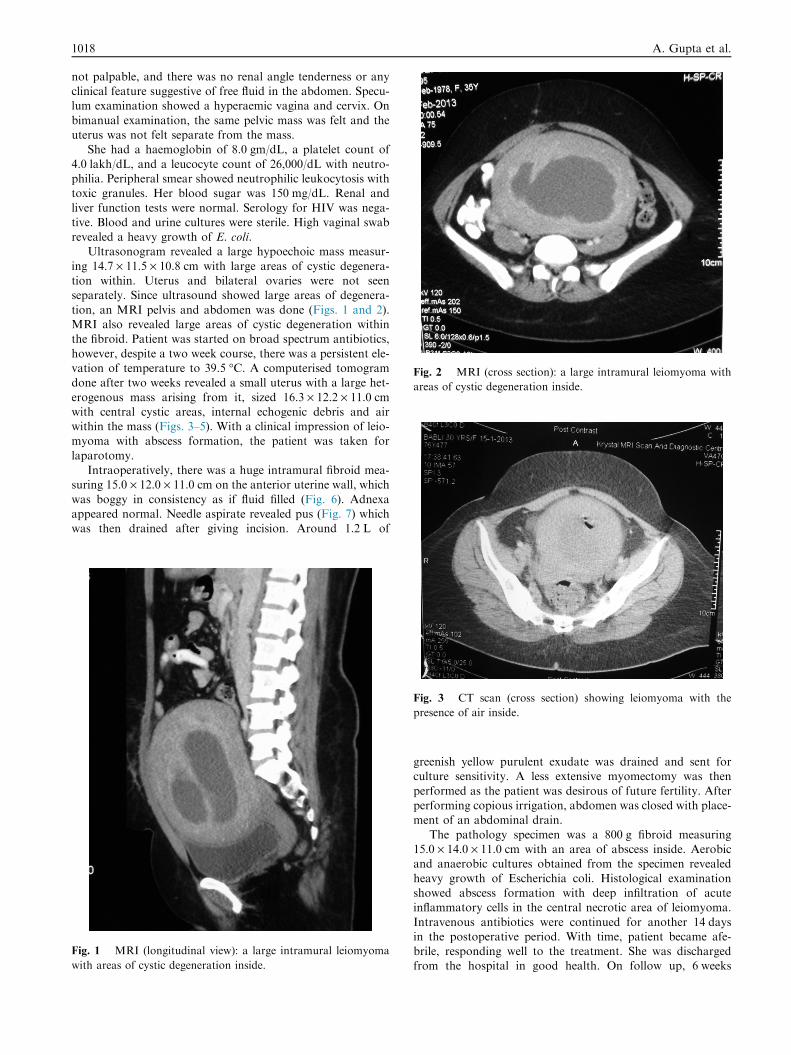
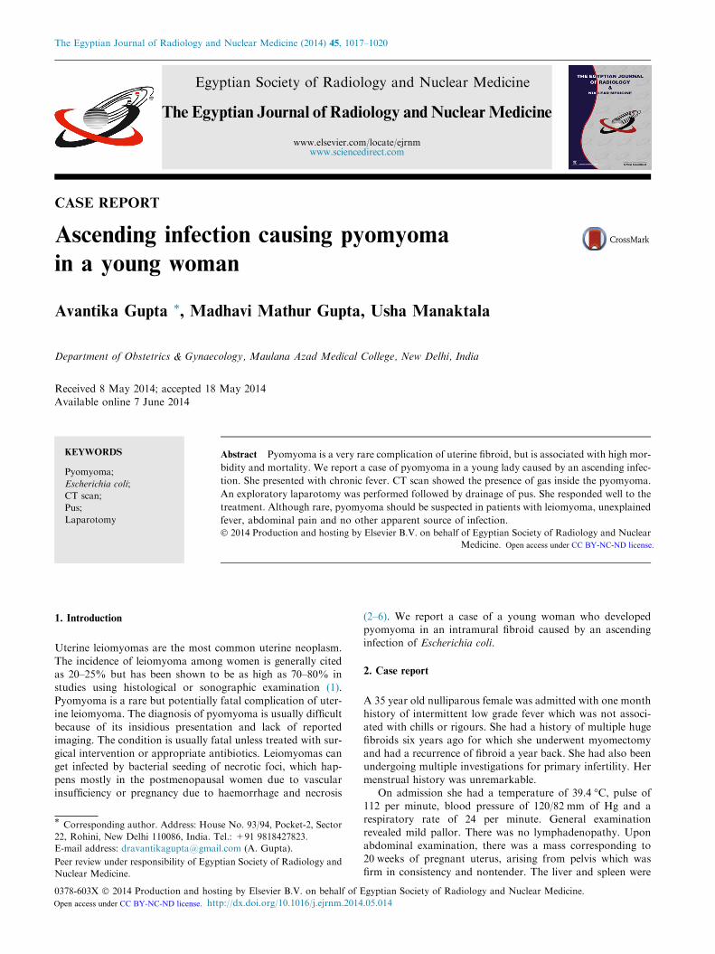
Fig. 2 MRI (cross section): a large intramural leiomyoma with
areas of cystic degeneration inside.
1018 A. Gupta et al.
not palpable, and there was no renal angle tenderness or anyclinical feature suggestive of free fluid in the abdomen. Specu-lum examination showed a hyperaemic vagina and cervix. On
bimanual examination, the same pelvic mass was felt and theuterus was not felt separate from the mass.
She had a haemoglobin of 8.0 gm/dL, a platelet count of
4.0 lakh/dL, and a leucocyte count of 26,000/dL with neutro-philia. Peripheral smear showed neutrophilic leukocytosis withtoxic granules. Her blood sugar was 150 mg/dL. Renal and
liver function tests were normal. Serology for HIV was nega-tive. Blood and urine cultures were sterile. High vaginal swabrevealed a heavy growth of E. coli.
Ultrasonogram revealed a large hypoechoic mass measur-
ing 14.7 · 11.5 · 10.8 cm with large areas of cystic degenera-tion within. Uterus and bilateral ovaries were not seenseparately. Since ultrasound showed large areas of degenera-
tion, an MRI pelvis and abdomen was done (Figs. 1 and 2).MRI also revealed large areas of cystic degeneration withinthe fibroid. Patient was started on broad spectrum antibiotics,
however, despite a two week course, there was a persistent ele-vation of temperature to 39.5 �C. A computerised tomogramdone after two weeks revealed a small uterus with a large het-
erogenous mass arising from it, sized 16.3 · 12.2 · 11.0 cmwith central cystic areas, internal echogenic debris and airwithin the mass (Figs. 3–5). With a clinical impression of leio-myoma with abscess formation, the patient was taken for
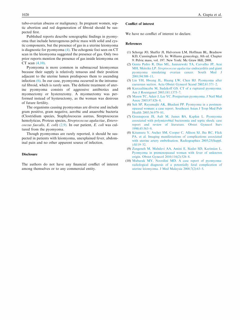
laparotomy.Intraoperatively, there was a huge intramural fibroid mea-
suring 15.0 · 12.0 · 11.0 cm on the anterior uterine wall, which
was boggy in consistency as if fluid filled (Fig. 6). Adnexaappeared normal. Needle aspirate revealed pus (Fig. 7) whichwas then drained after giving incision. Around 1.2 L of
Fig. 1 MRI (longitudinal view): a large intramural leiomyoma
with areas of cystic degeneration inside.
Fig. 3 CT scan (cross section) showing leiomyoma with the
presence of air inside.
greenish yellow purulent exudate was drained and sent forculture sensitivity. A less extensive myomectomy was then
performed as the patient was desirous of future fertility. Afterperforming copious irrigation, abdomen was closed with place-ment of an abdominal drain.
The pathology specimen was a 800 g fibroid measuring15.0 · 14.0 · 11.0 cm with an area of abscess inside. Aerobicand anaerobic cultures obtained from the specimen revealed
heavy growth of Escherichia coli. Histological examinationshowed abscess formation with deep infiltration of acuteinflammatory cells in the central necrotic area of leiomyoma.
Intravenous antibiotics were continued for another 14 daysin the postoperative period. With time, patient became afe-brile, responding well to the treatment. She was dischargedfrom the hospital in good health. On follow up, 6 weeks
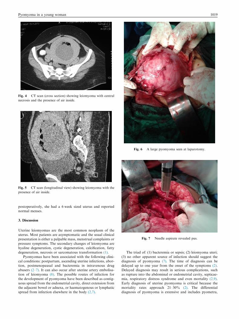
Fig. 4 CT scan (cross section) showing leiomyoma with central
necrosis and the presence of air inside.
Fig. 5 CT scan (longitudinal view) showing leiomyoma with the
presence of air inside.
Fig. 6 A large pyomyoma seen at laparotomy.
Fig. 7 Needle aspirate revealed pus.
Pyomyoma in a young woman 1019
postoperatively, she had a 6 week sized uterus and reportednormal menses.
3. Discussion
Uterine leiomyomas are the most common neoplasm of theuterus. Most patients are asymptomatic and the usual clinical
presentation is either a palpable mass, menstrual complaints orpressure symptoms. The secondary changes of leiomyoma arehyaline degeneration, cystic degeneration, calcification, fatty
degeneration, necrosis or sarcomatous transformation (1).Pyomyomas have been associated with the following clini-
cal conditions: postpartum, ascending uterine infections, abor-
tion, postmenopausal and bacteremia in intravenous drugabusers (2–7). It can also occur after uterine artery embolisa-tion of leiomyoma (8). The possible routes of infection for
the development of pyomyoma have been described as contig-uous spread from the endometrial cavity, direct extension fromthe adjacent bowel or adnexa, or haematogenous or lymphaticspread from infection elsewhere in the body (2,7).
The triad of: (1) bacteremia or sepsis; (2) leiomyoma uteri;(3) no other apparent source of infection should suggest thediagnosis of pyomyoma (7). The time of diagnosis can be
delayed up to one year from the onset of the symptoms (2).Delayed diagnosis may result in serious complications, suchas rupture into the abdominal or endometrial cavity, septicae-
mia, respiratory distress syndrome and even mortality (2,9).Early diagnosis of uterine pyomyoma is critical because themortality rates approach 21–30% (2). The differentialdiagnosis of pyomyoma is extensive and includes pyometra,
1020 A. Gupta et al.
tubo-ovarian abscess or malignancy. In pregnant women, sep-tic abortion and red degeneration of fibroid should be sus-pected first.
Published reports describe sonographic findings in pyomy-oma that include heterogenous pelvic mass with solid and cys-tic components, but the presence of gas in a uterine leiomyoma
is diagnostic for pyomyoma (4). The echogenic foci seen on CTscan in the leiomyoma suggested the presence of gas. Only twoprior reports mention the presence of gas inside leiomyoma on
CT scan (4,10).Pyomyoma is more common in submucosal leiomyomas
because their supply is relatively tenuous and their positionadjacent to the uterine lumen predisposes them to ascending
infection (6). In our case, pyomyoma occurred in the intramu-ral fibroid, which is rarely seen. The definite treatment of uter-ine pyomyoma consists of aggressive antibiotics and
myomectomy or hysterectomy. A myomectomy was per-formed instead of hysterectomy, as the woman was desirousof future fertility.
The organisms causing pyomyomas are diverse and includegram positive, gram negative, aerobic and anaerobic bacteria(Clostridium species, Staphylococcus aureus, Streptococcus
hemolyticus, Proteus species, Streptococcus agalactiae, Entero-coccus faecalis, E. coli) (2,9). In our patient, E. coli was cul-tured from the pyomyoma.
Though pyomyomas are rarely reported, it should be sus-
pected in patients with leiomyoma, unexplained fever, abdom-inal pain and no other apparent source of infection.
Disclosure
The authors do not have any financial conflict of interestamong themselves or to any commercial entity.
Conflict of interest
We have no conflict of interest to declare.
References
(1) Schorge JO, Shaffer JI, Halvorson LM, Hoffman BL, Bradnow
KD, Cunningham FG. In: Williams gynecology, 8th ed. Chapter
9: Pelvic mass, vol. 197. New York: Mc Graw Hill; 2008.
(2) Genta Pedro R, Dias ML, Janiszewski TA, Carvalho JP, Arai
MH, Meireles LP. Streptococcus agalactiae endocarditis and giant
pyomyoma simulating ovarian cancer. South Med J
2001;94:508–11.
(3) Lin YH, Hwang JL, Huang LW, Chen HJ. Pyomyoma after
caesarean section. Acta Obstet Gynecol Scand 2002;81:571–2.
(4) Karcaaltincaba M, Sudakoff GS. CT of a ruptured pyomyoma.
Am J Roentgenol 2003;181:1375–7.
(5) Mason TC, Adair J, Lee YC. Postpartum pyomyoma. J Natl Med
Assoc 2005;97:826–8.
(6) Sah SP, Rayamajhi AK, Bhadani PP. Pyomyoma in a postmen-
opausal woman: a case report. Southeast Asian J Trop Med Pub
Health 2005;36:979–81.
(7) Greenspoon JS, Ault M, James BA, Kaplan L. Pyomyoma
associated with polymicrobial bacteremia and septic shock: case
report and review of literature. Obstet Gynecol Surv
1990;45:563–9.
(8) Kitamura Y, Ascher SM, Cooper C, Allison SJ, Jha RC, Flick
PA, et al. Imaging manifestations of complications associated
with uterine artery embolisation. Radiographics 2005;25(Suppl.
):S119–32.
(9) Zangeneh M, Mahdavi AA, Amini E, Siadat SD, Karimian L.
Pyomyoma in premenopausal woman with fever of unknown
origin. Obstet Gynecol 2010;116(2):526–8.
(10) Mubarak MY, Noordini MD. A case report of pyomyoma:
radiological diagnosis of a potentially fatal complication of
uterine leiomyoma. J Med Malaysia 2008;7(2):63–5.