Embed Size (px)
DESCRIPTION
Tiroid
Citation preview
ORIGINAL STUDIES, REVIEWS,
AND SCHOLARLY DIALOGTHYROID CANCER AND NODULES
Revised American Thyroid Association ManagementGuidelines for Patients with Thyroid Nodules
and Differentiated Thyroid Cancer
The American Thyroid Association (ATA) Guidelines Taskforceon Thyroid Nodules and Differentiated Thyroid Cancer
David S. Cooper, M.D.1 (Chair)*, Gerard M. Doherty, M.D.,2 Bryan R. Haugen, M.D.,3
Richard T. Kloos, M.D.,4 Stephanie L. Lee, M.D., Ph.D.,5 Susan J. Mandel, M.D., M.P.H.,6
Ernest L. Mazzaferri, M.D.,7 Bryan McIver, M.D., Ph.D.,8 Furio Pacini, M.D.,9 Martin Schlumberger, M.D.,10
Steven I. Sherman, M.D.,11 David L. Steward, M.D.,12 and R. Michael Tuttle, M.D.13
Background: Thyroid nodules are a common clinical problem, and differentiated thyroid cancer is becomingincreasingly prevalent. Since the publication of the American Thyroid Association’s guidelines for the man-agement of these disorders was published in 2006, a large amount of new information has become available,prompting a revision of the guidelines.Methods: Relevant articles through December 2008 were reviewed by the task force and categorized by topic andlevel of evidence according to a modified schema used by the United States Preventative Services Task Force.Results: The revised guidelines for the management of thyroid nodules include recommendations regardinginitial evaluation, clinical and ultrasound criteria for fine-needle aspiration biopsy, interpretation of fine-needleaspiration biopsy results, and management of benign thyroid nodules. Recommendations regarding the initialmanagement of thyroid cancer include those relating to optimal surgical management, radioiodine remnantablation, and suppression therapy using levothyroxine. Recommendations related to long-term management ofdifferentiated thyroid cancer include those related to surveillance for recurrent disease using ultrasound andserum thyroglobulin as well as those related to management of recurrent and metastatic disease.Conclusions: We created evidence-based recommendations in response to our appointment as an independenttask force by the American Thyroid Association to assist in the clinical management of patients with thyroidnodules and differentiated thyroid cancer. They represent, in our opinion, contemporary optimal care for pa-tients with these disorders.
Thyroid nodules are a common clinical problem. Epi-demiologic studies have shown the prevalence of palpa-
ble thyroid nodules to be approximately 5% in women and 1%in men living in iodine-sufficient parts of the world (1,2). Incontrast, high-resolution ultrasound (US) can detect thyroid
nodules in 19–67% of randomly selected individuals withhigher frequencies in women and the elderly (3). The clinicalimportance of thyroid nodules rests with the need to excludethyroid cancer which occurs in 5–15% depending on age, sex,radiation exposure history, family history, and other factors
*Authors are listed in alphabetical order and were appointed by ATA to independently formulate the content of this manuscript. None ofthe scientific or medical content of the manuscript was dictated by the ATA.
1The Johns Hopkins University School of Medicine, Baltimore, Maryland.2University of Michigan Medical Center, Ann Arbor, Michigan.3University of Colorado Health Sciences Center, Denver, Colorado.4The Ohio State University, Columbus, Ohio.5Boston University Medical Center, Boston, Massachusetts.6University of Pennsylvania School of Medicine, Philadelphia, Pennsylvania.7University of Florida College of Medicine, Gainesville, Florida.8The Mayo Clinic, Rochester, Minnesota.9The University of Siena, Siena, Italy.
10Institute Gustave Roussy, Paris, France.11University of Texas M.D. Anderson Cancer Center, Houston, Texas.12University of Cincinnati Medical Center, Cincinnati, Ohio.13Memorial Sloan-Kettering Cancer Center, New York, New York.
THYROIDVolume 19, Number 11, 2009ª Mary Ann Liebert, Inc.DOI: 10.1089=thy.2009.0110
1167
(4,5). Differentiated thyroid cancer (DTC), which includespapillary and follicular cancer, comprises the vast majority(90%) of all thyroid cancers (6). In the United States, approx-imately 37,200 new cases of thyroid cancer will be diagnosedin 2009 (7). The yearly incidence has increased from 3.6 per100,000 in 1973 to 8.7 per 100,000 in 2002, a 2.4-fold increase( p< 0.001 for trend) and this trend appears to be continuing(8). Almost the entire change has been attributed to an in-crease in the incidence of papillary thyroid cancer (PTC),which increased 2.9-fold between 1988 and 2002. Moreover,49% of the rising incidence consisted of cancers measuring1 cm or smaller and 87% consisted of cancers measuring 2 cmor smaller (8). This tumor shift may be due to the increasinguse of neck ultrasonography and early diagnosis and treat-ment (9), trends that are changing the initial treatment andfollow-up for many patients with thyroid cancer.
In 1996, the American Thyroid Association (ATA) pub-lished treatment guidelines for patients with thyroid nodulesand DTC (10). Over the last decade, there have been manyadvances in the diagnosis and therapy of both thyroid nodulesand DTC. Controversy exists in many areas, including themost cost-effective approach in the diagnostic evaluation of athyroid nodule, the extent of surgery for small thyroid cancers,the use of radioactive iodine to ablate remnant tissue followingthyroidectomy, the appropriate use of thyroxine suppressiontherapy, and the role of human recombinant thyrotropin(rhTSH). In recognition of the changes that have taken place inthe overall management of these clinically important prob-lems, the ATA appointed a task force to re-examine the currentstrategies that are used to diagnose and treat thyroid nodulesand DTC, and to develop clinical guidelines using principles ofevidence-based medicine. Members of the taskforce includedexperts in thyroid nodule and thyroid cancer managementwith representation from the fields of endocrinology, surgery,and nuclear medicine. The medical opinions expressed hereare those of the authors; none were dictated by the ATA. Thefinal document was approved by the ATA Board of Directorsand endorsed (in alphabetical order) by the American Asso-ciation of Clinical Endocrinologists (AACE), American Collegeof Endocrinology, British Association of Head and NeckOncologists (BAHNO), The Endocrine Society, European As-sociation for Cranio-Maxillo-Facial Surgery (EACMFS), Eur-opean Association of Nuclear Medicine (EANM), EuropeanSociety of Endocrine Surgeons (ESES), European Society forPaediatric Endocrinology (ESPE), International Association ofEndocrine Surgeons (IAES), and Latin American Thyroid So-ciety (LATS).
Other groups have previously developed guidelines, in-cluding the American Association of Clinical Endocrinologistsand the American Association of Endocrine Surgeons (11), theBritish Thyroid Association and The Royal College of Physi-cians (12), and the National Comprehensive Cancer Network(13) that have provided somewhat conflicting recommenda-tions due to the lack of high quality evidence from random-ized controlled trials. The European Thyroid Association haspublished consensus guidelines for the management of DTC(14). The European Association of Nuclear Medicine has alsorecently published consensus guidelines for radioiodine (RAI)therapy of DTC (15).
The ATA guidelines taskforce used a strategy similar to thatemployed by the National Institutes of Health for its Consen-sus Development Conferences (http:==consensus.nih.gov=
aboutcdp.htm), and developed a series of clinically relevantquestions pertaining to thyroid nodule and thyroid cancer di-agnosis and treatment. These questions were as follows:
—Questions regarding thyroid nodules� What is the appropriate evaluation of clinically or inci-
dentally discovered thyroid nodule(s)?* What laboratory tests and imaging modalities are in-
dicated?* What is the role of fine-needle aspiration (FNA)?
� What is the best method of long-term follow up of pa-tients with thyroid nodules?
� What is the role of medical therapy of patients withbenign thyroid nodules?
� How should thyroid nodules in children and pregnantwomen be managed?
—Questions regarding the initial management of DTC� What is the role of preoperative staging with diagnostic
imaging and laboratory tests?� What is the appropriate operation for indeterminate
thyroid nodules and DTC?� What is the role of postoperative staging systems and
which should be used?� What is the role of postoperative RAI remnant ablation?� What is the role of thyrotropin (TSH) suppression
therapy?� Is there a role for adjunctive external beam irradiation or
chemotherapy?
—Questions regarding the long term management of DTC� What are the appropriate features of long-term man-
agement?� What is the role of serum thyroglobulin (Tg) assays?� What is the role of US and other imaging techniques
during follow-up?� What is the role of TSH suppression in long-term follow-
up?� What is the most appropriate management of patients
with metastatic disease?� How should Tg-positive, scan-negative patients be
managed?� What is the role of external radiation therapy?� What is the role of chemotherapy?
— What are directions for future research?
The initial ATA guidelines were published in 2006 (16).Because of the rapid growth of the literature on this topic,plans for revising the guidelines within 24–36 months ofpublication were made at the inception of the project. Re-levant articles on thyroid cancer were identified using thesame search criteria employed for the original guidelines (16).Individual task force members submitted suggestions forclarification of prior recommendations, as well as new infor-mation derived from studies published since 2004. Relevantliterature continued to be reviewed through December 2008.To begin the revision process, a half-day meeting was heldon June 2, 2007. The Task Force was broadened to includeEuropean experts and a head and neck surgeon. Three sub-sequent half-day meetings were held on October 5, 2007; July13, 2008; and October 5, 2008, to review these suggestions andfor additional comments to be considered. The meeting in July2008 also included a meeting with six additional surgeons in
1168 COOPER ET AL.
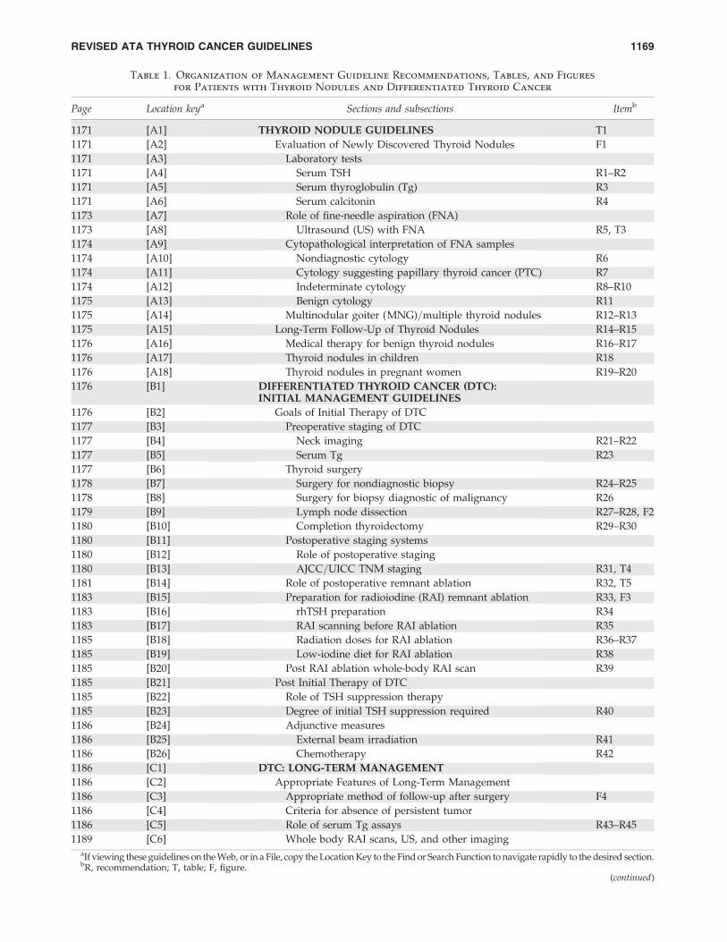
Table 1. Organization of Management Guideline Recommendations, Tables, and Figures
for Patients with Thyroid Nodules and Differentiated Thyroid Cancer
Page Location keya Sections and subsections Itemb
1171 [A1] THYROID NODULE GUIDELINES T1
1171 [A2] Evaluation of Newly Discovered Thyroid Nodules F1
1171 [A3] Laboratory tests
1171 [A4] Serum TSH R1–R2
1171 [A5] Serum thyroglobulin (Tg) R3
1171 [A6] Serum calcitonin R4
1173 [A7] Role of fine-needle aspiration (FNA)
1173 [A8] Ultrasound (US) with FNA R5, T3
1174 [A9] Cytopathological interpretation of FNA samples
1174 [A10] Nondiagnostic cytology R6
1174 [A11] Cytology suggesting papillary thyroid cancer (PTC) R7
1174 [A12] Indeterminate cytology R8–R10
1175 [A13] Benign cytology R11
1175 [A14] Multinodular goiter (MNG)=multiple thyroid nodules R12–R13
1175 [A15] Long-Term Follow-Up of Thyroid Nodules R14–R15
1176 [A16] Medical therapy for benign thyroid nodules R16–R17
1176 [A17] Thyroid nodules in children R18
1176 [A18] Thyroid nodules in pregnant women R19–R20
1176 [B1] DIFFERENTIATED THYROID CANCER (DTC):INITIAL MANAGEMENT GUIDELINES
1176 [B2] Goals of Initial Therapy of DTC
1177 [B3] Preoperative staging of DTC
1177 [B4] Neck imaging R21–R22
1177 [B5] Serum Tg R23
1177 [B6] Thyroid surgery
1178 [B7] Surgery for nondiagnostic biopsy R24–R25
1178 [B8] Surgery for biopsy diagnostic of malignancy R26
1179 [B9] Lymph node dissection R27–R28, F2
1180 [B10] Completion thyroidectomy R29–R30
1180 [B11] Postoperative staging systems
1180 [B12] Role of postoperative staging
1180 [B13] AJCC=UICC TNM staging R31, T4
1181 [B14] Role of postoperative remnant ablation R32, T5
1183 [B15] Preparation for radioiodine (RAI) remnant ablation R33, F3
1183 [B16] rhTSH preparation R34
1183 [B17] RAI scanning before RAI ablation R35
1185 [B18] Radiation doses for RAI ablation R36–R37
1185 [B19] Low-iodine diet for RAI ablation R38
1185 [B20] Post RAI ablation whole-body RAI scan R39
1185 [B21] Post Initial Therapy of DTC
1185 [B22] Role of TSH suppression therapy
1185 [B23] Degree of initial TSH suppression required R40
1186 [B24] Adjunctive measures
1186 [B25] External beam irradiation R41
1186 [B26] Chemotherapy R42
1186 [C1] DTC: LONG-TERM MANAGEMENT
1186 [C2] Appropriate Features of Long-Term Management
1186 [C3] Appropriate method of follow-up after surgery F4
1186 [C4] Criteria for absence of persistent tumor
1186 [C5] Role of serum Tg assays R43–R45
1189 [C6] Whole body RAI scans, US, and other imagingaIf viewing these guidelines on the Web, or in a File, copy the Location Key to the Find or Search Function to navigate rapidly to the desired section.bR, recommendation; T, table; F, figure.
(continued)
REVISED ATA THYROID CANCER GUIDELINES 1169
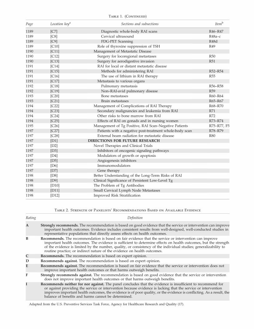
Table 1. (Continued)
Page Location keya Sections and subsections Itemb
1189 [C7] Diagnostic whole-body RAI scans R46–R47
1189 [C8] Cervical ultrasound R48a–c
1189 [C9] FDG-PET Scanning R48d
1189 [C10] Role of thyroxine suppression of TSH R49
1190 [C11] Management of Metastatic Disease
1190 [C12] Surgery for locoregional metastases R50
1190 [C13] Surgery for aerodigestive invasion R51
1191 [C14] RAI for local or distant metastatic disease
1191 [C15] Methods for administering RAI R52–R54
1191 [C16] The use of lithium in RAI therapy R55
1191 [C17] Metastasis to various organs
1192 [C18] Pulmonary metastasis R56–R58
1192 [C19] Non–RAI-avid pulmonary disease R59
1193 [C20] Bone metastases R60–R64
1193 [C21] Brain metastases R65–R67
1194 [C22] Management of Complications of RAI Therapy R68–R70
1194 [C23] Secondary malignancies and leukemia from RAI R71
1194 [C24] Other risks to bone marrow from RAI R72
1194 [C25] Effects of RAI on gonads and in nursing women R73–R74
1195 [C26] Management of Tg Positive, RAI Scan–Negative Patients R75–R77, F5
1197 [C27] Patients with a negative post-treatment whole-body scan R78–R79
1197 [C28] External beam radiation for metastatic disease R80
1197 [D1] DIRECTIONS FOR FUTURE RESEARCH
1197 [D2] Novel Therapies and Clinical Trials
1197 [D3] Inhibitors of oncogenic signaling pathways
1197 [D4] Modulators of growth or apoptosis
1197 [D5] Angiogenesis inhibitors
1197 [D6] Immunomodulators
1197 [D7] Gene therapy
1198 [D8] Better Understanding of the Long-Term Risks of RAI
1198 [D9] Clinical Significance of Persistent Low-Level Tg
1198 [D10] The Problem of Tg Antibodies
1198 [D11] Small Cervical Lymph Node Metastases
1198 [D12] Improved Risk Stratification
Table 2. Strength of Panelists’ Recommendations Based on Available Evidence
Rating Definition
A Strongly recommends. The recommendation is based on good evidence that the service or intervention can improveimportant health outcomes. Evidence includes consistent results from well-designed, well-conducted studies inrepresentative populations that directly assess effects on health outcomes.
B Recommends. The recommendation is based on fair evidence that the service or intervention can improveimportant health outcomes. The evidence is sufficient to determine effects on health outcomes, but the strengthof the evidence is limited by the number, quality, or consistency of the individual studies; generalizability toroutine practice; or indirect nature of the evidence on health outcomes.
C Recommends. The recommendation is based on expert opinion.
D Recommends against. The recommendation is based on expert opinion.
E Recommends against. The recommendation is based on fair evidence that the service or intervention does notimprove important health outcomes or that harms outweigh benefits.
F Strongly recommends against. The recommendation is based on good evidence that the service or interventiondoes not improve important health outcomes or that harms outweigh benefits.
I Recommends neither for nor against. The panel concludes that the evidence is insufficient to recommend foror against providing the service or intervention because evidence is lacking that the service or interventionimproves important health outcomes, the evidence is of poor quality, or the evidence is conflicting. As a result, thebalance of benefits and harms cannot be determined.
Adapted from the U.S. Preventive Services Task Force, Agency for Healthcare Research and Quality (17).
an effort to produce guidelines related to central neck dis-section that would be as authoritative as possible. The orga-nization of management guideline recommendations isshown in Table 1. It was agreed to continue to categorize thepublished data and strength of recommendations using amodified schema proposed by the U.S. Preventive ServicesTask Force (17) (Table 2).
[A1] THYROID NODULE GUIDELINES
A thyroid nodule is a discrete lesion within the thyroidgland that is radiologically distinct from the surroundingthyroid parenchyma. Some palpable lesions may not corre-spond to distinct radiologic abnormalities (18). Such abnor-malities do not meet the strict definition for thyroid nodules.Nonpalpable nodules detected on US or other anatomic im-aging studies are termed incidentally discovered nodules or‘‘incidentalomas.’’ Nonpalpable nodules have the same risk ofmalignancy as palpable nodules with the same size (19).Generally, only nodules >1 cm should be evaluated, sincethey have a greater potential to be clinically significant can-cers. Occasionally, there may be nodules <1 cm that requireevaluation because of suspicious US findings, associatedlymphadenopathy, a history of head and neck irradiation, or ahistory of thyroid cancer in one or more first-degree relatives.However, some nodules <1 cm lack these warning signs yeteventually cause morbidity and mortality. These are rare and,given unfavorable cost=benefit considerations, attempts todiagnose and treat all small thyroid cancers in an effort toprevent these rare outcomes would likely cause more harmthan good. Approximately 1–2% of people undergoing 2-deoxy-2[18F]fluoro-d-glucose positron emission tomography(18FDG-PET) imaging for other reasons have thyroid nodulesdiscovered incidentally. Since the risk of malignancy in these18FDG-positive nodules is about 33% and the cancers may bemore aggressive (20), such lesions require prompt evaluation(21–23). When seen, diffuse 18FDG uptake is likely related tounderlying autoimmune thyroiditis.
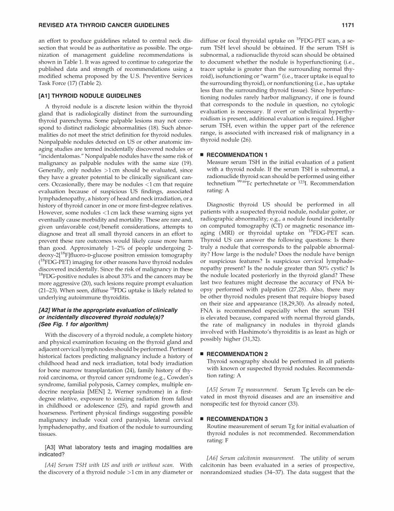
[A2] What is the appropriate evaluation of clinicallyor incidentally discovered thyroid nodule(s)?(See Fig. 1 for algorithm)
With the discovery of a thyroid nodule, a complete historyand physical examination focusing on the thyroid gland andadjacent cervical lymph nodes should be performed. Pertinenthistorical factors predicting malignancy include a history ofchildhood head and neck irradiation, total body irradiationfor bone marrow transplantation (24), family history of thy-roid carcinoma, or thyroid cancer syndrome (e.g., Cowden’ssyndrome, familial polyposis, Carney complex, multiple en-docrine neoplasia [MEN] 2, Werner syndrome) in a first-degree relative, exposure to ionizing radiation from falloutin childhood or adolescence (25), and rapid growth andhoarseness. Pertinent physical findings suggesting possiblemalignancy include vocal cord paralysis, lateral cervicallymphadenopathy, and fixation of the nodule to surroundingtissues.
[A3] What laboratory tests and imaging modalities areindicated?
[A4] Serum TSH with US and with or without scan. Withthe discovery of a thyroid nodule >1 cm in any diameter or
diffuse or focal thyroidal uptake on 18FDG-PET scan, a se-rum TSH level should be obtained. If the serum TSH issubnormal, a radionuclide thyroid scan should be obtainedto document whether the nodule is hyperfunctioning (i.e.,tracer uptake is greater than the surrounding normal thy-roid), isofunctioning or ‘‘warm’’ (i.e., tracer uptake is equal tothe surrounding thyroid), or nonfunctioning (i.e., has uptakeless than the surrounding thyroid tissue). Since hyperfunc-tioning nodules rarely harbor malignancy, if one is foundthat corresponds to the nodule in question, no cytologicevaluation is necessary. If overt or subclinical hyperthy-roidism is present, additional evaluation is required. Higherserum TSH, even within the upper part of the referencerange, is associated with increased risk of malignancy in athyroid nodule (26).
& RECOMMENDATION 1Measure serum TSH in the initial evaluation of a patientwith a thyroid nodule. If the serum TSH is subnormal, aradionuclide thyroid scan should be performed using eithertechnetium 99 mTc pertechnetate or 123I. Recommendationrating: A
Diagnostic thyroid US should be performed in allpatients with a suspected thyroid nodule, nodular goiter, orradiographic abnormality; e.g., a nodule found incidentallyon computed tomography (CT) or magnetic resonance im-aging (MRI) or thyroidal uptake on 18FDG-PET scan.Thyroid US can answer the following questions: Is theretruly a nodule that corresponds to the palpable abnormal-ity? How large is the nodule? Does the nodule have benignor suspicious features? Is suspicious cervical lymphade-nopathy present? Is the nodule greater than 50% cystic? Isthe nodule located posteriorly in the thyroid gland? Theselast two features might decrease the accuracy of FNA bi-opsy performed with palpation (27,28). Also, there maybe other thyroid nodules present that require biopsy basedon their size and appearance (18,29,30). As already noted,FNA is recommended especially when the serum TSHis elevated because, compared with normal thyroid glands,the rate of malignancy in nodules in thyroid glandsinvolved with Hashimoto’s thyroiditis is as least as high orpossibly higher (31,32).
& RECOMMENDATION 2Thyroid sonography should be performed in all patientswith known or suspected thyroid nodules. Recommenda-tion rating: A
[A5] Serum Tg measurement. Serum Tg levels can be ele-vated in most thyroid diseases and are an insensitive andnonspecific test for thyroid cancer (33).
& RECOMMENDATION 3Routine measurement of serum Tg for initial evaluation ofthyroid nodules is not recommended. Recommendationrating: F
[A6] Serum calcitonin measurement. The utility of serumcalcitonin has been evaluated in a series of prospective,nonrandomized studies (34–37). The data suggest that the
REVISED ATA THYROID CANCER GUIDELINES 1171
use of routine serum calcitonin for screening may detectC-cell hyperplasia and medullary thyroid cancer at anearlier stage and overall survival may be improved. How-ever, most studies rely on pentagastrin stimulation test-ing to increase specificity. This drug is no longer availablein the United States, and there remain unresolved issues
of sensitivity, specificity, assay performance and cost-effectiveness. A recent cost-effectiveness analysis suggestedthat calcitonin screening would be cost effective in theUnited States (38). However, the prevalence estimates ofmedullary thyroid cancer in this analysis included patientswith C-cell hyperplasia and micromedullary carcinoma,
123I or 99Tc Scana
Normal or High TSH History, Physical, TSH Low TSH
Diagnostic US
RESULTS of FNA
Elevated TSH
Evaluate and Rx for
Hyperthyroidism
Not Functioning
Hyperfunctioning
Nodule on US Do FNA
(See R5a–c)
No Nodule on US
Normal TSH
Evaluate and Rx for Hypo-
thyroidism
FNA not Indicated
Nondiagnostic
Malignant PTC
Suspicious for PTC
Benign
Indeterminate
Repeat US- Guided FNA Non-
diagnostic Close Follow-Up or Surgery (See
Text)
Pre-op US Surgery
Follicular Neoplasm
Hürthle Cell Neoplasm
Follow
Consider 123I Scan if TSH
Low Normal
Not Hyperfunctioning
Hyperfunctioning
WORKUP OF THYROID NODULEDETECTED BY PALPATION OR IMAGING
FIG. 1. Algorithm for the evaluation of patients with one or more thyroid nodules.aIf the scan does not show uniform distribution of tracer activity, ultrasound may be considered to assess for the presence
of a cystic component.
1172 COOPER ET AL.
which have an uncertain clinical significance. If the un-stimulated serum calcitonin determination has been ob-tained and the level is greater than 100 pg=mL, medullarycancer is likely present (39).
& RECOMMENDATION 4The panel cannot recommend either for or against theroutine measurement of serum calcitonin. Recommenda-tion rating: I
[A7] What is the role of FNA biopsy? FNA is the mostaccurate and cost-effective method for evaluating thyroidnodules. Retrospective studies have reported lower rates ofboth nondiagnostic and false-negative cytology specimensfrom FNA procedures performed via US guidance comparedto palpation (40,41). Therefore, for nodules with a higherlikelihood of either a nondiagnostic cytology (>25–50% cysticcomponent) (28) or sampling error (difficult to palpate orposteriorly located nodules), US-guided FNA is preferred (seeTable 3). If the diagnostic US confirms the presence of a pre-dominantly solid nodule corresponding to what is palpated,the FNA may be performed via palpation or US guidance.Traditionally FNA biopsy results are divided into four cate-gories: nondiagnostic, malignant (risk of malignancy at sur-gery >95%), indeterminate or suspicious for neoplasm, andbenign. The recent National Cancer Institute Thyroid Fine-Needle Aspiration State of the Science Conference proposed amore expanded classification for FNA cytology that adds twoadditional categories: suspicious for malignancy (risk of ma-lignancy 50–75%) and follicular lesion of undetermined sig-nificance (risk of malignancy 5–10%). The conference furtherrecommended that ‘‘neoplasm, either follicular or Hurthle cell
neoplasm’’ be substituted for ‘‘indeterminate’’ (risk of malig-nancy 15–25%) (42).
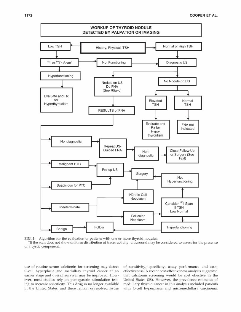
[A8] US for FNA decision making (see Table 3). Varioussonographic characteristics of a thyroid nodule have beenassociated with a higher likelihood of malignancy (43–48).These include nodule hypoechogenicity compared to thenormal thyroid parenchyma, increased intranodular vascu-larity, irregular infiltrative margins, the presence of micro-calcifications, an absent halo, and a shape taller than the widthmeasured in the transverse dimension. With the exception ofsuspicious cervical lymphadenopathy, which is a specific butinsensitive finding, no single sonographic feature or combi-nations of features is adequately sensitive or specific toidentify all malignant nodules. However, certain features andcombination of features have high predictive value for ma-lignancy. Furthermore, the most common sonographic ap-pearances of papillary and follicular thyroid cancer differ. APTC is generally solid or predominantly solid and hy-poechoic, often with infiltrative irregular margins and in-creased nodular vascularity. Microcalcifications, if present,are highly specific for PTC, but may be difficult to distinguishfrom colloid. Conversely, follicular cancer is more often iso- tohyperechoic and has a thick and irregular halo, but does nothave microcalcifications (49). Follicular cancers that are<2 cmin diameter have not been shown to be associated with met-astatic disease (50).
Certain sonographic appearances may also be highly pre-dictive of a benign nodule. A pure cystic nodule, although rare(<2% of all nodules), is highly unlikely to be malignant (47). Inaddition, a spongiform appearance, defined as an aggregationof multiple microcystic components in more than 50% of thenodule volume, is 99.7% specific for identification of a benign
Table 3. Sonographic and Clinical Features of Thyroid Nodules and Recommendations for FNA
Nodule sonographic or clinical features Recommended nodule threshold size for FNA
High-risk historya
Nodule WITH suspicious sonographic featuresb >5 mm Recommendation A
Nodule WITHOUT suspicious sonographic featuresb >5 mm Recommendation I
Abnormal cervical lymph nodes Allc Recommendation A
Microcalcifications present in nodule �1 cm Recommendation B
Solid nodule
AND hypoechoic >1 cm Recommendation B
AND iso- or hyperechoic �1–1.5 cm Recommendation C
Mixed cystic–solid nodule
WITH any suspicious ultrasound featuresb �1.5–2.0 cm Recommendation B
WITHOUT suspicious ultrasound features �2.0 cm Recommendation C
Spongiform nodule �2.0 cmd Recommendation C
Purely cystic nodule FNA not indicatede Recommendation E
aHigh-risk history: History of thyroid cancer in one or more first degree relatives; history of external beam radiation as a child; exposure toionizing radiation in childhood or adolescence; prior hemithyroidectomy with discovery of thyroid cancer, 18FDG avidity on PET scanning;MEN2=FMTC-associated RET protooncogene mutation, calcitonin>100 pg=mL. MEN, multiple endocrine neoplasia; FMTC, familial medullarythyroid cancer.
bSuspicious features: microcalcifications; hypoechoic; increased nodular vascularity; infiltrative margins; taller than wide on transverse view.cFNA cytology may be obtained from the abnormal lymph node in lieu of the thyroid nodule.dSonographic monitoring without biopsy may be an acceptable alternative (see text) (48).eUnless indicated as therapeutic modality (see text).
REVISED ATA THYROID CANCER GUIDELINES 1173
thyroid nodule (48,51,52). In a recent study, only 1 of 360malignant nodules demonstrated this appearance (48) and inanother report, a spongiform appearance had a negative pre-dictive value for malignancy of 98.5% (52). Elastography is anemerging and promising sonographic technique that requiresadditional validation with prospective studies (53).
Routine FNA is not recommended for subcentimeter nod-ules. However, the presence of a solid hypoechoic nodule withmicrocalcifications is highly suggestive of PTC. Although mostmicropapillary carcinomas may be incidental findings, a subsetmay be more clinically relevant, especially those >5 mm indiameter (54). These include nodules that have abnormallymph nodes detected clinically or with imaging at presenta-tion (55,56). Therefore, after imaging a subcentimeter nodulewith a suspicious appearance, sonographic assessment of lat-eral neck and central neck lymph nodes (more limited due tothe presence of the thyroid) must be performed. Detection ofabnormal lymph nodes should lead to FNA of the lymph node.Other groups of patients for whom consideration of FNA of asubcentimeter nodule may be warranted include those with ahigher likelihood of malignancy (high risk history): 1) familyhistory of PTC (57); 2) history of external beam radiation ex-posure as a child (58); 3) exposure to ionizing radiation inchildhood or adolescence (59); 4) history of prior hemi-thyroidectomy with discovery of thyroid cancer; and 5) 18FDG-PET–positive thyroid nodules.
Mixed cystic–solid nodules and predominantly cystic with>50% cystic component are generally evaluated by FNA withdirected biopsy of the solid component (especially the vas-cular component.) Cyst drainage may also be performed, es-pecially in symptomatic patients.
& RECOMMENDATION 5 (see Table 3)(a) FNA is the procedure of choice in the evaluation of
thyroid nodules. Recommendation rating: A(b) US guidance for FNA is recommended for those nod-
ules that are nonpalpable, predominantly cystic, orlocated posteriorly in the thyroid lobe. Recommenda-tion rating: B
[A9] What are the principles of the cytopathological inter-pretation of FNA samples?
[A10] Nondiagnostic cytology. Nondiagnostic biopsies arethose that fail to meet specified criteria for cytologic adequacythat have been previously established (the presence of at leastsix follicular cell groups, each containing 10–15 cells derivedfrom at least two aspirates of a nodule) (5). After an initialnondiagnostic cytology result, repeat FNA with US guidancewill yield a diagnostic cytology specimen in 75% of solidnodules and 50% of cystic nodules (28). Therefore, such bi-opsies need to be repeated using US guidance (60) and, ifavailable, on-site cytologic evaluation, which may substan-tially increase cytology specimen adequacy (61,62). However,up to 7% of nodules continue to yield nondiagnostic cytologyresults despite repeated biopsies and may be malignant at thetime of surgery (63,64).
& RECOMMENDATION 6(a) US guidance should be used when repeating the FNA
procedure for a nodule with an initial nondiagnosticcytology result. Recommendation rating: A
(b) Partially cystic nodules that repeatedly yield non-diagnostic aspirates need close observation or surgicalexcision. Surgery should be more strongly consideredif the cytologically nondiagnostic nodule is solid. Re-commendation rating: B
[A11] Cytology suggesting PTC.
& RECOMMENDATION 7If a cytology result is diagnostic of or suspicious for PTC,surgery is recommended (65). Recommendation rating: A
[A12] Indeterminate cytology (follicular or Hurthle cell neoplasmfollicular lesion of undetermined significance, atypia). Indetermi-nate cytology, reported as ‘‘follicular neoplasm’’ or ‘‘Hurthlecell neoplasm’’ can be found in 15–30% of FNA specimens (4)and carries a 20–30% risk of malignancy (42), while lesionsreported as atypia or follicular lesion of undetermined signifi-cance are variably reported and have 5–10% risk of malignancy(42). While certain clinical features such as male sex and nodulesize (>4 cm) (66), older patient age (67), or cytologic featuressuch as presence of atypia (68) can improve the diagnostic ac-curacy for malignancy in patients with indeterminate cytology,overall predictive values are still low. Many molecular markers(e.g., galectin-3 (69), cytokeratin, BRAF) have been evaluated toimprove diagnostic accuracy for indeterminate nodules (70–72). Recent large prospective studies have confirmed the abilityof genetic markers (BRAF, Ras, RET=PTC) and protein markers(galectin-3) to improve preoperative diagnostic accuary forpatients with indeterminate thyroid nodules (69,73,74). Manyof these markers are available for commercial use in referencelaboratories but have not yet been widely applied in clinicalpractice. It is likely that some combination of molecularmarkers will be used in the future to optimize management ofpatients with indeterminate cytology on FNA specimens.
Recently, 18FDG-PET scanning has been utilized in an ef-fort to distinguish those indeterminate nodules that are be-nign from those that are malignant (75–78). 18FDG-PET scansappear to have relatively high sensitivity for malignancy butlow specificity, but results vary among studies (79).
& RECOMMENDATION 8(a) The use of molecular markers (e.g., BRAF, RAS,
RET=PTC, Pax8-PPARg, or galectin-3) may be consid-ered for patients with indeterminate cytology on FNAto help guide management. Recommendation rating: C
(b) The panel cannot recommend for or against routineclinical use of 18FDG-PET scan to improve diagnosticaccuracy of indeterminate thyroid nodules. Recom-mendation rating: I
& RECOMMENDATION 9If the cytology reading reports a follicular neoplasm, a 123Ithyroid scan may be considered, if not already done, es-pecially if the serum TSH is in the low-normal range. If aconcordant autonomously functioning nodule is not seen,lobectomy or total thyroidectomy should be considered.Recommendation rating: C
& RECOMMENDATION 10If the reading is ‘‘suspicious for papillary carcinoma’’ or‘‘Hurthle cell neoplasm,’’ a radionuclide scan is not needed,
1174 COOPER ET AL.
and either lobectomy or total thyroidectomy is re-commended, depending on the lesion’s size and other riskfactors. Recommendation rating: A
[A13] Benign cytology.
& RECOMMENDATION 11If the nodule is benign on cytology, further immediate di-agnostic studies or treatment are not routinely required.Recommendation rating: A
[A14] How should multinodular thyroid glands or multi-nodular goiters be evaluated for malignancy? Patients withmultiple thyroid nodules have the same risk of malignancy asthose with solitary nodules (18,44). However, one large studyfound that a solitary nodule had a higher likelihood of malig-nancy than did a nonsolitary nodule ( p< 0.01), although therisk of malignancy per patient was the same and independentof the number of nodules (47). A diagnostic US should beperformed to delineate the nodules, but if only the ‘‘dominant’’or largest nodule is aspirated, the thyroid cancer may be missed(44). Radionuclide scanning should also be considered in pa-tients with multiple thyroid nodules, if the serum TSH is in thelow or low-normal range, with FNA being reserved for thosenodules that are shown to be hypofunctioning.
& RECOMMENDATION 12(a) In the presence of two or more thyroid nodules >1 cm,
those with a suspicious sonographic appearance (seetext and Table 3) should be aspirated preferentially.Recommendation rating: B
(b) If none of the nodules has a suspicious sonographicappearance and multiple sonographically similar coa-lescent nodules with no intervening normal paren-chyma are present, the likelihood of malignancy is lowand it is reasonable to aspirate the largest nodules onlyand observe the others with serial US examinations.Recommendation rating: C
& RECOMMENDATION 13A low or low-normal serum TSH concentration may sug-gest the presence of autonomous nodule(s). A technetium99 mTc pertechnetate or 123I scan should be performed anddirectly compared to the US images to determine func-tionality of each nodule >1–1.5 cm. FNA should then beconsidered only for those isofunctioning or nonfunctioningnodules, among which those with suspicious sonographicfeatures should be aspirated preferentially. Recommenda-tion rating: B
[A15] What are the best methods for long-termfollow-up of patients with thyroid nodules?
Thyroid nodules diagnosed as benign require follow-upbecause of a low, but not negligible, false-negative rate of upto 5% with FNA (41,80), which may be even higher withnodules >4 cm (81). While benign nodules may decrease insize, they often increase in size, albeit slowly (82). One studyof cytologically benign thyroid nodules <2 cm followed byultrasonography for about 38 months found that the rate ofthyroid nodule growth did not distinguish between benignand malignant nodules (83).
Nodule growth is not in and of itself pathognomonic ofmalignancy, but growth is an indication for repeat biopsy. Formixed cystic–solid nodules, the indication for repeat biopsyshould be based upon growth of the solid component. Fornodules with benign cytologic results, recent series reporta higher false-negative rate with palpation FNA (1–3%)(40,84,85) than with US FNA (0.6%) (40). Since the accuracy ofphysical examination for nodule size is likely inferior to that ofUS (30), it is recommended that serial US be used in follow-upof thyroid nodules to detect clinically significant changes insize. There is no consensus on the definition of nodule growth,however, or the threshold that would require rebiopsy. Somegroups suggest a 15% increase in nodule volume, while othersrecommend measuring a change in the mean nodule diameter(82,86). One reasonable definition of growth is a 20% increasein nodule diameter with a minimum increase in two or moredimensions of at least 2 mm. This approximates the 50% in-crease in nodule volume that was found by Brauer et al. (87) tobe the minimally significant reproducibly recorded change innodule size. These authors suggested that only volumechanges of at least 49% or more can be interpreted as noduleshrinkage or growth and consequently suggest that futureinvestigations should not describe changes in nodule volume<50% as significant. A 50% cutoff for nodule volume reduc-tion or growth, which is used in many studies, appears toappropriate and safe, since the false-negative rate for malig-nant thyroid nodules on repeat FNA is low (88,89).
& RECOMMENDATION 14(a) It is recommended that all benign thyroid nodules be
followed with serial US examinations 6–18 monthsafter the initial FNA. If nodule size is stable (i.e., nomore than a 50% change in volume or <20% increasein at least two nodule dimensions in solid nodules orin the solid portion of mixed cystic–solid nodules), theinterval before the next follow-up clinical examinationor US may be longer, e.g., every 3–5 years. Recom-mendation rating: C
(b) If there is evidence for nodule growth either by palpationor sonographically (more than a 50% change in volume ora 20% increase in at least two nodule dimensions witha minimal increase of 2 mm in solid nodules or in thesolid portion of mixed cystic–solid nodules), the FNAshould be repeated, preferably with US guidance. Re-commendation rating: B
Cystic nodules that are cytologically benign can be moni-tored for recurrence (fluid reaccumulation) which can be seenin 60–90% of patients (90,91). For those patients with subse-quent recurrent symptomatic cystic fluid accumulation,surgical removal, generally by hemithyroidectomy, or per-cutaneous ethanol injection (PEI) are both reasonable strate-gies. Four controlled studies demonstrated a 75–85% successrate after PEI compared with a 7–38% success rate in controlstreated by simple cyst evacuation or saline injection. Successwas achieved after an average of two PEI treatments. Com-plications included mild to moderate local pain, flushing,dizziness, and dysphonia (90–93).
& RECOMMENDATION 15Recurrent cystic thyroid nodules with benign cytologyshould be considered for surgical removal or PEI based on
REVISED ATA THYROID CANCER GUIDELINES 1175
compressive symptoms and cosmetic concerns. Recom-mendation rating: B
[A16] What is the role of medical therapy for benign thyroidnodules? Evidence from multiple randomized control trialsand three meta-analyses suggest that thyroid hormone in dosesthat suppress the serum TSH to subnormal levels may result ina decrease in nodule size and may prevent the appearance ofnew nodules in regions of the world with borderline low iodineintake. Data in iodine-sufficient populations are less compel-ling (94–96), with large studies suggesting that only about17–25% of thyroid nodules shrink more than 50% with le-vothyroxine (LT4) suppression of serum TSH (94–96).
& RECOMMENDATION 16Routine suppression therapy of benign thyroid nodules iniodine sufficient populations is not recommended. Re-commendation rating: F
& RECOMMENDATION 17Patients with growing nodules that are benign after repeatbiopsy should be considered for continued monitoring orintervention with surgery based on symptoms and clinicalconcern. There are no data on the use of LT4 in this sub-population of patients. Recommendation rating: I
[A17] How should thyroid nodules in children be man-aged? Thyroid nodules occur less frequently in childrenthan in adults. In one study in which approximately 5000children aged 11–18 years were assessed annually in thesouthwestern United States, palpable thyroid nodules oc-curred in approximately 20 per 1000 children, with an annualincidence of 7 new cases per 1000 children (97). Some studieshave shown the frequency of malignancy to be higher inchildren than adults, in the range of 15–20% (98–100), whereasother data have suggested that the frequency of thyroid can-cer in childhood thyroid nodules is similar to that of adults(101,102). FNA biopsy is sensitive and specific in the diagnosisof childhood thyroid nodules (99–101).
& RECOMMENDATION 18The diagnostic and therapeutic approach to one or morethyroid nodules in a child should be the same as it would bein an adult (clinical evaluation, serum TSH, US, FNA).Recommendation rating: A
[A18] How should thyroid nodules in pregnant women bemanaged? It is uncertain if thyroid nodules discovered inpregnant women are more likely to be malignant than thosefound in nonpregnant women (103), since there are no popu-lation-based studies on this question. The evaluation is the sameas for a nonpregnant patient, with the exception that a radio-nuclide scan is contraindicated. In addition, for patients withnodules diagnosed as DTC by FNA during pregnancy, delay-ing surgery until after delivery does not affect outcome (104).
& RECOMMENDATION 19For euthyroid and hypothyroid pregnant women withthyroid nodules, FNA should be performed. For womenwith suppressed serum TSH levels that persist after the firsttrimester, FNA may be deferred until after pregnancy andcessation of lactation, when a radionuclide scan can be
performed to evaluate nodule function. Recommendationrating: A
If the FNA cytology is consistent with PTC, surgery is re-commended. However, there is no consensus about whethersurgery should be performed during pregnancy or after de-livery. To minimize the risk of miscarriage, surgery duringpregnancy should be done in the second trimester before24 weeks gestation (105). However, PTC discovered duringpregnancy does not behave more aggressively than that di-agnosed in a similar-aged group of nonpregnant women(104,106). A retrospective study of pregnant women with DTCfound there to be no difference in either recurrence, or survivalrates, between women operated on during or after theirpregnancy (104). Further, retrospective data suggest thattreatment delays of less than 1 year from the time of thyroidcancer discovery do not adversely affect patient outcome (107).Finally, a recent study reported a higher rate of complicationsin pregnant women undergoing thyroid surgery comparedwith nonpregnant women (108). Some experts recommendthyroid hormone suppression therapy for pregnant womenwith FNA suspicious for or diagnostic of PTC, if surgery isdeferred until the postpartum period (109).
& RECOMMENDATION 20(a) A nodule with cytology indicating PTC discovered early
in pregnancy should be monitored sonographically andif it grows substantially (as defined above) by 24 weeksgestation, surgery should be performed at that point.However, if it remains stable by midgestation or if it isdiagnosed in the second half of pregnancy, surgery maybe performed after delivery. In patients with more ad-vanced disease, surgery in the second trimester is rea-sonable. Recommendation rating: C
(b) In pregnant women with FNA that is suspicious for ordiagnostic of PTC, consideration could be given toadministration of LT4 therapy to keep the TSH in therange of 0.1–1 mU=L. Recommendation rating: C
[B1] DIFFERENTIATED THYROID CANCER:INITIAL MANAGEMENT GUIDELINES
Differentiated thyroid cancer, arising from thyroid follicularepithelial cells, accounts for the vast majority of thyroid can-cers. Of the differentiated cancers, papillary cancer comprisesabout 85% of cases compared to about 10% that have follicularhistology, and 3% that are Hurthle cell or oxyphil tumors (110).In general, stage for stage, the prognoses of PTC and follicularcancer are similar (107,110). Certain histologic subtypes of PTChave a worse prognosis (tall cell variant, columnar cell variant,diffuse sclerosing variant), as do more highly invasive variantsof follicular cancer. These are characterized by extensive vas-cular invasion and invasion into extrathyroidal tissues orextensive tumor necrosis and=or mitoses. Other poorly dif-ferentiated aggressive tumor histologies include trabecular,insular, and solid subtypes (111). In contrast, minimally in-vasive follicular thyroid cancer, is characterized histologicallyby microscopic penetration of the tumor capsule withoutvascular invasion, and carries no excess mortality (112–115).
[B2] Goals of initial therapy of DTC
The goals of initial therapy of DTC are follows:
1176 COOPER ET AL.
1. To remove the primary tumor, disease that has ex-tended beyond the thyroid capsule, and involved cer-vical lymph nodes. Completeness of surgical resectionis an important determinant of outcome, while residualmetastatic lymph nodes represent the most commonsite of disease persistence=recurrence (116–118).
2. To minimize treatment-related morbidity. The extent ofsurgery and the experience of the surgeon both playimportant roles in determining the risk of surgicalcomplications (119,120).
3. To permit accurate staging of the disease. Because dis-ease staging can assist with initial prognostication,disease management, and follow-up strategies, accuratepostoperative staging is a crucial element in the man-agement of patients with DTC (121,122).
4. To facilitate postoperative treatment with radioactiveiodine, where appropriate. For patients undergoing RAIremnant ablation, or RAI treatment of residual or met-astatic disease, removal of all normal thyroid tissue isan important element of initial surgery (123). Near totalor total thyroidectomy also may reduce the risk for re-currence within the contralateral lobe (124).
5. To permit accurate long-term surveillance for diseaserecurrence. Both RAI whole-body scanning (WBS) andmeasurement of serum Tg are affected by residualnormal thyroid tissue. Where these approaches areutilized for long-term monitoring, near-total or total-thyroidectomy is required (125).
6. To minimize the risk of disease recurrence and meta-static spread. Adequate surgery is the most importanttreatment variable influencing prognosis, while radio-active iodine treatment, TSH suppression, and externalbeam irradiation each play adjunctive roles in at leastsome patients (125–128).
[B3] What is the role of preoperative staging with diag-nostic imaging and laboratory tests?
[B4] Neck imaging. Differentiated thyroid carcinoma(particularly papillary carcinoma) involves cervical lymphnodes in 20–50% of patients in most series using standardpathologic techniques (45,129–132), and may be present evenwhen the primary tumor is small and intrathyroidal (133). Thefrequency of micrometastases may approach 90%, dependingon the sensitivity of the detection method (134,135). However,the clinical implications of micrometastases are likely lesssignificant compared to macrometastases. Preoperative USidentifies suspicious cervical adenopathy in 20–31% of cases,potentially altering the surgical approach (136,137) in as manyas 20% of patients (138,139). However, preoperative USidentifies only half of the lymph nodes found at surgery, dueto the presence of the overlying thyroid gland (140).
Sonographic features suggestive of abnormal metastaticlymph nodes include loss of the fatty hilus, a rounded ratherthan oval shape, hypoechogenicity, cystic change, calcifica-tions, and peripheral vascularity. No single sonographic fea-ture is adequately sensitive for detection of lymph nodes withmetastatic thyroid cancer. A recent study correlated the sono-graphic features acquired 4 days preoperatively directly withthe histology of 56 cervical lymph nodes. Some of the mostspecific criteria were short axis>5 mm (96%), presence of cysticareas (100%), presence of hyperechogenic punctuations re-
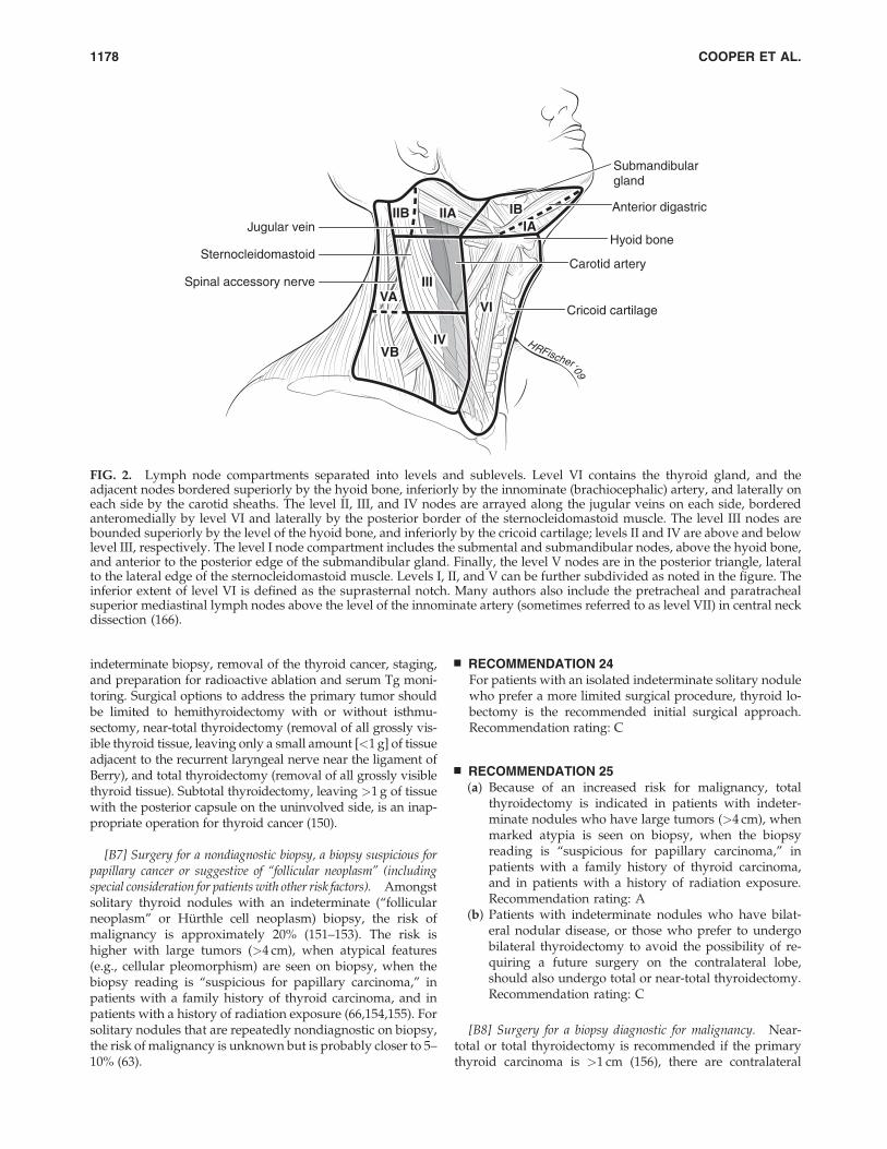
presenting either colloid or microcalcifications (100%), andperipheral vascularity (82%). Of these, the only one with suf-ficient sensitivity was peripheral vascularity (86%). All of theothers had sensitivities <60% and would not be adequate touse as single criterion for identification of malignant involve-ment (140). As shown by earlier studies (141,142), the featurewith the highest sensitivity was absence of a hilus (100%), butthis had a low specificity of only 29%. The location of the lymphnodes may also be useful for decision-making. Malignantlymph nodes are much more likely to occur in levels III, IV,and VI than in level II (140,142). Figure 2 illustrates the delin-eation of cervical lymph node Levels I through VI.
Confirmation of malignancy in lymph nodes with a sus-picious sonographic appearance is achieved by US-guidedFNA aspiration for cytology and=or measurement of Tg in theneedle washout. This FNA measurement of Tg is valid even inpatients with circulating Tg autoantibodies (143,144).
Accurate staging is important in determining the prognosisand tailoring treatment for patients with DTC. However,unlike many tumor types, the presence of metastatic diseasedoes not obviate the need for surgical excision of the primarytumor in DTC (145). Because metastatic disease may respondto RAI therapy, removal of the thyroid as well as the primarytumor and accessible locoregional disease remains an im-portant component of initial treatment even in metastaticdisease.
As US evaluation is uniquely operator dependent, alter-native imaging procedures may be preferable in some clinicalsettings, though the sensitivities of CT, MRI, and PET for thedetection of cervical lymph node metastases are all relativelylow (30–40%) (146). These alternative imaging modalities, aswell as laryngoscopy and endoscopy, may also be useful inthe assessment of large, rapidly growing, or retrosternal orinvasive tumors to assess the involvement of extrathyroidaltissues (147,148).
& RECOMMENDATION 21Preoperative neck US for the contralateral lobe and cervical(central and especially lateral neck compartments) lymphnodes is recommended for all patients undergoing thy-roidectomy for malignant cytologic findings on biopsy. US-guided FNA of sonographically suspicious lymph nodesshould be performed to confirm malignancy if this wouldchange management. Recommendation rating: B
& RECOMMENDATION 22Routine preoperative use of other imaging studies (CT,MRI, PET) is not recommended. Recommendation rating: E
[B5] Measurement of serum Tg. There is limited evidencethat high preoperative concentrations of serum Tg may pre-dict a higher sensitivity for postoperative surveillance withserum Tg (149). Evidence that this impacts patient manage-ment or outcomes is not yet available.
& RECOMMENDATION 23Routine preoperative measurement of serum Tg is not re-commended. Recommendation rating: E
[B6] What is the appropriate operation for indeterminatethyroid nodules and DTC? The goals of thyroid surgerycan include provision of a diagnosis after a nondiagnostic or
REVISED ATA THYROID CANCER GUIDELINES 1177
indeterminate biopsy, removal of the thyroid cancer, staging,and preparation for radioactive ablation and serum Tg moni-toring. Surgical options to address the primary tumor shouldbe limited to hemithyroidectomy with or without isthmu-sectomy, near-total thyroidectomy (removal of all grossly vis-ible thyroid tissue, leaving only a small amount [<1 g] of tissueadjacent to the recurrent laryngeal nerve near the ligament ofBerry), and total thyroidectomy (removal of all grossly visiblethyroid tissue). Subtotal thyroidectomy, leaving >1 g of tissuewith the posterior capsule on the uninvolved side, is an inap-propriate operation for thyroid cancer (150).
[B7] Surgery for a nondiagnostic biopsy, a biopsy suspicious forpapillary cancer or suggestive of ‘‘follicular neoplasm’’ (includingspecial consideration for patients with other risk factors). Amongstsolitary thyroid nodules with an indeterminate (‘‘follicularneoplasm’’ or Hurthle cell neoplasm) biopsy, the risk ofmalignancy is approximately 20% (151–153). The risk ishigher with large tumors (>4 cm), when atypical features(e.g., cellular pleomorphism) are seen on biopsy, when thebiopsy reading is ‘‘suspicious for papillary carcinoma,’’ inpatients with a family history of thyroid carcinoma, and inpatients with a history of radiation exposure (66,154,155). Forsolitary nodules that are repeatedly nondiagnostic on biopsy,the risk of malignancy is unknown but is probably closer to 5–10% (63).
& RECOMMENDATION 24For patients with an isolated indeterminate solitary nodulewho prefer a more limited surgical procedure, thyroid lo-bectomy is the recommended initial surgical approach.Recommendation rating: C
& RECOMMENDATION 25(a) Because of an increased risk for malignancy, total
thyroidectomy is indicated in patients with indeter-minate nodules who have large tumors (>4 cm), whenmarked atypia is seen on biopsy, when the biopsyreading is ‘‘suspicious for papillary carcinoma,’’ inpatients with a family history of thyroid carcinoma,and in patients with a history of radiation exposure.Recommendation rating: A
(b) Patients with indeterminate nodules who have bilat-eral nodular disease, or those who prefer to undergobilateral thyroidectomy to avoid the possibility of re-quiring a future surgery on the contralateral lobe,should also undergo total or near-total thyroidectomy.Recommendation rating: C
[B8] Surgery for a biopsy diagnostic for malignancy. Near-total or total thyroidectomy is recommended if the primarythyroid carcinoma is >1 cm (156), there are contralateral
HRFischer‘09
Sternocleidomastoid
Jugular vein
Carotid arterySpinal accessory nerve
Submandibulargland
Anterior digastric
Hyoid bone
Cricoid cartilage
IBIIA
III
IVVB
VAVI
IIBIA
FIG. 2. Lymph node compartments separated into levels and sublevels. Level VI contains the thyroid gland, and theadjacent nodes bordered superiorly by the hyoid bone, inferiorly by the innominate (brachiocephalic) artery, and laterally oneach side by the carotid sheaths. The level II, III, and IV nodes are arrayed along the jugular veins on each side, borderedanteromedially by level VI and laterally by the posterior border of the sternocleidomastoid muscle. The level III nodes arebounded superiorly by the level of the hyoid bone, and inferiorly by the cricoid cartilage; levels II and IV are above and belowlevel III, respectively. The level I node compartment includes the submental and submandibular nodes, above the hyoid bone,and anterior to the posterior edge of the submandibular gland. Finally, the level V nodes are in the posterior triangle, lateralto the lateral edge of the sternocleidomastoid muscle. Levels I, II, and V can be further subdivided as noted in the figure. Theinferior extent of level VI is defined as the suprasternal notch. Many authors also include the pretracheal and paratrachealsuperior mediastinal lymph nodes above the level of the innominate artery (sometimes referred to as level VII) in central neckdissection (166).
1178 COOPER ET AL.
thyroid nodules present or regional or distant metastases arepresent, the patient has a personal history of radiation therapyto the head and neck, or the patient has first-degree familyhistory of DTC. Older age (>45 years) may also be a criterionfor recommending near-total or total thyroidectomy evenwith tumors <1–1.5 cm, because of higher recurrence rates inthis age group (112,116,122,123,157). Increased extent of pri-mary surgery may improve survival for high-risk patients(158–160) and low-risk patients (156). A study of over 50,000patients with PTC found on multivariate analysis that totalthyroidectomy significantly improved recurrence and sur-vival rates for tumors >1.0 cm (156). When examined sepa-rately, even patients with 1.0–2.0 cm tumors who underwentlobectomy, had a 24% higher risk of recurrence and a 49%higher risk of thyroid cancer mortality ( p¼ 0.04 and p< 0.04,respectively). Other studies have also shown that rates of re-currence are reduced by total or near total thyroidectomyamong low-risk patients (122,161,162).
& RECOMMENDATION 26For patients with thyroid cancer >1 cm, the initial surgicalprocedure should be a near-total or total thyroidectomyunless there are contraindications to this surgery. Thyroidlobectomy alone may be sufficient treatment for small(<1 cm), low-risk, unifocal, intrathyroidal papillary carci-nomas in the absence of prior head and neck irradiation orradiologically or clinically involved cervical nodal metas-tases. Recommendation rating: A
[B9] Lymph node dissection. Regional lymph node metas-tases are present at the time of diagnosis in 20–90% of patientswith papillary carcinoma and a lesser proportion of patientswith other histotypes (129,139). Although PTC lymph nodemetastases are reported by some to have no clinically impor-tant effect on outcome in low risk patients, a study of theSurveillance, Epidemiology, and End Results (SEER) databasefound, among 9904 patients with PTC, that lymph node me-tastases, age>45 years, distant metastasis, and large tumor sizesignificantly predicted poor outcome on multivariate analysis(163). All-cause survival at 14 years was 82% for PTC withoutlymph node and 79% with lymph node metastases ( p< 0.05).Another recent SEER registry study concluded that cervicallymph node metastases conferred an independent risk of de-creased survival, but only in patients with follicular cancer andpatients with papillary cancer over age 45 years (164). Also, therisk of regional recurrence is higher in patients with lymphnode metastases, especially in those patients with multiplemetastases and=or extracapsular nodal extension (165).
In many patients, lymph node metastases in the centralcompartment (166) do not appear abnormal preoperativelywith imaging (138) or by inspection at the time of surgery.Central compartment dissection (therapeutic or prophylactic)can be achieved with low morbidity in experienced hands(167–171), and may convert some patients from clinical N0 topathologic N1a, upstaging patients over age 45 from Ameri-can Joint Committee on Cancer (AJCC) stage I to III (172). A
recent consensus conference statement discusses the relevantanatomy of the central neck compartment, delineates the no-dal subgroups within the central compartment commonlyinvolved with thyroid cancer, and defines the terminologyrelevant to central compartment neck dissection (173).
Comprehensive bilateral central compartment node dis-section may improve survival compared to historic controlsand reduce risk for nodal recurrence (174). In addition, se-lective unilateral paratracheal central compartment nodedissection increases the proportion of patients who appeardisease free with unmeasureable Tg levels 6 months aftersurgery (175). Other studies of central compartment dissec-tion have demonstrated higher morbidity, primarily recurrentlaryngeal nerve injury and transient hypoparathyroidism,with no reduction in recurrence (176,177). In another study,comprehensive (bilateral) central compartment dissectiondemonstrated higher rates of transient hypoparathyroidismcompared to selective (unilateral) dissection with no reduc-tion in rates of undetectable or low Tg levels (178). Althoughsome lymph node metastases may be treated with radioactiveiodine, several treatments may be necessary, depending uponthe histology, size, and number of metastases (179).
& RECOMMENDATION 27*(a) Therapeutic central-compartment (level VI) neck dis-
section for patients with clinically involved central orlateral neck lymph nodes should accompany totalthyroidectomy to provide clearance of disease from thecentral neck. Recommendation rating: B
(b) Prophylactic central-compartment neck dissection(ipsilateral or bilateral) may be performed in patientswith papillary thyroid carcinoma with clinically unin-volved central neck lymph nodes, especially for ad-vanced primary tumors (T3 or T4). Recommendationrating: C
(c) Near-total or total thyroidectomy without prophylacticcentral neck dissection may be appropriate for small(T1 or T2), noninvasive, clinically node-negative PTCsand most follicular cancer. Recommendation rating: C
These recommendations (R27a–c) should be interpreted inlight of available surgical expertise. For patients with small,noninvasive, apparently node-negative tumors, the balance ofrisk and benefit may favor simple near-total thyroidectomywith close intraoperative inspection of the central compart-ment with compartmental dissection only in the presence ofobviously involved lymph nodes. This approach may increasethe chance of future locoregional recurrence, but overall thisapproach may be safer in less experienced surgical hands.
Lymph nodes in the lateral neck (compartments II–V), levelVII (anterior mediastinum), and rarely in Level I may also beinvolved by thyroid cancer (129,180). For those patients inwhom nodal disease is evident clinically, on preoperative USand nodal FNA or Tg measurement, or at the time of surgery,surgical resection may reduce the risk of recurrence andpossibly mortality (56,139,181). Functional compartmental
*R27a, 27b, 27c, and 28 were developed in collaboration with an ad hoc committee of endocrinologists (David S. Cooper, M.D., Richard T.Kloos, M.D., Susan J. Mandel, M.D., M.P.H., and R. Michael Tuttle, M.D.), otolaryngology-head and neck surgeons (Gregory Randolph, M.D.,David Steward, M.D., David Terris, M.D. and Ralph Tufano, M.D.), and endocrine surgeons (Sally Carty, M.D., Gerard M. Doherty, M.D.,Quan-Yang Duh, M.D., and Robert Udelsman, M.D., M.B.A.)
REVISED ATA THYROID CANCER GUIDELINES 1179
en-bloc neck dissection is favored over isolated lymphade-nectomy (‘‘berry picking’’) with limited data suggesting im-proved mortality (118,182–184).
& RECOMMENDATION 28*Therapeutic lateral neck compartmental lymph node dis-section should be performed for patients with biopsy-proven metastatic lateral cervical lymphadenopathy.Recommendation rating: B
[B10] Completion thyroidectomy. Completion thyroidec-tomy may be necessary when the diagnosis of malignancy ismade following lobectomy for an indeterminate or non-diagnostic biopsy. Some patients with malignancy may re-quire completion thyroidectomy to provide completeresection of multicentric disease (185), and to allow RAItherapy. Most (186,187) but not all (185) studies of papillarycancer have observed a higher rate of cancer in the oppositelobe when multifocal (two or more foci), as opposed to uni-focal, disease is present in the ipsilateral lobe. The surgicalrisks of two-stage thyroidectomy (lobectomy followed bycompletion thyroidectomy) are similar to those of a near-totalor total thyroidectomy (188).
& RECOMMENDATION 29Completion thyroidectomy should be offered to those pa-tients for whom a near-total or total thyroidectomy wouldhave been recommended had the diagnosis been availablebefore the initial surgery. This includes all patients withthyroid cancer except those with small (<1 cm), unifocal,intrathyroidal, node-negative, low-risk tumors. Ther-apeutic central neck lymph node dissection should be in-cluded if the lymph nodes are clinically involved.Recommendation rating: B
& RECOMMENDATION 30Ablation of the remaining lobe with radioactive iodine hasbeen used as an alternative to completion thyroidectomy(189). It is unknown whether this approach results in sim-ilar long-term outcomes. Consequently, routine radioactiveiodine ablation in lieu of completion thyroidectomy is notrecommended. Recommendation rating: D
[B11] What is the role of postoperative staging systemsand which should be used?
[B12] The role of postoperative staging. Postoperative stag-ing for thyroid cancer, as for other cancer types, is used: 1) topermit prognostication for an individual patient with DTC;2) to tailor decisions regarding postoperative adjunctive ther-apy, including RAI therapy and TSH suppression, to assess thepatient’s risk for disease recurrence and mortality; 3) to makedecisions regarding the frequency and intensity of follow-up,directing more intensive follow-up towards patients at highestrisk; and 4) to enable accurate communication regarding apatient among health care professionals. Staging systems alsoallow evaluation of differing therapeutic strategies applied tocomparable groups of patients in clinical studies.
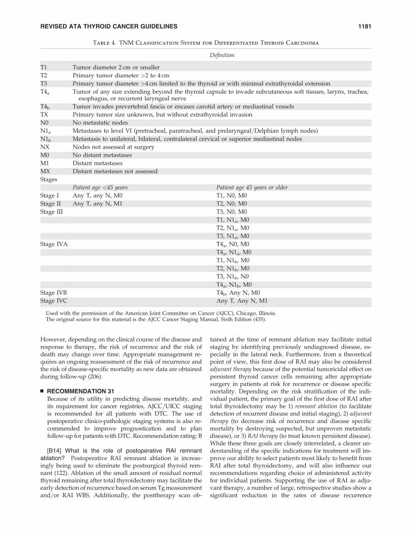
[B13] AJCC=UICC TNM staging. Application of theAJCC=International Union against Cancer (AJCC=UICC)classification system based on pTNM parameters and age isrecommended for tumors of all types, including thyroidcancer (121,190), because it provides a useful shorthandmethod to describe the extent of the tumor (191) (Table 4). Thisclassification is also used for hospital cancer registries andepidemiologic studies. In thyroid cancer, the AJCC=UICCstage does not take account of several additional independentprognostic variables and may risk misclassification of somepatients. Numerous other schemes have been developed in aneffort to achieve more accurate risk factor stratification, in-cluding CAEORTC, AGES, AMES, U of C, MACIS, OSU,MSKCC, and NTCTCS systems. (107,116,122,159,192–195).These schemes take into account a number of factors identi-fied as prognostic for outcome in multivariate analysis ofretrospective studies, with the most predictive factors gener-ally being regarded as the presence of distant metastases, theage of the patient, and the extent of the tumor. These and otherrisk factors are weighted differently among these systemsaccording to their importance in predicting outcome, but noscheme has demonstrated clear superiority (195). Each of theschemes allows accurate identification of the majority (70–85%) of patients at low-risk of mortality (T1–3, M0 patients),allowing the follow-up and management of these patients tobe less intensive than the higher-risk minority (T4 and M1patients), who may benefit from a more aggressive manage-ment strategy (195). Nonetheless, none of the examinedstaging classifications is able to account for more than a smallproportion of the uncertainty in either short-term, disease-specific mortality or the likelihood of remaining disease free(121,195,196). AJCC=IUCC staging was developed to predictrisk for death, not recurrence. For assessment of risk of re-currence, a three-level stratification can be used:
� Low-risk patients have the following characteristics:1) no local or distant metastases; 2) all macroscopic tu-mor has been resected; 3) there is no tumor invasion oflocoregional tissues or structures; 4) the tumor does nothave aggressive histology (e.g., tall cell, insular, colum-nar cell carcinoma) or vascular invasion; 5) and, if 131I isgiven, there is no 131I uptake outside the thyroid bed onthe first posttreatment whole-body RAI scan (RxWBS)(197–199).
� Intermediate-risk patients have any of the following:1) microscopic invasion of tumor into the perithyroidalsoft tissues at initial surgery; 2) cervical lymph nodemetastases or 131I uptake outside the thyroid bed on theRxWBS done after thyroid remnant ablation (200,201);or 3) tumor with aggressive histology or vascular inva-sion (202–204).
� High-risk patients have 1) macroscopic tumor invasion,2) incomplete tumor resection, 3) distant metastases, andpossibly 4) thyroglobulinemia out of proportion to whatis seen on the posttreatment scan (205).
Since initial staging is based on clinico-pathologic factorsthat are available shortly after diagnosis and initial therapy,the AJCC stage of the patient does not change over time.
*See footnote, page 1179.
1180 COOPER ET AL.
However, depending on the clinical course of the disease andresponse to therapy, the risk of recurrence and the risk ofdeath may change over time. Appropriate management re-quires an ongoing reassessment of the risk of recurrence andthe risk of disease-specific mortality as new data are obtainedduring follow-up (206).
& RECOMMENDATION 31Because of its utility in predicting disease mortality, andits requirement for cancer registries, AJCC=UICC stagingis recommended for all patients with DTC. The use ofpostoperative clinico-pathologic staging systems is also re-commended to improve prognostication and to planfollow-up for patients with DTC. Recommendation rating: B
[B14] What is the role of postoperative RAI remnantablation? Postoperative RAI remnant ablation is increas-ingly being used to eliminate the postsurgical thyroid rem-nant (122). Ablation of the small amount of residual normalthyroid remaining after total thyroidectomy may facilitate theearly detection of recurrence based on serum Tg measurementand=or RAI WBS. Additionally, the posttherapy scan ob-
tained at the time of remnant ablation may facilitate initialstaging by identifying previously undiagnosed disease, es-pecially in the lateral neck. Furthermore, from a theoreticalpoint of view, this first dose of RAI may also be consideredadjuvant therapy because of the potential tumoricidal effect onpersistent thyroid cancer cells remaining after appropriatesurgery in patients at risk for recurrence or disease specificmortality. Depending on the risk stratification of the indi-vidual patient, the primary goal of the first dose of RAI aftertotal thyroidectomy may be 1) remnant ablation (to facilitatedetection of recurrent disease and initial staging), 2) adjuvanttherapy (to decrease risk of recurrence and disease specificmortality by destroying suspected, but unproven metastaticdisease), or 3) RAI therapy (to treat known persistent disease).While these three goals are closely interrelated, a clearer un-derstanding of the specific indications for treatment will im-prove our ability to select patients most likely to benefit fromRAI after total thyroidectomy, and will also influence ourrecommendations regarding choice of administered activityfor individual patients. Supporting the use of RAI as adju-vant therapy, a number of large, retrospective studies show asignificant reduction in the rates of disease recurrence
Table 4. TNM Classification System for Differentiated Thyroid Carcinoma
Definition
T1 Tumor diameter 2 cm or smaller
T2 Primary tumor diameter >2 to 4 cm
T3 Primary tumor diameter >4 cm limited to the thyroid or with minimal extrathyroidal extension
T4a Tumor of any size extending beyond the thyroid capsule to invade subcutaneous soft tissues, larynx, trachea,esophagus, or recurrent laryngeal nerve
T4b Tumor invades prevertebral fascia or encases carotid artery or mediastinal vessels
TX Primary tumor size unknown, but without extrathyroidal invasion
N0 No metastatic nodes
N1a Metastases to level VI (pretracheal, paratracheal, and prelaryngeal=Delphian lymph nodes)
N1b Metastasis to unilateral, bilateral, contralateral cervical or superior mediastinal nodes
NX Nodes not assessed at surgery
M0 No distant metastases
M1 Distant metastases
MX Distant metastases not assessed
Stages
Patient age <45 years Patient age 45 years or older
Stage I Any T, any N, M0 T1, N0, M0
Stage II Any T, any N, M1 T2, N0, M0
Stage III T3, N0, M0
T1, N1a, M0
T2, N1a, M0
T3, N1a, M0
Stage IVA T4a, N0, M0
T4a, N1a, M0
T1, N1b, M0
T2, N1b, M0
T3, N1b, N0
T4a, N1b, M0
Stage IVB T4b, Any N, M0
Stage IVC Any T, Any N, M1
Used with the permission of the American Joint Committee on Cancer (AJCC), Chicago, Illinois.The original source for this material is the AJCC Cancer Staging Manual, Sixth Edition (435).
REVISED ATA THYROID CANCER GUIDELINES 1181
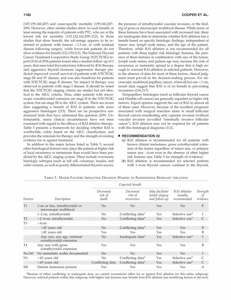
(107,159,160,207) and cause-specific mortality (159,160,207–209). However, other similar studies show no such benefit, atleast among the majority of patients with PTC, who are at thelowest risk for mortality (110,122,162,209–212). In thosestudies that show benefit, the advantage appears to be re-stricted to patients with tumors >1.5 cm, or with residualdisease following surgery, while lower-risk patients do notshow evidence for benefit (122,159,213). The National ThyroidCancer Treatment Cooperative Study Group (NTCTCSG) re-port (214) of 2936 patients found after a median follow-up of 3years, that near-total thyroidectomy followed by RAI therapyand aggressive thyroid hormone suppression therapy pre-dicted improved overall survival of patients with NTCTCSGstage III and IV disease, and was also beneficial for patientswith NTCTCSG stage II disease. No impact of therapy wasobserved in patients with stage I disease. It should be notedthat the NTCTCSG staging criteria are similar but not iden-tical to the AJCC criteria. Thus, older patients with micro-scopic extrathyroidal extension are stage II in the NTCTCSGsystem, but are stage III in the AJCC system. There are recentdata suggesting a benefit of RAI in patients with moreaggressive histologies (215). There are no prospective ran-domized trials that have addressed this question (209). Un-fortunately, many clinical circumstances have not beenexamined with regard to the efficacy of RAI ablative therapy.Table 5 presents a framework for deciding whether RAI isworthwhile, solely based on the AJCC classification, andprovides the rationale for therapy and the strength of existingevidence for or against treatment.
In addition to the major factors listed in Table 5, severalother histological features may place the patient at higher riskof local recurrence or metastases than would have been pre-dicted by the AJCC staging system. These include worrisomehistologic subtypes (such as tall cell, columnar, insular, andsolid variants, as well as poorly differentiated thyroid cancer),
the presence of intrathyroidal vascular invasion, or the find-ing of gross or microscopic multifocal disease. While many ofthese features have been associated with increased risk, thereare inadequate data to determine whether RAI ablation has abenefit based on specific histologic findings, independent oftumor size, lymph node status, and the age of the patient.Therefore, while RAI ablation is not recommended for allpatients with these higher risk histologic features, the pres-ence of these features in combination with size of the tumor,lymph node status, and patient age may increase the risk ofrecurrence or metastatic spread to a degree that is high en-ough to warrant RAI ablation in selected patients. However,in the absence of data for most of these factors, clinical judg-ment must prevail in the decision-making process. For mi-croscopic multifocal papillary cancer, when all foci are<1 cm,recent data suggest that RAI is of no benefit in preventingrecurrence (216,217).
Nonpapillary histologies (such as follicular thyroid cancerand Hurthle cell cancer) are generally regarded as higher risktumors. Expert opinion supports the use of RAI in almost allof these cases. However, because of the excellent prognosisassociated with surgical resection alone in small follicularthyroid cancers manifesting only capsular invasion (withoutvascular invasion (so-called ‘‘minimally invasive follicularcancer’’), RAI ablation may not be required for all patientswith this histological diagnosis (112).
& RECOMMENDATION 32(a) RAI ablation is recommended for all patients with
known distant metastases, gross extrathyroidal exten-sion of the tumor regardless of tumor size, or primarytumor size >4 cm even in the absence of other higherrisk features (see Table 5 for strength of evidence).
(b) RAI ablation is recommended for selected patientswith 1–4 cm thyroid cancers confined to the thyroid,
Table 5. Major Factors Impacting Decision Making in Radioiodine Remnant Ablation
Expected benefit
Factors Description
Decreasedrisk ofdeath
Decreasedrisk of
recurrence
May facilitateinitial stagingand follow-up
RAI ablationusually
recommended
Strengthof
evidence
T1 1 cm or less, intrathyroidal ormicroscopic multifocal
No No Yes No E
1–2 cm, intrathyroidal No Conflicting dataa Yes Selective usea I
T2 >2–4 cm, intrathyroidal No Conflicting dataa Yes Selective usea C
T3 >4 cm
<45 years old No Conflicting dataa Yes Yes B
�45 years old Yes Yes Yes Yes B
Any size, any age, minimalextrathyroidal extension
No Inadequate dataa Yes Selective usea I
T4 Any size with grossextrathyroidal extension
Yes Yes Yes Yes B
Nx,N0 No metastatic nodes documented No No Yes No I
N1 <45 years old No Conflicting dataa Yes Selective usea C
>45 years old Conflicting data Conflicting dataa Yes Selective usea C
M1 Distant metastasis present Yes Yes Yes Yes A
aBecause of either conflicting or inadequate data, we cannot recommend either for or against RAI ablation for this entire subgroup.However, selected patients within this subgroup with higher risk features may benefit from RAI ablation (see modifying factors in the text).
1182 COOPER ET AL.
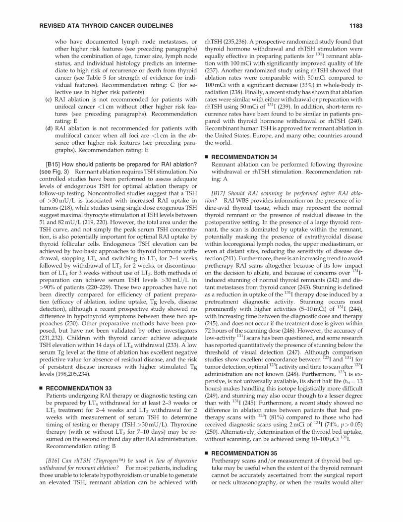
who have documented lymph node metastases, orother higher risk features (see preceding paragraphs)when the combination of age, tumor size, lymph nodestatus, and individual histology predicts an interme-diate to high risk of recurrence or death from thyroidcancer (see Table 5 for strength of evidence for indi-vidual features). Recommendation rating: C (for se-lective use in higher risk patients)
(c) RAI ablation is not recommended for patients withunifocal cancer <1 cm without other higher risk fea-tures (see preceding paragraphs). Recommendationrating: E
(d) RAI ablation is not recommended for patients withmultifocal cancer when all foci are <1 cm in the ab-sence other higher risk features (see preceding para-graphs). Recommendation rating: E
[B15] How should patients be prepared for RAI ablation?(see Fig. 3) Remnant ablation requires TSH stimulation. Nocontrolled studies have been performed to assess adequatelevels of endogenous TSH for optimal ablation therapy orfollow-up testing. Noncontrolled studies suggest that a TSHof >30 mU=L is associated with increased RAI uptake intumors (218), while studies using single dose exogenous TSHsuggest maximal thyrocyte stimulation at TSH levels between51 and 82 mU=L (219, 220). However, the total area under theTSH curve, and not simply the peak serum TSH concentra-tion, is also potentially important for optimal RAI uptake bythyroid follicular cells. Endogenous TSH elevation can beachieved by two basic approaches to thyroid hormone with-drawal, stopping LT4 and switching to LT3 for 2–4 weeksfollowed by withdrawal of LT3 for 2 weeks, or discontinua-tion of LT4 for 3 weeks without use of LT3. Both methods ofpreparation can achieve serum TSH levels >30 mU=L in>90% of patients (220–229). These two approaches have notbeen directly compared for efficiency of patient prepara-tion (efficacy of ablation, iodine uptake, Tg levels, diseasedetection), although a recent prospective study showed nodifference in hypothyroid symptoms between these two ap-proaches (230). Other preparative methods have been pro-posed, but have not been validated by other investigators(231,232). Children with thyroid cancer achieve adequateTSH elevation within 14 days of LT4 withdrawal (233). A lowserum Tg level at the time of ablation has excellent negativepredictive value for absence of residual disease, and the riskof persistent disease increases with higher stimulated Tglevels (198,205,234).
& RECOMMENDATION 33Patients undergoing RAI therapy or diagnostic testing canbe prepared by LT4 withdrawal for at least 2–3 weeks orLT3 treatment for 2–4 weeks and LT3 withdrawal for 2weeks with measurement of serum TSH to determinetiming of testing or therapy (TSH >30 mU=L). Thyroxinetherapy (with or without LT3 for 7–10 days) may be re-sumed on the second or third day after RAI administration.Recommendation rating: B
[B16] Can rhTSH (Thyrogen�) be used in lieu of thyroxinewithdrawal for remnant ablation? For most patients, includingthose unable to tolerate hypothyroidism or unable to generatean elevated TSH, remnant ablation can be achieved with
rhTSH (235,236). A prospective randomized study found thatthyroid hormone withdrawal and rhTSH stimulation wereequally effective in preparing patients for 131I remnant abla-tion with 100 mCi with significantly improved quality of life(237). Another randomized study using rhTSH showed thatablation rates were comparable with 50 mCi compared to100 mCi with a significant decrease (33%) in whole-body ir-radiation (238). Finally, a recent study has shown that ablationrates were similar with either withdrawal or preparation withrhTSH using 50 mCi of 131I (239). In addition, short-term re-currence rates have been found to be similar in patients pre-pared with thyroid hormone withdrawal or rhTSH (240).Recombinant human TSH is approved for remnant ablation inthe United States, Europe, and many other countries aroundthe world.
& RECOMMENDATION 34Remnant ablation can be performed following thyroxinewithdrawal or rhTSH stimulation. Recommendation rat-ing: A
[B17] Should RAI scanning be performed before RAI abla-tion? RAI WBS provides information on the presence of io-dine-avid thyroid tissue, which may represent the normalthyroid remnant or the presence of residual disease in thepostoperative setting. In the presence of a large thyroid rem-nant, the scan is dominated by uptake within the remnant,potentially masking the presence of extrathyroidal diseasewithin locoregional lymph nodes, the upper mediastinum, oreven at distant sites, reducing the sensitivity of disease de-tection (241). Furthermore, there is an increasing trend to avoidpretherapy RAI scans altogether because of its low impacton the decision to ablate, and because of concerns over 131I-induced stunning of normal thyroid remnants (242) and dis-tant metastases from thyroid cancer (243). Stunning is definedas a reduction in uptake of the 131I therapy dose induced by apretreatment diagnostic activity. Stunning occurs mostprominently with higher activities (5–10 mCi) of 131I (244),with increasing time between the diagnostic dose and therapy(245), and does not occur if the treatment dose is given within72 hours of the scanning dose (246). However, the accuracy oflow-activity 131I scans has been questioned, and some researchhas reported quantitatively the presence of stunning below thethreshold of visual detection (247). Although comparisonstudies show excellent concordance between 123I and 131I fortumor detection, optimal 123I activity and time to scan after 123Iadministration are not known (248). Furthermore, 123I is ex-pensive, is not universally available, its short half life (t½¼ 13hours) makes handling this isotope logistically more difficult(249), and stunning may also occur though to a lesser degreethan with 131I (245). Furthermore, a recent study showed nodifference in ablation rates between patients that had pre-therapy scans with 123I (81%) compared to those who hadreceived diagnostic scans using 2 mCi of 131I (74%, p> 0.05)(250). Alternatively, determination of the thyroid bed uptake,without scanning, can be achieved using 10–100mCi 131I.
& RECOMMENDATION 35Pretherapy scans and=or measurement of thyroid bed up-take may be useful when the extent of the thyroid remnantcannot be accurately ascertained from the surgical reportor neck ultrasonography, or when the results would alter
REVISED ATA THYROID CANCER GUIDELINES 1183
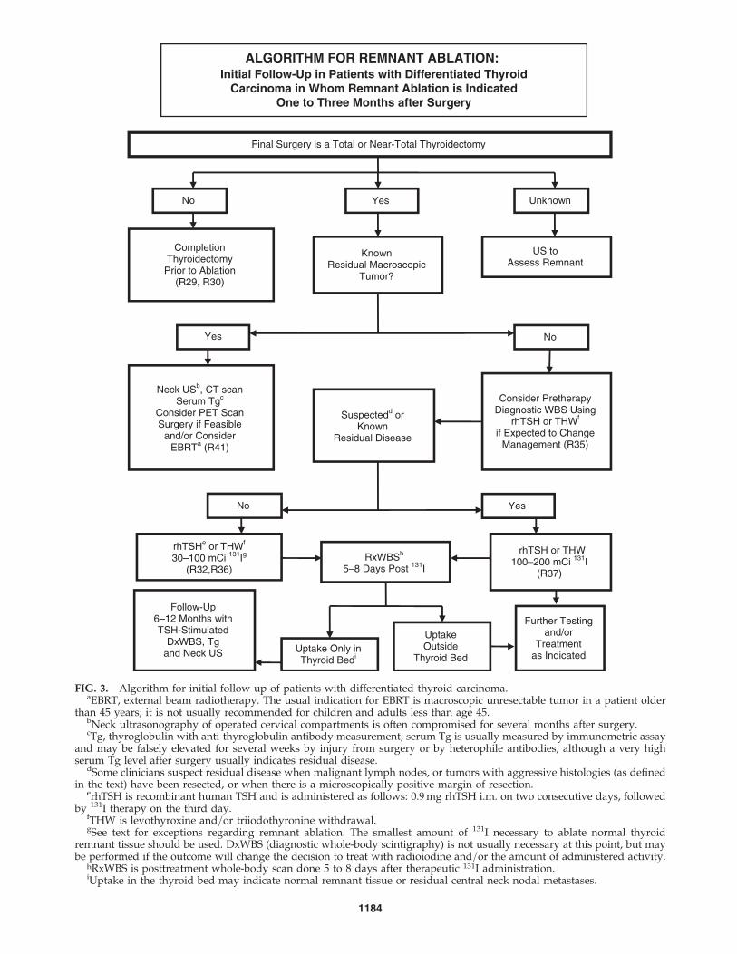
ALGORITHM FOR REMNANT ABLATION: Initial Follow-Up in Patients with Differentiated Thyroid
Carcinoma in Whom Remnant Ablation is IndicatedOne to Three Months after Surgery
Uptake Only in Thyroid Bedi
Final Surgery is a Total or Near-Total Thyroidectomy
Completion Thyroidectomy Prior to Ablation
(R29, R30)
Unknown Yes No
Known Residual Macroscopic
Tumor?
US to Assess Remnant
Neck USb, CT scan Serum Tgc
Consider PET Scan Surgery if Feasible
and/or Consider EBRTa (R41)
Consider Pretherapy Diagnostic WBS Using
rhTSH or THWf
if Expected to Change Management (R35)
Suspectedd or Known
Residual Disease
rhTSHe or THWf
30–100 mCi 131Ig
(R32,R36) RxWBSh
5–8 Days Post 131I
rhTSH or THW 100–200 mCi 131I
(R37)
Uptake Outside
Thyroid Bed
Follow-Up 6–12 Months with TSH-Stimulated
DxWBS, Tg and Neck US
Further Testing and/or
Treatment as Indicated
Yes No
Yes No
FIG. 3. Algorithm for initial follow-up of patients with differentiated thyroid carcinoma.aEBRT, external beam radiotherapy. The usual indication for EBRT is macroscopic unresectable tumor in a patient older
than 45 years; it is not usually recommended for children and adults less than age 45.bNeck ultrasonography of operated cervical compartments is often compromised for several months after surgery.cTg, thyroglobulin with anti-thyroglobulin antibody measurement; serum Tg is usually measured by immunometric assay
and may be falsely elevated for several weeks by injury from surgery or by heterophile antibodies, although a very highserum Tg level after surgery usually indicates residual disease.
dSome clinicians suspect residual disease when malignant lymph nodes, or tumors with aggressive histologies (as definedin the text) have been resected, or when there is a microscopically positive margin of resection.
erhTSH is recombinant human TSH and is administered as follows: 0.9 mg rhTSH i.m. on two consecutive days, followedby 131I therapy on the third day.
fTHW is levothyroxine and=or triiodothyronine withdrawal.gSee text for exceptions regarding remnant ablation. The smallest amount of 131I necessary to ablate normal thyroid
remnant tissue should be used. DxWBS (diagnostic whole-body scintigraphy) is not usually necessary at this point, but maybe performed if the outcome will change the decision to treat with radioiodine and=or the amount of administered activity.
hRxWBS is posttreatment whole-body scan done 5 to 8 days after therapeutic 131I administration.iUptake in the thyroid bed may indicate normal remnant tissue or residual central neck nodal metastases.
1184
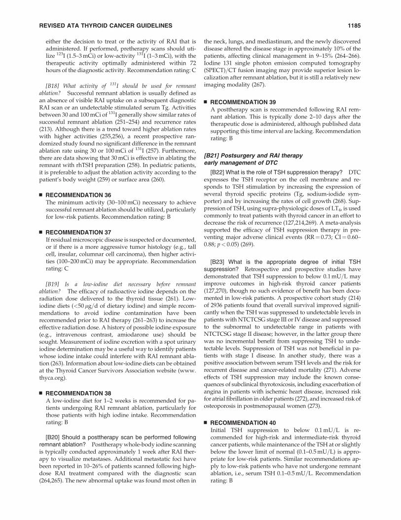
either the decision to treat or the activity of RAI that isadministered. If performed, pretherapy scans should uti-lize 123I (1.5–3 mCi) or low-activity 131I (1–3 mCi), with thetherapeutic activity optimally administered within 72hours of the diagnostic activity. Recommendation rating: C
[B18] What activity of 131I should be used for remnantablation? Successful remnant ablation is usually defined asan absence of visible RAI uptake on a subsequent diagnosticRAI scan or an undetectable stimulated serum Tg. Activitiesbetween 30 and 100 mCi of 131I generally show similar rates ofsuccessful remnant ablation (251–254) and recurrence rates(213). Although there is a trend toward higher ablation rateswith higher activities (255,256), a recent prospective ran-domized study found no significant difference in the remnantablation rate using 30 or 100 mCi of 131I (257). Furthermore,there are data showing that 30 mCi is effective in ablating theremnant with rhTSH preparation (258). In pediatric patients,it is preferable to adjust the ablation activity according to thepatient’s body weight (259) or surface area (260).
& RECOMMENDATION 36The minimum activity (30–100 mCi) necessary to achievesuccessful remnant ablation should be utilized, particularlyfor low-risk patients. Recommendation rating: B
& RECOMMENDATION 37If residual microscopic disease is suspected or documented,or if there is a more aggressive tumor histology (e.g., tallcell, insular, columnar cell carcinoma), then higher activi-ties (100–200 mCi) may be appropriate. Recommendationrating: C
[B19] Is a low-iodine diet necessary before remnantablation? The efficacy of radioactive iodine depends on theradiation dose delivered to the thyroid tissue (261). Low-iodine diets (<50 mg=d of dietary iodine) and simple recom-mendations to avoid iodine contamination have beenrecommended prior to RAI therapy (261–263) to increase theeffective radiation dose. A history of possible iodine exposure(e.g., intravenous contrast, amiodarone use) should besought. Measurement of iodine excretion with a spot urinaryiodine determination may be a useful way to identify patientswhose iodine intake could interfere with RAI remnant abla-tion (263). Information about low-iodine diets can be obtainedat the Thyroid Cancer Survivors Association website (www.thyca.org).
& RECOMMENDATION 38A low-iodine diet for 1–2 weeks is recommended for pa-tients undergoing RAI remnant ablation, particularly forthose patients with high iodine intake. Recommendationrating: B
[B20] Should a posttherapy scan be performed followingremnant ablation? Posttherapy whole-body iodine scanningis typically conducted approximately 1 week after RAI ther-apy to visualize metastases. Additional metastatic foci havebeen reported in 10–26% of patients scanned following high-dose RAI treatment compared with the diagnostic scan(264,265). The new abnormal uptake was found most often in
the neck, lungs, and mediastinum, and the newly discovereddisease altered the disease stage in approximately 10% of thepatients, affecting clinical management in 9–15% (264–266).Iodine 131 single photon emission computed tomography(SPECT)=CT fusion imaging may provide superior lesion lo-calization after remnant ablation, but it is still a relatively newimaging modality (267).
& RECOMMENDATION 39A posttherapy scan is recommended following RAI rem-nant ablation. This is typically done 2–10 days after thetherapeutic dose is administered, although published datasupporting this time interval are lacking. Recommendationrating: B
[B21] Postsurgery and RAI therapyearly management of DTC
[B22] What is the role of TSH suppression therapy? DTCexpresses the TSH receptor on the cell membrane and re-sponds to TSH stimulation by increasing the expression ofseveral thyroid specific proteins (Tg, sodium-iodide sym-porter) and by increasing the rates of cell growth (268). Sup-pression of TSH, using supra-physiologic doses of LT4, is usedcommonly to treat patients with thyroid cancer in an effort todecrease the risk of recurrence (127,214,269). A meta-analysissupported the efficacy of TSH suppression therapy in pre-venting major adverse clinical events (RR¼ 0.73; CI¼ 0.60–0.88; p< 0.05) (269).
[B23] What is the appropriate degree of initial TSHsuppression? Retrospective and prospective studies havedemonstrated that TSH suppression to below 0.1 mU=L mayimprove outcomes in high-risk thyroid cancer patients(127,270), though no such evidence of benefit has been docu-mented in low-risk patients. A prospective cohort study (214)of 2936 patients found that overall survival improved signifi-cantly when the TSH was suppressed to undetectable levels inpatients with NTCTCSG stage III or IV disease and suppressedto the subnormal to undetectable range in patients withNTCTCSG stage II disease; however, in the latter group therewas no incremental benefit from suppressing TSH to unde-tectable levels. Suppression of TSH was not beneficial in pa-tients with stage I disease. In another study, there was apositive association between serum TSH levels and the risk forrecurrent disease and cancer-related mortality (271). Adverseeffects of TSH suppression may include the known conse-quences of subclinical thyrotoxicosis, including exacerbation ofangina in patients with ischemic heart disease, increased riskfor atrial fibrillation in older patients (272), and increased risk ofosteoporosis in postmenopausal women (273).
& RECOMMENDATION 40Initial TSH suppression to below 0.1 mU=L is re-commended for high-risk and intermediate-risk thyroidcancer patients, while maintenance of the TSH at or slightlybelow the lower limit of normal (0.1–0.5 mU=L) is appro-priate for low-risk patients. Similar recommendations ap-ply to low-risk patients who have not undergone remnantablation, i.e., serum TSH 0.1–0.5 mU=L. Recommendationrating: B
REVISED ATA THYROID CANCER GUIDELINES 1185
[B24] Is there a role for adjunctive external beam irradiationor chemotherapy?
[B25] External beam irradiation. External beam irradiationis used infrequently in the management of thyroid cancerexcept as a palliative treatment for locally advanced, other-wise unresectable disease (274). There are reports of responsesamong patients with locally advanced disease (275,276) andimproved relapse-free and cause-specific survival in patientsover age 60 with extrathyroidal extension but no gross re-sidual disease (277). It remains unknown whether externalbeam radiation might reduce the risk for recurrence in theneck following adequate primary surgery and=or RAI treat-ment in patients with aggressive histologic subtypes (278).
& RECOMMENDATION 41The use of external beam irradiation to treat the primarytumor should be considered in patients over age 45 withgrossly visible extrathyroidal extension at the time of sur-gery and a high likelihood of microscopic residual disease,and for those patients with gross residual tumor in whomfurther surgery or RAI would likely be ineffective. The se-quence of external beam irradiation and RAI therapy de-pends on the volume of gross residual disease and thelikelihood of the tumor being RAI responsive. Re-commendation rating: B
[B26] Chemotherapy. There are no data to support the useof adjunctive chemotherapy in the management of DTC.Doxorubicin may act as a radiation sensitizer in some tumorsof thyroid origin (279), and could be considered for patientswith locally advanced disease undergoing external beam ra-diation.
& RECOMMENDATION 42There is no role for the routine adjunctive use of chemo-therapy in patients with DTC. Recommendation rating: F
[C1] DTC: LONG-TERM MANAGEMENT GUIDELINES
[C2] What are the appropriate featuresof long-term management?
Accurate surveillance for possible recurrence in patientsthought to be free of disease is a major goal of long-termfollow-up. Tests with high negative predictive value allowidentification of patients unlikely to experience disease re-currence, so that less aggressive management strategies canbe used that may be more cost effective and safe. Similarly,patients with a higher risk of recurrence are monitored moreaggressively because it is believed that early detection of re-current disease offers the best opportunity for effectivetreatment. A large study (280), found that the residual lifespan in disease-free patients treated with total or near-totalthyroidectomy and 131I for remnant ablation and, in somecases, high dose 131I for residual disease, was similar to that inthe general Dutch population. In contrast, the life expectancyfor patients with persistent disease was reduced to 60% of thatin the general population but varied widely depending upontumor features. Age was not a factor in disease-specific mor-tality when patients were compared with aged matched in-dividuals in the Dutch population. Treatment thus appearssafe and does not shorten life expectancy. Although an in-
creased incidence of second tumors in thyroid cancer patientshas been recognized (157,281) this elevated risk was not foundto be associated with the use of 131I in another study (282), andRAI therapy in low-risk patients did not affect median overallsurvival in another (210). Patients with persistent or recurrentdisease are offered treatment to cure or to delay future mor-bidity or mortality. In the absence of such options, therapies topalliate by substantially reducing tumor burden or prevent-ing tumor growth are utilized, with special attention paid totumors threatening critical structures.
A second goal of long-term follow-up is to monitor thy-roxine suppression or replacement therapy, to avoid under-replacement or overly aggressive therapy (283).
[C3] What is the appropriate methodfor following patients after surgerywith or without remnant ablation?
See Fig. 4 for an algorithm for the first 6–12 months ofmanagement.
[C4] What are the criteria for absence of persistenttumor? In patients who have undergone total or near-totalthyroidectomy and thyroid remnant ablation, disease-freestatus comprises all of the following:
1) no clinical evidence of tumor,2) no imaging evidence of tumor (no uptake outside the
thyroid bed on the initial posttreatment WBS, or, ifuptake outside the thyroid bed had been present, noimaging evidence of tumor on a recent diagnostic scanand neck US), and
3) undetectable serum Tg levels during TSH suppressionand stimulation in the absence of interfering antibodies.
[C5] What is the role of serum Tg assays in the follow up ofDTC? Measurement of serum Tg levels is an importantmodality to monitor patients for residual or recurrent disease.Most laboratories currently use immunometric assays tomeasure serum Tg, and it is important that these assays arecalibrated against the CRM-457 international standard. De-spite improvements in standardization of thyroglobuin as-says, there is still a twofold difference between some assays(149), leading to the recommendation that measurements inindividual patients over time be performed in the same assay.Immunometric assays are prone to interference from Tgautoantibodies, which commonly cause falsely low serumTg measurements. Radioimmunoassays may be less proneto antibody interference, but are not as widely available,and their role in the clinical care of patients is uncertain. Inthe absence of antibody interference, serum Tg has a highdegree of sensitivity and specificity to detect thyroid cancer,especially after total thyroidectomy and remnant ablation,with the highest degrees of sensitivity noted following thyroidhormone withdrawal or stimulation using rhTSH (284). SerumTg measurements obtained during thyroid hormone sup-pression of TSH, and, less commonly during TSH stimula-tion, may fail to identify patients with relatively small amountsof residual tumor (197,285,286). Conversely, even TSH-stimulated Tg measurement may fail to identify patients withclinically significant tumor, due to anti-Tg antibodies or lesscommonly to defective or absent production and secretion of
1186 COOPER ET AL.
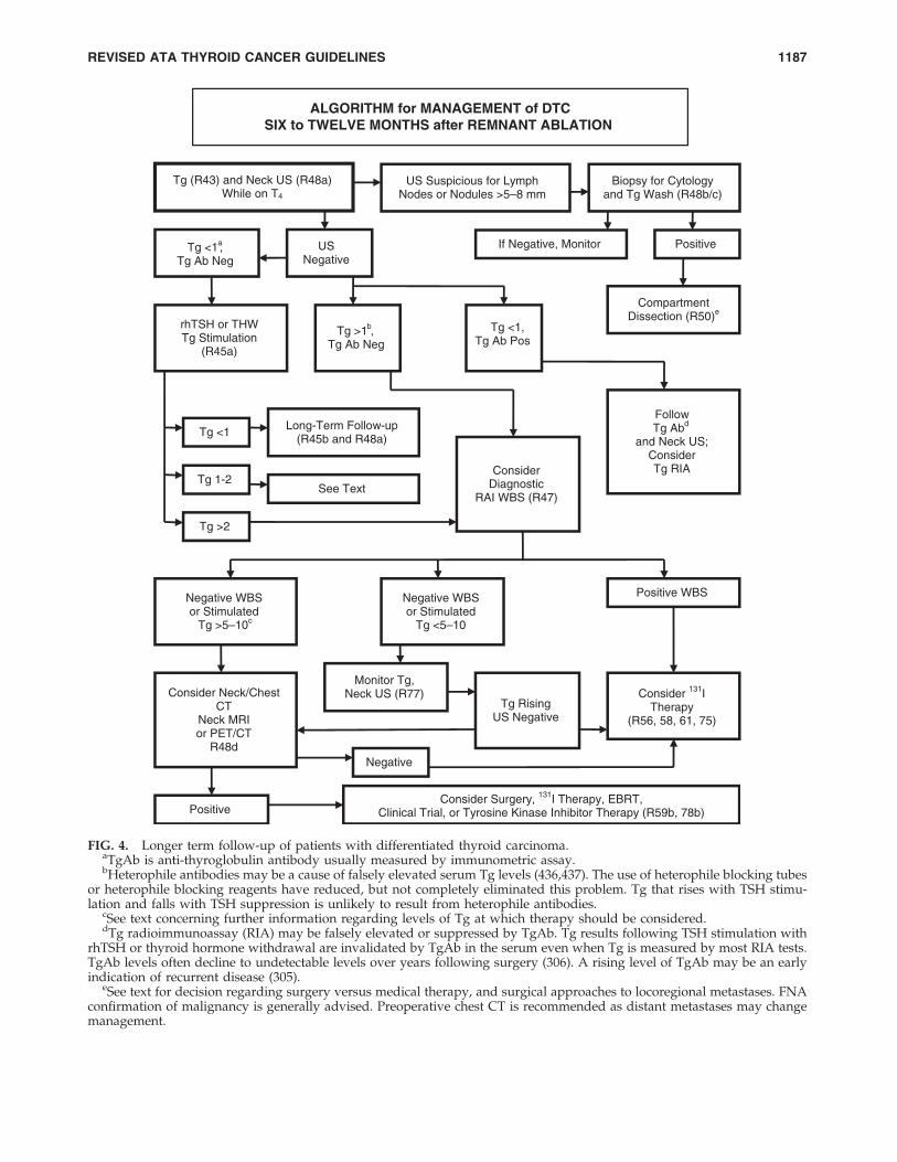
ALGORITHM for MANAGEMENT of DTCSIX to TWELVE MONTHS after REMNANT ABLATION
Tg (R43) and Neck US (R48a) While on T4
US Suspicious for Lymph Nodes or Nodules >5–8 mm
US Negative
Biopsy for Cytology and Tg Wash (R48b/c)
If Negative, Monitor Positive Tg <1a, Tg Ab Neg
rhTSH or THW Tg Stimulation
(R45a)
Tg <1
Tg >2
Tg 1-2
Long-Term Follow-up (R45b and R48a)
See Text
Tg >1 ,b
Tg Ab Neg
Tg <1,Tg Ab Pos
Consider Diagnostic
RAI WBS (R47)
Follow Tg Abd
and Neck US; Consider Tg RIA
Compartment Dissection (R50)e
Negative WBS or Stimulated
Tg >5–10c
Negative WBS or Stimulated
Tg <5–10
Positive WBS
Consider Neck/Chest CT
Neck MRI or PET/CT
R48d
Consider 131I Therapy
(R56, 58, 61, 75)
Negative
Positive
Tg Rising US Negative
Monitor Tg, Neck US (R77)
Consider Surgery, 131I Therapy, EBRT, Clinical Trial, or Tyrosine Kinase Inhibitor Therapy (R59b, 78b)
FIG. 4. Longer term follow-up of patients with differentiated thyroid carcinoma.aTgAb is anti-thyroglobulin antibody usually measured by immunometric assay.bHeterophile antibodies may be a cause of falsely elevated serum Tg levels (436,437). The use of heterophile blocking tubes
or heterophile blocking reagents have reduced, but not completely eliminated this problem. Tg that rises with TSH stimu-lation and falls with TSH suppression is unlikely to result from heterophile antibodies.
cSee text concerning further information regarding levels of Tg at which therapy should be considered.dTg radioimmunoassay (RIA) may be falsely elevated or suppressed by TgAb. Tg results following TSH stimulation with
rhTSH or thyroid hormone withdrawal are invalidated by TgAb in the serum even when Tg is measured by most RIA tests.TgAb levels often decline to undetectable levels over years following surgery (306). A rising level of TgAb may be an earlyindication of recurrent disease (305).
eSee text for decision regarding surgery versus medical therapy, and surgical approaches to locoregional metastases. FNAconfirmation of malignancy is generally advised. Preoperative chest CT is recommended as distant metastases may changemanagement.
REVISED ATA THYROID CANCER GUIDELINES 1187
immunoreactive Tg by tumor cells (286). Tg levels should beinterpreted in light of the pretest probability of clinically sig-nificant residual tumor. An aggressive or poorly differentiatedtumor may be present despite low basal or stimulated Tg; incontrast, a minimally elevated stimulated Tg may occur inpatients at low risk for clinically significant morbidity (287).Nevertheless, a single rhTSH-stimulated serum Tg<0.5 ng=mLin the absence of anti-Tg antibody has an approximately98–99.5% likelihood of identifying patients completely free oftumor on follow-up (288,289).
Follow-up of low-risk patients who have undergone totalor near-total thyroidectomy alone without 131I remnant ab-lation or hemithyroidectomy alone may represent a chal-lenge. A cohort of 80 consecutive patients with very low-riskpapillary thyroid microcarcinoma who had undergone near-total thyroidectomy without postoperative RAI treatmentwas studied over 5 years (290). The rhTSH-stimulated serumTg levels were �1 ng=mL in 45 patients (56%) and >1 ng=mLin 35 (44%) patients in whom rhTSH-stimulated Tg levelswere as high as 25 ng=mL. The diagnostic WBS (DxWBS)revealed uptake in the thyroid bed but showed no patho-logical uptake in any patient, and thyroid bed uptake corre-lated with the rhTSH-stimulated serum Tg levels ( p< 0.0001).Neck ultrasonography identified lymph node metastasesin both Tg-positive and Tg-negative patients. The authorsconcluded that for follow-up of this group of patients: 1)WBS was ineffective in detecting metastases; 2) neck ultra-sonography as the main surveillance tool was highly sensitivein detecting node metastases; and 3) detectable rhTSH-stimulated serum Tg levels mainly depended upon the size ofthyroid remnants.
Initial follow-up for low-risk patients (about 85% of post-operative patients) who have undergone total or near-totalthyroidectomy and 131I remnant ablation should be basedmainly on TSH-suppressed Tg and cervical US, followedby TSH-stimulated serum Tg measurements if the TSH-suppressed Tg testing is undetectable (197,285). However, aTg assay with a functional sensitivity of 0.1 ng=mL may re-duce the need to perform TSH-stimulated Tg measurementsduring the initial follow-up of some patients. In one study ofthis assay, a T4-suppressed serum Tg <0.1 ng=mL was onlyrarely (2.5%) associated with an rhTSH-stimulated Tg>2 ng=mL; however, 61.5% of the patients had baseline Tgelevation>0.1 ng=mL, but only one patient was found to haveresidual tumor (291). In another study of the same assay (292),a TSH-suppressed serum Tg level was >0.1 ng=mL in 14% ofpatients, but the false-positive rate was 35% using an rhTSH-stimulated Tg cutoff of >2 ng=mL, raising the possibility ofunnecessary testing and treatment. The only prospectivestudy also documented increased sensitivity of detection ofdisease at the expense of reduced specificity (293).
Approximately 20% of patients who are clinically free ofdisease with serum Tg levels <1 ng=mL during thyroid hor-mone suppression of TSH (285) will have a serum Tg level>2 ng=mL after rhTSH or thyroid hormone withdrawal at 12months after initial therapy with surgery and RAI. In this pa-tient population, one third will have identification of persistentor recurrent disease and of increasing Tg levels, and the othertwo thirds will remain free of clinical disease and will havestable or decreasing stimulated serum Tg levels over time (294).There is good evidence that a Tg cutoff level above 2 ng=mLfollowing rhTSH stimulation is highly sensitive in identifying
patients with persistent tumor (285,295–300). However, theresults of serum Tg measurements made on the same serumspecimen differ among assay methods (149). Therefore, the Tgcutoff may differ significantly among medical centers andlaboratories. Further, the clinical significance of minimallydetectable Tg levels is unclear, especially if only detected fol-lowing TSH stimulation. In these patients, the trend in serumTg over time will typically identify patients with clinicallysignificant residual disease. A rising unstimulated or stimu-lated serum Tg indicates disease that is likely to become clini-cally apparent (294,301).
The presence of anti-Tg antibodies, which occur in ap-proximately 25% of thyroid cancer patients (302) and 10% ofthe general population (303), will falsely lower serum Tg de-terminations in immunometric assays (304). The use of re-covery assays in this setting to detect significant interference iscontroversial (201,304). Serial serum anti-Tg antibody quan-tification using the same methodology may serve as an im-precise surrogate marker of residual normal thyroid tissue ortumor (305, 306).
& RECOMMENDATION 43Serum Tg should be measured every 6–12 months by animmunometric assay that is calibrated against the CRM-457 standard. Ideally, serum Tg should be assessed in thesame laboratory and using the same assay, during follow-up of patients with DTC who have undergone total or neartotal thyroidectomy with or without thyroid remnant ab-lation. Thyroglobulin antibodies should be quantitativelyassessed with every measurement of serum Tg. Recom-mendation rating: A
& RECOMMENDATION 44Periodic serum Tg measurements and neck ultrasonographyshould be considered during follow-up of patients with DTCwho have undergone less than total thyroidectomy, and inpatients who have had a total thyroidectomy but not RAIablation. While specific cutoff levels during TSH suppres-sion or stimulation that optimally distinguish normal re-sidual thyroid tissue from persistent thyroid cancer areunknown, rising Tg values over time are suspicious forgrowing thyroid tissue or cancer. Recommendation rating: B
& RECOMMENDATION 45(a) In low-risk patients who have had remnant ablation and
negative cervical US and undetectable TSH-suppressedTg within the first year after treatment, serum Tg shouldbe measured after thyroxine withdrawal or rhTSH stim-ulation approximately 12 months after the ablation toverify absence of disease. Recommendation rating: A
The timing or necessity of subsequent stimulated testing isuncertain for those found to be free of disease, because there isinfrequent benefit in this patient cohort from repeated TSH-stimulated Tg testing (289).
(b) Low-risk patients who have had remnant abla-tion, negative cervical US, and undetectable TSH-stimulated Tg can be followed primarily with yearlyclinical examination and Tg measurements on thyroidhormone replacement. Recommendation rating: B
1188 COOPER ET AL.
[C6] What are the roles of diagnostic whole-body RAI scans,US, and other imaging techniques during follow-up of DTC?
[C7] Diagnostic whole-body RAI scans. There are two mainissues that affect the use of DxWBS during follow-up: stun-ning (described above) and accuracy. A DxWBS is most usefulduring follow-up when there is little or no remaining normalthyroid tissue. Disease not visualized on the DxWBS,regardless of the activity of 131I employed, may occasionallybe visualized on the RxWBS images done after larger, thera-peutic amounts of 131I (285,307–310). Following RAI ablation,when the posttherapy scan does not reveal uptake outside thethyroid bed, subsequent DxWBS have low sensitivity and areusually not necessary in low-risk patients who are clinicallyfree of residual tumor and have an undetectable serum Tglevel on thyroid hormone and negative cervical US(197,285,309,311).
& RECOMMENDATION 46After the first RxWBS performed following RAI remnantablation, low-risk patients with an undetectable Tg onthyroid hormone with negative antithyrogolublin anti-bodies and a negative US do not require routine DxWBSduring follow-up. Recommendation rating: F
& RECOMMENDATION 47DxWBS, either following thyroid hormone withdrawalor rhTSH, 6–12 months after remnant ablation may be ofvalue in the follow-up of patients with high or intermedi-ate risk of persistent disease (see risk stratification systemunder AJCC=UICC TNM staging), but should be donewith 123I or low activity 131I. Recommendation rating: C
[C8] Cervical ultrasonography. Cervical ultrasonography ishighly sensitive in the detection of cervical metastases in pa-tients with DTC (139,290,312). Recent data suggest thatmeasurement of Tg in the needle washout fluid enhances thesensitivity of FNA of cervical nodes that are suspicious on US(313,314). Cervical metastases occasionally may be detectedby neck ultrasonography even when TSH-stimulated serumTg levels remain undetectable (201,296).
& RECOMMENDATION 48(a) Following surgery, cervical US to evaluate the thyroid
bed and central and lateral cervical nodal compartmentsshould be performed at 6–12 months and then periodi-cally, depending on the patient’s risk for recurrent dis-ease and Tg status. Recommendation rating: B
(b) If a positive result would change management, ultra-sonographically suspicious lymph nodes greater than5–8 mm in the smallest diameter should be biopsied forcytology with Tg measurement in the needle washoutfluid. Recommendation rating: A
(c) Suspicious lymph nodes less than 5–8 mm in largest di-ameter may be followed without biopsy with consider-ation for intervention if there is growth or if the nodethreatens vital structures. Recommendation rating: C
[C9] 18FDG-PET scanning. For many years, the primaryclinical application of 18FDG-PET scanning in thyroid cancerwas to localize disease in Tg-positive (>10 ng=mL), RAI scan–negative patients (315). When used for this indication, insur-
ance providers have usually required documentation that thepatient had a follicular derived thyroid cancer with sup-pressed or stimulated Tg >10 ng=mL in the setting of a neg-ative DxWBS. Still, the impact of 18FDG-PET imaging onbiochemical cure, survival, or progression-free survival in thissetting are not well defined.
More recently, publications provide data that support theuse of 18FDG-PET scanning for indications beyond simpledisease localization in Tg-positive, RAI scan–negative pa-tients (315,316).
Current additional clinical uses of 18FDG-PET scanningmay include:
� Initial staging and follow-up of high-risk patients withpoorly differentiated thyroid cancers unlikely to con-centrate RAI in order to identify sites of disease that maybe missed with RAI scanning and conventional imaging.
� Initial staging and follow-up of invasive or metastaticHurthle cell carcinoma.
� As a powerful prognostic tool for identifying whichpatients with known distant metastases are at highestrisk for disease-specific mortality.
� As a selection tool to identify those patients unlikely torespond to additional RAI therapy.
� As a measurement of posttreatment response followingexternal beam irradiation, surgical resection, emboliza-tion, or systemic therapy.
As can be seen from the list of indications above, low-riskpatients are very unlikely to require 18FDG-PET scanning aspart of initial staging or follow-up. Additionally, inflamma-tory lymph nodes, suture granulomas, and increased muscleactivity are common causes of false-positive 18FDG-PETfindings. Therefore, cytologic or histologic confirmation isrequired before one can be certain that an 18FDG-positive le-sion represents metastatic disease.
The sensitivity of 18FDG-PET scanning may be marginallyimproved with TSH stimulation (especially in patients withlow Tg values), but the clinical benefit of identifying theseadditional small foci is yet to be proven (316).
(d) In addition to its proven role in the localization ofdisease in Tg-positive, RAI scan–negative patients,18FDG-PET scanning may be employed 1) as part ofinitial staging in poorly differentiated thyroid cancersand invasive Hurthle cell carcinomas, especially thosewith other evidence of disease on imaging or becauseof elevated serum Tg levels, and 2) as a prognostic toolin patients with metastatic disease to identify thosepatients at highest risk for rapid disease progressionand disease-specific mortality, 3) and as an evaluationof posttreatment response following systemic or localtherapy of metastatic or locally invasive disease. Re-commendation rating: C
[C10] What is the role of thyroxine TSH suppressionduring thyroid hormone therapy in the long-term follow-up ofDTC? A meta-analysis has suggested an association (269)between thyroid hormone suppression therapy and reductionof major adverse clinical events. The appropriate degree ofTSH suppression by LT4 is still unknown, especially in high-risk patients rendered free of disease. One study found that aconstantly suppressed TSH (�0.05 mU=L) was associated with
REVISED ATA THYROID CANCER GUIDELINES 1189
a longer relapse-free survival than when serum TSH levelswere always 1 mU=L or greater, and that the degree of TSHsuppression was an independent predictor of recurrence inmultivariate analysis (270). Conversely, another large studyfound that disease stage, patient age, and 131I therapy inde-pendently predicted disease progression, but that the degreeof TSH suppression did not (127). A third study showed thatduring LT4 therapy the mean Tg levels were significantlyhigher when TSH levels were normal than when TSH levelswere suppressed (<0.5 mU=L) but only in patients with localor distant relapse (317). A fourth study of 2936 patients foundthat overall survival improved significantly when the TSHwas suppressed to <0.1 mU=L in patients with NTCTCSGstage III or IV disease and to 0.1 to about 0.5 range in patientswith NTCTCSG stage II disease; however, there was no in-cremental benefit from suppressing TSH to undetectable levelsin stage II patients and suppression of TSH was of no benefit inpatients with stage I disease (214). Another recent study foundthat a serum TSH threshold of 2 mU=L differentiated best be-tween patients free of disease and those with relapse or cancer-related mortality (271). No prospective studies have beenperformed examining the risk of recurrence and death fromthyroid cancer associated with varying serum TSH levels,based on the criteria for the absence of tumor at 6–12 monthspostsurgery and RAI ablation outlined above in [C3].
& RECOMMENDATION 49(a) In patients with persistent disease, the serum TSH
should be maintained below 0.1 mU=L indefinitelyin the absence of specific contraindications. Recom-mendation rating: B
(b) In patients who are clinically and biochemically freeof disease but who presented with high risk disease,consideration should be given to maintaining TSH-suppressive therapy to achieve serum TSH levels of0.1–0.5 mU=L for 5–10 years. Recommendation rating: C
(c) In patients free of disease, especially those at low risk forrecurrence, the serum TSH may be kept within the lownormal range (0.3–2 mU=L). Recommendation rating: B
(d) In patients who have not undergone remnant ablationwho are clinically free of disease and have undetect-able suppressed serum Tg and normal neck US, theserum TSH may be allowed to rise to the low normalrange (0.3–2 mU=L). Recommendation rating: C
[C11] What is the most appropriate managementof DTC patients with metastatic disease?
Metastases discovered during follow-up are likely mani-festations of persistent disease that has survived initial treat-ment. Some patients will have a reduction in tumor burdenwith additional treatments that may offer a survival or palli-ative benefit (318–322). The preferred hierarchy of treatmentfor metastatic disease (in order) is surgical excision of locor-egional disease in potentially curable patients, 131I therapy forRAI-avid disease, external beam radiation, watchful waitingwith patients with stable or slowly progressive asymptomaticdisease, and experimental trials, especially for patients withsignificantly progressive macroscopic refractory disease. Ex-perimental trials may be tried before external beam radiationin special circumstances, in part because of the morbidity ofexternal beam radiation and its relative lack of efficacy. A
small fraction of patients may benefit from radiofrequencyablation (323), ethanol ablation (324), or chemo-embolization(325). Additionally, surgical therapy in selected incurablepatients is important to prevent complications in targetedareas, such as the central nervous system (CNS) and centralneck compartment. Conversely, watchful waiting may beappropriate for selected patients with stable asymptomaticlocal metastatic disease, and most patients with stableasymptomatic non-CNS distant metastatic disease.
[C12] What is the surgical management of locoregionalmetastases? Surgery is favored for locoregional (i.e., cervicallymph nodes and=or soft tissue tumor in the neck) recurrences,when distant metastases are not present. Approximately onethird to one half of patients may become free of disease in short-term follow-up (288). It is not clear that treatment of locor-egional disease is beneficial in the setting of untreatable distantmetastases, except for possible palliation of symptoms or pre-vention of airway or aerodigestive obstruction. Impalpablemetastatic lymph nodes, visualized on US or other anatomicimaging modality, that have survived initial 131I therapyshould be considered for resection. Conversely, the benefit toremoving asymptomatic small (<5–8 mm) metastatic lymphnodes towards improving gross clinical disease recurrences ordisease-specific survival is unproven. When surgery is elected,most surgeons endorse comprehensive or selective ipsilateralcompartmental dissection of previously unexplored compart-ments with clinically significant persistent or recurrent disease(i.e., lymph nodes >0.8 cm in diameter,) while sparing vitalstructures (e.g., ipsilateral central neck dissection [level VI],selective neck dissection levels II–IV, or modified neck dissec-tion [levels II–V sparing the spinal accessory nerve, the internaljugular vein, and sternocleidomastoid muscle] (326) as op-posed to ‘‘berry picking,’’ limited lymph node resection pro-cedures, or ethanol ablation (324), because microscopic lymphnode metastases are commonly more extensive than wouldappear from imaging studies alone (183,327,328). Conversely,compartmental surgical dissections may not be feasible in thesetting of compartments that have been previously exploreddue to extensive scarring, and only a more limited or targetedlymph node resection may be possible.
& RECOMMENDATION 50(a) Therapeutic comprehensive compartmental lateral
and=or central neck dissection, sparing uninvolvedvital structures, should be performed for patients withpersistent or recurrent disease confined to the neck.Recommendation rating: B
(b) Limited compartmental lateral and=or central com-partmental neck dissection may be a reasonablealternative to more extensive comprehensive dissec-tion for patients with recurrent disease within com-partments having undergone prior comprehensivedissection and=or external beam radiotherapy. Re-commendation rating: C
[C13] What is the surgical management of aerodigestiveinvasion? For tumors that invade the upper aerodigestivetract, surgery combined with additional therapy such as 131Iand=or external beam radiation is generally advised (329,330).Patient outcome is related to complete resection of all gross
1190 COOPER ET AL.
disease with the preservation of function, with techniquesranging from shaving tumor off the trachea or esophagus forsuperficial invasion, to more aggressive techniques when thetrachea is more deeply invaded (e.g., direct intraluminal in-vasion) including tracheal resection and anastomosis (331–333)or laryngopharyngoesophagectomy. Patients who are notcurable may undergo less aggressive local treatment in cases ofasphyxia or significant hemoptysis, and as a preliminary stepprior to subsequent radical or palliative treatments (330).
& RECOMMENDATION 51When technically feasible, surgery for aerodigestive invasivedisease is recommended in combination with RAI and=orexternal beam radiotherapy. Recommendation rating: B
[C14] What is the nature of RAI therapy for locoregional ordistant metastatic disease? For regional nodal metastasesdiscovered on DxWBS, RAI may be employed, althoughsurgery is typically used in the presence of bulky disease ordisease amenable to surgery found on anatomic imaging suchas US, CT scanning, or MRI. Radioiodine is also used ad-junctively following surgery for regional nodal disease oraerodigestive invasion if residual RAI avid disease is presentor suspected.
[C15] Dose and methods of administering 131I for locoregionalor metastatic disease. Despite the apparent effectiveness of131I therapy in many patients, the optimal therapeutic activityremains uncertain and controversial (334). There are threeapproaches to 131I therapy: empiric fixed amounts, therapydetermined by the upper bound limit of blood and bodydosimetry, and quantitative tumor dosimetry (335). Dosi-metric methods are often reserved for patients with distantmetastases or unusual situations such as renal insufficiency(336,337) or when therapy with rhTSH stimulation is deemednecessary. Comparison of outcome among these methodsfrom published series is difficult (334). No prospective ran-domized trial to address the optimal therapeutic approachhas been published. Arguments in favor of higher activitiescite a positive relationship between the total 131I uptake pertumor mass and outcome (225), while others have not con-firmed this relationship (338). In the future, the use of 123I or131I with modern SPECT=CT or 124I PET-based dosimetry mayfacilitate whole-body and lesional dosimetry (339,340).
The maximum tolerated radiation absorbed dose (MTRD),commonly defined as 200 rads (cGy) to the blood, is poten-tially exceeded in a significant number of patients undergoingempiric treatment with various amounts of 131I. In one study(341) 1–22% of patients treated with 131I according to dosim-etry calculations would have theoretically exceeded theMTRD had they been empirically treated with 100–300 mCi of131I. Another study (342) found that an empirically adminis-tered 131I activity of 200 mCi would exceed the MTRD in8–15% of patients younger than age 70 and 22–38% of patientsaged 70 years and older. Administering 250 mCi empiricallywould have exceeded the MTRD in 22% of patients youngerthan 70 and 50% of patients 70 and older.
& RECOMMENDATION 52(a) In the treatment of locoregional or metastatic disease,
no recommendation can be made about the superiority
of one method of RAI administration over another(empiric high dose vs. blood and=or body dosimetryvs. lesional dosimetry.) Recommendation rating: I
(b) Empirically administered amounts of 131I exceeding200 mCi that often potentially exceed the maximumtolerable tissue dose should be avoided in patientsover age 70 years. Recommendation rating: A
No randomized trial comparing thyroid hormone with-drawal therapy to rhTSH-mediated therapy for treatment ofmetastatic disease has been reported but there is, despite agrowing body of nonrandomized studies regarding this use(343–352), one small comparative study that showed the radi-ation dose to metastatic foci is lower with rhTSH than thatfollowing withdrawal (353). Many of these case reports andseries report disease stabilization or improvement in somepatients following rhTSH-mediated 131I therapy. The use ofrhTSH does not eliminate and may even increase the possibilityof rapid swelling of metastatic lesions (348,354–356).
& RECOMMENDATION 53There are currently insufficient outcome data to recom-mend rhTSH-mediated therapy for all patients with meta-static disease being treated with 131I. Recommendationrating: D
& RECOMMENDATION 54Recombinant human TSH–mediated therapy may be in-dicated in selected patients with underlying comorbiditiesmaking iatrogenic hypothyroidism potentially risky, inpatients with pituitary disease who are unable to raise theirserum TSH, or in patients in whom a delay in therapymight be deleterious. Such patients should be given thesame or higher activity that would have been given hadthey been prepared with hypothyroidism or a dosime-trically determined activity. Recommendation rating: C
[C16] Use of lithium in 131I therapy. Lithium inhibits io-dine release from the thyroid without impairing iodine up-take, thus enhancing 131I retention in normal thyroid andtumor cells (357). One study (358) found that lithium in-creased the estimated 131I radiation dose in metastatic tumorsan average of more than twofold, but primarily in those tu-mors that rapidly cleared iodine. On the other hand, anothermore recent study was unable to document any clinical ad-vantage of lithium therapy on outcome in patients withmetastatic disease, despite an increase in RAI uptake in tumordeposits (359).
& RECOMMENDATION 55Since there are no outcome data that demonstrate a betteroutcome of patients treated with lithium as an adjunct to131I therapy, the data are insufficient to recommend lithiumtherapy. Recommendation rating: I
[C17] How should distant metastatic disease to variousorgans be treated? The overall approach to treatment ofdistant metastatic thyroid cancer is based upon the followingobservations and oncologic principles:
1. Morbidity and mortality are increased in patients withdistant metastases, but individual prognosis depends
REVISED ATA THYROID CANCER GUIDELINES 1191
upon factors including histology of the primary tumor,distribution and number of sites of metastasis (e.g.,brain, bone, lung), tumor burden, age at diagnosisof metastases, and 18FDG and RAI avidity (320,351,360–366).
2. Improved survival is associated with responsiveness tosurgery and=or RAI (320,351,360–366).
3. In the absence of demonstrated survival benefit, certaininterventions can provide significant palliation or re-duce morbidity (325,367–369).
4. In the absence of improved survival, palliative benefit,or reduced potential morbidity, the value of empirictherapeutic intervention is significantly limited by thepotential for toxicity.
5. Treatment of a specific metastatic area must be con-sidered in light of the patient’s performance status andother sites of disease; e.g., 5–20% of patients with dis-tant metastases die from progressive cervical disease(366,370).
6. Longitudinal re-evaluation of patient status and con-tinuing re-assessment of potential benefit and risk ofintervention is required.
7. The overall poor outcome of patients with radio-graphically evident or symptomatic metastases that donot respond to RAI, the complexity of multidisciplinarytreatment considerations and the availability of pro-spective clinical trials should encourage the clinician torefer such patients to tertiary centers with particularexpertise.
[C18] Treatment of pulmonary metastases. In the manage-ment of the patient with pulmonary metastases, key criteriafor therapeutic decisions include 1) size of metastatic lesions(macronodular typically detected by chest radiography; mi-cronodular typically detected by CT; lesions beneath the res-olution of CT); 2) avidity for RAI and, if applicable, responseto prior RAI therapy; and 3) stability (or lack thereof ) ofmetastatic lesions. Pulmonary pneumonitis and fibrosis arerare complications of high-dose radioactive iodine treatment.Dosimetry studies with a limit of 80 mCi whole-body reten-tion at 48 hours and 200 cGy to the red bone marrow shouldbe considered in patients with diffuse 131I pulmonary uptake(371). If pulmonary fibrosis is suspected, then appropriateperiodic pulmonary function testing and consultation shouldbe obtained. The presence of pulmonary fibrosis may limit theability to further treat metastatic disease with RAI.
& RECOMMENDATION 56Pulmonary micrometastases should be treated with RAItherapy, and repeated every 6–12 months as long as diseasecontinues to concentrate RAI and respond clinically, be-cause the highest rates of complete remission are reportedin these subgroups (360,365,372,373). Recommendationrating: A
& RECOMMENDATION 57The selection of RAI activity to administer for pulmonarymicrometastases can be empiric (100–200 mCi) or estimatedby dosimetry to limit whole-body retention to 80 mCi at48 hours and 200 cGy to the red bone marrow. Recom-mendation rating: B
Macronodular pulmonary metastases may also be treatedwith RAI if demonstrated to be iodine avid. How many dosesof RAI to give and how often to give it is a decision that mustbe individualized based on the disease response to treatment,the rate of disease progression in between treatments, age ofthe patient, the presence or absence of other metastatic lesions,and the availability of other treatment options includingclinical trials (360,365).
& RECOMMENDATION 58Radioiodine-avid macronodular metastases should be trea-ted with RAI and treatment should be repeated when ob-jective benefit is demonstrated (decrease in the size of thelesions, decreasing Tg), but complete remission is not com-mon and survival remains poor. The selection of RAI activityto administer can be made empirically (100–200 mCi) orestimated by lesional dosimetry or dosimetry to limit whole-body retention to 80 mCi at 48 hours and 200 cGy to the redbone marrow. Recommendation rating: B
[C19] Non–RAI-avid pulmonary disease. Radioiodine is ofno benefit in patients with non–RAI-avid disease. In thesetting of a negative diagnostic RAI scan, micronodularpulmonary metastases may demonstrate a positive post-treatment scan and measurable benefit to RAI therapy,whereas this is unlikely in the setting of macronodular me-tastases. In one study, administration of 200–300 mCi of RAIto 10 patients with pulmonary macrometastases who hadnegative 3 mCi diagnostic scans was associated with a five-fold increase in the median TSH-suppressed Tg, and deathwas reported in several patients within 4 years of treatment(374). Although not specifically limited to pulmonary lesions,patients with increasing volumes of 18FDG-avid disease seenon PET scans were less likely to respond to RAI and morelikely to die during a 3-year follow-up compared with 18FDG-negative patients (375). Another study found that RAI ther-apy of metastatic lesions that were positive on 18FDG-PETscanning was of no benefit (376). In other studies of 18FDG-PET imaging, however, insufficient details exist in patientsknown to have pulmonary metastases to assess the utility ofthis modality to predict treatment response or prognosis(377). A study (378) that retrospectively examined the clinicalcourse of 400 thyroid cancer patients with distant metastaseswho had undergone 18FDG-PET scanning found that al-though age, initial tumor stage, histology, Tg level, RAI up-take, and PET outcomes all correlated with survival byunivariate analysis, only age and PET results were strongpredictors of survival. There were significant inverse rela-tionships between survival and both the glycolytic rate of themost active lesion and the number of 18FDG-avid lesions. Thestudy found tumors that did not concentrate 18FDG had asignificantly better prognosis after a median follow-up ofabout 8 years than did tumors that avidly concentrated18FDG.
Most studies evaluating systemic therapy for metastaticdisease have focused on patients with pulmonary metastases.Traditional cytotoxic chemotherapeutic agents, such asdoxorubicin and cisplatin, are generally associated with nomore than 25% partial response rates, complete remission hasbeen rare, and toxicities from these treatments are consider-able (379). Doxorubicin monotherapy, which remains the onlytreatment for metastatic thyroid carcinoma approved by the
1192 COOPER ET AL.
U.S. Food and Drug Administration, is occasionally effectivewhen dosed appropriately (60–75 mg=m2 every 3 weeks)(380–383), but durable responses are uncommon. Most stud-ies of combination chemotherapy show no increased responseover single agent doxorubicin and increased toxicity (384).Some specialists recommend consideration of single agentdoxorubicin or paclitaxel, or a combination of these agents,based on limited data in anaplastic thyroid carcinoma (385).One recent study evaluated the effect of combination che-motherapy (carboplatinum and epirubicin) under TSH stim-ulation (endogenous or rhTSH) (386), demonstrating anoverall rate of complete and partial response of 37%. Thesedata need to be confirmed prior to consideration for generaluse. Recently published phase II trials suggest that anti-angiogenic therapies may produce partial response rates ofup to 31% and stabilize another 40–50% of patients withprogressive metastatic disease (387–391). Clinical benefitlasting at least 24 weeks was observed in about half ofpatients. The orally available anti-angiogenic tyrosine kinaseinhibitors (axitinib, motesanib, and sorafenib) have numerouscommon side effects, including hypertension, diarrhea,fatigue, skin rashes and erythema, and weight loss, and var-ious drug-specific toxicities have been reported as well. Theseside effects, although often mild and responsive to supportivecare measures, justify suggesting that treatment with theseagents should be limited to specialists experienced in theiruse. Similar results are also being reported with use of suni-tinib, but phase II studies are still ongoing. Serum TSH levelsmay increase with the use of these agents. Serum TSH shouldbe monitored, and the thyroxine dose increased as needed.Multiple other agents are in clinical trials, targeting pathwaysinvolved in angiogenesis, cell cycle regulation, and tumordifferentiation.
If the patient qualifies for a clinical trial, they should con-sider bypassing traditional chemotherapy and moving di-rectly to clinical trials. However, often patients cannotparticipate in clinical trials because of the time and expenserequired, or failure to meet strict eligibility criteria. Mostavailable trials can be found listed at www.clinicaltrials.gov, www.nci.nih.gov, www.centerwatch.com, or www.thyroid.org.
& RECOMMENDATION 59(a) Evidence of benefit of routine treatment of non–RAI-
avid pulmonary metastases is insufficient to recom-mend any specific systemic therapy. For manypatients, metastatic disease is slowly progressive andpatients can often be followed conservatively on TSH-suppressive therapy with minimal evidence of radio-graphic or symptomatic progression. For selectedpatients, however, other treatment options need to beconsidered, such as metastasectomy, endobronchiallaser ablation, or external beam radiation for palliationof symptomatic intrathoracic lesions (e.g., obstructingor bleeding endobronchial masses), and pleural orpericardial drainage for symptomatic effusions. Re-ferral for participation in clinical trials should be con-sidered. Recommendation rating: C
(b) Referral for participation in clinical trials should beconsidered for patients with progressive or symp-tomatic metastatic disease. For those patients who donot participate in clinical trials, treatment with tyrosine
kinase inhibitors should be considered. Recommenda-tion rating: B
[C20] Treatment of bone metastases. In the management ofthe patient with bone metastases, key criteria for therapeuticdecisions include 1) the presence of or the risk for pathologicfracture, particularly in a weight-bearing structure; 2) risk forneurologic compromise from vertebral lesions; 3) presence ofpain; 4) avidity of RAI uptake; and 5) potential significantmarrow exposure from radiation arising from RAI-avid pelvicmetastases.
& RECOMMENDATION 60Complete surgical resection of isolated symptomatic me-tastases has been associated with improved survival andshould be considered, especially in patients <45 years oldwith slowly progressive disease (320,363). Recommenda-tion rating: B
& RECOMMENDATION 61RAI therapy of iodine-avid bone metastases has been as-sociated with improved survival and should be employed(320,365), although RAI is rarely curative. The RAI activityadministered can be given empirically (100–200 mCi) ordetermined by dosimetry (225). Recommendation rating: B
& RECOMMENDATION 62When skeletal metastatic lesions arise in locations whereacute swelling may produce severe pain, fracture, or neu-rologic complications, external radiation and the concom-itant use of glucocorticoids to minimize potential TSH-induced and=or radiation-related tumor expansion shouldbe strongly considered (392). Recommendation rating: C
& RECOMMENDATION 63Painful lesions that cannot be resected can also be treatedby several options individually or in combination, includ-ing RAI, external beam radiotherapy, intra-arterial embo-lization (325,393), radiofrequency ablation (394), periodicpamidronate or zoledronate infusions (with monitoring fordevelopment of possible mandibular osteonecrosis) (369),or verteboplasty or kyphoplasty (395). While many of thesemodalities have been shown to relieve bone pain in cancer,they have not necessarily been reported to have been usedin thyroid cancer patients. Recommendation rating: C
& RECOMMENDATION 64Evidence is insufficient to recommend treatment ofasymptomatic, non–RAI-responsive, stable lesions that donot threaten nearby critical structures. Recommendationrating: I
[C21] Treatment of brain metastases. Brain metastases typ-ically occur in older patients with more advanced disease andare associated with a poor prognosis (351). Surgical resectionand external beam radiotherapy traditionally have been themainstays of therapy (351,396). There are few data showingefficacy of RAI.
& RECOMMENDATION 65Complete surgical resection of CNS metastases shouldbe considered regardless of RAI avidity, because it is
REVISED ATA THYROID CANCER GUIDELINES 1193
associated with significantly longer survival. Recommen-dation rating: B
& RECOMMENDATION 66CNS lesions that are not amenable to surgery should beconsidered for external beam irradiation. Optimally, verytargeted approaches (such as radiosurgery) are employedto limit the radiation exposure of the surrounding braintissue. Whole brain and spine irradiation could be consid-ered if multiple metastases are present. Recommendationrating: C
& RECOMMENDATION 67If CNS metastases do concentrate RAI, then RAI could beconsidered. If RAI is being considered, prior external beamradiotherapy and concomitant glucocorticoid therapy arestrongly recommended to minimize the effects of a poten-tial TSH-induced increase in tumor size and the subsequentinflammatory effects of the RAI (392). Recommendationrating: C
[C22] What is the management of complicationsof RAI therapy?
While RAI appears to be a reasonably safe therapy, it isassociated with a cumulative dose-related low risk of early-and late-onset complications such as salivary gland damage,dental caries (397), nasolacrimal duct obstruction (398), andsecondary malignancies (157,281,399,400). Therefore, it isimportant to ensure that the benefits of RAI therapy, espe-cially repeated courses, outweigh the potential risks. There isprobably no dose of RAI that is completely safe nor is thereany maximum cumulative dose that could not be used inselected situations. However, with higher individual andcumulative doses there are increased risks of side effects asdiscussed previously.
For acute transient loss of taste or change in taste and sia-ladentitis, recommended measures to prevent damage to thesalivary glands have included amifostine, hydration, sourcandies, and cholinergic agents (401), but evidence is insuffi-cient to recommend for or against these modalities. One re-cent study suggested sour candy may actually increasesalivary gland damage when given within 1 hour of RAItherapy, as compared to its use until 24 hours posttherapy(402). For chronic salivary gland complications, such as drymouth and dental caries, cholinergic agents may increasesalivary flow (401).
& RECOMMENDATION 68The evidence is insufficient to recommend for or againstthe routine use of preventive measures to prevent salivarygland damage after RAI therapy. Recommendation rating: I
& RECOMMENDATION 69Patients with xerostomia are at increased risk of dentalcaries and should discuss preventive strategies with theirdentists. Recommendation rating: C
& RECOMMENDATION 70Surgical correction should be considered for nasolacrimaloutflow obstruction, which often presents as excessive
tearing (epiphora) but also predisposes to infection. Re-commendation rating: B
[C23] What is the risk of second malignancies and leu-kemia from RAI therapy? Most long-term follow-up studiesvariably report a very low risk of secondary malignancies(bone and soft tissue malignancies, including breast, colo-rectal, kidney, and salivary cancers, and myeloma and leu-kemia) in long-term survivors (157,281). A meta-analysis oftwo large multicenter studies showed that the risk of secondmalignancies was significantly increased at 1.19 (95% CI:1.04–1.36; p< 0.010), relative to thyroid cancer survivors nottreated with RAI (403). The risk of leukemia was also signif-icantly increased in thyroid cancer survivors treated withRAI, with a relative risk of 2.5 (95% CI: 1.13–5.53; p< 0.024)(403). The risk of secondary malignancies is dose related (157),with an excess absolute risk of 14.4 solid cancers and of 0.8leukemias per gigabecquerel of 131I at 10,000 person-years offollow-up. Cumulative 131I activities above 500–600 mCi areassociated with a significant increase in risk. There appears tobe an increased risk of breast cancer in women with thyroidcancer (281,399,404). It is unclear whether this is due toscreening bias, RAI therapy, or other factors. An elevated riskof breast cancer with 131I was not observed in another study(282). The use of laxatives may decrease radiation exposure ofthe bowel, and vigorous oral hydration will reduce exposureof the bladder and gonads (15).
& RECOMMENDATION 71Because there is no evidence demonstrating a benefit ofmore intensive screening, all thyroid cancer patients shouldbe encouraged to seek age-appropriate screenings forcancer according to routine health maintenance recom-mendations. Patients who receive a cumulative 131I activityin excess of 500–600 mCi should be advised that they mayhave a small excess risk of developing leukemia and solidtumors in the future. Recommendation rating: C
[C24] What are other risks to the bone marrow from RAItherapy? Published data indicate that when administeredactivities are selected to remain below 200 cGy to the bonemarrow, minimal transient effects are noted in white bloodcell and platelet counts (371). However, persistent mild dec-rements in white blood cell count and=or platelets are notuncommon in patients who have received multiple RAItherapies. Further, radiation to the bone marrow is impactedby several factors, including renal function.
& RECOMMENDATION 72Patients receiving therapeutic doses of RAI should havebaseline CBC and assessment of renal function. Recom-mendation rating: C
[C25] What are the effects of RAI on gonadal function andin nursing women? Women about to receive radioactiveiodine therapy should first undergo pregnancy testing. Go-nadal tissue is exposed to radiation from RAI in the blood,urine, and feces. Temporary amenorrhea=oligomenorrhealasting 4–10 months occurs in 20–27% of menstruatingwomen after 131I therapy for thyroid cancer. Although thenumbers of patients studied are small, long-term rates of in-
1194 COOPER ET AL.
fertility, miscarriage, and fetal malformation do not appear tobe elevated in women after RAI therapy (405–407). One largeretrospective study suggested that pregnancy should bepostponed for 1 year after therapy because of an increase inmiscarriage rate (408), although this was not confirmed inanother similarly designed study (409). Ovarian damage fromRAI therapy may result in menopause occurring approxi-mately 1 year earlier than the general population, but thisresult was not associated with cumulative dose administeredor the age at which the therapy was given (410). In men, RAItherapy may be associated with a temporary reduction insperm counts and elevated serum follicle-stimulating hor-mone (FSH) levels (411,412). Higher cumulative activities(500–800 mCi) in men are associated with an increased risk ofpersistent elevation of serum FSH levels, but fertility and risksof miscarriage or congenital abnormalities in subsequentpregnancies are not changed with moderate RAI activities(*200 mCi) (413,414). Permanent male infertility is unlikelywith a single ablative activity of RAI, but theoretically therecould be cumulative damage with multiple treatments. It hasbeen suggested that sperm banking be considered in men whomay receive cumulative RAI activities �400 mCi (412). Go-nadal radiation exposure is reduced with good hydration,frequent micturition to empty the bladder, and avoidance ofconstipation (415).
& RECOMMENDATION 73Women receiving RAI therapy should avoid pregnancy for6–12 months. Recommendation rating: C
& RECOMMENDATION 74(a) Radioactive iodine should not be given to nursing
women. Depending on the clinical situation, RAItherapy could be deferred until a time when lactatingwomen have stopped breast-feeding for at least 6–8weeks. Recommendation rating: B
(b) Dopaminergic agents might be useful in decreasingbreast exposure in recently lactating women, althoughcaution should be exercised given the risk of seriousside effects associated with their routine use to sup-press postpartum lactation. Recommendation rating: C
[C26] What is the management of Tg-positive,RAI scan–negative patients?
If the unstimulated Tg is or becomes detectable, or in-creases over time, or if stimulated Tg levels rise to greaterthan 2 ng=mL, imaging of the neck and chest should beperformed to search for metastatic disease, typically withneck US and with thin cut (5–7 mm) helical chest CT. Iodi-nated contrast should be avoided if RAI therapy is plannedwithin the subsequent few months, although intravenouscontrast may aid in identification of cervical and mediastinaldisease. In addition, for patients with a prior history ofmetastatic cervical lymph nodes in the anterior compart-ments, cross-sectional imaging with either neck CT or MRIshould be considered to evaluate the retropharyngeal lymphnodes that cannot be imaged by sonography. If imaging isnegative for disease that is potentially curable by surgery, orthe serum Tg appears out of proportion to the identifiedsurgically resectable disease, then whole-body 18FDG-PETimaging may be obtained if the stimulated serum Tg is
>10 ng=mL. If the 18FDG PET scan is negative, then empirictherapy with RAI (100–200 mCi) should be considered to aidlocalization or for therapy of surgically incurable disease(Fig. 5). This approach may identify the location of persistentdisease in approximately 50% of patients (307,416) with awide range of reported success. Some investigators havereported a fall in serum Tg after empiric RAI therapy inpatients with negative DxWBS (417,418), but there is no ev-idence for improved survival with empiric therapy in thissetting (374,418). On the other hand, Tg levels may declinewithout specific therapy during the first years of follow-up(418).
When the RxWBS after empiric 131I therapy is negative,18FDG-PET scanning is indicated if not already obtained. In-tegrated 18FDG-PET=CT is able to improve diagnostic accu-racy of 18FDG-PET in patients with iodine-negative tumors. Ina study of 40 such patients, in whom PET and CT images werescored blindly, the diagnostic accuracy was 93% for inte-grated 18FDG-PET=CT and 78% for PET alone ( p< 0.5) (419).In 74% of the patients with suspicious 18FDG foci, integrated18FDG-PET=CT added relevant information to the side-by-side interpretation of PET and CT images by precisely local-izing the lesions. 18FDG-PET=CT fusion studies led to achange of therapy in 48% of the patients. In another study,18FDG-PET=CT changed the clinical management of 44% of 61patients, including surgery, radiation therapy, or chemo-therapy (420). The rate of PET scan positivity is low (11–13%)in patients with stimulated Tg levels <10 ng=mL (421,422).Some have argued that 18FDG-PET scanning should be per-formed prior to empiric RAI therapy (423), since tumors thatare 18FDG-PET positive do not generally concentrate RAI(376), and RAI therapy is unlikely to alter the poorer outcomein such patients (378).
A cutoff value of Tg above which a patient should betreated with an empiric dose of RAI is difficult to determine,due in part to the wide variation in available Tg assays (in-cluding those used in reports suggesting benefit of suchtherapy) and the differences in Tg levels based on method anddegree of TSH stimulation or suppression. Recent studieshave reported primarily on patients with Tg levels after T4
withdrawal of 10 ng=mL or higher, and it has been suggestedthat a corresponding level after rhTSH stimulation would be5 ng=mL (308,374,416,418,424). A Tg level that is rising maywarrant greater concern for the need for empiric therapy, al-though data regarding the appropriate rate of change areminimal (301). However a detectable but low Tg level at 9–12months following remnant ablation may not warrant furthertherapy.
& RECOMMENDATION 75Empiric radioactive iodine therapy (100–200 mCi) might beconsidered in patients with elevated (Tg levels after T4
withdrawal of 10 ng=mL or higher, or a level of 5 ng=mL orhigher after rhTSH stimulation) or rising serum Tg levels inwhom imaging has failed to reveal a potential tumor source.If the posttherapy scan is negative, no further RAI therapyshould be administered. Recommendation rating: C
& RECOMMENDATION 76If persistent nonresectable disease is localized after anempiric dose of RAI, and there is objective evidence of
REVISED ATA THYROID CANCER GUIDELINES 1195
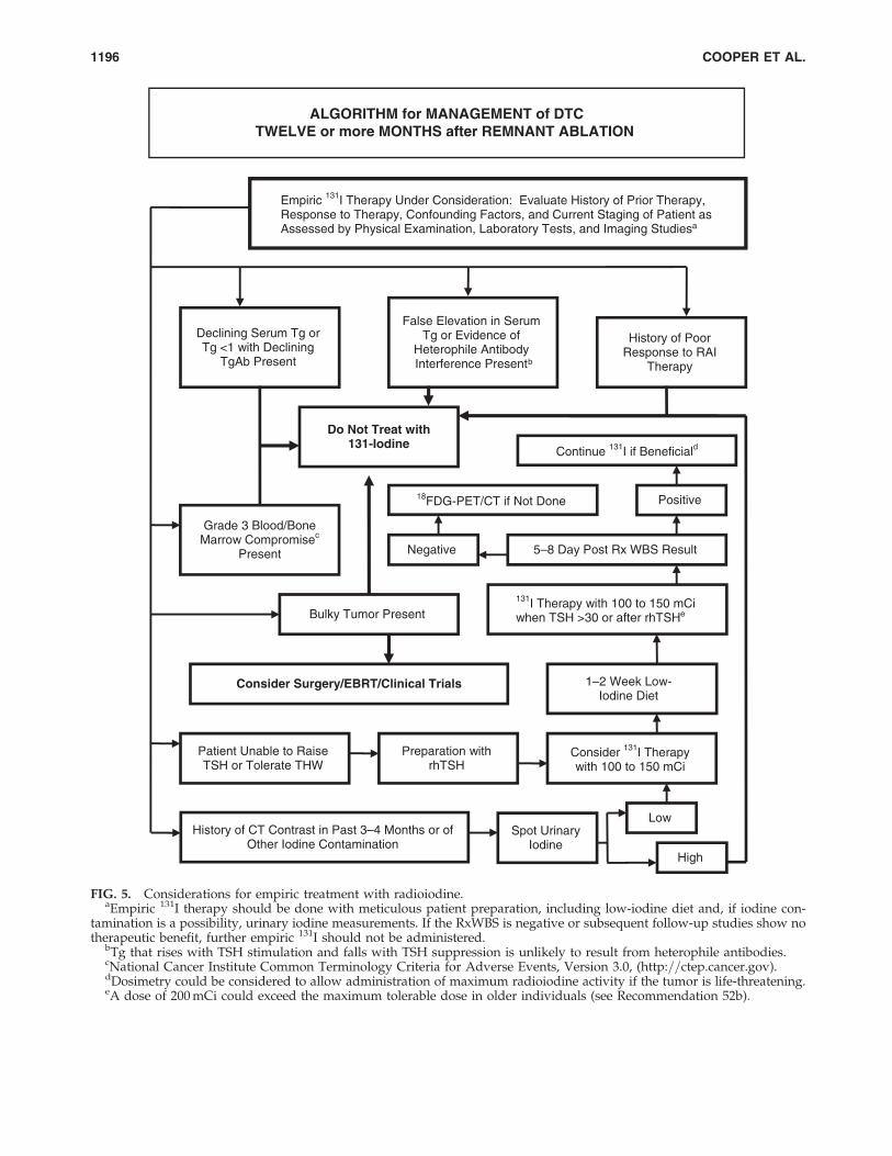
ALGORITHM for MANAGEMENT of DTCTWELVE or more MONTHS after REMNANT ABLATION
Declining Serum Tg or Tg <1 with Declining
TgAb Present
History of Poor Response to RAI
Therapy
Bulky Tumor Present
Grade 3 Blood/Bone Marrow Compromisec
Present
Do Not Treat with131-Iodine
Consider Surgery/EBRT/Clinical Trials
Patient Unable to Raise TSH or Tolerate THW
History of CT Contrast in Past 3–4 Months or of Other Iodine Contamination
Spot Urinary Iodine
Preparation with rhTSH
Consider 131I Therapy with 100 to 150 mCi
1–2 Week Low- Iodine Diet
Low
High
5–8 Day Post Rx WBS Result Negative
Positive
Continue 131I if Beneficiald
False Elevation in Serum Tg or Evidence of
Heterophile Antibody Interference Present b
Empiric 131I Therapy Under Consideration: Evaluate History of Prior Therapy, Response to Therapy, Confounding Factors, and Current Staging of Patient as Assessed by Physical Examination, Laboratory Tests, and Imaging Studiesa
131I Therapy with 100 to 150 mCi when TSH >30 or after rhTSH e
18FDG-PET/CT if Not Done
FIG. 5. Considerations for empiric treatment with radioiodine.aEmpiric 131I therapy should be done with meticulous patient preparation, including low-iodine diet and, if iodine con-
tamination is a possibility, urinary iodine measurements. If the RxWBS is negative or subsequent follow-up studies show notherapeutic benefit, further empiric 131I should not be administered.
bTg that rises with TSH stimulation and falls with TSH suppression is unlikely to result from heterophile antibodies.cNational Cancer Institute Common Terminology Criteria for Adverse Events, Version 3.0, (http:==ctep.cancer.gov).dDosimetry could be considered to allow administration of maximum radioiodine activity if the tumor is life-threatening.eA dose of 200 mCi could exceed the maximum tolerable dose in older individuals (see Recommendation 52b).
1196 COOPER ET AL.
significant tumor reduction, then RAI therapy should berepeated until the tumor has been eradicated or the tumorno longer responds to treatment. The risk of repeatedtherapeutic doses of RAI must be balanced against uncer-tain long-term benefits. Recommendation rating: C
& RECOMMENDATION 77In the absence of structurally evident disease, stimulatedserum Tg<10 ng=mL with thyroid hormone withdrawal or<5 ng=mL with rhTSH can be followed with continued LT4
therapy alone, reserving additional therapies for thosepatients with rising serum Tg levels over time or otherevidence of structural disease progression. Recommenda-tion rating: C
[C27] What is the management of patients with a negativeRxWBS?
& RECOMMENDATION 78(a) If an empiric dose (100–200 mCi) of RAI fails to localize
the persistent disease, 18FDG-PET=CT scanning shouldbe considered, especially in patients with unstimulatedserum Tg levels >10–20 ng=mL or in those with ag-gressive histologies, in order to localize metastatic le-sions that may require treatment or continued closeobservation (425,426). Recommendation rating: B
Stimulation with endogenous TSH following thyroxinewithdrawal or rhTSH (316) and CT fusion (427) may mini-mally enhance the sensitivity and specificity of 18FDG-PETscanning.
(b) Tg-positive, RxWBS-negative patients with diseasethat is incurable with surgery and is structurally evi-dent or visualized on 18FDG-PET=CT scan can bemanaged with thyroid hormone suppression therapy,external beam radiotherapy, chemotherapy, radio-frequency ablation, chemo-embolization, or monitor-ing without additional therapy if stable. Clinical trialsshould also be considered. Recommendation rating: C
& RECOMMENDATION 79Tg-positive, RxWBS-negative patients with no structuralevidence of disease can be followed with serial structuralimaging studies and serial Tg measurements, with bothperformed more frequently if the Tg level is rising. Whenand how often to repeat 18FDG-PET=CT imaging in thissetting is less certain. Recommendation rating: C
[C28] What is the role of external beam radiotherapyin treatment of metastatic disease?
& RECOMMENDATION 80External beam radiation should be used in the managementof unresectable gross residual or recurrent cervical disease,painful bone metastases, or metastatic lesions in criticallocations likely to result in fracture, neurological, or com-pressive symptoms that are not amenable to surgery (e.g.,vertebral metastases, CNS metastases, selected mediastinalor subcarinal lymph nodes, pelvic metastases) (277). Re-commendation rating: B
[D1] WHAT ARE DIRECTIONS FOR FUTURE RESEARCH?
[D2] Novel therapies and clinical trials
While surgery and the judicious use of RAI, as described inthese guidelines, is sufficient treatment for the majority ofpatients with DTC, a minority of these patients experienceprogressive, life-threatening growth and metastatic spread ofthe disease. The recent explosion of knowledge regarding themolecular and cellular pathogenesis of cancer has led to thedevelopment of a range of targeted therapies, now undergoingclinical evaluation. Efficacy has already been demonstrated forseveral agents in phase II studies, including axitinib, motesanib,sorafenib, pazopanib, and thalidomide, whereas many othersare in ongoing trials. Randomized phase III trials to demon-strate improved survival, improved progression free survival,or superiority of one therapy over another have not been per-formed, however, and none of these drugs have been specifi-cally approved for treatment of metastatic thyroid carcinoma.These therapies can be grouped into a number of categories.
[D3] Inhibitors of oncogenic signaling pathways. Tyrosinekinase inhibitors of interest in thyroid carcinoma usually targettransmembrane tyrosine kinase receptors that initiate signal-ing through the MAP kinase pathway. This signaling pathwayis activated in the majority of PTCs. Inhibitors of RET, RAS,RAF, and MEK kinases target various members of the samesignaling pathway. Several of these agents are in developmentwith several clinical trials completed or underway. Specificoncogene targeting for follicular thyroid cancer and Hurthle cellcancer awaits better understanding of the pathways involvedin initiation of these tumor types, although responses in pa-tients with these subtypes have been reported in clinical trials.
[D4] Modulators of growth or apoptosis. Key componentsof growth and apoptotic pathways are targeted by PPARgactivators, including COX2 inhibitors; rexinoids, which acti-vate RXR; bortezomib, which inactivates the cancer pro-teasome; and derivatives of geldanomycin, which target thehsp-90 protein. Clinical trials in thyroid cancer of each of theseagents are available.
[D5] Angiogenesis inhibitors. Targeting of vascular en-dothelial growth factor (VEGF) receptors and other membersof the signaling cascades responsible for neoangiogenesis maylimit the growth of cancers by restricting their blood supply.Many of the kinase inhibitors that have been studied to dateare very potent inhibitors of the tyrosine kinase of the VEGFreceptors. Trials of several of these agents are currently un-derway in all subtypes of thyroid cancer.
[D6] Immunomodulators. Stimulation of the immune re-sponse to cancer may be achieved by augmenting the activityof antigen-presenting dendritic cells. This approach hasshown possible benefits in phase I clinical trials, but has notyet been studied in thyroid cancer. The apparent immuno-genicity of thyroid cells makes this an attractive approach forfuture clinical trials.
[D7] Gene therapy. Preclinical studies have demon-strated some efficacy in thyroid cancer cell lines. Approachesinclude introducing toxic genes under the control of thyroid-specific promoters, or restoration of the p53 tumor suppressor
REVISED ATA THYROID CANCER GUIDELINES 1197
gene in anaplastic thyroid cancer cell lines. Problems withgene delivery limit the clinical utility of these approaches,which have not yet reached clinical trials in thyroid cancer.
Each of these targeted approaches holds promise for ourfuture ability to treat patients with life-threatening diseaseunresponsive to traditional therapy. In the meantime, forappropriate patients, entry into one of the available clinicaltrials may be an attractive option.
[D8] Better understanding of the long-term risks of RAI
With the more widespread use of RAI in the managementof thyroid cancer, and the normal life expectancy of mostpatients with the disease, it is imperative that we have a betterunderstanding of the long-term risks associated with its use.Research that focuses on how to minimize the impact of RAIon the salivary glands in order to prevent sialadenitis andxerostomia would provide a significant benefit to patients. Abetter understanding of the long-term effects of RAI on re-productive issues in men and women is also an importanttopic. Finally, while the risk of second malignancies appearssmall following the usual activities of RAI used for remnantablation, we need better understanding of the long-term risksfor salivary gland tumors, bladder tumors, and colon cancerswhen repeated doses of RAI are needed in young patientswho are potentially long-term survivors of thyroid cancer.
[D9] Clinical significance of persistent lowlevels of serum Tg
After initial surgery and RAI therapy some patients willhave persistently detectable stimulated serum Tg when eval-uated 9–12 months later. Most of these patients have stimu-lated Tg levels in the range of 1–10 ng=mL, levels typicallyassociated with a small volume of tissue. Some of these patientsdemonstrate a subsequent spontaneous fall in Tg over time,others remain stable, while still others demonstrate rising Tglevels. The optimal management of these patients is unknown.How often should they undergo neck US or stimulated serumTg testing? Will sensitive Tg assays combined with neck USreplace stimulation testing? Which (if any) of these patientsshould undergo chest CT, PET, or empiric RAI therapy? Canwe improve our abilities to predict and monitor which patientsare likely to be harmed by their disease as opposed to thosewho will live unaffected by theirs? Does metastatic disease insmall local lymph nodes have the potential to metastasize todistant sites during observation while on TSH suppressiontherapy? The current impetus to test and treat all of these pa-tients is based on the argument that early diagnosis may lead toearly treatment of residual disease when treatment is morelikely to be effective, as opposed to less effective treatmentwhen the tumor is more bulky, more extensive, or has spread toinoperable locations. However, there is no current proof thataggressive treatment of minimal residual disease improvespatient outcome. This is brought into focus by the fact that onlyabout 5% of all PTC patients die of their disease, yet 15–20% oflow-risk PTC patients are likely to have persistent diseasebased on persistent measurable Tg with stimulation testing.
[D10] The problem of Tg antibodies
Anti-Tg antibodies are a common clinical problem inpatients with DTC (305). The presence of these antibodies
usually interferes with serum Tg measurement and recoveryassays do not appear to accurately predict this interference(305,428). Decreasing antibody levels are correlated with‘‘disease-free’’ status while increasing levels suggest persistentdisease (306,429). However, there are clear exceptions to this‘‘rule.’’ These patients are therefore a challenge to manage orstudy because one often can not be certain of their diseasestatus. This problem limits definitive investigation which, inturn, hampers development of evidence-based guidelinessuch as these to assist clinicians. Measurement of Tg mRNA inthe blood may be a sensitive marker for persistent thyroidcells even in the presence of anti-Tg antibodies (430–432), butRNA extraction is not well standardized and some studiesquestion the specificity of this marker (433,434). Futurestudies optimizing the measurement of Tg mRNA and per-haps other thyroid-related substances in blood from DTCpatients with anti-Tg antibodies are needed to better monitorthis challenging subgroup of DTC patients. This goal wouldalso be enhanced by development of Tg assays that havelimited interference by anti-Tg antibodies and by methods toclear anti-Tg antibodies prior to Tg measurement.
[D11] Small cervical lymph node metastases
The rates of cervical lymph node metastases generally rangefrom about 20% to 50% in most large series of DTC, with higherrates in children or when micrometastases are considered. Thelocation and number of lymph node metastases is often diffi-cult to identify before, during, or after surgery, especially mi-crometastases. Although postoperative 131I given to ablate thethyroid remnant undoubtedly destroys some micrometastases,the most common site of recurrence is in cervical lymph nodes,which comprise the majority of all recurrences. Future researchmust consider the dilemma of minimizing iatrogenic patientharm versus preventing cancer morbidity and (perhaps) mor-tality. Perhaps techniques will be developed to safely removeor destroy small cervical nodal metastases, which in some caseswould otherwise progress to overt, clinically significant me-tastases. Conversely, the clinical significance of very small(<0.5 cm) nodal metastases needs to be clarified by long-termfollow-up studies. Development of a cost-effective method todetermine which metastases can be safely followed withoutintervention would be of great benefit.
[D12] Improved risk stratification
Current risk stratification schemes rely almost exclusivelyon clinical, pathological, and radiological data obtained dur-ing the initial evaluation and therapy of the patient. However,none of the commonly used risk stratification schemes ade-quately incorporate the prognostic implications of the verydetailed pathological descriptions that are provided (e.g.,various histological subtypes of thyroid cancer, frequent mi-toses, areas of tumor necrosis, minor degrees of extrathyroidalextension, or capsular invasion) or the molecular character-istics of the primary tumor. Furthermore, current stagingsystems are static representations of the patient at the time ofpresentation and are not easily modifiable over time as newdata become available during follow-up. Therefore, a riskstratification system that incorporates all the important in-formation available at presentation and also evolves over timeas new data become available would be useful in providing
1198 COOPER ET AL.
ongoing risk assessments that would optimize managementthroughout the life of the patient.
Acknowledgments
The taskforce wishes to thank Ms. Bobbi Smith, ExecutiveDirector, American Thyroid Association, and Ms. SheriSlaughter, Assistant to the Taskforce, for their constant helpand support. We also wish to thank Sally Carty, M.D., Quan-Yang Duh, M.D., Gregory Randolph, M.D., David Steward,M.D., David Terris, M.D., Ralph Tufano, M.D., and RobertUdelsman, M.D., for their help in developing recommenda-tions related to central neck dissection.
Disclaimer
It is our goal in formulating these guidelines, and the ATA’sgoal in providing support for the development of theseguidelines, that they assist in the clinical care of patients, andshare what we believe is current, rational, and optimal med-ical practice. In some circumstances, it may be apparent thatthe level of care recommended may be best provided in lim-ited centers with specific expertise. Finally, it is not the intentof these guidelines to replace individual decision making, thewishes of the patient or family, or clinical judgment.
Disclosure Statement
These guidelines were funded by the American ThyroidAssociation without support from any commercial sources.
GMD is a consultant for MedTronic ENT. BRH has receivedhonoraria from Genzyme and grant=research support fromVeracyte. RTK has received grant=research support fromGenzyme, Bayer-Onyx, Eisai, and Veracyte; is a consultant forGenzyme, Bayer-Onyx, Abbott, and Veracyte; and is on theSpeakers Bureau for Genzyme and Abbott. He has received nohonoraria for commercial speaking since November 2006 andall commercial consulting since that time has been approved bythe ATA Board of Directors, the ATA Ethics Committee, andhas been without financial compensation. SLL has receivedgrant=research support from Bayer and is a consultant forAbbott, Onyx, and Bayer. SJM has received grant=researchsupport from Veracyte and has been a CME speaker for Gen-zyme. ELM is on the Speakers Bureau for Genzyme. FP hasreceived grant=research support from Amgen, Exelixis, andAstraZeneca and is a consultant and on the Speakers Bureaufor Genzyme. MS has received grant=research support fromGenzyme, Amgen, AstraZeneca, Bayer, Exelixis, and Eisai; is aconsultant for Genzyme, AstraZeneca, Bayer, and Exelixis; andis on the Speakers Bureau for Genzyme, AstraZeneca, andExelixis. SIS has received grant=research support from Gen-zyme, Amgen, AstraZeneca, and Eisai; is a consultant forAstraZeneca, Eisai, Exelixis, Plexxikon, Oxigene, Semalore,Celgene, and Eli Lily; is on the Speakers Bureau for Genzyme;and has received honoraria from Abbott. DLS has receivedgrant=research support from Veracyte, Wyeth, Astra-Zeneca,and Gyrus. RMT is a consultant for Genzyme, Abbott, and EliLily, and has received honoraria from Genzyme and Abbott.DSC and BM report that no competing financial interests exist.
References
1. Tunbridge WMG, Evered DC, Hall R, Appleton D, BrewisM, Clark F, Evans JG, Young E, Bird T, Smith PA 1977 The
spectrum of thyroid disease in a community: the Whick-ham Survey. Clin Endocrinol (Oxf ) 7:481–493.
2. Vander JB, Gaston EA, Dawber TR 1968 The significance ofnontoxic thyroid nodules. Ann Intern Med 69:537–540.
3. Tan GH, Gharib H 1997 Thyroid incidentalomas: manage-ment approaches to nonpalpable nodules discovered inci-dentally on thyroid imaging. Ann Intern Med 126:226–231.
4. Hegedus L 2004 Clinical practice. The thyroid nodule. NEngl J Med 351:1764–1771.
5. Mandel SJ 2004 A 64-year-old woman with a thyroid nod-ule. JAMA 292:2632–2642.
6. Sherman SI 2003 Thyroid carcinoma. Lancet 361:501–511.7. Jemal A, Siegel R, Ward E, Hao Y, Xu J, Thun MJ 2009
Cancer statistics, 2009. CA Cancer J Clin. Published onlinebefore print May 27, 2009.
8. Davies L, Welch HG 2006 Increasing incidence of thyroidcancer in the United States, 1973–2002. JAMA 295:2164–2167.
9. Leenhardt L, Bernier MO, Boin-Pineau MH, Conte DB,Marechaud R, Niccoli-Sire P, Nocaudie M, Orgiazzi J,Schlumberger M, Wemeau JL, Cherie-Challine L, De Va-thaire F 2004 Advances in diagnostic practices affect thyroidcancer incidence in France. Eur J Endocrinol 150:133–139.
10. Singer PA, Cooper DS, Daniels GH, Ladenson PW,Greenspan FS, Levy EG, Braverman LE, Clark OH,McDougall IR, Ain KV, Dorfman SG 1996 Treatmentguidelines for patients with thyroid nodules and well-dif-ferentiated thyroid cancer. American Thyroid Association.Arch Intern Med 156:2165–2172.
11. American Association of Clinical Endocrinologists 2001AACE=AAES medical=surgical guidelines for clinicalpractice: management of thyroid carcinoma. Endocr Pract7:202–220.
12. British Thyroid Association and Royal College of Physi-cians. 2007 Guidelines for the management of thyroidcancer, 2nd Edition. www.british-thyroid-association.org.
13. National Comprehensive Cancer Network. 2009 Thyroidcarcinoma. www.nccn.org=professionals=physician_gls=PDF=thyroid.pdf. Accessed January 28, 2009.
14. Pacini F, Schlumberger M, Dralle H, Elisei R, Smit JW,Wiersinga W 2006 European consensus for the manage-ment of patients with differentiated thyroid carcinoma ofthe follicular epithelium. Eur J Endocrinol 154:787–803.
15. Luster M, Clarke SE, Dietlein M, Lassmann M, Lind P,Oyen WJ, Tennvall J, Bombardieri E 2008 Guidelines forradioiodine therapy of differentiated thyroid cancer. Eur JNucl Med Mol Imaging 35:1941–1959.
16. Cooper DS, Doherty GM, Haugen BR, Kloos RT, Lee SL,Mandel SJ, Mazzaferri EL, McIver B, Sherman SI, Tuttle RM2006 The American Thyroid Association Guidelines Task-force. Management guidelines for patients with thyroid nod-ules and differentiated thyroid cancer. Thyroid 16:109–142.
17. U.S. Preventive Services Task Force Ratings: Strength ofRecommendations and Quality of Evidence. Guide toClinical Preventive Services, Third Edition: Periodic Up-dates, 2000–2003. Agency for Healthcare Research andQuality, Rockville, MD.
18. Marqusee E, Benson CB, Frates MC, Doubilet PM, LarsenPR, Cibas ES, Mandel SJ 2000 Usefulness of ultrasonogra-phy in the management of nodular thyroid disease. AnnIntern Med 1339:696–700.
19. Hagag P, Strauss S, Weiss M 1998 Role of ultrasound-guided fine-needle aspiration biopsy in evaluation of non-palpable thyroid nodules. Thyroid 8:989–995.
REVISED ATA THYROID CANCER GUIDELINES 1199
20. Are C, Hsu JF, Ghossein RA, Schoder H, Shah JP, Shaha AR2007 Histological aggressiveness of fluorodeoxyglucosepositron-emission tomogram (FDG-PET)-detected inciden-tal thyroid carcinomas. Ann Surg Oncol 14:3210–3215.
21. Bogsrud TV, Karantanis D, Nathan MA, Mullan BP, Wi-seman GA, Collins DA, Kasperbauer JL, Strome SE, Read-ing CC, Hay ID, Lowe VJ 2007 The value of quantifying18F-FDG uptake in thyroid nodules found incidentally onwhole-body PET-CT. Nucl Med Commun 28:373–381.
22. Kang KW, Kim SK, Kang HS, Lee ES, Sim JS, Lee IG, JeongSY, Kim SW 2003 Prevalence and risk of cancer of focalthyroid incidentaloma identified by 18F-fluorodeox-yglucose positron emission tomography for metastasisevaluation and cancer screening in healthy subjects. J ClinEndocrinol Metab 88:4100–4104.
23. Choi JY, Lee KS, Kim HJ, Shim YM, Kwon OJ, Park K, BaekCH, Chung JH, Lee KH, Kim BT 2006 Focal thyroid lesionsincidentally identified by integrated 18F-FDG PET=CT:clinical significance and improved characterization. J NuclMed 47:609–615.
24. Curtis RE, Rowlings PA, Deeg HJ, Shriner DA, Socie G,Travis LB, Horowitz MM, Witherspoon RP, Hoover RN,Sobocinski KA, Fraumeni JF, Boice JD, Schoch HG, Sale GE,Storb R, Travis WD, Kolb HJ, Gale RP, Passweg JR 1997Solid cancers after bone marrow transplantation. N Engl JMed 336:897–904.
25. Pacini F, Vorontsova T, Demidchik E, Molinaro E, Agate, L,Romei C, Shavrova E, Cherstvoy ED, Ivashkevitch Y, Ku-chinskaya E, Schlumberger M, Ronga G, Filesi M, PincheraA 1997 Post-Chernobyl thyroid carcinoma in Belarus chil-dren and adolescents: comparison with naturally occurringthyroid carcinoma in Italy and France. J Clin EndocrinolMetab 81:3563–3569.
26. Boelaert K, Horacek J, Holder RL, Watkinson JC, SheppardMC, Franklyn JA 2006 Serum thyrotropin concentration asa novel predictor of malignancy in thyroid nodules inves-tigated by fine-needle aspiration. J Clin Endo Metab91:4295–4301.
27. Hall TL, Layfield LJ, Philippe A, Rosenthal D 1989 Sourcesof diagnostic error in fine needle aspiration of the thyroid.Cancer 63:718–725.
28. Alexander EK, Heering JP, Benson CB, Frates MC, DoubiletPM, Cibas ES, Marqusee E 2002 Assessment of non-diagnostic ultrasound-guided fine needle aspiration ofthyroid nodules. J Clin Endocrinol Metab 87:4924–4927.
29. Brander A, Viikinkoski P, Tuuhea J, Voutilainen L, Kivi-saari L 1992 Clinical versus ultrasound examination of thethyroid gland in common clinical practice. J Clin Ultra-sound 20:37–42.
30. Tan GH, Gharib H, Reading CC 1995 Solitary thyroidnodule. Arch Intern Med 155:2418–2423.
31. Singh B, Shaha AR, Trivedi H, Carew JF, Poluri A, Shah JP1999 Coexistent Hashimoto’s thyroiditis with papillarythyroid carcinoma: impact on presentation, management,and outcome. Surgery 126:1070–1077.
32. Repplinger D, Bargren A, Zhang YW, Adler JT, HaymartM, Chen H 2008 Is Hashimoto’s thyroiditis a risk factor forpapillary thyroid cancer? J Surg Res 150:49–52.
33. Pacini F, Pinchera A, Giani C, Grasso L, Doveri F, BaschieriL 1980. Serum thyroglobulin in thyroid carcinoma andother thyroid disorders. J Endocrinol Invest 3:283–292.
34. Elisei R, Bottici V, Luchetti F, Di Coscio G, Romei C, GrassoL, Miccoli P, Iacconi P, Basolo F, Pinchera A, Pacini F 2004Impact of routine measurement of serum calcitonin on the
diagnosis and outcome of medullary thyroid cancer: ex-perience in 10,864 patients with nodular thyroid disorders.J Clin Endocrinol Metab 89:163–168.
35. Hahm JR, Lee MS, Min YK, Lee MK, Kim KW, Nam SJ,Yang JH, Chung JH 2001 Routine measurement of serumcalcitonin is useful for early detection of medullary thyroidcarcinoma in patients with nodular thyroid diseases.Thyroid 11:73–80.
36. Niccoli P, Wion-Barbot N, Caron P, Henry JF, de Micco C,Saint Andre JP, Bigorgne JC, Modigliani E, Conte-Devolx B1997 Interest of routine measurement of serum calcitonin:study in a large series of thyroidectomized patients. TheFrench Medullary Study Group. J Clin Endocrinol Metab82:338–341.
37. Costante G, Meringolo D, Durante C, Bianchi D, Nocera M,Tumino S Crocetti U, Attard M, Maranghi M, TorlontanoM, Filetti S 2007 Predictive value of serum calcitonin levelsfor preoperative diagnosis of medullary thyroid carcinomain a cohort of 5817 consecutive patients with thyroid nod-ules. J Clin Endocrinol Metab 92:450–455.
38. Cheung K, Roman SA, Wang TS, Walker HD, Sosa JA 2008Calcitonin measurement in the evaluation of thyroid nod-ules in the United States: a cost-effectiveness and decisionanalysis. J Clin Endocrinol Metab 93:2173–2180.
39. Gagel RF, Hoff AO, Cote GJ 2005 Medullary thyroid car-cinoma. In Werner and Ingbar’s The Thyroid. LippincottWilliams and Wilkins, Philadelphia, pp 967–988.
40. Danese D, Sciacchitano S, Farsetti A, Andreoli M, Ponte-corvi A 1998 Diagnostic accuracy of conventional versussonography-guided fine-needle aspiration biopsy of thy-roid nodules. Thyroid 8:15–21.
41. Carmeci C, Jeffrey RB, McDougall IR, Nowels KW, WeigelRJ 1998 Ultrasound-guided fine-needle aspiration biopsy ofthyroid masses. Thyroid 8:283–289.
42. Baloch ZW, LiVolsi VA, Asa SL, Rosai J, Merino MJ, Ran-dolph G, Vielh P, DeMay RM, Sidawy MK, Frable WJ 2008Diagnostic terminology and morphologic criteria for cyto-logic diagnosis of thyroid lesions: a synopsis of the NationalCancer Institute Thyroid Fine-Needle Aspiration State of theScience Conference. Diagn Cytopathol 36:425–437.
43. Leenhardt L, Hejblum G, Franc B, Fediaevsky LD, Delbot T,Le Guillouzic D, Menegaux F, Guillausseau C, Hoang C,Turpin G, Aurengo A 1999 Indications and limits of ultra-sound-guided cytology in the management of nonpalpablethyroid nodules. J Clin Endocrinol Metab 84:24–28.
44. Papini E, Guglielmi R, Bianchini A, Crescenzi A, TaccognaS, Nardi F, Panunzi C, Rinaldi R, Toscano V, Pacella CM2002 Risk of malignancy in nonpalpable thyroid nodules:predictive value of ultrasound and color-Doppler features.J Clin Endocrinol Metab 87:1941–1946.
45. Nam-Goong IS, Kim HY, Gong G, Lee HK, Hong SJ, KimWB, Shong YK 2004 Ultrasonography-guided fine-needleaspiration of thyroid incidentaloma: correlation withpathological findings. Clin Endocrinol (Oxf ) 60:21–28.
46. Cappelli C, Castellano M, Pirola I, Cumetti D, Agosti B,Gandossi E, Agabiti Rosei E 2007 The predictive value ofultrasound findings in the management of thyroid nodules.QJM 100:29–35.
47. Frates MC, Benson CB, Doubilet PM, Kunreuther E, Con-treras M, Cibas ES, Orcutt J, Moore FD Jr, Larsen PR,Marqusee E, Alexander EK 2006 Prevalence and distribu-tion of carcinoma in patients with solitary and multiplethyroid nodules on sonography. J Clin Endocrinol Metab91:3411–3417.
1200 COOPER ET AL.
48. Moon WJ, Jung SL, Lee JH, Na DG, Baek JH, Lee YH, Kim J,Kim HS, Byun JS, Lee DH; Thyroid Study Group, KoreanSociety of Neuro- and Head and Neck Radiology 2008 Be-nign and malignant thyroid nodules: US differentiation—multicenter retrospective study. Radiology 247:762–770.
49. Jeh SK, Jung SL, Kim BS, Lee YS 2007 Evaluating the degreeof conformity of papillary carcinoma and follicular carci-noma to the reported ultrasonographic findings of malig-nant thyroid tumor. Korean J Radiol 8:192–197.
50. Machens A, Holzhausen HJ, Dralle H 2005 The prognosticvalue of primary tumor size in papillary and follicularthyroid carcinoma. Cancer 103:2269–2273.
51. Moon WJ, Kwag HJ, Na DG 2009 Are there any specificultrasound findings of nodular hyperplasia (‘‘leave mealone’’ lesion) to differentiate it from follicular adenoma?Acta Radiologica 50:383–388.
52. Bonavita JA, Mayo J, Babb J, Bennett G, Oweity T, MacariM, Yee J 2009 Pattern recognition of benign nodules at ul-trasound of the thyroid: which nodules can be left alone?AJR Am J Roentgenol 193:207–213.
53. Rago T, Santini F, Scutari M, Pinchera A, Vitti P 2007Elastography: new developments in ultrasound for pre-dicting malignancy in thyroid nodules. J Clin EndocrinolMetab 92:2917–2922.
54. Noguchi S, Yamashita H, Uchino S, Watanabe S 2008 Pa-pillary microcarcinoma. World J Surg 32:747–753.
55. Wada N, Duh QY, Sugino K, Iwasaki H, Kameyama K,Mimura T, Ito K, Takami H, Takanashi Y 2003 Lymphnode metastasis from 259 papillary thyroid microcarci-nomas: frequency, pattern of occurrence and recurrence,and optimal strategy for neck dissection. Ann Surg 237:
399–407.56. Ito Y, Tomoda C, Uruno T, Takamura Y, Miya A, Ko-
bayashi K, Matsuzuka F, Kuma K, Miyauchi A 2004 Pre-operative ultrasonographic examination for lymph nodemetastasis: usefulness when designing lymph node dis-section for papillary microcarcinoma of the thyroid. WorldJ Surg 28:498–501.
57. Hemminki K, Eng C, Chen B 2005 Familial risks for non-medullary thyroid cancer. J Clin Endocrinol Metab90:5747–5753.
58. Schneider AB, Ron E, Lubin J, Stovall M, Gierlowski TC1993 Dose-response relationships for radiation-inducedthyroid cancer and thyroid nodules: evidence for the pro-longed effects of radiation on the thyroid. J Clin EndocrinolMetab 77:362–369.
59. Shibata Y, Yamashita S, Masyakin VB, Panasyuk GD, Na-gataki S 2001 15 years after Chernobyl: new evidence ofthyroid cancer. Lancet 358:1965–1966.
60. Braga M, Cavalcanti TC, Collaco LM, Graf H 2001 Efficacyof ultrasound-guided fine-needle aspiration biopsy in thediagnosis of complex thyroid nodules. J Clin EndocrinolMetab 86:4089–4091.
61. Redman R, Zalaznick H, Mazzaferri EL, Massoll NA 2006The impact of assessing specimen adequacy and number ofneedle passes for fine-needle aspiration biopsy of thyroidnodules. Thyroid 16:55–60.
62. Baloch ZW, Tam D, Langer J, Mandel S, LiVolsi VA, GuptaPK 2000 Ultrasound-guided fine-needle aspiration biopsyof the thyroid: role of on-site assessment and multiple cy-tologic preparations. Diagn Cytopathol 23:425–429.
63. de los Santos ET, Keyhani-Rofagha S, Cunningham JJ,Mazzaferri EL 1990 Cystic thyroid nodules. The dilemma ofmalignant lesions. Arch Intern Med 150:1422–1427.
64. Yeh MW, Demircan O, Ituarte P, Clark OH 2004 False-negative fine-needle aspiration cytology results delaytreatment and adversely affect outcome in patients withthyroid carcinoma. Thyroid 14:207–215.
65. Gharib H, Goellner JR, Johnson DA 1993 Fine-needle as-piration cytology of the thyroid. A 12-year experience with11,000 biopsies. Clin Lab Med 13:699–709.
66. Tuttle RM, Lemar H, Burch HB 1998 Clinical features as-sociated with an increased risk of thyroid malignancy inpatients with follicular neoplasia by fine-needle aspiration.Thyroid 8:377–383.
67. Tyler DS, Winchester DJ, Caraway NP, Hickey RC, EvansDB 1994 Indeterminate fine-needle aspiration biopsy of thethyroid: identification of subgroups at high risk for inva-sive carcinoma. Surgery 116:1054–1060.
68. Kelman AS, Rathan A, Leibowitz J, Burstein DE, Haber RS2001 Thyroid cytology and the risk of malignancy in thy-roid nodules: importance of nuclear atypia in indetermi-nate specimens. Thyroid 11:271–277.
69. Bartolazzi A, Orlandi F, Saggiorato E, Volante M, Arecco F,Rossetto R, Palestini N, Ghigo E, Papotti M, Bussolati G,Martegani MP, Pantellini F, Carpi A, Giovagnoli MR,Monti S, Toscano V, Sciacchitano S, Pennelli GM, Mian C,Pelizzo MR, Rugge M, Troncone G, Palombini L, Chiap-petta G, Botti G, Vecchione A, Bellocco R; Italian ThyroidCancer Study Group (ITCSG) 2008 Galectin-3-expressionanalysis in the surgical selection of follicular thyroid nod-ules with indeterminate fine-needle aspiration cytology: aprospective multicentre study. Lancet Oncol 9:543–549.
70. Segev DL, Clark DP, Zeiger MA, Umbricht C 2003 Beyondthe suspicious thyroid fine needle aspirate. A review. ActaCytol 47:709–722.
71. Haugen BR, Woodmansee WW, McDermott MT 2002Towards improving the utility of fine-needle aspirationbiopsy for the diagnosis of thyroid tumors. Clin Endo 56:
281–290.72. Sapio MR, Posca D, Raggioli A, Guerra A, Marotta V,
Deandrea M, Motta M, Limone PP, Troncone G, Caleo A,Rossi G, Fenzi G, Vitale M 2007 Detection of RET=PTC,TRK and BRAF mutations in preoperative diagnosis ofthyroid nodules with indeterminate cytological findings.Clin Endocrinol (Oxf ) 66:678–683.
73. Nikiforov YE, Nikiforov YE, Steward DL, Robinson-SmithTM, Haugen BR, Klopper JP, Zhu Z, Fagin JA, Falciglia M,Weber K, Nikiforova MN 2009 Molecular testing for mu-tations in improving the fine-needle aspiration diagnosis ofthyroid nodules. J Clin Endocrinol Metab 94:2092–2098.
74. Franco C, Martınez V, Allamand JP, Medina F, GlasinovicA, Osorio M, Schachter D 2009 Molecular markers in thy-roid fine-needle aspiration biopsy: a prospective study.Appl Immunohistochem Mol Morphol 17:211–215.
75. Mitchell JC, Grant F, Evenson AR, Parker JA, HasselgrenPO, Parangi S 2005. Preoperative evaluation of thyroidnodules with 18FDG-PET=CT. Surgery 138:1166–1174;discussion 1174–1175.
76. de Geus-Oei LF, Pieters GF, Bonenkamp JJ, Mudde AH,Bleeker-Rovers CP, Corstens FH, Oyen WJ 2006 18F-FDGPET reduces unnecessary hemithyroidectomies for thyroidnodules with inconclusive cytologic results. J Nucl Med47:770–775.
77. Kim JM, Ryu JS, Kim TY, Kim WB, Kwon GY, Gong G,Moon DH, Kim SC, Hong SJ, Shong YK 2007 18F-fluorodeoxyglucose positron emission tomography doesnot predict malignancy in thyroid nodules cytologically
REVISED ATA THYROID CANCER GUIDELINES 1201
diagnosed as follicular neoplasm. J Clin Endocrinol Metab92:1630–1634.
78. Sebastianes FM, Cerci JJ, Zanoni PH, Soares J Jr, ChibanaLK, Tomimori EK, de Camargo RY, Izaki M, Giorgi MC,Eluf-Neto J, Meneghetti JC, Pereira MA 2007 Role of 18F-fluorodeoxyglucose positron emission tomography in pre-operative assessment of cytologically indeterminate thyroidnodules. J Clin Endocrinol Metab 92:4485–4488.
79. Hales NW, Krempl GA, Medina JE 2008 Is there a rolefor fluorodeoxyglucose positron emission tomography=computed tomography in cytologically indeterminate thy-roid nodules? Am J Otolaryngol 29:113–118.
80. Ylagan LR, Farkas T, Dehner LP 2004 Fine needle aspira-tion of the thyroid: a cytohistologic correlation and study ofdiscrepant cases. Thyroid 14:35–41.
81. McCoy KL, Jabbour N, Ogilvie JB, Ohori NP, Carty SE, YimJH 2007 The incidence of cancer and rate of false-negativecytology in thyroid nodules greater than or equal to 4 cm insize. Surgery 142:837–844.
82. Alexander EK, Hurwitz S, Heering JP, Benson CB, FratesMC, Doubilet PM, Cibas ES, Larsen PR, Marqusee E 2003Natural history of benign solid and cystic thyroid nodules.Ann Int Med 138:315–318.
83. Asanuma K, Kobayashi S, Shingu K, Hama Y, Yokoyama S,Fujimori M, Amano J 2001 The rate of tumour growth doesnot distinguish between malignant and benign thyroidnodules. Eur J Surg 167:102–105.
84. Erdogan MF, Kamel N, Aras D, Akdogan A, Baskal N,Erdogan G 1998 Value of re-aspirations in benign nodularthyroid disease. Thyroid 8:1087–1090.
85. Orlandi A, Puscar A, Capriata E, Fideleff H 2005 Repeatedfine-needle aspiration of the thyroid in benign nodularthyroid disease: critical evaluation of long-term follow-up.Thyroid 15:274–278.
86. Papini E, Petrucci L, Guglielmi R, Panunzi C, Rinaldi R,Bacci V, Crescenzi A, Nardi F, Fabbrini R, Pacella CM 1998Long-term changes in nodular goiter: a 5-year prospectiverandomized trial of levothyroxine suppressive therapy forbenign cold thyroid nodules. J Clin Endocrinol Metab83:780–783.
87. Brauer VF, Eder P, Miehle K, Wiesner TD, Hasenclever H,Paschke R 2005 Interobserver variation for ultrasound deter-mination of thyroid nodule volumes. Thyroid 15:1169–1175.
88. Brander AE, Viikinkoski VP, Nickels JI, Kivisaari LM 2000Importance of thyroid abnormalities detected at USscreening: a 5-year follow-up. Radiology 215:801–806.
89. Oertel YC, Miyahara-Felipe L, Mendoza MG, Yu K 2007Value of repeated fine needle aspirations of the thyroid: ananalysis of over ten thousand FNAs. Thyroid 17:1061–1066.
90. Bennedbaek FN, Hegedus L 2003 Treatment of recurrentthyroid cysts with ethanol: a randomized double-blindcontrolled trial. J Clin Endocrinol Metab 88:5773–5777.
91. Valcavi R, Frasoldati A 2004 Ultrasound-guided percuta-neous ethanol injection therapy in thyroid cystic nodules.Endocr Pract 10:269–275.
92. Antonelli A, Campatelli A, Di Vito A, Alberti B, Baldi V,Salvioni G, Fallahi P, Baschieri L 1994 Comparison betweenethanol sclerotherapy and emptying with injection of salinein treatment of thyroid cysts. Clin Investig 72:971–974.
93. Verde G, Papini E, Pacella CM, Gallotti C, Delpiano S,Strada S, Fabbrini R, Bizzarri G, Rinaldi R, Panunzi C, GeiliD 1994 Ultrasound guided percutaneous ethanol injectionin the treatment of cystic thyroid nodules. Clin Endocrinol(Oxf ) 41:719–724.
94. Zelmanovitz F, Genro S, Gross JL 1998 Suppressive ther-apy with levothyroxine for solitary thyroid nodules: adouble-blind controlled clinical study and cumulativemeta-analyses. J Clin Endocrinol Metab 83:3881–3885.
95. Wemeau JL, Caron P, Schvartz C, Schlienger JL, Orgiazzi J,Cousty C, Vlaeminck-Guillem V 2002 Effects of thyroid-stimulating hormone suppression with levothyroxine inreducing the volume of solitary thyroid nodules andimproving extranodular nonpalpable changes: a random-ized, double-blind, placebo-controlled trial by the FrenchThyroid Research Group. J Clin Endocrinol Metab 87:4928–4934.
96. Castro MR, Caraballo PJ, Morris JC 2002 Effectiveness ofthyroid hormone suppressive therapy in benign solitarythyroid nodules: a meta-analysis. J Clin Endocrinol Metab87:4154–4159.
97. Rallison ML, Dobyns BM, Keating FR Jr, Rall JE, Tyler FH1975 Thyroid nodularity in children. JAMA 233:1069–1072.
98. Raab SS, Silverman JF, Elsheikh TM, Thomas PA, WakelyPE 1995 Pediatric thyroid nodules: disease demographicsand clinical management as determined by fine needle as-piration biopsy. Pediatrics 95:46–49.
99. Corrias A, Einaudi S, Chiorboli E, Weber G, Crino A, An-dreo M, Cesaretti G, de Sanctis L, Messina MF, Segni M,Cicchetti M, Vigone M, Pasquino AM, Spera S, de Luca F,Mussa GC, Bona G 2001 Accuracy of fine needle aspirationbiopsy of thyroid nodules in detecting malignancy inchildhood: comparison with conventional clinical, labora-tory, and imaging approaches. J Clin Endocrinol Metab86:4644–4648.
100. Hung W 1999 Solitary thyroid nodules in 93 children andadolescents, a 35-years experience. Horm Res 52:15–18.
101. Gharib H, Zimmerman D, Goellner JR, Bridley SM, LeBlancSM 1995 Fine-needle aspiration biopsy: Use in diagnosisand management of pediatric thyroid diseases. Endo Pract1:9–13.
102. Arda IS, Yildirim S, Demirhan B, Firat S 2001 Fine needleaspiration biopsy of thyroid nodules. Arch Dis Child85:313–317.
103. Tan GH, Gharib H, Goellner JR, van Heerden JA, Bahn RS1996 Management of thyroid nodules in pregnancy. ArchIntern Med 156:2317–2320.
104. Moosa M, Mazzaferri EL 1997 Outcome of differentiatedthyroid cancer diagnosed in pregnant women. J Clin En-docrinol Metab 82:2862–2866.
105. Mestman JH, Goodwin TM, Montoro MM 1995 Thyroiddisorders of pregnancy. Endocrinol Metab Clin North Am24:41–71.
106. Herzon FS, Morris DM, Segal MN, Rauch G, Parnell T 1994Coexistent thyroid cancer and pregnancy. Arch Otolar-yngol Head Neck Surg 120:1191–1193.
107. Mazzaferri EL, Jhiang SM 1994 Long-term impact of initialsurgical and medical therapy on papillary and follicularthyroid cancer. Am J Med 97:418–428.
108. Kuy S, Roman SA, Desai R, Sosa JA 2009 Outcomesfollowing thyroid and parathyroid surgery in pregnantwomen. Arch Surg 144:399–406.
109. Rosen IB, Korman M, Walfish PG 1997 Thyroid nodulardisease in pregnancy: current diagnosis and management.Clin Obstet Gynecol 40:81–89.
110. Hundahl SA, Fleming ID, Fremgen AM, Menck HR 1998 ANational Cancer Data Base report on 53,856 cases of thy-roid carcinoma treated in the U.S., 1985–1995. Cancer83:2638–2648.
1202 COOPER ET AL.
111. Volante M, Landolfi S, Chiusa L, Palestini N, Motta M,Codegone A, Torchio B, Papotti MG 2004 Poorly differen-tiated carcinomas of the thyroid with trabecular, insular,and solid patterns: a clinicopathologic study of 183 pa-tients. Cancer 100:950–957.
112. van Heerden JA, Hay ID, Goellner JR, Salomao D, EbersoldJR, Bergstralh EJ, Grant CS 1992 Follicular thyroid carci-noma with capsular invasion alone: a nonthreatening ma-lignancy. Surgery. 112:1130–6.
113. Sanders LE, Silverman M 1998 Follicular and Hurthle cellcarcinoma: predicting outcome and directing therapy.Surgery 124:967–974.
114. Lo CY, Chan WF, Lam KY, Wan KY 2005 Follicular thyroidcarcinoma: the role of histology and staging systems inpredicting survival. Ann Surg 242:708–715.
115. D’Avanzo A, Treseler P, Ituarte PH, Wong M, Streja L,Greenspan FS, Siperstein AE, Duh QY, Clark OH 2004.Follicular thyroid carcinoma: histology and prognosis.Cancer 100:1123–1129.
116. Hay ID, Bergstralh EJ, Goellner JR, Ebersold JR, Grant CS1993 Predicting outcome in papillary thyroid carcinoma:development of a reliable prognostic scoring system in acohort of 1779 patients surgically treated at one institutionduring 1940 through 1989. Surgery 114:1050–1057; discus-sion 1057–1058.
117. Shah MD, Hall FT, Eski SJ, Witterick IJ, Walfish PG, Free-man JL 2003 Clinical course of thyroid carcinoma after neckdissection. Laryngoscope 113:2102–2107.
118. Wang TS, Dubner S, Sznyter LA, Heller KS 2004 Incidenceof metastatic well-differentiated thyroid cancer in cervicallymph nodes. Arch Otolaryngol Head Neck Surg 130:110–113.
119. Sosa JA, Bowman HM, Tielsch JM, Powe NR, Gordon TA,Udelsman R 1998 The importance of surgeon experience forclinical and economic outcomes from thyroidectomy. AnnSurg 228:320–330.
120. Friedman M, Pacella BL, Jr 1990 Total versus subtotalthyroidectomy. Arguments, approaches, and recommen-dations. Otolaryngol Clin North Am 23:413–427.
121. Brierley JD, Panzarella T, Tsang RW, Gospodarowicz MK,O’Sullivan B 1997 A comparison of different staging sys-tems predictability of patient outcome. Thyroid carcinomaas an example. Cancer 79:2414–2423.
122. Hay ID, Thompson GB, Grant CS, Bergstralh EJ, DvorakCE, Gorman CA, Maurer MS, McIver B, Mullan BP, ObergAL, Powell CC, van Heerden JA, Goellner JR 2002 Papillarythyroid carcinoma managed at the Mayo Clinic during sixdecades (1940–1999): temporal trends in initial therapy andlong-term outcome in 2444 consecutively treated patients.World J Surg 26:879–885.
123. Lin JD, Chao TC, Huang MJ, Weng HF, Tzen KY 1998 Useof radioactive iodine for thyroid remnant ablation in well-differentiated thyroid carcinoma to replace thyroid re-operation. Am J Clin Oncol 21:77–81.
124. Esnaola NF, Cantor SB, Sherman SI, Lee JE, Evans DB 2001Optimal treatment strategy in patients with papillary thy-roid cancer: a decision analysis. Surgery 130:921–930.
125. Mazzaferri EL 1999 An overview of the management ofpapillary and follicular thyroid carcinoma. Thyroid 9:421–427.
126. Mazzaferri EL 2000 Long-term outcome of patients withdifferentiated thyroid carcinoma: effect of therapy. EndocrPract 6:469–476.
127. Cooper DS, Specker B, Ho M, Sperling M, Ladenson PW,Ross DS, Ain KB, Bigos ST, Brierley JD, Haugen BR, Klein I,
Robbins J, Sherman SI, Taylor T, Maxon HR 3rd 1998Thyrotropin suppression and disease progression in pa-tients with differentiated thyroid cancer: results from theNational Thyroid Cancer Treatment Cooperative Registry.Thyroid 8:737–744.
128. Kim TH, Yang DS, Jung KY, Kim CY, Choi MS 2003 Valueof external irradiation for locally advanced papillary thy-roid cancer. Int J Radiat Oncol Biol Phys 55:1006–1012.
129. Grebe SK, Hay ID 1996 Thyroid cancer nodal metastases:biologic significance and therapeutic considerations. SurgOncol Clin N Am 5:43–63.
130. Scheumann GF, Gimm O, Wegener G, Hundeshagen H,Dralle H 1994 Prognostic significance and surgical man-agement of locoregional lymph node metastases in papil-lary thyroid cancer. World J Surg 18:559–568.
131. Ito Y, Uruno T, Nakano K, Takamura Y, Miya A, KobayashiK, Yokozawa T, Matsuzuka F, Uchimi T, Kuwano M, MiyoshiE, Matsuura N, Kuma K, Miyauchi A 2003 An observationtrial without surgical treatment in patients with papillarymicrocarcinoma of the thyroid. Thyroid 13:381–387.
132. Chow SM, Law SC, Chan JK, Au SK, Yau S, Lau WH 2003Papillary microcarcinoma of the thyroid-Prognostic signif-icance of lymph node metastasis and multifocality. Cancer98:31–40.
133. Hay ID, Grant CS, van Heerden JA, Goellner JR, EbersoldJR, Bergstralh EJ 1992 Papillary thyroid microcarcinoma: astudy of 535 cases observed in a 50-year period. Surgery112:1139–1146; discussion 1146–1147.
134. Qubain SW, Nakano S, Baba M, Takao S, Aikou T 2002Distribution of lymph node micrometastasis in pN0 well-differentiated thyroid carcinoma. Surgery 131:249–256.
135. Arturi F, Russo D, Giuffrida D, Ippolito A, Perrotti N,Vigneri R, Filetti S 1997 Early diagnosis by genetic analysisof differentiated thyroid cancer metastases in small lymphnodes. J Clin Endocrinol Metab 82:1638–1641.
136. Solorzano CC, Carneiro DM, Ramirez M, Lee TM, IrvinGL3rd 2004 Surgeon-performed ultrasound in the man-agement of thyroid malignancy. Am Surg 70:576–580; dis-cussion 580–582.
137. Shimamoto K, Satake H, Sawaki A, Ishigaki T, FunahashiH, Imai T 1998 Preoperative staging of thyroid papillarycarcinoma with ultrasonography. Eur J Radiol 29:4–10.
138. Stulak JM, Grant CS, Farley DR, Thompson GB, vanHeerden JA, Hay ID, Reading CC, Charboneau JW 2006Value of preoperative ultrasonography in the surgicalmanagement of initial and reoperative papillary thyroidcancer. Arch Surg 141:489–494.
139. Kouvaraki MA, Shapiro SE, Fornage BD, Edeiken-MonroBS, Sherman SI, Vassilopoulou-Sellin R, Lee JE, Evans DB2003 Role of preoperative ultrasonography in the surgicalmanagement of patients with thyroid cancer. Surgery134:946–954; discussion 954–955.
140. Leboulleux S, Girard E, Rose M, Travagli JP, Sabbah N,Caillou B, Hartl DM, Lassau N, Baudin E, Schlumberger M2007 Ultrasound criteria of malignancy for cervical lymphnodes in patients followed up for differentiated thyroidcancer. J Clin Endocrinol Metab 92:3590–3594.
141. Frasoldati A, Valcavi R 2004 Challenges in neck ultraso-nography: lymphadenopathy and parathyroid glands. En-docr Pract 10:261–268.
142. Kuna SK, Bracic I, Tesic V, Kuna K, Herceg GH, Dodig D2006 Ultrasonographic differentiation of benign from ma-lignant neck lymphadenopathy in thyroid cancer. J Ultra-sound Med 25:1531–1537.
REVISED ATA THYROID CANCER GUIDELINES 1203
143. Frasoldati A, Pesenti M, Gallo M, Caroggio A, Salvo D,Valcavi R 2003 Diagnosis of neck recurrences in patientswith differentiated thyroid carcinoma. Cancer 97:90–96.
144. Boi F, Baghino G, Atzeni F, Lai ML, Faa G, Mariotti S 2006The diagnostic value for differentiated thyroid carcinomametastases of thyroglobulin (Tg) measurement in washoutfluid from fine-needle aspiration biopsy of neck lymphnodes is maintained in the presence of circulating anti-Tgantibodies. J Clin Endocrinol Metab 91:1364–1369.
145. Stephenson BM, Wheeler MH, Clark OH 1994 The role oftotal thyroidectomy in the management of differentiatedthyroid cancer. Curr Opin Gen Surg 53–59.
146. Jeong HS, Baek CH, Son YI, Choi JY, Kim HJ, Ko YH,Chung JH, Baek HJ 2006 Integrated 18F-FDG PET=CT forthe initial evaluation of cervical node level of patients withpapillary thyroid carcinoma: comparison with ultrasoundand contrast-enhanced CT. Clin Endocrinol (Oxf ) 65:
402–407.147. Kresnik E, Gallowitsch HJ, Mikosch P, Stettner H,
Igerc I, Gomez I, Kumnig G, Lind P 2003 Fluorine-18-fluorodeoxyglucose positron emission tomography in thepreoperative assessment of thyroid nodules in an endemicgoiter area. Surgery 133:294–299.
148. Zbaren P, Becker M, Lang H 1997 Pretherapeutic staging ofhypopharyngeal carcinoma. Clinical findings, computedtomography, and magnetic resonance imaging comparedwith histopathologic evaluation. Arch Otolaryngol HeadNeck Surg 123:908–913.
149. Spencer CA, Bergoglio LM, Kazarosyan M, Fatemi S, Lo-Presti JS 2005 Clinical impact of thyroglobulin (Tg) and Tgautoantibody method differences on the management ofpatients with differentiated thyroid carcinomas. J Clin En-docrinol Metab 90:5566–5575.
150. Duren M, Yavuz N, Bukey Y, Ozyegin MA, Gundogdu S,Acbay O, Hatemi H, Uslu I, Onsel C, Aksoy F, Oz F,Unal G, Duren E 2000 Impact of initial surgical treatmenton survival of patients with differentiated thyroid cancer:experience of an endocrine surgery center in an iodine-deficient region. World J Surg 24:1290–1294.
151. Gharib H, Goellner JR, Zinsmeister AR, Grant CS, VanHeerden JA 1984. Fine-needle aspiration biopsy of thethyroid. The problem of suspicious cytologic findings. AnnIntern Med 101:25–28.
152. Baloch ZW, Fleisher S, LiVolsi VA, Gupta PK 2002 Diag-nosis of ‘‘follicular neoplasm’’: a gray zone in thyroid fine-needle aspiration cytology. Diagn Cytopathol 26:41–44.
153. Sclabas GM, Staerkel GA, Shapiro SE, Fornage BD, Sher-man SI, Vassillopoulou-Sellin R, Lee JE, Evans DB 2003Fine-needle aspiration of the thyroid and correlation withhistopathology in a contemporary series of 240 patients.Am J Surg 186:702–709; discussion 709–710.
154. Goldstein RE, Netterville JL, Burkey B, Johnson JE 2002Implications of follicular neoplasms, atypia, and lesionssuspicious for malignancy diagnosed by fine-needle aspi-ration of thyroid nodules. Ann Surg 235:656–662.
155. Schlinkert RT, van Heerden JA, Goellner JR, Gharib H,Smith SL, Rosales RF, Weaver AL 1997 Factors that predictmalignant thyroid lesions when fine-needle aspirationis ‘‘suspicious for follicular neoplasm’’. Mayo Clin Proc72:913–916.
156. Bilimoria KY, Bentrem DJ, Ko CY, Stewart AK, WinchesterDP, Talamonti MS, Sturgeon C 2007 Extent of surgery af-fects survival for papillary thyroid cancer. Ann Surg246:375–381.
157. Rubino C, de Vathaire F, Dottorini ME, Hall P, Schvartz C,Couette JE, Dondon MG, Abbas MT, Langlois C, Schlum-berger M 2003 Second primary malignancies in thyroidcancer patients. Br J Cancer 89:1638–1644.
158. Mazzaferri EL, Young RL 1981 Papillary thyroid carcino-ma: a 10 year follow-up report of the impact of therapy in576 patients. Am J Med 70:511–518.
159. DeGroot LJ, Kaplan EL, McCormick M, Straus FH 1990Natural history, treatment, and course of papillary thyroidcarcinoma. J Clin Endocrinol Metab 71:414–424.
160. Samaan NA, Schultz PN, Hickey RC, Goepfert H, HaynieTP, Johnston DA, Ordonez NG 1992 The results of variousmodalities of treatment of well differentiated thyroid car-cinomas: a retrospective review of 1599 patients. J ClinEndocrinol Metab 75:714–720.
161. Shaha AR, Shah JP, Loree TR 1997 Differentiated thyroidcancer presenting initially with distant metastasis. Am JSurg 174:474–476.
162. Sanders LE, Cady B 1998 Differentiated thyroid cancer:reexamination of risk groups and outcome of treatment.Arch Surg 133:419–425.
163. Podnos YD, Smith D, Wagman LD, Ellenhorn JD 2005 Theimplication of lymph node metastasis on survival in pa-tients with well-differentiated thyroid cancer. Am Surg71:731–734.
164. Zaydfudim V, Feurer ID, Griffin MR, Phay JE 2008 Theimpact of lymph node involvement on survival in patientswith papillary and follicular thyroid carcinoma. Surgery144:1070–1077; discussion 1077–1078.
165. Leboulleux S, Rubino C, Baudin E, Caillou B, Hartl DM,Bidart JM, Travagli JP, Schlumberger M 2005 Prognosticfactors for persistent or recurrent disease of papillary thy-roid carcinoma with neck lymph node metastases and=ortumor extension beyond the thyroid capsule at initial di-agnosis. J Clin Endocrinol Metab 90:5723–5729.
166. Robbins KT, Shaha AR, Medina JE, Califano JA, Wolf GT,Ferlito A, Som PM, Day TA; Committee for Neck Dissec-tion Classification, American Head and Neck Society 2008Consensus statement on the classification and terminologyof neck dissection. Arch Otolaryngol Head Neck Surg134:536–538.
167. Olson JA, Jr., DeBenedetti MK, Baumann DS, Wells SA, Jr1996 Parathyroid autotransplantation during thyroidec-tomy. Results of long-term follow-up. Ann Surg 223:472–478; discussion 478–480.
168. Gimm O, Rath FW, Dralle H 1998 Pattern of lymph nodemetastases in papillary thyroid carcinoma. Br J Surg85:252–254.
169. Henry JF, Gramatica L, Denizot A, Kvachenyuk A, PucciniM, Defechereux T 1998 Morbidity of prophylactic lymphnode dissection in the central neck area in patients withpapillary thyroid carcinoma. Langenbecks Arch Surg383:167–169.
170. Cheah WK, Arici C, Ituarte PH, Siperstein AE, Duh QY,Clark OH 2002 Complications of neck dissection for thy-roid cancer. World J Surg 26:1013–1016.
171. White ML, Gauger PG, Doherty GM 2007 Central lymphnode dissection in differentiated thyroid cancer. World JSurg 31:895–904.
172. Bonnet S, Hartl D, Leboulleux S, Baudin E, Lumbroso JD,Al Ghuzlan A, Chami L, Schlumberger M, Travagli JP 2009Prophylactic lymph node dissection for papillary thyroidcancer less than 2 cm: implications for radioiodine treat-ment. J Clin Endocrinol Metab 94:1162–1167.
1204 COOPER ET AL.
173. The ATA Surgery Working Group 2009 Consensus State-ment on the Terminology and Classification of CentralNeck Dissection for Thyroid Cancer. Thyroid 19:1153–
1158.174. Tisell LE, Nilsson B, Molne J, Hansson G, Fjalling M,
Jansson S, Wingren U 1996 Improved survival of patientswith papillary thyroid cancer after surgical microdissec-tion. World J Surg 20:854–859.
175. Sywak M, Cornford L, Roach P, Stalberg P, Sidhu S, Del-bridge L 2006 Routine ipsilateral level VI lymphade-nectomy reduces postoperative thyroglobulin levels inpapillary thyroid cancer. Surgery 140:1000–1007.
176. Roh JL, Park JY, Park CI 2007 Total thyroidectomy plusneck dissection in differentiated papillary thyroid carci-noma patients: pattern of nodal metastasis, morbidity, re-currence, and postoperative levels of serum parathyroidhormone. Ann Surg 245:604–610.
177. Cavicchi O, Piccin O, Caliceti U, De Cataldis A, Pasquali R,Ceroni AR 2007 Transient hypoparathyroidism followingthyroidectomy: a prospective study and multivariateanalysis of 604 consecutive patients. Otolaryngol HeadNeck Surg 137:654–658.
178. Lee YS, Kim SW, Kim SW, Kim SK, Kang HS, Lee ES,Chung KW 2007 Extent of routine central lymph nodedissection with small papillary thyroid carcinoma. World JSurg 31:1954–1959.
179. Kozak OV, Muzichenko LV, Trembach AM, Voit NU,Turicina VV 2006 First treatment activity and outcome ofradioiodine therapy in thyroid cancer patients with me-tastases in lymph nodes: mathematical correlation andclinical implications. Exp Oncol 28:75–79.
180. Machens A, Hinze R, Thomusch O, Dralle H 2002 Patternof nodal metastasis for primary and reoperative thyroidcancer. World J Surg 26:22–28.
181. Gemsenjager E, Perren A, Seifert B, Schuler G, Schweizer I,Heitz PU 2003 Lymph node surgery in papillary thyroidcarcinoma. J Am Coll Surg 197:182–190.
182. Kupferman ME, Patterson M, Mandel SJ, LiVolsi V, WeberRS 2004 Patterns of lateral neck metastasis in papillarythyroid carcinoma. Arch Otolaryngol Head Neck Surg130:857–860.
183. Kupferman ME, Patterson DM, Mandel SJ, LiVolsi V, WeberRS 2004 Safety of modified radical neck dissection for dif-ferentiated thyroid carcinoma. Laryngoscope 114:403–406.
184. Goropoulos A, Karamoshos K, Christodoulou A, Ntitsias T,Paulou K, Samaras A, Xirou P, Efstratiou I 2004 Value ofthe cervical compartments in the surgical treatment ofpapillary thyroid carcinoma. World J Surg 28:1275–1281.
185. Pacini F, Elisei R, Capezzone M, Miccoli P, Molinaro E,Basolo F, Agate L, Bottici V, Raffaelli M, Pinchera A 2001Contralateral papillary thyroid cancer is frequent at com-pletion thyroidectomy with no difference in low- and high-risk patients. Thyroid 11:877–881.
186. Pasieka JL, Thompson NW, McLeod MK, Burney RE, Ma-cha M 1992 The incidence of bilateral well-differentiatedthyroid cancer found at completion thyroidectomy. World JSurg 16:711–716; discussion 716–717.
187. Kim ES, Kim TY, Koh JM, Kim YI, Hong SJ, Kim WB,Shong YK 2004 Completion thyroidectomy in patients withthyroid cancer who initially underwent unilateral opera-tion. Clin Endocrinol (Oxf ) 61:145–148.
188. Erdem E, Gulcelik MA, Kuru B, Alagol H 2003 Comparisonof completion thyroidectomy and primary surgery for dif-ferentiated thyroid carcinoma. Eur J Surg Oncol 29:747–749.
189. Randolph GW, Daniels GH 2002 Radioactive iodine lobeablation as an alternative to completion thyroidectomy forfollicular carcinoma of the thyroid. Thyroid 12:989–996.
190. Loh KC, Greenspan FS, Gee L, Miller TR, Yeo PP 1997Pathological tumor-node-metastasis (pTNM) staging forpapillary and follicular thyroid carcinomas: a retrospectiveanalysis of 700 patients. J Clin Endocrinol Metab 82:3553–3562.
191. Wittekind C, Compton CC, Greene FL, Sobin LH 2002TNM residual tumor classification revisited. Cancer94:2511–2516.
192. Byar DP, Green SB, Dor P, Williams ED, Colon J, van GilseHA, Mayer M, Sylvester RJ, van Glabbeke M 1979 Aprognostic index for thyroid carcinoma. A study of theE.O.R.T.C. Thyroid Cancer Cooperative Group. Eur JCancer 15:1033–1041.
193. Cady B, Rossi R 1988 An expanded view of risk-groupdefinition in differentiated thyroid carcinoma. Surgery104:947–953.
194. Shaha AR, Loree TR, Shah JP 1995 Prognostic factors andrisk group analysis in follicular carcinoma of the thyroid.Surgery 118:1131–1136; discussion 1136–1138.
195. Sherman SI, Brierley JD, Sperling M, Ain KB, Bigos ST,Cooper DS, Haugen BR, Ho M, Klein I, Ladenson PW,Robbins J, Ross DS, Specker B, Taylor T, Maxon HR 3rd
1998 Prospective multicenter study of thyroid carcinomatreatment: initial analysis of staging and outcome. NationalThyroid Cancer Treatment Cooperative Study RegistryGroup. Cancer 83:1012–1021.
196. Eustatia-Rutten CF, Corssmit EP, Biermasz NR, PereiraAM, Romijn JA, Smit JW 2006 Survival and death causes indifferentiated thyroid carcinoma. J Clin Endocrinol Metab91:313–319.
197. Schlumberger M, Berg G, Cohen O, Duntas L, Jamar F,Jarzab B, Limbert E, Lind P, Pacini F, Reiners C, SanchezFranco F, Toft A, Wiersinga WM 2004 Follow-up of low-risk patients with differentiated thyroid carcinoma: a Eu-ropean perspective. Eur J Endocrinol 150:105–112.
198. Toubeau M, Touzery C, Arveux P, Chaplain G, Vaillant G,Berriolo A, Riedinger JM, Boichot C, Cochet A, Brunotte F2004 Predictive value for disease progression of serumthyroglobulin levels measured in the postoperative periodand after (131)I ablation therapy in patients with differen-tiated thyroid cancer. J Nucl Med 45:988–994.
199. Rouxel A, Hejblum G, Bernier MO, Boelle PY, Menegaux F,Mansour G, Hoang C, Aurengo A, Leenhardt L 2004Prognostic factors associated with the survival of patientsdeveloping loco-regional recurrences of differentiatedthyroid carcinomas. J Clin Endocrinol Metab 89:5362–5368.
200. Cailleux AF, Baudin E, Travagli JP, Ricard M, Schlumber-ger M 2000 Is diagnostic iodine-131 scanning useful aftertotal thyroid ablation for differentiated thyroid cancer? JClin Endocrinol Metab 85:175–178.
201. Bachelot A, Cailleux AF, Klain M, Baudin E, Ricard M,Bellon N, Caillou B, Travagli JP, Schlumberger M 2002Relationship between tumor burden and serum thyro-globulin level in patients with papillary and follicularthyroid carcinoma. Thyroid 12:707–711.
202. Wenig BM, Thompson LD, Adair CF, Shmookler B, HeffessCS 1998 Thyroid papillary carcinoma of columnar cell type:a clinicopathologic study of 16 cases. Cancer 82:740–753.
203. Prendiville S, Burman KD, Ringel MD, Shmookler BM,Deeb ZE, Wolfe K, Azumi N, Wartofsky L, Sessions RB
REVISED ATA THYROID CANCER GUIDELINES 1205
2000 Tall cell variant: an aggressive form of papillary thy-roid carcinoma. Otolaryngol Head Neck Surg 122:352–357.
204. Akslen LA, Livolsi VA 2000 Prognostic significance ofhistologic grading compared with subclassification ofpapillary thyroid carcinoma. Cancer 88:1902–1908.
205. Kim TY, Kim WB, Kim ES, Ryu JS, Yeo JS, Kim SC, HongSJ, Shong YK 2005 Serum thyroglobulin levels at the time of131I remnant ablation just after thyroidectomy are usefulfor early prediction of clinical recurrence in low-risk pa-tients with differentiated thyroid carcinoma. J Clin En-docrinol Metab 90:1440–1445.
206. Tuttle RM, Leboeuf R 2008 Follow up approaches in thy-roid cancer: a risk adapted paradigm. Endocrinol MetabClin North Am 37:419–435.
207. Mazzaferri EL, Jhiang SM 1994 Differentiated thyroidcancer long-term impact of initial therapy. Trans Am ClinClimatol Assoc 106:151–168; discussion 168–170.
208. Taylor T, Specker B, Robbins J, Sperling M, Ho M, Ain K,Bigos ST, Brierley J, Cooper D, Haugen B, Hay I, HertzbergV, Klein I, Klein H, Ladenson P, Nishiyama R, Ross D,Sherman S, Maxon HR 1998 Outcome after treatment ofhigh-risk papillary and non-Hurthle-cell follicular thyroidcarcinoma. Ann Intern Med 129:622–627.
209. Sawka AM, Brierley JD, Tsang RW, Thabane L, Rotstein L,Gafni A, Straus S, Goldstein DP 2008 An updatedsystematic review and commentary examining the effec-tiveness of radioactive iodine remnant ablation in well-differentiated thyroid cancer. Endocrinol Metab Clin NorthAm. 37:457–480.
210. Kim S, Wei JP, Braveman JM, Brams DM 2004 Predictingoutcome and directing therapy for papillary thyroid carci-noma. Arch Surg 139:390–394; discussion 393–394.
211. Sugitani I, Fujimoto Y 1999 Symptomatic versus asymp-tomatic papillary thyroid microcarcinoma: a retrospectiveanalysis of surgical outcome and prognostic factors. EndocrJ.46:209–216.
212. Lundgren CI, Hall P, Dickman PW, Zedenius J 2007 In-fluence of surgical and postoperative treatment on survivalin differentiated thyroid cancer. Br J Surg 94:571–577.
213. Mazzaferri EL1997 Thyroid remnant 131I ablation forpapillary and follicular thyroid carcinoma. Thyroid 7:265–271.
214. Jonklaas J, Sarlis NJ, Litofsky D, Ain KB, Bigos ST, BrierleyJD, Cooper DS, Haugen BR, Ladenson PW, Magner J,Robbins J, Ross DS, Skarulis M, Maxon HR, Sherman SI2006 Outcomes of patients with differentiated thyroid car-cinoma following initial therapy. Thyroid 16:1229–1242.
215. Jung TS, Kim TY, Kim KW, Oh YL, Park do J, Cho BY,Shong YK, Kim WB, Park YJ, Jung JH, Chung JH 2007Clinical features and prognostic factors for survival in pa-tients with poorly differentiated thyroid carcinoma andcomparison to the patients with the aggressive variants ofpapillary thyroid carcinoma. Endocr J. 54:265–274.
216. Hay ID, Hutchinson ME, Gonzalez-Losada T, McIver B,Reinalda ME, Grant CS, Thompson GB, Sebo TJ, GoellnerJR 2009 Papillary thyroid microcarcinoma: a study of 900cases observed in a 60-year period. Surgery 144:980–987.
217. Ross DS, Litofsky D, Ain KB, Brierley JD, Cooper DS,Haugen BR, Jonklaas J, Ladenson PW, Magner J, Robbins J,Skarulis MC, Steward DL, Maxon HR, Sherman SI 2009Recurrence after treatment of micropapillary thyroid can-cer. Thyroid 19:1043–1048.
218. Edmonds CJ, Hayes S, Kermode JC., Thompson BD 1977Measurement of serum TSH and thyroid hormones in the
management of treatment of thyroid carcinoma withradioiodine. Br J Radiol 50:799–807.
219. Torres MS, Ramirez L, Simkin PH, Braverman LE, EmersonCH 2001 Effect of various doses of recombinant humanthyrotropin on the thyroid radioactive iodine uptake andserum levels of thyroid hormones and thyroglobulin innormal subjects. J Clin Endocrinol Metab 86:1660–1664.
220. Hershman JM, Edwards CL 1972 Serum thyrotropin (TSH)levels after thyroid ablation compared with TSH levels af-ter exogenous bovine TSH: implications for 131-I treatmentof thyroid carcinoma. J Clin Endocrinol Metab 34:814–818.
221. Martin ND 1978 Endogenous serum TSH levels and met-astatic survey scans in thyroid cancer patients using triio-dothyronine withdrawal. Clin Nucl Med 3:401–403.
222. Hilts SV, Hellman D, Anderson J, Woolfenden J, Van An-twerp J, Patton D 1979 Serial TSH determination after T3withdrawal or thyroidectomy in the therapy of thyroidcarcinoma. J Nucl Med 20:928–932.
223. Goldman JM, Line BR, Aamodt RL, Robbins J 1980 Influ-ence of triiodothyronine withdrawal time on 131I uptakepostthyroidectomy for thyroid cancer. J Clin EndocrinolMetab 50:734–739.
224. Schneider AB, Line B, Goldman JM, Robbins J 1981 Se-quential serum thyroglobulin determinations, 131I scans,and 131I uptakes after triiodothyronine withdrawal inpatients with thyroid cancer. J Clin Endocrinol Metab53:1199–1206.
225. Maxon HR, Thomas SR, Hertzberg VS, Kereiakes JG, ChenIW, Sperling MI, Saenger EL 1983 Relation between effec-tive radiation dose and outcome of radioiodine therapy forthyroid cancer. N Engl J Med 309:937–941.
226. Liel Y 2002 Preparation for radioactive iodine administra-tion in differentiated thyroid cancer patients. Clin En-docrinol(Oxf ) 57:523–527.
227. Sanchez R, Espinosa-de-los-Monteros AL, Mendoza V, BreaE, Hernandez I, Sosa E, Mercado M 2002 Adequate thyroid-stimulating hormone levels after levothyroxine discontin-uation in the follow-up of patients with well-differentiatedthyroid carcinoma. Arch Med Res 33:478–481.
228. Grigsby PW, Siegel BA, Bekker S, Clutter WE, Moley JF2004 Preparation of patients with thyroid cancer for 131Iscintigraphy or therapy by 1–3 weeks of thyroxine dis-continuation. J Nucl Med 45:567–570.
229. Serhal DI, Nasrallah MP, Arafah BM 2004 Rapid rise inserum thyrotropin concentrations after thyroidectomy orwithdrawal of suppressive thyroxine therapy in prepara-tion for radioactive iodine administration to patients withdifferentiated thyroid cancer. J Clin Endocrinol Metab89:3285–3289.
230. Leboeuf R, Perron P, Carpentier AC, Verreault J, LangloisMF 2007 L-T3 preparation for whole-body scintigraphy: arandomized-controlled trial. Clin Endocrinol (Oxf ) 67:839–844.
231. Guimaraes V, DeGroot LJ 1996 Moderate hypothyroidismin preparation for whole body 131I scintiscans and thyro-globulin testing. Thyroid 6:69–73.
232. Maxon HR 1999 Detection of residual and recurrent thyroidcancer by radionuclide imaging. Thyroid 9:443–446.
233. Kuijt WJ, Huang SA 2005 Children with differentiatedthyroid cancer achieve adequate hyperthyrotropinemiawithin 14 days of levothyroxine withdrawal. J Clin En-docrinol Metab 90:6123–6125.
234. Heemstra KA, Liu YY, Stokkel M, Kievit J, Corssmit E,Pereira AM, Romijn JA, Smit JW 2007 Serum thyroglobulin
1206 COOPER ET AL.
concentrations predict disease-free remission and death indifferentiated thyroid carcinoma. Clin Endocrinol (Oxf )66:58–64.
235. Robbins RJ, Larson SM, Sinha N, Shaha A, Divgi C, Pen-tlow KS, Ghossein R, Tuttle RM 2002 A retrospective re-view of the effectiveness of recombinant human TSH as apreparation for radioiodine thyroid remnant ablation. JNucl Med 43:1482–1488.
236. Pacini F, Molinaro E, Castagna MG, Lippi F, Ceccarelli C,Agate L, Elisei R, Pinchera A 2002 Ablation of thyroidresidues with 30 mCi (131)I: a comparison in thyroid cancerpatients prepared with recombinant human TSH or thyroidhormone withdrawal. J Clin Endocrinol Metab 87:4063–4068.
237. Pacini F, Ladenson PW, Schlumberger M, Driedger A,Luster M, Kloos RT, Sherman S, Haugen B, Corone C,Molinaro E, Elisei R, Ceccarelli C, Pinchera A, Wahl RL,Leboulleux S, Ricard M, Yoo J, Busaidy NL, Delpassand E,Hanscheid H, Felbinger R, Lassmann M, Reiners C 2006Radioiodine ablation of thyroid remnants after preparationwith recombinant human thyrotropin in differentiatedthyroid carcinoma: results of an international, randomized,controlled study. J Clin Endocrinol Metab 91:926–932.
238. Pilli T, Brianzoni E, Capoccetti F, Castagna MG, Fattori S,Poggiu A, Rossi G, Ferretti F, Guarino E, Burroni L,Vattimo A, Cipri C, Pacini F 2007 A comparison of 1850(50 mCi) and 3700 MBq (100 mCi) 131-iodine administereddoses for recombinant thyrotropin-stimulated postopera-tive thyroid remnant ablation in differentiated thyroidcancer. J Clin Endocrinol Metab 92:3542–3546.
239. Chianelli M, Todino V, Graziano F, Panunzi C, Pace D,Guglielmi R, Signore A, Papini E 2009 Low dose (2.0 GBq;54 mCi) radioiodine postsurgical remnant ablation in thy-roid cancer: comparison between hormone withdrawal anduse of rhTSH in low risk patients. Eur J Endocrinol 160:
431–436.240. Tuttle RM, Brokhin M, Omry G, Martorella AJ, Larson SM,
Grewal RK, Fleisher M, Robbins RJ 2008. Recombinanthuman TSH-assisted radioactive iodine remnant ablationachieves short-term clinical recurrence rates similar to thoseof traditional thyroid hormone withdrawal. J Nucl Med49:764–770.
241. Carril JM, Quirce R, Serrano J, Banzo I, Jimenez-Bonilla JF,Tabuenca O, Barquın RG 1997 Total-body scintigraphywith thallium-201 and iodine-131 in the follow-up of dif-ferentiated thyroid cancer. J Nucl Med 38:686–692.
242. Muratet JP, Giraud P, Daver A, Minier JF, Gamelin E, LarraF 1997 Predicting the efficacy of first iodine-131 treatmentin differentiated thyroid carcinoma. J Nucl Med 38:1362–1368.
243. Leger AF, Pellan M, Dagousset F, Chevalier A, Keller I,Clerc J 2005 A case of stunning of lung and bone metastasesof papillary thyroid cancer after a therapeutic dose(3.7 GBq) of 131I and review of the literature: implicationsfor sequential treatments. Br J Radiol 78:428–432.
244. Park HM, Park YH, Zhou XH 1997 Detection of thyroidremnant=metastasis without stunning: an ongoing di-lemma. Thyroid 7:277–280.
245. Hilditch TE, Dempsey MF, Bolster AA, McMenemin RM,Reed NS 2002 Self-stunning in thyroid ablation: evidencefrom comparative studies of diagnostic 131I and 123I. Eur JNucl Med Mol Imaging 29:783–788.
246. Morris LF, Waxman AD, Braunstein GD 2001 The nonim-pact of thyroid stunning: remnant ablation rates in 131I-
scanned and nonscanned individuals. J Clin EndocrinolMetab 86:3507–3511
247. Lassmann M, Luster M, Hanscheid H, Reiners C 2004 Im-pact of (131)I diagnostic activities on the biokinetics ofthyroid remnants. J Nucl Med 45:619–625.
248. Anderson GS, Fish S, Nakhoda K, Zhuang H, Alava A,Mandel SJ 2003 Comparison of I-123 and I-131 for whole-body imaging after stimulation by recombinant humanthyrotropin: a preliminary report. Clin Nucl Med 28:93–96.
249. Gerard SK, Cavalieri RR 2002 I-123 diagnostic thyroid tu-mor whole-body scanning with imaging at 6, 24, and 48hours. Clin Nucl Med 27:1–8.
250. Silberstein EB 2007 Comparison of outcomes after (123)Iversus (131)I pre-ablation imaging before radioiodine ab-lation in differentiated thyroid carcinoma. J Nucl Med48:1043–1046.
251. Rosario PW, Reis JS, Barroso AL, Rezende LL, Padrao EL,Fagundes TA 2004 Efficacy of low and high 131I doses forthyroid remnant ablation in patients with differentiatedthyroid carcinoma based on post-operative cervical uptake.Nucl Med Commun 25:1077–1081.
252. Bal C, Padhy AK, Jana S, Pant GS, Basu AK 1996 Pro-spective randomized clinical trial to evaluate the optimaldose of 131 I for remnant ablation in patients with differ-entiated thyroid carcinoma. Cancer 77:2574–2580.
253. Creutzig H 1987 High or low dose radioiodine ablation ofthyroid remnants? Eur J Nucl Med 12:500–502.
254. Johansen K, Woodhouse NJ, Odugbesan O1991 Compar-ison of 1073 MBq and 3700 MBq iodine-131 in postoperativeablation of residual thyroid tissue in patients with differ-entiated thyroid cancer. J Nucl Med 32:252–254.
255. Doi SA, Woodhouse NJ 2000 Ablation of the thyroid rem-nant and 131I dose in differentiated thyroid cancer. ClinEndocrinol (Oxf ) 52:765–773.
256. Hackshaw A, Harmer C, Mallick U, Haq M, Franklyn JA2007 131I activity for remnant ablation in patients withdifferentiated thyroid cancer: a systematic review. J ClinEndocrinol Metab 92:28–38.
257. Maenpaa HO, Heikkonen J, Vaalavirta L, Tenhunen M,Joensuu H 2008 Low vs. high radioiodine activity to ablatethe thyroid after thyroidectomy for cancer: a randomizedstudy. PLoS ONE 3:e1885.
258. Barbaro D, Boni G, Meucci G, Simi U, Lapi P, Orsini P,Pasquini C, Piazza F, Caciagli M, Mariani G 2003 Radio-iodine treatment with 30 mCi after recombinant humanthyrotropin stimulation in thyroid cancer: effectiveness forpostsurgical remnants ablation and possible role of iodinecontent in L-thyroxine in the outcome of ablation. J ClinEndocrinol Metab 88:4110–4115.
259. Franzius C, Dietlein M, Biermann M, Fruhwald M, LindenT, Bucsky P, Reiners C, Schober O 2007 Procedure guide-line for radioiodine therapy and 131iodine whole-bodyscintigraphy in paediatric patients with differentiated thy-roid cancer. Nuklearmedizin 46:224–231.
260. Jarzab B, Handkiewicz-Junak D, Wloch J 2005 Juveniledifferentiated thyroid carcinoma and the role of radio-iodine in its treatment: a qualitative review. Endocr RelatCancer 12:773–803.
261. Maxon HR, Thomas SR, Boehringer A, Drilling J, SperlingMI, Sparks JC, Chen IW 1983 Low iodine diet in I-131 ab-lation of thyroid remnants. Clin Nucl Med 8:123–126.
262. Goslings BM 1975 Proceedings: Effect of a low iodine dieton 131-I therapy in follicular thyroid carcinomata. J En-docrinol 64:30P.
REVISED ATA THYROID CANCER GUIDELINES 1207
263. Pluijmen MJ, Eustatia-Rutten C, Goslings BM, Stokkel MP,Arias AM, Diamant M, Romijn JA, Smit JW 2003 Effects oflow-iodide diet on postsurgical radioiodide ablation ther-apy in patients with differentiated thyroid carcinoma. ClinEndocrinol (Oxf ) 58:428–435.
264. Fatourechi V, Hay ID, Mullan BP, Wiseman GA, Eghbali-Fatourechi GZ, Thorson LM, Gorman CA 2000 Are post-therapy radioiodine scans informative and do theyinfluence subsequent therapy of patients with differenti-ated thyroid cancer? Thyroid 10:573–577.
265. Sherman SI, Tielens ET, Sostre S, Wharam MD Jr, LadensonPW 1994 Clinical utility of posttreatment radioiodine scansin the management of patients with thyroid carcinoma. JClin Endocrinol Metab 78:629–634.
266. Souza Rosario PW, Barroso AL, Rezende LL, Padrao EL,Fagundes TA, Penna GC, Purisch S 2004 Post I-131 therapyscanning in patients with thyroid carcinoma metastases: anunnecessary cost or a relevant contribution? Clin Nucl Med29:795–798.
267. Wong KK, Zarzhevsky N, Cahill JM, Frey KA, Avram AM2008 Incremental value of diagnostic 131I SPECT=CT fu-sion imaging in the evaluation of differentiated thyroidcarcinoma. AJR Am J Roentgenol 191:1785–1794.
268. Brabant G 2008 Thyrotropin suppressive therapy in thyroidcarcinoma: what are the targets? J Clin Endocrinol Metab93:1167–1169.
269. McGriff NJ, Csako G, Gourgiotis L, Lori CG, Pucino F,Sarlis NJ 2002 Effects of thyroid hormone suppressiontherapy on adverse clinical outcomes in thyroid cancer.Ann Med 34:554–564.
270. Pujol P, Daures JP, Nsakala N, Baldet L, Bringer J, Jaffiol C1996 Degree of thyrotropin suppression as a prognosticdeterminant in differentiated thyroid cancer. J Clin En-docrinol Metab 81:4318–4323.
271. Hovens GC, Stokkel MP, Kievit J, Corssmit EP, Pereira AM,Romijn JA, Smit JW 2007 Associations of serum thyrotropinconcentrations with recurrence and death in differentiatedthyroid cancer. J Clin Endocrinol Metab 92:2610–2615.
272. Sawin CT, Geller A, Wolf PA, Belanger AJ, Baker E, Ba-charach P, Wilson PW, Benjamin EJ, D’Agostino RB 1994Low serum thyrotropin concentrations as a risk factor foratrial fibrillation in older persons. N Engl J Med 33:1249–1252.
273. Toft AD 2001 Clinical practice. Subclinical hyperthyroid-ism. N Engl J Med 345:512–516.
274. Wilson PC, Millar BM, Brierley JD 2004 The management ofadvanced thyroid cancer. Clin Oncol (R Coll Radiol).16:561–568.
275. Ford D, Giridharan S, McConkey C, Hartley A, Brammer C,Watkinson JC, Glaholm J 2003 External beam radiotherapyin the management of differentiated thyroid cancer. ClinOncol (R Coll Radiol). 15:337–341.
276. Terezakis SA, Lee KS, Ghossein RA, Rivera M,Tuttle RM, Wolden SL, Zelefsky MJ, Wong RJ, Patel SG,Pfister DG, Shaha AR, Lee NY 2008 Role of external beamradiotherapy in patients with advanced or recurrent non-anaplastic thyroid cancer: Memorial Sloan-KetteringCancer Center experience. Int J Radiat Oncol Biol Phys73:795–801.
277. Brierley J, Tsang R, Panzarella T, Bana N 2005 Prognosticfactors and the effect of treatment with radioactive iodineand external beam radiation on patients with differentiatedthyroid cancer seen at a single institution over 40 years.Clin Endocrinol (Oxf ) 63:418–427.
278. Sanders EM Jr, LiVolsi VA, Brierley J, Shin J, Randolph GW2007 An evidence-based review of poorly differentiatedthyroid cancer. World J Surg 31:934–945.
279. Kim JH, Leeper RD 1987 Treatment of locally advancedthyroid carcinoma with combination doxorubicin and ra-diation therapy. Cancer 60:2372–2375.
280. Links TP, van Tol KM, Jager PL, Plukker JT, Piers DA,Boezen HM, Dullaart RP, de Vries EG, Sluiter WJ 2005 Lifeexpectancy in differentiated thyroid cancer: a novel ap-proach to survival analysis. Endocr Relat Cancer 12:273–280.
281. Brown AP, Chen J, Hitchcock YJ, Szabo A, Schrieve DC,Tward JD 2008 The risk of second primary malignancies upto three decades after the treatment of differentiated thy-roid cancer. J Clin Endocrinol Metab 93:504–515.
282. Berthe E, Henry-Amar M, Michels JJ, Rame JP, Berthet P,Babin E, Icard P, Samama G, Galateau-Salle F, MahoudeauJ, Bardet S 2004 Risk of second primary cancer followingdifferentiated thyroid cancer. Eur J Nucl Med Mol Imaging31:685–691.
283. Biondi B, Filetti S, Schlumberger M 2005 Thyroid-hormonetherapy and thyroid cancer: a reassessment. Nat Clin PractEndocrinol Metab 1:32–40.
284. Eustatia-Rutten CF, Smit JW, Romijn JA, van der Kleij-Corssmit EP, Pereira AM, Stokkel MP, Kievit J 2004 Diag-nostic value of serum thyroglobulin measurements in thefollow-up of differentiated thyroid carcinoma, a structuredmeta-analysis. Clin Endocrinol (Oxf ) 61:61–74.
285. Mazzaferri EL, Robbins RJ, Braverman LE, Pacini F, Hau-gen B, Wartofsky L, Haugen BR, Sherman SI, Cooper DS,Braunstein GD, Lee S, Davies TF, Arafah BM, LadensonPW, Pinchera A 2003 Authors’ response: a consensus reportof the role of serum thyroglobulin as a monitoring methodfor low-risk patients with papillary thyroid carcinoma. JClin Endocrinol Metab;88:4508–4509.
286. Bachelot A, Leboulleux S, Baudin E, Hartl DM, Caillou B,Travagli JP, Schlumberger M 2005 Neck recurrencefrom thyroid carcinoma: serum thyroglobulin and high-dose total body scan are not reliable criteria for cureafter radioiodine treatment. Clin Endocrinol (Oxf ) 62:376–379.
287. Robbins RJ, Srivastava S, Shaha A, Ghossein R, Larson SM,Fleisher M, Tuttle RM 2004 Factors influencing the Basaland recombinant human thyrotropin-stimulated serumthyroglobulin in patients with metastatic thyroid carci-noma. J Clin Endocrinol Metab 89:6010–6016.
288. Kloos RT, Mazzaferri EL 2005 A single recombinant humanthyrotrophin-stimulated serum thyroglobulin measure-ment predicts differentiated thyroid carcinoma metastasesthree to five years later. J Clin Endocrinol Metab 90:5047–5057.
289. Castagna MG, Brilli L, Pilli T, Montanaro A, Cipri C,Fioravanti C, Sestini F, Capezzone M, Pacini F 2008Limited value of repeat recombinant thyrotropin (rhTSH)-stimulated thyroglobulin testing in differentiated thyroidcarcinoma patients with previous negative rhTSH-stimulated thyroglobulin and undetectable basal serumthyroglobulin levels. J Clin Endocrinol Metab 93:76–81.
290. Torlontano M, Crocetti U, Augello G, D’Aloiso L, BonfittoN, Varraso A, Dicembrino F, Modoni S, Frusciante V, DiGiorgio A, Bruno R, Filetti S, Trischitta V 2006 Comparativeevaluation of recombinant human thyrotropin-stimulatedthyroglobulin levels, 131I whole-body scintigraphy, andneck ultrasonography in the follow-up of patients with
1208 COOPER ET AL.
papillary thyroid microcarcinoma who have not undergoneradioiodine therapy. J Clin Endocrinol Metab 91:60–63.
291. Smallridge RC, Meek SE, Morgan MA, Gates GS, Fox TP,Grebe S, Fatourechi V 2007 Monitoring thyroglobulin ina sensitive immunoassay has comparable sensitivity torecombinant human TSH-stimulated thyroglobulin infollow-up of thyroid cancer patients. J Clin EndocrinolMetab 92:82–87.
292. Iervasi A, Iervasi G, Ferdeghini M, Solimeo C, Bottoni A,Rossi L, Colato C, Zucchelli GC 2007 Clinical relevance ofhighly sensitive Tg assay in monitoring patients treated fordifferentiated thyroid cancer. Clin Endocrinol (Oxf )67:434–441.
293. Schlumberger M, Hitzel A, Toubert ME, Corone C, TroalenF, Schlageter MH, Claustrat F, Koscielny S, Taieb D, Tou-beau M, Bonichon F, Borson-Chazot F, Leenhardt L,Schvartz C, Dejax C, Brenot-Rossi I, Torlontano M, Te-nenbaum F, Bardet S, Bussiere F, Girard JJ, Morel O,Schneegans O, Schlienger JL, Prost A, So D, ArchambeaudF, Ricard M, Benhamou E 2007 Comparison of seven serumthyroglobulin assays in the follow-up of papillary andfollicular thyroid cancer patients. J Clin Endocrinol Metab92:2487–2495.
294. Baudin E, Do Cao C, Cailleux AF, Leboulleux S, TravagliJP, Schlumberger M 2003 Positive predictive value of serumthyroglobulin levels, measured during the first year offollow-up after thyroid hormone withdrawal, in thyroidcancer patients. J Clin Endocrinol Metab 88:1107–1111.
295. Haugen BR, Pacini F, Reiners C, Schlumberger M, La-denson PW, Sherman SI, Cooper DS, Graham KE, Braver-man LE, Skarulis MC, Davies TF, DeGroot LJ, MazzaferriEL, Daniels GH, Ross DS, Luster M, Samuels MH, BeckerDV, Maxon HR 3rd, Cavalieri RR, Spencer CA, McEllin K,Weintraub BD, Ridgway EC 1999 A comparison of re-combinant human thyrotropin and thyroid hormonewithdrawal for the detection of thyroid remnant or cancer. JClin Endocrinol Metab 84:3877–3885.
296. David A, Blotta A, Bondanelli M, Rossi R, Roti E, Braver-man LE, Busutti L, degli Uberti EC 2001 Serum thyro-globulin concentrations and (131)I whole-body scan resultsin patients with differentiated thyroid carcinoma after ad-ministration of recombinant human thyroid-stimulatinghormone. J Nucl Med 42:1470–1475.
297. Mazzaferri EL, Kloos RT 2002 Is diagnostic iodine-131scanning with recombinant human TSH (rhTSH) useful inthe follow-up of differentiated thyroid cancer after thyroidablation? J Clin Endocrinol Metab 87:1490–1498.
298. Haugen BR, Ridgway EC, McLaughlin BA, McDermott MT2002 Clinical comparison of whole-body radioiodine scanand serum thyroglobulin after stimulation with recombi-nant human thyrotropin. Thyroid 12:37–43.
299. Lima N, Cavaliere H, Tomimori E, Knobel M, Medeiros-Neto G 2002 Prognostic value of serial serum thyroglobulindeterminations after total thyroidectomy for differentiatedthyroid cancer. J Endocrinol Invest 25:110–115.
300. Wartofsky L 2002 Management of low-risk well-differen-tiated thyroid cancer based only on thyroglobulin mea-surement after recombinant human thyrotropin. Thyroid12:583–590.
301. Schaap J, Eustatia-Rutten CF, Stokkel M, Links TP, Dia-mant M, van der Velde EA, Romijn JA, Smit JW 2002 Doesradioiodine therapy have disadvantageous effects in non-iodine accumulating differentiated thyroid carcinoma? ClinEndocrinol (Oxf ) 57:117–124.
302. Spencer CA, LoPresti JS, Fatemi S, Nicoloff JT 1999 Detec-tion of residual and recurrent differentiated thyroid carci-noma by serum thyroglobulin measurement. Thyroid9:435–441. Review.
303. Hollowell JG, Staehling NW, Flanders WD, Hannon WH,Gunter EW, Spencer CA, Braverman LE 2002 Serum TSH,T(4), and thyroid antibodies in the United States population(1988 to 1994): National Health and Nutrition ExaminationSurvey (NHANES III). J Clin Endocrinol Metab 87:489–499.
304. Spencer CA 2004 Challenges of serum thyroglobulin (thy-roglobulin) measurement in the presence of thyroglobulinautoantibodies. J Clin Endocrinol Metab 89:3702–3704.
305. Spencer CA, Takeuchi M, Kazarosyan M, Wang CC, Gut-tler RB, Singer PA, Fatemi S, LoPresti JS, Nicoloff JT 1998Serum thyroglobulin autoantibodies: prevalence, influenceon serum thyroglobulin measurement, and prognostic sig-nificance in patients with differentiated thyroid carcinoma.J Clin Endocrinol Metab 83:1121–1127.
306. Chiovato L, Latrofa F, Braverman LE, Pacini F, CapezzoneM, Masserini L, Grasso L, Pinchera A 2003 Disappearanceof humoral thyroid autoimmunity after complete removalof thyroid antigens. Ann Intern Med 139:346–351.
307. Schlumberger M, Mancusi F, Baudin E, Pacini F 1997 131-ITherapy for elevated thyroglobulin levels. Thyroid 7:273–276.
308. Mazzaferri EL, Kloos RT 2001 Current approaches to pri-mary therapy for papillary and follicular thyroid cancer. JClin Endocrinol Metab 86:1447–1463.
309. Pacini F, Capezzone M, Elisei R, Ceccarelli C, Taddei D,Pinchera A 2002 Diagnostic 131-iodine whole-body scanmay be avoided in thyroid cancer patients who have un-detectable stimulated serum thyroglobulin levels after ini-tial treatment. J Clin Endocrinol Metab 87:1499–1501.
310. Koh JM, Kim ES, Ryu JS, Hong SJ, Kim WB, Shong YK 2003Effects of therapeutic doses of 131I in thyroid papillarycarcinoma patients with elevated thyroglobulin level andnegative 131I whole-body scan: comparative study. ClinEndocrinol (Oxf ) 58:421–427.
311. Torlontano M, Crocetti U, D’Aloiso L, Bonfitto N, Di GiorgioA, Modoni S, Valle G, Frusciante V, Bisceglia M, Filetti S,Schlumberger M, Trischitta V 2003 Serum thyroglobulin and131I whole body scan after recombinant human TSH stim-ulation in the follow-up of low-risk patients with differen-tiated thyroid cancer. Eur J Endocrinol 148:19–24.
312. Pacini F, Molinaro E, Castagna MG, Agate L, Elisei R,Ceccarelli C, Lippi F, Taddei D, Grasso L, Pinchera A 2003Recombinant human thyrotropin-stimulated serum thyro-globulin combined with neck ultrasonography has thehighest sensitivity in monitoring differentiated thyroidcarcinoma. J Clin Endocrinol Metab 88:3668–3673.
313. Snozek CL, Chambers EP, Reading CC, Sebo TJ, SistrunkJW, Singh RJ, Grebe SK 2007 Serum thyroglobulin, high-resolution ultrasound, and lymph node thyroglobulin indiagnosis of differentiated thyroid carcinoma nodal me-tastases. J Clin Endocrinol Metab 92:4278–4281.
314. Cunha N, Rodrigues F, Curado F, Ilheu O, Cruz C, Nai-denov P, Rascao MJ, Ganho J, Gomes I, Pereira H, Real O,Figueiredo P, Campos B, Valido F 2007 Thyroglobulin de-tection in fine-needle aspirates of cervical lymph nodes: atechnique for the diagnosis of metastatic differentiatedthyroid cancer. Eur J Endocrinol 157:101–107.
315. Larson SM, Robbins R 2002 Positron emission tomography inthyroid cancer management. Semin Roentgenol 37:169–174.
316. Leboulleux S, Schroeder PR, Busaidy NL, Auperin A,Corone C, Jacene HA, Ewertz ME, Bournaud C, Wahl RL,
REVISED ATA THYROID CANCER GUIDELINES 1209
Sherman SI, Ladenson PW, Schlumberger M 2009 Assess-ment of the incremental value of recombinant TSH stimu-lation before FDG PET=CT imaging to localize residualdifferentiated thyroid cancer. J Clin Endocrinol Metab94:1310–1316.
317. Wang PW, Wang ST, Liu RT, Chien WY, Tung SC, Lu YC,Chen HY, Lee CH 1999 Levothyroxine suppression ofthyroglobulin in patients with differentiated thyroid carci-noma. J Clin Endocrinol Metab 84:4549–4553.
318. Leeper RD 1973 The effect of 131 I therapy on survival ofpatients with metastatic papillary or follicular thyroid car-cinoma. J Clin Endocrinol Metab 36:1143–1152.
319. Beierwaltes WH, Nishiyama RH, Thompson NW, Copp JE,Kubo A 1982 Survival time and ‘‘cure’’ in papillary andfollicular thyroid carcinoma with distant metastases: sta-tistics following University of Michigan therapy. J NuclMed 23:561–568.
320. Bernier MO, Leenhardt L, Hoang C, Aurengo A, Mary JY,Menegaux F, Enkaoua E, Turpin G, Chiras J, Saillant G,Hejblum G 2001 Survival and therapeutic modalities inpatients with bone metastases of differentiated thyroidcarcinomas. J Clin Endocrinol Metab 86:1568–1573.
321. Sampson E, Brierly JD, Le LW, Rotstein L, Tsang RW 2007Clinical management and outcome of papillary and folli-cular (differentiated) thyroid cancer presenting with distantmetastasis at diagnosis. Cancer 110:1451–1456.
322. Durante C, Haddy N, Baudin E, Leboulleux S, Hartl D,Travagli JP, Caillou B, Ricard M, Lumbroso JD, De VathaireF, Schlumberger M 2006 Long term outcome of 444 patientswith distant metastases from papillary and follicular thy-roid carcinoma: benefits and limits of radioiodine therapy.J Clin Endocrinol Metab 92:450–455.
323. Dupuy DE, Monchik JM, Decrea C, Pisharodi L 2001Radiofrequency ablation of regional recurrence from well-differentiated thyroid malignancy. Surgery 130:971–977.
324. Lewis BD, Hay ID, Charboneau JW, McIver B, Reading CC,Goellner JR 2002 Percutaneous ethanol injection fortreatment of cervical lymph node metastases in patients withpapillary thyroid carcinoma. Am J Roentgenol 178:699–704.
325. Eustatia-Rutten CF, Romijn JA, Guijt MJ, Vielvoye GJ, vanden Berg R, Corssmit EP, Pereira AM, Smit JW 2003 Out-come of palliative embolization of bone metastases in dif-ferentiated thyroid carcinoma. J Clin Endocrinol Metab88:3184–3189.
326. Uchino S, Noguchi S, Yamashita H, Watanabe S 2004Modified radical neck dissection for differentiated thyroidcancer: operative technique. World J Surg 28:1199–1203.
327. Noguchi S, Yamashita H, Murakami N, Nakayama I, TodaM, Kawamoto H 1996 Small carcinomas of the thyroid.A long-term follow-up of 867 patients. Arch Surg 131:
187–191.328. Marchesi M, Biffoni M, Biancari F, Berni A, Campana FP
2003 Predictors of outcome for patients with differentiatedand aggressive thyroid carcinoma. Eur J Surg Suppl 588:
46–50.329. Ge JH, Zhao RL, Hu JL, Zhou WA 2004 Surgical treatment
of advanced thyroid carcinoma with aero-digestive inva-sion. Zhonghua Er Bi Yan Hou Ke Za Zhi 39:237–240.
330. Avenia N, Ragusa M, Monacelli M, Calzolari F, Daddi N,Di Carlo L, Semeraro A, Puma F 2004 Locally advancedthyroid cancer: therapeutic options. Chir Ital 56:501–508.
331. McCaffrey JC 2000 Evaluation and treatment of aero-digestive tract invasion by well-differentiated thyroid car-cinoma. Cancer Control 7:246–252.
332. Musholt TJ, Musholt PB, Behrend M, Raab R, ScheumannGF, Klempnauer J 1999 Invasive differentiated thyroid car-cinoma: tracheal resection and reconstruction procedures inthe hands of the endocrine surgeon. Surgery 126:1078–1087;discussion 1087–1088.
333. Czaja JM, McCaffrey TV 1997 The surgical management oflaryngotracheal invasion by well-differentiated papillarythyroid carcinoma. Arch Otolaryngol Head Neck Surg123:484–490.
334. Van Nostrand D, Atkins F, Yeganeh F, Acio E, Bursaw R,Wartofsky L 2002 Dosimetrically determined doses ofradioiodine for the treatment of metastatic thyroid carci-noma. Thyroid 12:121–134.
335. Robbins RJ, Schlumberger MJ 2005 The evolving role of(131)I for the treatment of differentiated thyroid carcinoma.J Nucl Med 46:28S–37S.
336. Holst JP, Burman KD, Atkins F, Umans JG, Jonklaas J 2005Radioiodine therapy for thyroid cancer and hyperthyroid-ism in patients with end-stage renal disease on hemodial-ysis. Thyroid 15:1321–1331.
337. Driedger AA, Quirk S, McDonald TJ, Ledger S, Gray D,Wall W, Yoo J 2006 A pragmatic protocol for I-131 rhTSH-stimulated ablation therapy in patients with renal failure.Clin Nucl Med 31:454–457.
338. Samuel AM, Rajashekharrao B, Shah DH 1998 Pulmonarymetastases in children and adolescents with well-differentiated thyroid cancer. J Nucl Med 39:1531–1536.
339. Sgouros G, Kolbert KS, Sheikh A, Pentlow KS, Mun EF,Barth A, Robbins RJ, Larson SM 2004 Patient-specificdosimetry for 131I thyroid cancer therapy using 124I PETand 3-dimensional-internal dosimetry (3D-ID) software.J Nucl Med 45:1366–1372.
340. Jentzen W, Freudenberg L, Eising EG, Sonnenschein W,Knust J, Bockisch A 2008 Optimized 124I PET dosimetryprotocol for radioiodine therapy of differentiated thyroidcancer. J Nucl Med 49:1017–1023.
341. Kulkarni K, Nostrand DV, Atkins F, Aiken M, Burman K,Wartofsky L 2006 The relative frequency in which empiricdosages of radioiodine would potentially overtreat or un-dertreat patients who have metastatic well-differentiatedthyroid cancer. Thyroid 16:1019–1023.
342. Tuttle RM, Leboeuf R, Robbins RJ, Qualey R, Pentlow K,Larson SM, Chan CY 2006 Empiric radioactive iodinedosing regimens frequently exceed maximum toleratedactivity levels in elderly patients with thyroid cancer. JNucl Med 47:1587–1591.
343. Rudavsky AZ, Freeman LM 1997 Treatment of scan-negative, thyroglobulin-positive metastatic thyroid cancerusing radioiodine 131I and recombinant human thyroidstimulating hormone. J Clin Endocrinol Metab 82:11–14.
344. Ringel MD, Ladenson PW 1996 Diagnostic accuracy of 131Iscanning with recombinant human thyrotropin versusthyroid hormone withdrawal in a patient with metastaticthyroid carcinoma and hypopituitarism. J Clin EndocrinolMetab 81:1724–1725.
345. Luster M, Lassmann M, Haenscheid H, Michalowski U,Incerti C, Reiners C 2000 Use of recombinant human thy-rotropin before radioiodine therapy in patients with ad-vanced differentiated thyroid carcinoma. J Clin EndocrinolMetab 85:3640–3645.
346. Mariani G, Ferdeghini M, Augeri C, Villa G, Taddei GZ,Scopinaro G, Boni G, Bodei L, Rabitti C, Molinari E, BianchiR 2000 Clinical experience with recombinant human thy-rotrophin (rhTSH) in the management of patients with
1210 COOPER ET AL.
differentiated thyroid cancer. Cancer Biother Radiopharm15:211–217.
347. Perros P 1999 Recombinant human thyroid-stimulatinghormone (rhTSH) in the radioablation of well-differentiatedthyroid cancer: preliminary therapeutic experience. J En-docrinol Invest 22:30–34.
348. Lippi F, Capezzone M, Angelini F, Taddei D, Molinaro E,Pinchera A, Pacini F 2001 Radioiodine treatment of meta-static differentiated thyroid cancer in patients on L-thyroxine, using recombinant human TSH. Eur JEndocrinol 144:5–11.
349. Pellegriti G, Scollo C, Giuffrida D, Vigneri R, Squatrito S,Pezzino V 2001 Usefulness of recombinant human thyro-tropin in the radiometabolic treatment of selected patientswith thyroid cancer. Thyroid 11:1025–1030.
350. Adler ML, Macapinlac HA, Robbins RJ 1998 Radioiodinetreatment of thyroid cancer with the aid of recombinanthuman thyrotropin. Endocr Pract 4:282–286.
351. Chiu AC, Delpassand ES, Sherman SI 1997 Prognosis andtreatment of brain metastases in thyroid carcinoma. J ClinEndocrinol Metab 82:3637–3642.
352. Lau WF, Zacharin MR, Waters K, Wheeler G, Johnston V,Hicks RJ 2006 Management of paediatric thyroid carcino-ma: recent experience with recombinant human thyroidstimulating hormone in preparation for radioiodine ther-apy. Intern Med J.36:564–570.
353. Potzi C, Moameni A, Karanikas G, Preitfellner J, BechererA, Pirich C, Dudczak R 2006 Comparison of iodine uptakein tumour and nontumour tissue under thyroid hormonedeprivation and with recombinant human thyrotropin inthyroid cancer patients. Clin Endocrinol (Oxf ) 65:519–523.
354. Vargas GE, Uy H, Bazan C, Guise TA, Bruder JM 1999Hemiplegia after thyrotropin alfa in a hypothyroid patientwith thyroid carcinoma metastatic to the brain. J Clin En-docrinol Metab 84:3867–3871.
355. Robbins RJ, Voelker E, Wang W, Macapinlac HA, LarsonSM 2000 Compassionate use of recombinant human thy-rotropin to facilitate radioiodine therapy: case report andreview of literature. Endocr Pract 6:460–464.
356. Braga M, Ringel MD, Cooper DS 2001 Sudden enlargementof local recurrent thyroid tumor after recombinant humanTSH administration. J Clin Endocrinol Metab 86:5148–5151.
357. Pons F, Carrio I, Estorch M, Ginjaume M, Pons J, Milian R1987 Lithium as an adjuvant of iodine-131 uptake whentreating patients with well-differentiated thyroid carci-noma. Clin Nucl Med 12:644–647.
358. Koong SS, Reynolds JC, Movius EG, Keenan AM, Ain KB,Lakshmanan MC, Robbins J 1999 Lithium as a potentialadjuvant to 131I therapy of metastatic, well differentiatedthyroid carcinoma. J Clin Endocrinol Metab 84:912–916.
359. Liu YY, van der Pluijm G, Karperien M, Stokkel MP, Per-eira AM, Morreau J, Kievit J, Romijn JA, Smit JW 2006Lithium as adjuvant to radioiodine therapy in differenti-ated thyroid carcinoma: clinical and in vitro studies. ClinEndocrinol (Oxf ) 64:617–624.
360. Ronga G, Filesi M, Montesano T, Di Nicola AD, Pace C,Travascio L, Ventroni G, Antonaci A, Vestri AR 2004 Lungmetastases from differentiated thyroid carcinoma. A 40years’ experience. Q J Nucl Med Mol Imaging 48:12–19.
361. Lin JD, Chao TC, Chou SC, Hsueh C 2004 Papillary thyroidcarcinomas with lung metastases. Thyroid 14:1091–1096.
362. Shoup M, Stojadinovic A, Nissan A, Ghossein RA, Freed-man S, Brennan MF, Shah JP, Shaha AR 2003 Prognosticindicators of outcomes in patients with distant metastases
from differentiated thyroid carcinoma. J Am Coll Surg197:191–197.
363. Zettinig G, Fueger BJ, Passler C, Kaserer K, Pirich C,Dudczak R, Niederle B 2002 Long-term follow-up of pa-tients with bone metastases from differentiated thyroidcarcinoma—surgery or conventional therapy? Clin En-docrinol (Oxf ) 56:377–382.
364. Pittas AG, Adler M, Fazzari M, Tickoo S, Rosai J, LarsonSM, Robbins RJ 2000 Bone metastases from thyroid carci-noma: clinical characteristics and prognostic variables inone hundred forty-six patients. Thyroid 10:261–268.
365. Schlumberger M, Challeton C, De Vathaire F, Travagli J-P,Gardet P, Lumbroso J-D, Francese C, Fontaine F, Ricard M,Parmentier C 1996 Radioactive iodine treatment and ex-ternal radiotherapy for lung and bone metastases fromthyroid carcinoma. J Nucl Med 37:598–605.
366. Dinneen SF, Valimaki MJ, Bergstralh EJ, Goellner JR, Gor-man CA, Hay ID 1995 Distant metastases in papillarythyroid carcinoma: 100 cases observed at one institutionduring 5 decades. J Clin Endocrinol Metab 80:2041–2045.
367. Foote RL, Brown PD, Garces YI, McIver B, Kasperbauer JL2003 Is there a role for radiation therapy in the manage-ment of Hurthle cell carcinoma? Int J Radiat Oncol BiolPhys 56:1067–1072.
368. Pak H, Gourgiotis L, Chang WI, Guthrie LC, Skarulis MC,Reynolds JC, Merino MJ, Schrump DS, Libutti SK, Alex-ander HR, Jr, Sarlis NJ 2003 Role of metastasectomy inthe management of thyroid carcinoma: the NIH experience.J Surg Oncol 82:10–18.
369. Vitale G, Fonderico F, Martignetti A, Caraglia M, CiccarelliA, Nuzzo V, Abbruzzese A, Lupoli G 2001 Pamidronateimproves the quality of life and induces clinical remissionof bone metastases in patients with thyroid cancer. Br JCancer 84:1586–1590.
370. Kitamura Y, Shimizu K, Nagahama M, Sugino K, Ozaki O,Mimura T, Ito K, Tanaka S 1999 Immediate causes of deathin thyroid carcinoma: clinicopathological analysis of 161fatal cases. J Clin Endocrinol Metab 84:4043–4049.
371. Benua RS, Cicale NR, Sonenberg M, Rawson RW 1962 Therelation of radioiodine dosimetry to results and complica-tions in the treatment of metastatic thyroid cancer. AJR87:171.
372. Ilgan S, Karacalioglu AO, Pabuscu Y, Atac GK, Arslan N,Ozturk E, Gunalp B, Ozguven MA 2004 Iodine-131 treat-ment and high-resolution CT: results in patients with lungmetastases from differentiated thyroid carcinoma. Eur JNucl Med Mol Imaging 3:825–830.
373. Hod N, Hagag P, Baumer M, Sandbank J, Horne T 2005Differentiated thyroid carcinoma in children and youngadults: evaluation of response to treatment. Clin Nucl Med30:387–390.
374. Fatourechi V, Hay ID, Javedan H, Wiseman GA, MullanBP, Gorman CA 2002 Lack of impact of radioiodine therapyin thyroglobulin-positive, diagnostic whole- body scan-negative patients with follicular cell-derived thyroid can-cer. J Clin Endocrinol Metab 87:1521–1526.
375. Wang W, Larson SM, Fazzari M, Tickoo SK, Kolbert K,Sgouros G, Yeung H, Macapinlac H, Rosai J, Robbins RJ2000 Prognostic value of [18F]fluorodeoxyglucose positronemission tomographic scanning in patients with thyroidcancer. J Clin Endocrinol Metab 85:1107–1113.
376. Wang W, Larson SM, Tuttle RM, Kalaigian H, Kolbert K,Sonenberg M, Robbins RJ 2001 Resistance of [18f ]-fluorodeoxyglucose-avid metastatic thyroid cancer lesions
REVISED ATA THYROID CANCER GUIDELINES 1211
to treatment with high-dose radioactive iodine. Thyroid11:1169–1175.
377. Hooft L, Hoekstra OS, Deville W, Lips P, Teule GJ, BoersM, van Tulder MW 2001 Diagnostic accuracy of 18F-fluorodeoxyglucose positron emission tomography in thefollow-up of papillary or follicular thyroid cancer. J ClinEndocrinol Metab 86:3779–3786.
378. Robbins RJ, Wan Q, Grewal RK, Reibke R, Gonen M,Strauss HW, Tuttle RM, Drucker W, Larson SM 2006 Real-time prognosis for metastatic thyroid carcinoma based on2-[18F]fluoro-2-deoxy-D-glucose-positron emission tomog-raphy scanning. J Clin Endocrinol Metab 91:498–505.
379. Sarlis NJ 2001 Metastatic thyroid cancer unresponsive toconventional therapies: novel management approachesthrough translational clinical research. Curr Drug TargetsImmune Endocr Metabol Disord 1:103–115.
380. Gottlieb JA, Hill CS, Jr., Ibanez ML, Clark RL 1972 Che-motherapy of thyroid cancer. An evaluation of experiencewith 37 patients. Cancer 30:848–853.
381. Gottlieb JA, Hill CS, Jr 1974 Chemotherapy of thyroidcancer with adriamycin. Experience with 30 patients. NEngl J Med 290:93–197.
382. O’Bryan RM, Baker LH, Gottlieb JE, Rivkin SE, BalcerzakSP, Grumet GN, Salmon SE, Moon TE, Hoogstraten B 1977Dose response evaluation of adriamycin in human neo-plasia. Cancer 39:1940–1948.
383. Pacini F, Vitti P, Martino E, Giani C, Bambini G, PincheraA, Bascheri L 1984 Treatment of refractory thyroid cancerwith adriamycin. Drugs Experimental Clinical Research10:911–916.
384. Haugen BR 1999 Management of the patient with pro-gressive radioiodine non-responsive disease. Semin SurgOncol 16:34–41.
385. Ain KB, Egorin MJ, DeSimone PA 2000 Treatment of ana-plastic thyroid carcinoma with paclitaxel: phase 2 trial us-ing ninety-six-hour infusion. Collaborative AnaplasticThyroid Cancer Health Intervention Trials (CATCHIT)Group. Thyroid 10:587–594.
386. Santini F, Bottici V, Elisei R, Montanelli L, Mazzeo S, BasoloF, Pinchera A, Pacini F 2002 Cytotoxic effects of carbopla-tinum and epirubicin in the setting of an elevated serumthyrotropin for advanced poorly differentiated thyroidcancer. J Clin Endocrinol Metab 87:4160–4165.
387. Ain KB, Lee C, Williams KD 2007 Phase II trial of tha-lidomide for therapy of radioiodine-unresponsive andrapidly progressive thyroid carcinomas. Thyroid 17:663–670.
388. Cohen EE, Rosen LS, Vokes EE, Kies MS, Forastiere AA,Worden FP, Kane MA, Sherman E, Kim S, Bycott P, Tor-torici M, Shalinsky DR, Liau KF, Cohen RB 2008 Axitinib isan active treatment for all histologic subtypes of advancedthyroid cancer: results from a phase II study. J Clin Oncol26:4708–4713.
389. Gupta-Abramson V, Troxel AB, Nellore A, Puttaswamy K,Redlinger M, Ransone K, Mandel SJ, Flaherty KT, LoevnerLA, O’Dwyer PJ, Brose MS 2008 Phase II trial of sorafenibin advanced thyroid cancer. J Clin Oncol 26:4714–4719.
390. Sherman SI, Wirth LJ, Droz JP, Hofmann M, Bastholt L,Martins RG, Licitra L, Eschenberg MJ, Sun YN, Juan T,Stepan DE, Schlumberger MJ 2008 Motesanib diphosphatein progressive differentiated thyroid cancer. N Engl J Med359:31–42.
391. Kloos RT, Ringel MD, Knopp MV, Hall NC, King M, Ste-vens R, Liang J, Wakely PE Jr, Vasko VV, Saji M, Ritten-
berry J, Wei L, Arbogast D, Collamore M, Wright JJ, GreverM, Shah MH 2009 Phase II trial of sorafenib in metastaticthyroid cancer. J Clin Oncol 27:1675–1684.
392. Luster M, Lippi F, Jarzab B, Perros P, Lassmann M, ReinersC, Pacini F 2005 rhTSH-aided radioiodine ablation andtreatment of differentiated thyroid carcinoma: a compre-hensive review. Endocr Relat Cancer 12:49–64.
393. Van Tol KM, Hew JM, Jager PL, Vermey A, Dullaart RP,Links TP 2000 Embolization in combination with radio-iodine therapy for bone metastases from differentiatedthyroid carcinoma. Clin Endocrinol (Oxf ) 52:653–659.
394. Posteraro AF, Dupuy DE, Mayo-Smith WW 2004 Radio-frequency ablation of bony metastatic disease. Clin Radiol59:803–811.
395. Masala S, Fiori R, Massari F, Simonetti G 2003 Vertebro-plasty and kyphoplasty: new equipment for malignantvertebral fractures treatment. J Exp Clin Cancer Res 22:
75–79.396. McWilliams RR, Giannini C, Hay ID, Atkinson JL, Stafford
SL, Buckner JC 2003 Management of brain metastases fromthyroid carcinoma: a study of 16 pathologically confirmedcases over 25 years. Cancer 98:356–362.
397. Walter MA, Turtschi CP, Schindler C, Minnig P, Muller-Brand J, Muller B 2007 The dental safety profile of high-dose radioiodine therapy for thyroid cancer: long-termresults of a longitudinal cohort study. J Nucl Med 48:1620–1625.
398. Kloos RT, Duvuuri V, Jhiang SM, Cahill KV, Foster JA,Burns JA 2002 Nasolacrimal drainage system obstructionfrom radioactive iodine therapy for thyroid carcinoma. JClin Endocrinol Metab 87:5817–5820.
399. Sandeep TC, Strachan MW, Reynolds RM, Brewster DH,Scelo G, Pukkala E, Hemminki K, Anderson A, Tracey E,Friis S, McBride ML, Kee-Seng C, Pompe-Kirn V, KliewerEV, Tonita JM, Jonasson JG, Martos C, Boffetta P, BrennanP 2006 Second primary cancers in thyroid cancer patients: amultinational record linkage study. J Clin Endocrinol Me-tab 91:1819–1825.
400. Subramanian S, Goldstein DP, Parlea L, Thabane L, EzzatS, Ibrahim-Zada I, Straus S, Brierley JD, Tsang RW, GafniA, Rotstein L, Sawka AM 2007 Second primary malignancyrisk in thyroid cancer survivors: a systematic review andmeta-analysis. Thyroid 17:1277–1288.
401. Mandel SJ, Mandel L 2003 Radioactive iodine and the sal-ivary glands. Thyroid 13:265–271.
402. Nakada K, Ishibashi T, Takei T, Hirata K, Shinohara K,Katoh S, Zhao S, Tamaki N, Noguchi Y, Noguchi S 2005Does lemon candy decrease salivary gland damage afterradioiodine therapy for thyroid cancer? J Nucl Med 46:
261–266.403. Sawka AM, Thabane L, Parlea L, Ibrahim-Zada I, Tsang
RW, Brierley JD, Straus S, Ezzat S, Goldstein DP 2009Second primary malignancy risk after radioactive iodinetreatment for thyroid cancer: a systematic review and meta-analysis. Thyroid 19:451–457.
404. Chen AY, Levy L, Goepfert H, Brown BW, Spitz MR,Vassilopoulou-Sellin R 2001 The development of breastcarcinoma in women with thyroid carcinoma. Cancer92:225–231.
405. Vini, L, Hyer S, Al-Saadi A, Pratt B, Harmer C 2002 Prog-nosis for fertility and ovarian function after treatment withradioiodine for thyroid cancer. Postgrad Med J 78:92.
406. Dottorini ME, Lomuscio G, Mazzucchelli L, Vignati A,Colombo L 1995 Assessment of female fertility and carci-
1212 COOPER ET AL.
nogenesis after iodine-131 therapy for differentiated thy-roid carcinoma. J Nucl Med 36:21–27.
407. Sawka AM, Lakra DC, Lea J, Alshehri B, Tsang RW,Brierley JD, Straus S, Thabane L, Gafni A, Ezzat S, GeorgeSR, Goldstein DP 2008 A systematic review examining theeffects of therapeutic radioactive iodine on ovarian functionand future pregnancy in female thyroid cancer survivors.Clin Endocrinol (Oxf ) 69:479–490.
408. Schlumberger M, De Vathaire F, Ceccarelli C, Delisle MJ,Francese C, Couette JE, Pinchera A, Parmentier C 1996Exposure to radioactive iodine-131 for scintigraphy ortherapy does not preclude pregnancy in thyroid cancerpatients. J Nucl Med 37:606–612.
409. Garsi JP, Schlumberger M, Rubino C, Ricard M, Labbe M,Paoletti C, Ceccarelli C, Schvartz C, Henri-Amar M, Cou-ette JE, de Vathaire F 2008 Therapeutic administration of131I for differentiated thyroid cancer, radiation dose toovaries and outcome of pregnancies. J Nucl Med 49:
845–852.410. Ceccarelli C, Benicivelli W, Morciano D, Pinchera A, Pacini
F 2001 I-131 therapy for differentiated thyroid cancer leadsto an earlier onset of menopause: Results of a retrospectivestudy. J Clin Endocrinol Metab 86:3512.
411. Wichers M, Benz E, Palmedo H, Biersack HJ, Grunwald F,Klingmuller D 2000 Testicular function after radioiodinetherapy for thyroid carcinoma. Eur J Nucl Med 27:503–507.
412. Hyer S, Vini L, O’Connell M, Pratt B, Harmer C 2002 Tes-ticular dose and fertility in men following I(131) therapy forthyroid cancer. Clin Endocrinol (Oxf ) 56:755–758.
413. Lushbaugh CC, Casarett GW 1976 The effects of gonadalirradiation in clinical radiation therapy: a review. Cancer37:1111–1125.
414. Sarkar SD, Beierwaltes WH, Gill SP, Cowley BJ 1976 Sub-sequent fertility and birth histories of children and ado-lescents treated with I-131 for thyroid cancer. J Nucl Med17:460–464.
415. Mazzaferri E 2002 Gonadal damage from 131I therapy forthyroid cancer. Clin Endocrinol 57:313–314.
416. van Tol KM, Jager PL, de Vries EG, Piers DA, Boezen HM,Sluiter WJ, Dullaart RP, Links TP 2003 Outcome in patientswith differentiated thyroid cancer with negative diagnosticwhole-body scanning and detectable stimulated thyro-globulin. Eur J Endocrinol 148:589–596.
417. Pineda JD, Lee T, Ain K, Reynolds JC, Robbins J 1995Iodine-131 therapy for thyroid cancer patients with ele-vated thyroglobulin and negative diagnostic scan. J ClinEndocrinol Metab 80:1488–1492.
418. Pacini F, Agate L, Elisei R, Capezzone M, Ceccarelli C,Lippi F, Molinaro E, Pinchera A 2001 Outcome of differ-entiated thyroid cancer with detectable serum thyroglob-ulin and negative diagnostic (131)I whole body scan:comparison of patients treated with high (131)I activitiesversus untreated patients. J Clin Endocrinol Metab 86:4092–4097.
419. Palmedo H, Bucerius J, Joe A, Strunk H, Hortling N, MeykaS, Roedel R, Wolff M, Wardelmann E, Biersack HJ, Jaeger U2006 Integrated PET=CT in differentiated thyroid cancer:diagnostic accuracy and impact on patient management. JNucl Med 47:616–624.
420. Shammas A, Degirmenci B, Mountz JM, McCook BM,Branstetter B, Bencherif B, Joyce JM, Carty SE, Kuffner HA,Avril N 2007 18F-FDG PET=CT in patients with suspectedrecurrent or metastatic well-differentiated thyroid cancer. JNucl Med 48:221–226.
421. Dietlein M, Scheidhauer K, Voth E, Theissen P, Schicha H1997 Fluorine-18 fluorodeoxyglucose positron emissiontomography and iodine-131 whole-body scintigraphy inthe follow-up of differentiated thyroid cancer. Eur J NuclMed 24:1342–1348.
422. Schluter B, Bohuslavizki KH, Beyer W, Plotkin M, BuchertR, Clausen M 2001 Impact of FDG PET on patients withdifferentiated thyroid cancer who present with elevatedthyroglobulin and negative 131I scan. J Nucl Med 42:71–76.
423. Kloos RT 2008 Approach to the patient with a positive se-rum thyroglobulin and a negative radioiodine scan afterinitial therapy for differentiated thyroid cancer. J Clin En-docrinol Metab 93:1519–1525.
424. Kabasakal L, Selcuk NA, Shafipour H, Ozmen O, Onsel C,Uslu I 2004 Treatment of iodine-negative thyroglobulin-positive thyroid cancer: differences in outcome in patientswith macrometastases and patients with micrometastases.Eur J Nucl Med Mol Imaging 31:1500–1504.
425. Wang W, Macapinlac H, Larson SM, Yeh SD, Akhurst T,Finn RD, Rosai J, Robbins RJ 1999 [18F]-2-fluoro-2-deoxy-D-glucose positron emission tomography localizes residualthyroid cancer in patients with negative diagnostic (131)Iwhole body scans and elevated serum thyroglobulin levels.J Clin Endocrinol Metab 84:2291–2302.
426. Helal BO, Merlet P, Toubert ME, Franc B, Schvartz C,Gauthier-Koelesnikov H, Prigent A, Syrota A 2001 Clinicalimpact of (18)F-FDG PET in thyroid carcinoma patientswith elevated thyroglobulin levels and negative (131)Iscanning results after therapy. J Nucl Med 42:1464–1469.
427. Nahas Z, Goldenberg D, Fakhry C, Ewertz M, Zeiger M,Ladenson PW, Wahl R, Tufano RP 2005 The role of positronemission tomography=computed tomography in the man-agement of recurrent papillary thyroid carcinoma. Lar-yngoscope 115:237–243.
428. Rosario PW, Maia FF, Fagundes TA, Vasconcelos FP, Car-doso LD, Purisch S 2004 Antithyroglobulin antibodies inpatients with differentiated thyroid carcinoma: methods ofdetection, interference with serum thyroglobulin measure-ment and clinical significance. Arq Bras Endocrinol Meta-bol 48:487–492.
429. Chung JK, Park YJ, Kim TY, So Y, Kim SK, Park DJ, Lee DS,Lee MC, Cho BY 2002 Clinical significance of elevated levelof serum antithyroglobulin antibody in patients with dif-ferentiated thyroid cancer after thyroid ablation. Clin En-docrinol (Oxf ) 57:215–221.
430. Chinnappa P, Taguba L, Arciaga R, Faiman C, Siperstein A,Mehta AE, Reddy SK, Nasr C, Gupta MK 2004 Detection ofthyrotropin-receptor messenger ribonucleic acid (mRNA)and thyroglobulin mRNA transcripts in peripheral blood ofpatients with thyroid disease: sensitive and specific mark-ers for thyroid cancer. J Clin Endocrinol Metab 89:3705–3709.
431. Li D, Butt A, Clarke S, Swaminathana R 2004 Real-timequantitative PCR measurement of thyroglobulin mRNA inperipheral blood of thyroid cancer patients and healthysubjects. Ann N Y Acad Sci 1022:147–151.
432. Grammatopoulos D, Elliott Y, Smith SC, Brown I, GrieveRJ, Hillhouse EW, Levine MA, Ringel MD 2003 Measure-ment of thyroglobulin mRNA in peripheral blood as anadjunctive test for monitoring thyroid cancer. Mol Pathol56:162–166.
433. Elisei R, Vivaldi A, Agate L, Molinaro E, Nencetti C, GrassoL, Pinchera A, Pacini F 2004 Low specificity of blood thy-roglobulin messenger ribonucleic acid assay prevents its
REVISED ATA THYROID CANCER GUIDELINES 1213
use in the follow-up of differentiated thyroid cancer pa-tients. J Clin Endocrinol Metab 89:33–39.
434. Bellantone R, Lombardi CP, Bossola M, Ferrante A, PrinciP, Boscherini M, Maussier L, Salvatori M, Rufini V, Reale F,Romano L, Tallini G, Zelano G, Pontecorvi A 2001 Validityof thyroglobulin mRNA assay in peripheral blood ofpostoperative thyroid carcinoma patients in predicting tu-mor recurrences varies according to the histologic type:results of a prospective study. Cancer 92:2273–2279.
435. Greene FL (ed) 2002 AJCC Cancer Staging Manual, 6th ed.Springer-Verlag, New York.
436. Preissner CM, Dodge LA, O’Kane DJ, Singh RJ, Grebe SK2005 Prevalence of heterophilic antibody interference ineight automated tumor marker immunoassays. Clin Chem51:208–210.
437. Preissner CM, O’Kane DJ, Singh RJ, Morris JC, Grebe SK2003 Phantoms in the assay tube: heterophile antibody in-terferences in serum thyroglobulin assays. J Clin En-docrinol Metab 88:3069–3074.
Address correspondence to:David S. Cooper, M.D.
Division of EndocrinologyThe Johns Hopkins University School of Medicine
1830 East Monument Street Suite 333Baltimore, MD 21287
E-mail: [email protected]
1214 COOPER ET AL.
This article has been cited by:
1. N. G. Iyer, Ashok R. Shaha, Carl E. Silver, Kenneth O. Devaney, Alessandra Rinaldo, Phillip K. Pellitteri, Alfio Ferlito. 2010.Thyroid incidentalomas: to treat or not to treat. European Archives of Oto-Rhino-Laryngology 267:7, 1019-1026. [CrossRef]
2. M. Hermann, K. Tonninger, F. Kober, E.-M. Furtlehner, A. Schultheis, N. Neuhold. 2010. Minimal-invasives follikuläresSchilddrüsenkarzinom. Der Chirurg . [CrossRef]
3. Cüneyd Anil , Sibel Goksel , Alptekin Gursoy . 2010. Hashimoto's Thyroiditis Is Not Associated with Increased Risk of ThyroidCancer in Patients with Thyroid Nodules: A Single-Center Prospective StudyHashimoto's Thyroiditis Is Not Associated withIncreased Risk of Thyroid Cancer in Patients with Thyroid Nodules: A Single-Center Prospective Study. Thyroid 20:6, 601-606.[Abstract] [Full Text] [PDF] [PDF Plus]
4. Elizabete R. Miranda , Eduardo L. Padrão , Barbara C. Silva , Luiz De Marco , Marta S. Sarquis . 2010. Papillary ThyroidCarcinoma with Brain Metastases: An Unusual 10-Year-Survival CasePapillary Thyroid Carcinoma with Brain Metastases: AnUnusual 10-Year-Survival Case. Thyroid 20:6, 657-661. [Abstract] [Full Text] [PDF] [PDF Plus]
5. Joanna Klubo-Gwiezdzinska , Douglas Van Nostrand , Kenneth D. Burman , Vasyl Vasko , Stanley Chia , Tom Deng , KanchanKulkarni , Leonard Wartofsky . 2010. Salivary Gland Malignancy and Radioiodine Therapy for Thyroid CancerSalivary GlandMalignancy and Radioiodine Therapy for Thyroid Cancer. Thyroid 20:6, 647-651. [Abstract] [Full Text] [PDF] [PDF Plus]
6. 2010. CorrectionCorrection. Thyroid 20:6, 674-675. [Citation] [Full Text] [PDF] [PDF Plus]7. Michael Wehmeier , Thorsten Petrich , Korbinian Brand , Ralf Lichtinghagen , Eric Hesse . 2010. Oncofetal Fibronectin
mRNA Is Highly Abundant in the Blood of Patients with Papillary Thyroid Carcinoma and Correlates with High-SerumThyroid-Stimulating Hormone LevelsOncofetal Fibronectin mRNA Is Highly Abundant in the Blood of Patients with PapillaryThyroid Carcinoma and Correlates with High-Serum Thyroid-Stimulating Hormone Levels. Thyroid 20:6, 607-613. [Abstract][Full Text] [PDF] [PDF Plus]
8. Carole Spencer , Shireen Fatemi , Peter Singer , John Nicoloff , Jonathan LoPresti . 2010. Serum Basal Thyroglobulin Measuredby a Second-Generation Assay Correlates with the Recombinant Human Thyrotropin–Stimulated Thyroglobulin Responsein Patients Treated for Differentiated Thyroid CancerSerum Basal Thyroglobulin Measured by a Second-Generation AssayCorrelates with the Recombinant Human Thyrotropin–Stimulated Thyroglobulin Response in Patients Treated for DifferentiatedThyroid Cancer. Thyroid 20:6, 587-595. [Abstract] [Full Text] [PDF] [PDF Plus]
9. Anastasios Gkountouvas , Ifigeneia Kostoglou-Athanassiou , Eirini Veniou , Panagiotis Repousis , Nikolaos Ziras , PhilipposKaldrimidis . 2010. Hematologic Toxicity in Patients Treated with Sunitinib for Advanced Thyroid CancerHematologic Toxicity inPatients Treated with Sunitinib for Advanced Thyroid Cancer. Thyroid 20:6, 597-600. [Abstract] [Full Text] [PDF] [PDF Plus]
10. Ian D. Hay. 2010. Managing Patients with a Preoperative Diagnosis of AJCC/UICC Stage I (T1N0M0) Papillary ThyroidCarcinoma: East Versus West, Whose Policy is Best?. World Journal of Surgery 34:6, 1291-1293. [CrossRef]
11. Babita Panigrahi, Sanziana A. Roman, Julie Ann Sosa. 2010. Medullary Thyroid Cancer: Are Practice Patterns in the UnitedStates Discordant From American Thyroid Association Guidelines?. Annals of Surgical Oncology 17:6, 1490-1498. [CrossRef]
12. Marcin Barczyński, Aleksander Konturek, Alicja Hubalewska-Dydejczyk, Filip Gołkowski, Stanisław Cichoń, Wojciech Nowak.2010. Five-year Follow-up of a Randomized Clinical Trial of Total Thyroidectomy versus Dunhill Operation versus BilateralSubtotal Thyroidectomy for Multinodular Nontoxic Goiter. World Journal of Surgery 34:6, 1203-1213. [CrossRef]
13. Jordi L. Reverter, Eulàlia Colomé, Susana Holgado, Eva Aguilera, Berta Soldevila, Lourdes Mateo, Anna Sanmartí. 2010. Bonemineral density and bone fracture in male patients receiving long-term suppressive levothyroxine treatment for differentiatedthyroid carcinoma. Endocrine 37:3, 467-472. [CrossRef]
14. Jandee Lee, Kuk Young Nah, Ra Mi Kim, Yeun Hee Ahn, Euy-Young Soh, Woong Youn Chung. 2010. Differences in postoperativeoutcomes, function, and cosmesis: open versus robotic thyroidectomy. Surgical Endoscopy . [CrossRef]
15. Gregory W. Randolph . 2010. The Importance of Pre- and Postoperative Laryngeal Examination for Thyroid SurgeryTheImportance of Pre- and Postoperative Laryngeal Examination for Thyroid Surgery. Thyroid 20:5, 453-458. [Citation] [Full Text][PDF] [PDF Plus]
16. Omer Turker , Ismail Dogan , Kamil Kumanlioglu . 2010. Radioiodine Accumulation in a Large AdnexalCystadenofibromaRadioiodine Accumulation in a Large Adnexal Cystadenofibroma. Thyroid 20:5, 561-562. [Abstract] [Full Text][PDF] [PDF Plus]
17. Gui-Zhou Xiao. 2010. Central Lymph Node Metastasis as a Predictor of Lateral Node Involvement in Papillary ThyroidCarcinoma: Reply to Letter. World Journal of Surgery . [CrossRef]
18. Fabian Pitoia , Laura S. Ward . 2010. Differences Between Latin American and American Associations' Thyroid CancerGuidelinesDifferences Between Latin American and American Associations' Thyroid Cancer Guidelines. Thyroid 20:4, 361-362.[Citation] [Full Text] [PDF] [PDF Plus]
19. Clodagh S. O'Gorman , Jill Hamilton , Marianna Rachmiel , Abha Gupta , Bo Ye Ngan , Denis Daneman . 2010. Thyroid Cancerin Childhood: A Retrospective Review of Childhood CourseThyroid Cancer in Childhood: A Retrospective Review of ChildhoodCourse. Thyroid 20:4, 375-380. [Abstract] [Full Text] [PDF] [PDF Plus]
20. David S. Cooper . 2010. Response to Pitoia and WardResponse to Pitoia and Ward. Thyroid 20:4, 362-362. [Citation] [Full Text][PDF] [PDF Plus]
21. John H. Yim , Sally E. Carty . 2010. Thyroid Surgery and Surgeons: The Common InterestThyroid Surgery and Surgeons: TheCommon Interest. Thyroid 20:4, 357-358. [Citation] [Full Text] [PDF] [PDF Plus]
22. Kristien Boelaert. 2010. Thyroid gland: Revised guidelines for the management of thyroid cancer. Nature Reviews Endocrinology6:4, 185-186. [CrossRef]
23. L. J. Layfield, E. S. Cibas, Z. Baloch. 2010. Thyroid fine needle aspiration cytology: a review of the National Cancer Institutestate of the science symposium. Cytopathology 21:2, 75-85. [CrossRef]
24. P. V. Pradeep, S. Kuldeep. 2010. Central Lymph Node Metastasis: Is It a Reliable Indicator of Node Involvement in PapillaryThyroid Carcinoma? Letter to the Editor. World Journal of Surgery . [CrossRef]
25. R. Michael Tuttle , Norma Lopez , Rebecca Leboeuf , Shaye M. Minkowitz , Ravinder Grewal , Matvey Brokhin , Gal Omry ,Steve Larson . 2010. Radioactive Iodine Administered for Thyroid Remnant Ablation Following Recombinant Human ThyroidStimulating Hormone Preparation Also Has an Important Adjuvant Therapy FunctionRadioactive Iodine Administered forThyroid Remnant Ablation Following Recombinant Human Thyroid Stimulating Hormone Preparation Also Has an ImportantAdjuvant Therapy Function. Thyroid 20:3, 257-263. [Abstract] [Full Text] [PDF] [PDF Plus]
26. Elizabeth N. Pearce , Alex Stagnaro-Green . 2010. Hypothyroidism in Pregnancy: Do Guidelines Alter Practice?Hypothyroidismin Pregnancy: Do Guidelines Alter Practice?. Thyroid 20:3, 241-242. [Citation] [Full Text] [PDF] [PDF Plus]
27. Bernadette Biondi , David S. Cooper . 2010. Benefits of Thyrotropin Suppression Versus the Risks of Adverse Effects inDifferentiated Thyroid CancerBenefits of Thyrotropin Suppression Versus the Risks of Adverse Effects in Differentiated ThyroidCancer. Thyroid 20:2, 135-146. [Abstract] [Full Text] [PDF] [PDF Plus]
28. John C. Morris . 2010. Resveratrol, Thyroid Cancer, and Iodide: Drink Up?Resveratrol, Thyroid Cancer, and Iodide: Drink Up?.Thyroid 20:2, 125-126. [Citation] [Full Text] [PDF] [PDF Plus]
29. Ralf Paschke, Kurt Werner Schmid, Roland Gärtner, Klaus Mann, Henning Dralle, Christian Reiners. 2010. Epidemiologie,Pathophysiologie, leitliniengerechte Diagnostik und Therapie des Schilddrüsenknotens. Medizinische Klinik 105:2, 80-87.[CrossRef]
30. Carrie C. Lubitz , William C. Faquin , Jingyun Yang , Michal Mekel , Randall D. Gaz , Sareh Parangi , Gregory W. Randolph ,Richard A. Hodin , Antonia E. Stephen . 2010. Clinical and Cytological Features Predictive of Malignancy in Thyroid FollicularNeoplasmsClinical and Cytological Features Predictive of Malignancy in Thyroid Follicular Neoplasms. Thyroid 20:1, 25-31.[Abstract] [Full Text] [PDF] [PDF Plus] [Supplementary material]
31. Kyung Tae Park, Soon-Hyun Ahn, Ji-Hun Mo, Young Joo Park, Do Joong Park, Sang Il Choi, So-Yeon Park. 2010. Role of coreneedle biopsy and ultrasonographic finding in management of indeterminate thyroid nodules. Head & Neck n/a-n/a. [CrossRef]
32. Nam Seop Lee, Ja Seong Bae, So-Ryeong Jeong, Chan Kwon Jung, Dong Jun Lim, Woo Chan Park, Jeong Soo Kim, SeungNam Kim. 2010. Risk Factors of Lymph Node Metastasis in Papillary Thyroid Microcarcinoma. Journal of the Korean SurgicalSociety 78:2, 82. [CrossRef]
33. Jung Jin Cho. 2010. Screening of Thyroid Cancer and Management of Thyroid Incidentaloma. Korean Journal of Family Medicine31:2, 87. [CrossRef]
34. Angelo Carpi, Jeffrey I. Mechanick, Sven Saussez, Andrea Nicolini. 2010. Thyroid tumor marker genomics and proteomics:Diagnostic and clinical implications. Journal of Cellular Physiology n/a-n/a. [CrossRef]
35. Jennifer A. Sipos . 2009. Advances in Ultrasound for the Diagnosis and Management of Thyroid CancerAdvances in Ultrasoundfor the Diagnosis and Management of Thyroid Cancer. Thyroid 19:12, 1363-1372. [Abstract] [Full Text] [PDF] [PDF Plus]
36. Marina N. Nikiforova , Yuri E. Nikiforov . 2009. Molecular Diagnostics and Predictors in Thyroid CancerMolecular Diagnosticsand Predictors in Thyroid Cancer. Thyroid 19:12, 1351-1361. [Abstract] [Full Text] [PDF] [PDF Plus]
37. Martin Schlumberger , Steven I. Sherman . 2009. Clinical Trials for Progressive Differentiated Thyroid Cancer: Patient Selection,Study Design, and Recent AdvancesClinical Trials for Progressive Differentiated Thyroid Cancer: Patient Selection, Study Design,and Recent Advances. Thyroid 19:12, 1393-1400. [Abstract] [Full Text] [PDF] [PDF Plus]
38. Douglas Van Nostrand . 2009. The Benefits and Risks of I-131 Therapy in Patients with Well-Differentiated Thyroid CancerTheBenefits and Risks of I-131 Therapy in Patients with Well-Differentiated Thyroid Cancer. Thyroid 19:12, 1381-1391. [Abstract][Full Text] [PDF] [PDF Plus]
39. Rebecca S. Sippel , Herbert Chen . 2009. Controversies in the Surgical Management of Newly Diagnosed and Recurrent/ResidualThyroid CancerControversies in the Surgical Management of Newly Diagnosed and Recurrent/Residual Thyroid Cancer. Thyroid19:12, 1373-1380. [Abstract] [Full Text] [PDF] [PDF Plus]
40. Caroline S. Kim , Xuguang Zhu . 2009. Lessons from Mouse Models of Thyroid CancerLessons from Mouse Models of ThyroidCancer. Thyroid 19:12, 1317-1331. [Abstract] [Full Text] [PDF] [PDF Plus]
41. Charles H. Emerson . 2009. Guidelines for Guidelines: Content, Accountability, Peer Review, and Intellectual OwnershipGuidelinesfor Guidelines: Content, Accountability, Peer Review, and Intellectual Ownership. Thyroid 19:11, 1137-1138. [Citation] [FullText] [PDF] [PDF Plus]
42. Leonard Wartofsky . 2009. Highlights of the American Thyroid Association Guidelines for Patients with Thyroid Nodules orDifferentiated Thyroid Carcinoma: The 2009 RevisionHighlights of the American Thyroid Association Guidelines for Patientswith Thyroid Nodules or Differentiated Thyroid Carcinoma: The 2009 Revision. Thyroid 19:11, 1139-1143. [Citation] [FullText] [PDF] [PDF Plus]
43. Efisio Puxeddu , Sebastiano Filetti . 2009. The 2009 American Thyroid Association Guidelines for Management of ThyroidNodules and Differentiated Thyroid Cancer: Progress on the Road from Consensus- to Evidence-Based PracticeThe 2009American Thyroid Association Guidelines for Management of Thyroid Nodules and Differentiated Thyroid Cancer: Progress onthe Road from Consensus- to Evidence-Based Practice. Thyroid 19:11, 1145-1147. [Citation] [Full Text] [PDF] [PDF Plus]
44. Sally E. Carty , David S. Cooper , Gerard M. Doherty , Quan-Yang Duh , Richard T. Kloos , Susan J. Mandel , GregoryW. Randolph , Brendan C. Stack , Jr. , David L. Steward , David J. Terris , Geoffrey B. Thompson , Ralph P. Tufano ,R. Michael Tuttle , Robert Udelsman . 2009. Consensus Statement on the Terminology and Classification of Central NeckDissection for Thyroid CancerThe American Thyroid Association Surgery Working Group with Participation from the AmericanAssociation of Endocrine Surgeons, American Academy of Otolaryngology—Head and Neck Surgery, and American Head andNeck SocietyConsensus Statement on the Terminology and Classification of Central Neck Dissection for Thyroid Cancer. Thyroid19:11, 1153-1158. [Abstract] [Full Text] [PDF] [PDF Plus]
45. Martha A. Zeiger. 2009. Evolution in the surgical management of well-differentiated thyroid cancer or not: To dissect or notdissect the central lymph node compartment. Journal of Surgical Oncology n/a-n/a. [CrossRef]