Embed Size (px)
Citation preview
ATS Critical Care Training Forum
Management of Patients with Severe ARDS Due to
COVID-19 with ECMO
Lauren Sullivan, MD
Pulmonary and Critical Care Fellow, UCSD
Case 1: RB
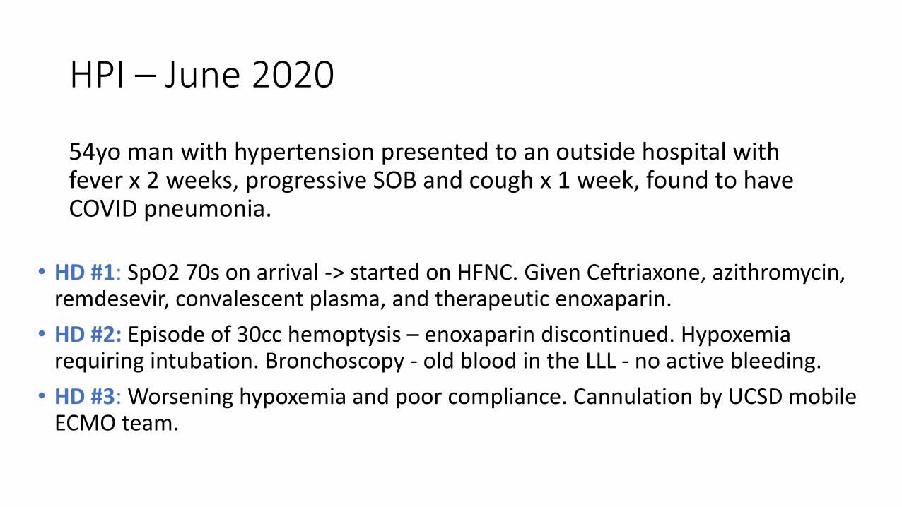
HPI – June 2020
54yo man with hypertension presented to an outside hospital with fever x 2 weeks, progressive SOB and cough x 1 week, found to have COVID pneumonia.
• HD #1: SpO2 70s on arrival -> started on HFNC. Given Ceftriaxone, azithromycin, remdesevir, convalescent plasma, and therapeutic enoxaparin.
• HD #2: Episode of 30cc hemoptysis – enoxaparin discontinued. Hypoxemia requiring intubation. Bronchoscopy - old blood in the LLL - no active bleeding.
• HD #3: Worsening hypoxemia and poor compliance. Cannulation by UCSD mobile ECMO team.
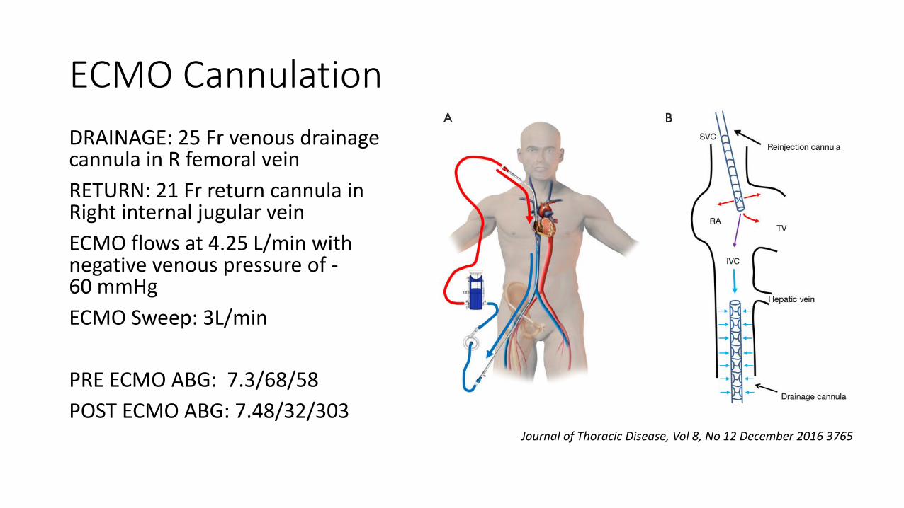
ECMO Cannulation
DRAINAGE: 25 Fr venous drainage cannula in R femoral vein
RETURN: 21 Fr return cannula in Right internal jugular vein
ECMO flows at 4.25 L/min with negative venous pressure of -60 mmHg
ECMO Sweep: 3L/min
PRE ECMO ABG: 7.3/68/58
POST ECMO ABG: 7.48/32/303Journal of Thoracic Disease, Vol 8, No 12 December 2016 3765
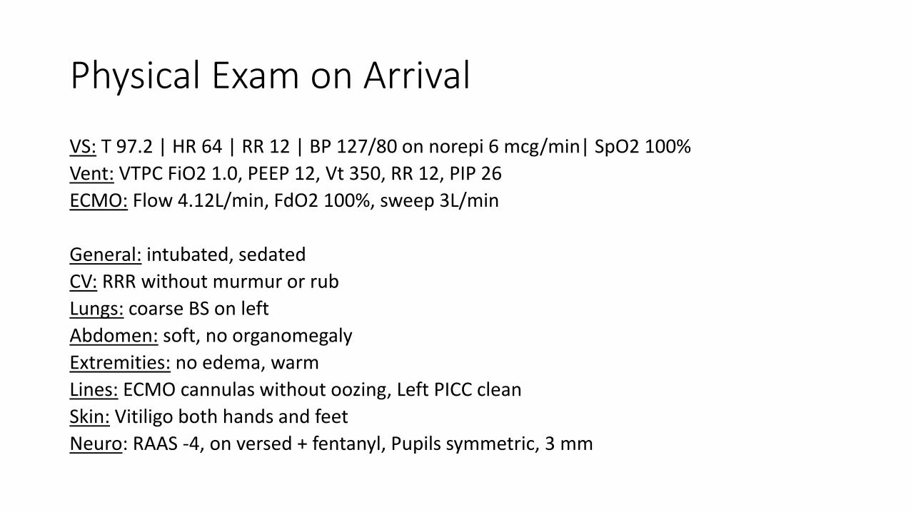
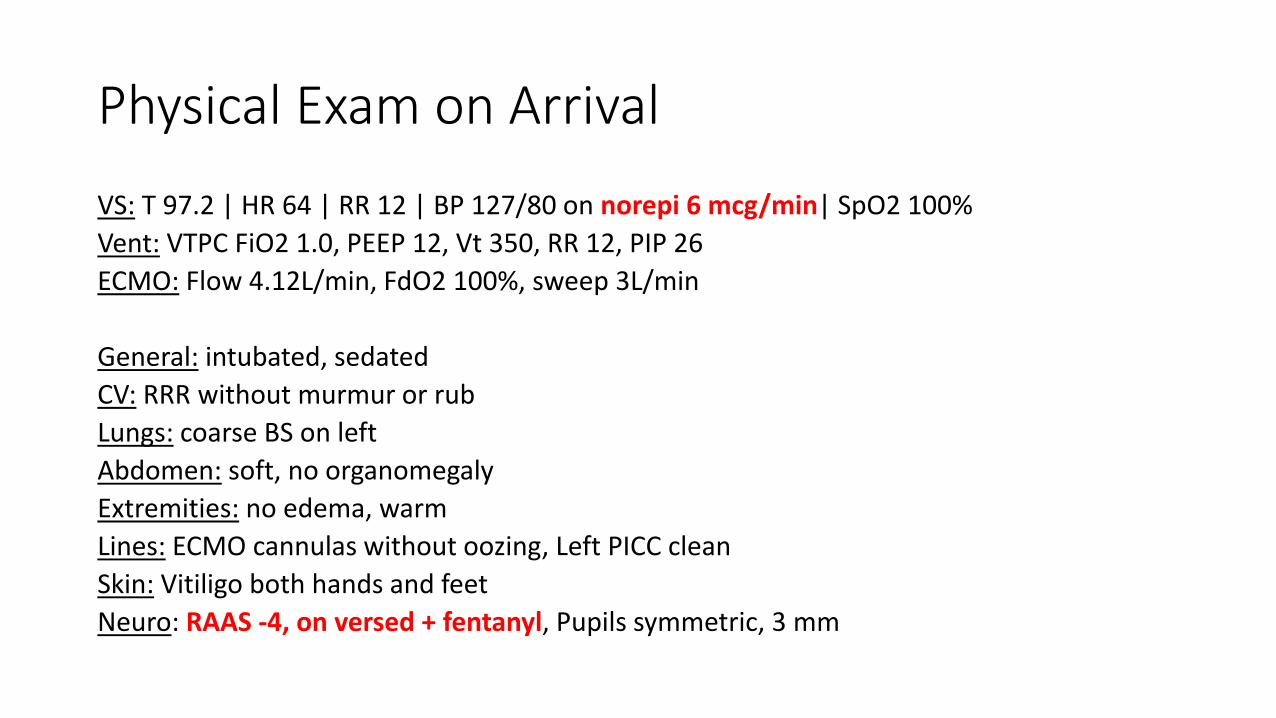
Physical Exam on Arrival
VS: T 97.2 | HR 64 | RR 12 | BP 127/80 on norepi 6 mcg/min| SpO2 100%
Vent: VTPC FiO2 1.0, PEEP 12, Vt 350, RR 12, PIP 26
ECMO: Flow 4.12L/min, FdO2 100%, sweep 3L/min
General: intubated, sedated
CV: RRR without murmur or rub
Lungs: coarse BS on left
Abdomen: soft, no organomegaly
Extremities: no edema, warm
Lines: ECMO cannulas without oozing, Left PICC clean
Skin: Vitiligo both hands and feet
Neuro: RAAS -4, on versed + fentanyl, Pupils symmetric, 3 mm
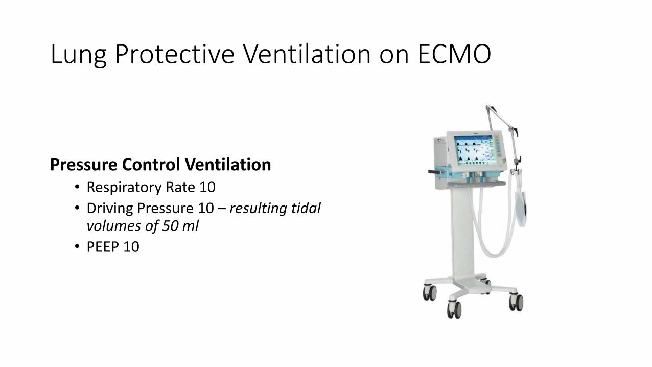
Lung Protective Ventilation on ECMO
Pressure Control Ventilation• Respiratory Rate 10
• Driving Pressure 10 – resulting tidal volumes of 50 ml
• PEEP 10
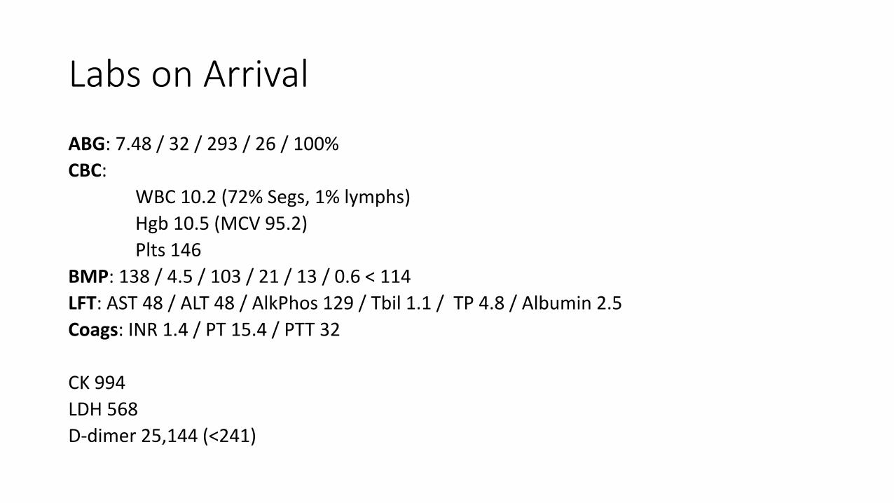
Labs on Arrival
ABG: 7.48 / 32 / 293 / 26 / 100%
CBC:
WBC 10.2 (72% Segs, 1% lymphs)
Hgb 10.5 (MCV 95.2)
Plts 146
BMP: 138 / 4.5 / 103 / 21 / 13 / 0.6 < 114
LFT: AST 48 / ALT 48 / AlkPhos 129 / Tbil 1.1 / TP 4.8 / Albumin 2.5
Coags: INR 1.4 / PT 15.4 / PTT 32
CK 994
LDH 568
D-dimer 25,144 (<241)
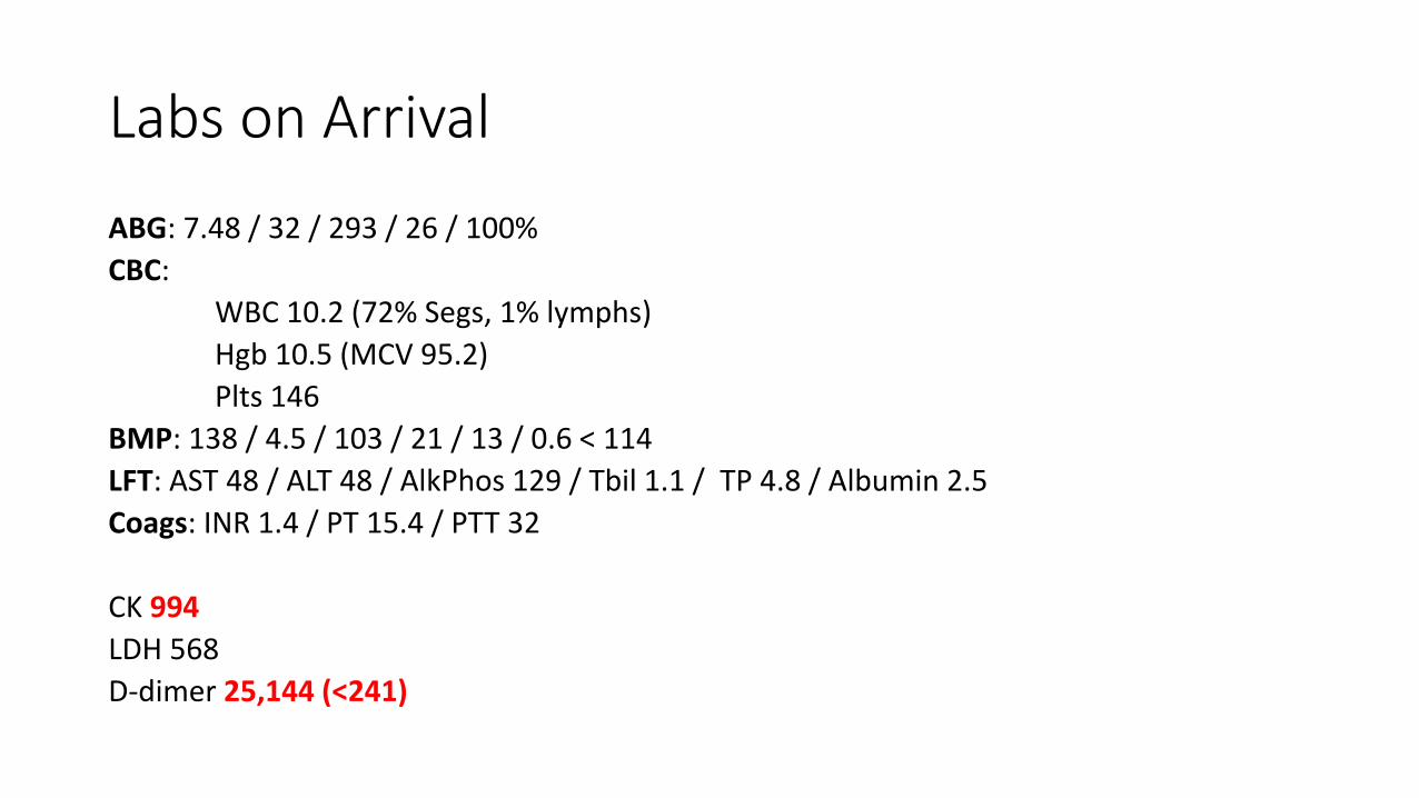
Labs on Arrival
ABG: 7.48 / 32 / 293 / 26 / 100%
CBC:
WBC 10.2 (72% Segs, 1% lymphs)
Hgb 10.5 (MCV 95.2)
Plts 146
BMP: 138 / 4.5 / 103 / 21 / 13 / 0.6 < 114
LFT: AST 48 / ALT 48 / AlkPhos 129 / Tbil 1.1 / TP 4.8 / Albumin 2.5
Coags: INR 1.4 / PT 15.4 / PTT 32
CK 994
LDH 568
D-dimer 25,144 (<241)
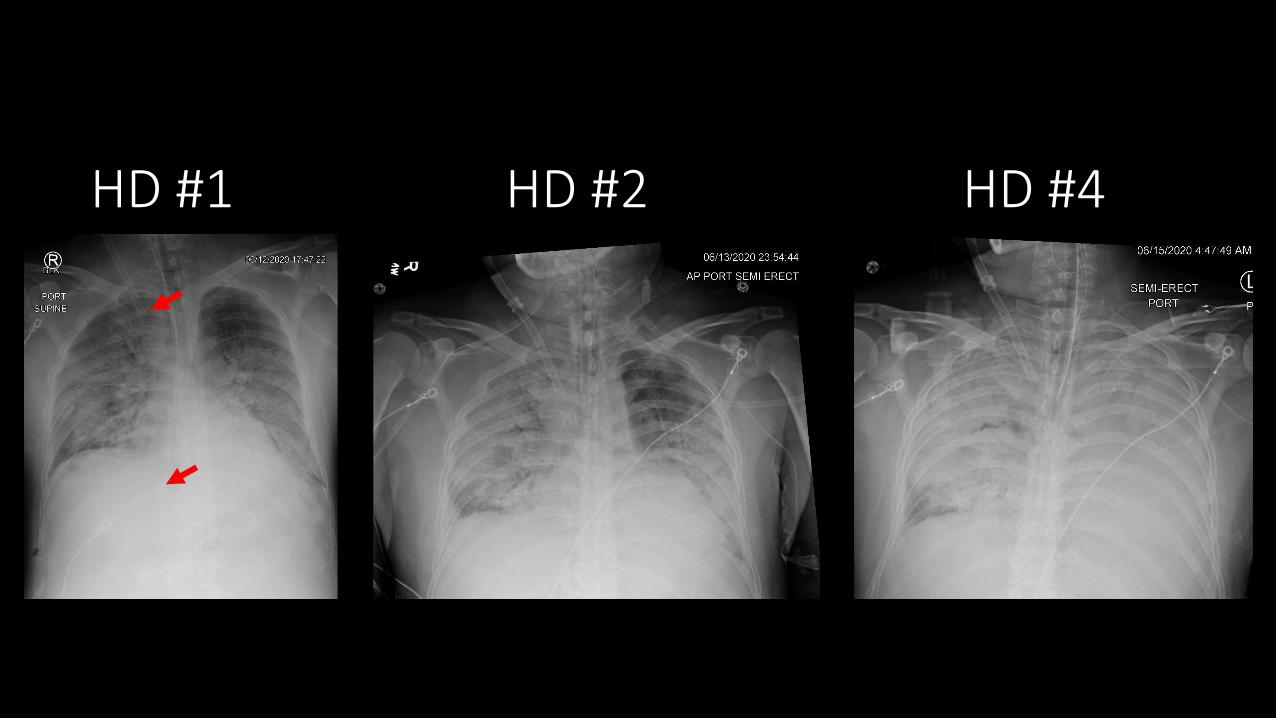
HD #1 HD #2 HD #4
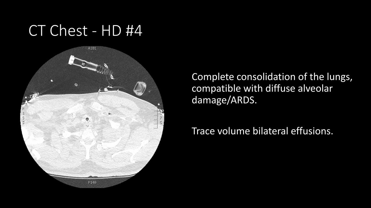
CT Chest - HD #4
Complete consolidation of the lungs, compatible with diffuse alveolar damage/ARDS.
Trace volume bilateral effusions.
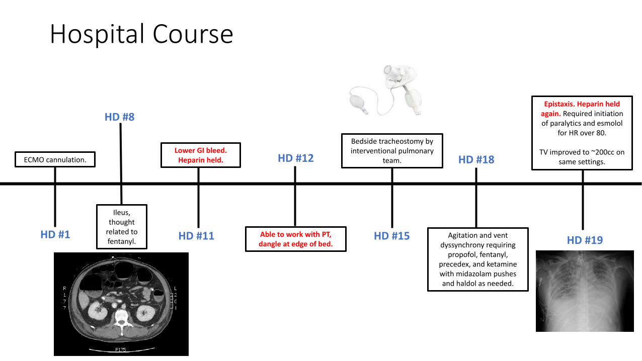
Hospital Course
HD #1
HD #8
Ileus, thought
related to fentanyl.
HD #11
Lower GI bleed. Heparin held. HD #12
Able to work with PT, dangle at edge of bed.
HD #15
Bedside tracheostomy by interventional pulmonary
team. HD #18
Agitation and vent dyssynchrony requiring
propofol, fentanyl, precedex, and ketamine with midazolam pushes and haldol as needed.
ECMO cannulation.
HD #19
Epistaxis. Heparin held again. Required initiation of paralytics and esmolol
for HR over 80.
TV improved to ~200cc on same settings.
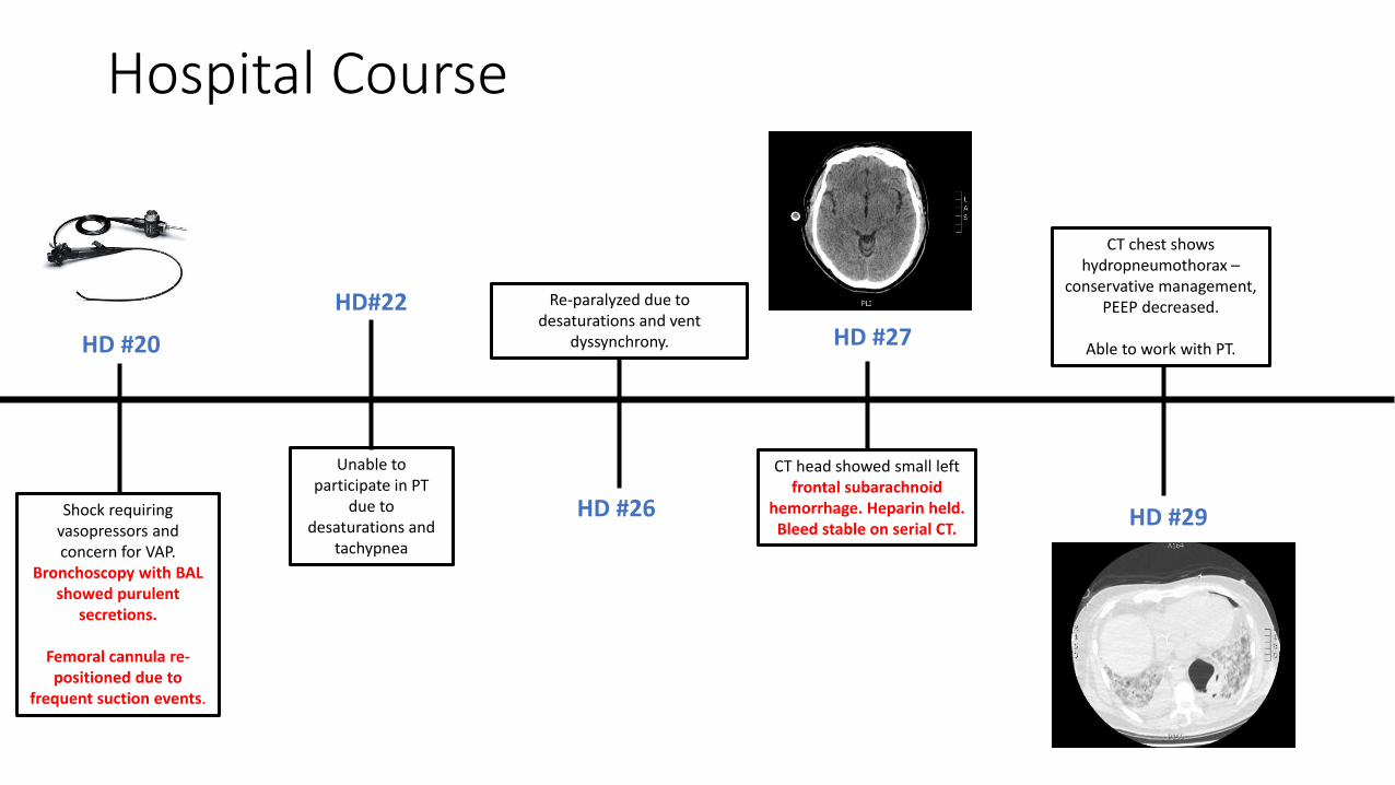
Hospital Course
HD #20
HD#22
Unable to participate in PT
due to desaturations and
tachypnea
HD #26
Re-paralyzed due to desaturations and vent
dyssynchrony. HD #27
CT head showed small left frontal subarachnoid
hemorrhage. Heparin held. Bleed stable on serial CT.
HD #29
CT chest shows hydropneumothorax –
conservative management, PEEP decreased.
Able to work with PT.
Shock requiring vasopressors and concern for VAP.
Bronchoscopy with BAL showed purulent
secretions.
Femoral cannula re-positioned due to
frequent suction events.
Hospital Course
HD #41
HD#44
Able to walk with PT. Tolerating trach
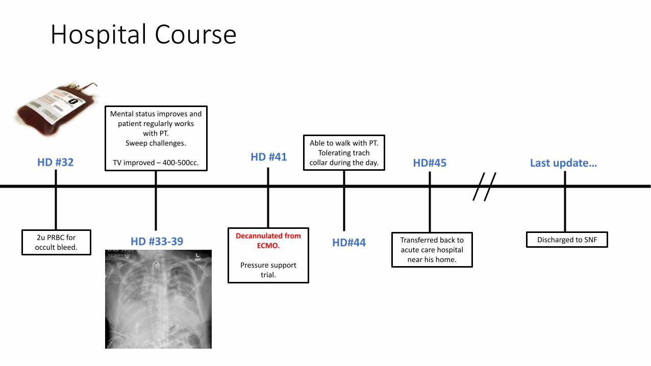
collar during the day.HD #32
2u PRBC for occult bleed.
Decannulated from ECMO.
Pressure support trial.
HD #33-39
Mental status improves and patient regularly works
with PT. Sweep challenges.
TV improved – 400-500cc. HD#45
Transferred back to acute care hospital
near his home.
Last update…
Discharged to SNF
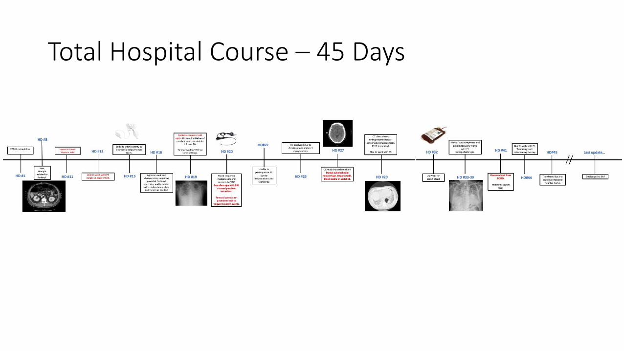
Total Hospital Course – 45 Days
Case 2: CE
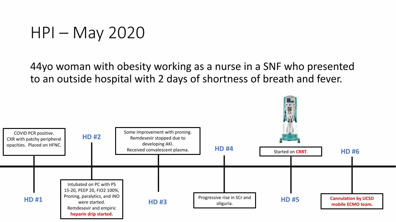
HPI – May 2020
44yo woman with obesity working as a nurse in a SNF who presented to an outside hospital with 2 days of shortness of breath and fever.
HD #1
COVID PCR positive.CXR with patchy peripheral opacities. Placed on HFNC.
HD #2
Intubated on PC with PS 15-20, PEEP 20, FiO2 100%, Proning, paralytics, and iNO
were started.Remdesevir and empiric
heparin drip started.
HD #3
Some improvement with proning.Remdesevir stopped due to
developing AKI. Received convalescent plasma. HD #4
Progressive rise in SCr and oliguria.
HD #5
Started on CRRT. HD #6
Cannulation by UCSD mobile ECMO team.
ECMO Cannulation
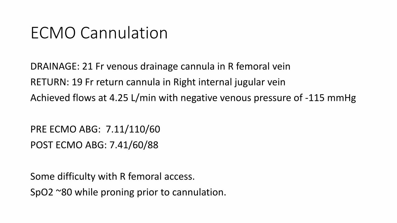
DRAINAGE: 21 Fr venous drainage cannula in R femoral vein
RETURN: 19 Fr return cannula in Right internal jugular vein
Achieved flows at 4.25 L/min with negative venous pressure of -115 mmHg
PRE ECMO ABG: 7.11/110/60
POST ECMO ABG: 7.41/60/88
Some difficulty with R femoral access.
SpO2 ~80 while proning prior to cannulation.
Physical Exam on Arrival
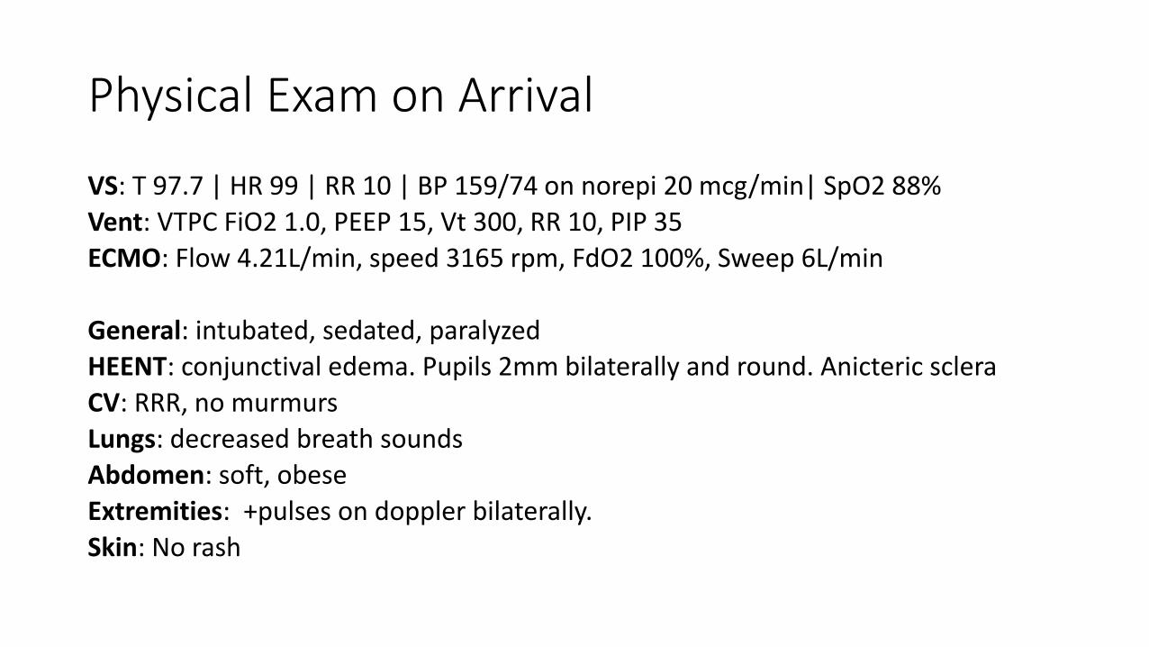
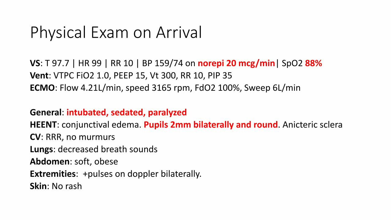
VS: T 97.7 | HR 99 | RR 10 | BP 159/74 on norepi 20 mcg/min| SpO2 88%
Vent: VTPC FiO2 1.0, PEEP 15, Vt 300, RR 10, PIP 35
ECMO: Flow 4.21L/min, speed 3165 rpm, FdO2 100%, Sweep 6L/min
General: intubated, sedated, paralyzed
HEENT: conjunctival edema. Pupils 2mm bilaterally and round. Anicteric sclera
CV: RRR, no murmurs
Lungs: decreased breath sounds
Abdomen: soft, obese
Extremities: +pulses on doppler bilaterally.
Skin: No rash
Physical Exam on Arrival
VS: T 97.7 | HR 99 | RR 10 | BP 159/74 on norepi 20 mcg/min| SpO2 88%
Vent: VTPC FiO2 1.0, PEEP 15, Vt 300, RR 10, PIP 35
ECMO: Flow 4.21L/min, speed 3165 rpm, FdO2 100%, Sweep 6L/min
General: intubated, sedated, paralyzed
HEENT: conjunctival edema. Pupils 2mm bilaterally and round. Anicteric sclera
CV: RRR, no murmurs
Lungs: decreased breath sounds
Abdomen: soft, obese
Extremities: +pulses on doppler bilaterally.
Skin: No rash
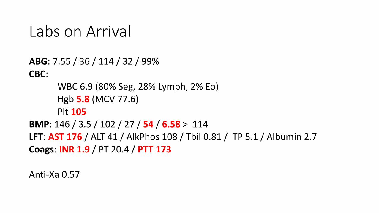
Labs on Arrival
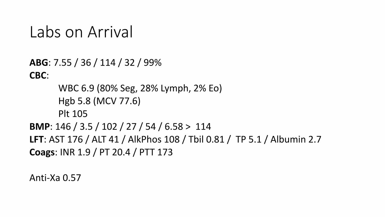
ABG: 7.55 / 36 / 114 / 32 / 99%CBC:
WBC 6.9 (80% Seg, 28% Lymph, 2% Eo)Hgb 5.8 (MCV 77.6)Plt 105
BMP: 146 / 3.5 / 102 / 27 / 54 / 6.58 > 114LFT: AST 176 / ALT 41 / AlkPhos 108 / Tbil 0.81 / TP 5.1 / Albumin 2.7Coags: INR 1.9 / PT 20.4 / PTT 173
Anti-Xa 0.57
Labs on Arrival
ABG: 7.55 / 36 / 114 / 32 / 99%CBC:
WBC 6.9 (80% Seg, 28% Lymph, 2% Eo)Hgb 5.8 (MCV 77.6)Plt 105
BMP: 146 / 3.5 / 102 / 27 / 54 / 6.58 > 114LFT: AST 176 / ALT 41 / AlkPhos 108 / Tbil 0.81 / TP 5.1 / Albumin 2.7Coags: INR 1.9 / PT 20.4 / PTT 173
Anti-Xa 0.57
HD #1
Hospital Course
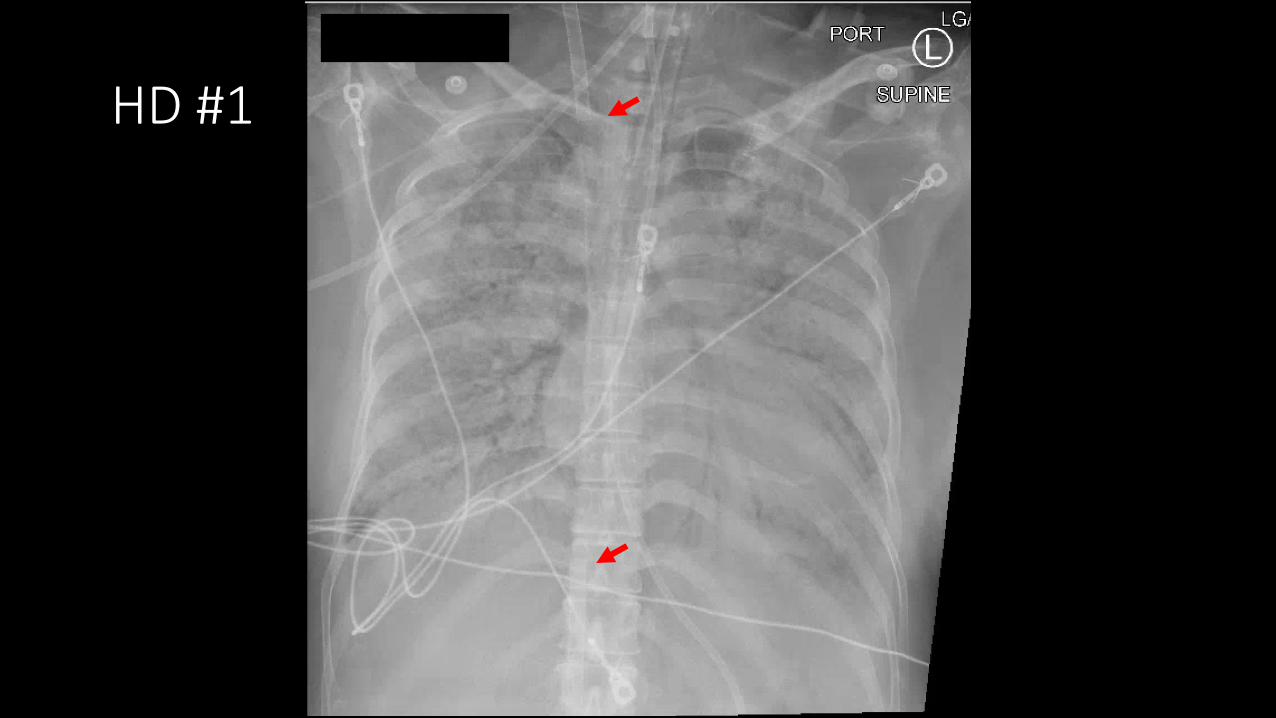
• HD #1:• PEEP maintained at 15 given low PaO2. Vasopressors given for shock. Given
2U PRBC for Hgb of 5.8 – thought possibly related to blood loss from ECMO cannulation. CRRT was re-started.
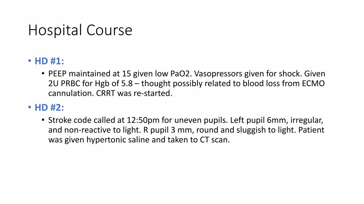
• HD #2:• Stroke code called at 12:50pm for uneven pupils. Left pupil 6mm, irregular,
and non-reactive to light. R pupil 3 mm, round and sluggish to light. Patient was given hypertonic saline and taken to CT scan.
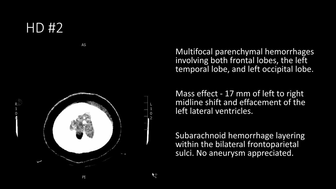
HD #2
Multifocal parenchymal hemorrhages involving both frontal lobes, the left temporal lobe, and left occipital lobe.
Mass effect - 17 mm of left to right midline shift and effacement of the left lateral ventricles.
Subarachnoid hemorrhage layering within the bilateral frontoparietal sulci. No aneurysm appreciated.
Hospital Course
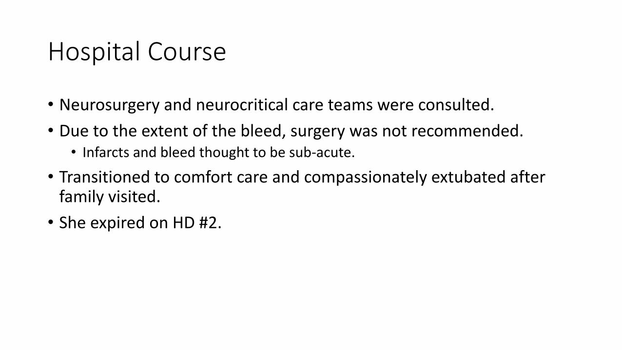
• Neurosurgery and neurocritical care teams were consulted.
• Due to the extent of the bleed, surgery was not recommended.• Infarcts and bleed thought to be sub-acute.
• Transitioned to comfort care and compassionately extubated after family visited.
• She expired on HD #2.
Reflections from 2 ECMO Cases
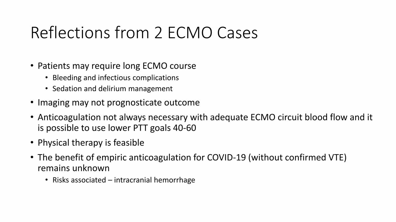
• Patients may require long ECMO course• Bleeding and infectious complications
• Sedation and delirium management
• Imaging may not prognosticate outcome
• Anticoagulation not always necessary with adequate ECMO circuit blood flow and it is possible to use lower PTT goals 40-60
• Physical therapy is feasible
• The benefit of empiric anticoagulation for COVID-19 (without confirmed VTE) remains unknown • Risks associated – intracranial hemorrhage