Embed Size (px)
Citation preview
Aus der
Klinik für Dermatologie, Venerologie und Allergologie / Immunologisches Zentrum
des Städtischen Klinikums Dessau
DISSERTATION
Towards the establishment and characterization
of a human skin explant co-culture model with SZ95 sebocytes
zur Erlangung des akademischen Grades
Doctor medicinae (Dr. med.)
vorgelegt der Medizinischen Fakultät
Charité – Universitätsmedizin Berlin
von
Georgios Nikolakis
aus Athen, Griechenland
Datum der Promotion: 12.09.2014
1
“Τὸν ἀγῶνα τὸν καλὸν ἀγωνίσθηκα, τὸν δρόμον
ἐτελείωσα, τὴν πίστιν ἐτήρησα“
“Ich habe den guten Kampf gekämpft, ich habe den Lauf vollendet, ich habe
den Glauben bewahrt”
2. Timotheus 4.7
To my parents, Dimitrios and Kalliopi
To Miriam
Table of Contents 2
Table of Contents
Table of Contents .................................................................................................... 2
Abstract ................................................................................................................... 5
Introduction ............................................................................................................. 7
1.1 Three dimensional (3D) skin models and their functions .......................................... 7
1.2 Skin explant models .................................................................................................. 8
1.3 In vitro engineered skin models .............................................................................. 10
1.4 In vitro engineered skin models: the rationale for integration of cutaneous
appendages .......................................................................................................................... 12
1.5 Advantages and disadvantages of 3D skin model types .......................................... 14
1.6 Methods of culturing primary sebocytes ................................................................. 16
1.7 Difficulties of maintaining sebaceous glands and sebocytes in culture .................. 17
1.8 The solution of sebocyte cell lines .......................................................................... 18
1.9 Common functions of the sebaceous gland ............................................................. 20
1.10 Sebaceous gland as a target of circulating hormones and as a site of steroid
hormone synthesis ............................................................................................................... 22
1.11 Sebocytes as target of various other hormones ....................................................... 23
1.12 Sebaceous gland and its role in endogenous and adaptive immunity ...................... 25
Aim of Study ......................................................................................................... 29
Materials and Methods .......................................................................................... 30
2.1 Materials, media, solutions and equipment ............................................................. 30
2.2 Cell culture methods ................................................................................................ 33
2.2.1 Cell culture basics.................................................................................................... 33
2.2.2 Cell lines .................................................................................................................. 33
2.2.3 Freezing, thawing, subculturing and counting of cells ............................................ 35
2.3 Oil red staining ........................................................................................................ 41
2.4 Measurement of free LDH release........................................................................... 42
2.5 Apoptosis detection – TUNEL reaction .................................................................. 43
Table of Contents 3
2.6 IL-6 and IL-8 enzyme-linked immunosorbent assay (ELISA) ................................ 44
2.7 Skin preparation for histology ................................................................................. 46
2.8 Hematoxylin eosin staining ..................................................................................... 47
2.9 Immunocytochemistry and immunohistochemistry ................................................ 48
2.10 DNA fragmentation evaluation ............................................................................... 55
2.11 Skin explant stratum corneum and vital epidermis thickness .................................. 55
2.12 Statistical analysis ................................................................................................... 55
2.13 Protein analysis methods ......................................................................................... 55
2.13.1 Skin protein isolation ............................................................................................... 56
2.13.2 Protein quantitation ................................................................................................. 56
2.13.3 Western blot ............................................................................................................ 57
Results ................................................................................................................... 61
3.1 Morphological evaluation after hematoxylin eosin staining (SG degeneration) ..... 61
3.2 Biochemical evidence of sebaceous gland degeneration ......................................... 64
3.3 Morphological evaluation of the epidermis after hematoxylin eosin staining......... 65
3.4 Stratum corneum and vital epidermis thickness ...................................................... 69
3.5 DNA fragmentation evaluation ............................................................................... 72
3.6 LDH release ............................................................................................................. 75
3.7 IL-6 and IL-8 secretion ............................................................................................ 77
3.8 SZ95 Sebocytes – Morphology, Oil red and immunocytochemistry findings ........ 83
3.9 Expression of Ki67 antigen as an epidermal vitality marker ................................... 89
3.10 Specificity of SZ95 lipid accumulation through direct contact with the dermis of
skin explants ........................................................................................................................ 92
3.11 Evaluation of apoptosis with western blots ............................................................. 94
Discussion ............................................................................................................. 96
4.1 Integration of the variable “sebaceous gland” in a new skin explant model ........... 96
4.2 Choice of medium ................................................................................................... 96
4.3 Sebaceous gland maintenance ex vivo .................................................................... 98
4.4 Direct contact of SZ95 sebocytes with skin explant dermis promotes skin explant
epidermal integrity .............................................................................................................. 99
Table of Contents 4
4.5 Explanation for IL-6 downregulation after co-culture with SZ95 sebocytes ........ 101
4.6 The insufficiency of LDH release as a marker of cell death for co-cultures ......... 103
4.7 Sebaceous differentiation and lipid accumulation through skin explant molecular
interaction offers a more in-vivo like phenotype............................................................... 103
4.8 The crucial role of molecular interaction of the epithelial and mesenchymal
component of the skin ....................................................................................................... 104
4.9 Role of the culture setting in the SZ95 sebocyte-mediated skin explant vitality .. 106
4.10 Skin explant culture to the next level: hypotheses and future perspectives ........... 107
4.11 Potential effects of SZ95 sebocytes on skin explant keratinocyte apoptosis ......... 109
Conclusion ........................................................................................................... 110
Abbreviation list .................................................................................................. 112
References ........................................................................................................... 114
Curriculum Vitae ................................................................................................. 138
Publication List .................................................................................................... 142
Eidesstattliche Versicherung ............................................................................... 148
Acknowledgements ............................................................................................. 149
Abstract 5
Towards the establishment and characterization
of a human skin explant co-culture model with SZ95 sebocytes
Zur Etablierung und Charakterisierung
eines humanen Ko-Kultur-Hautmodells mit SZ95 Sebozyten
Georgios Nikolakis
Abstract The maintenance of normal human sebocytes in organ culture and in vitro is extremely
difficult and barely reproducible, mainly because the cells are programmed to differentiate
and undergo holocrine secretion, i.e. cell membrane rupture and release of their content.
Therefore, we developed a skin explant model, where skin specimens are co-cultured for 6
days with a monolayer cell culture of the immortalized human SZ95 sebaceous gland cells
(sebocytes). Through model variation the molecular cross-talk between SZ95 sebocytes and
the skin specimens is possible both through direct cell-tissue as well as humoral contact. The
structural integrity of the skin explant epidermis was facilitated through the co-culture in
direct contact. Parallel co-cultures with human fibroblasts provided evidence for the cell type
specificity of the aforementioned results. Interestingly, the presence of SZ95 sebocytes in the
culture system in direct contact with the skin specimens reduced the secretion of IL-6 by the
latter. Immunohistochemical labelling with antibodies raised against IL-6 and IL-8, showed
that IL-6 was expressed in both the epidermal as well as the dermal component of the skin
explants, while IL-8 was not expressed from the epidermal keratinocytes. Stratum corneum
excessive thickness and size of the vital non-cornified epidermis were modified towards
normalization after direct contact co-culture of the explants with SZ95 sebocytes. In addition,
DNA fragmentation (TUNEL technique) showed decreased apoptosis and Ki67
immunostaining a partial conservation of Ki67 expression in basal epidermal keratinocytes of
the skin specimens co-cultured with SZ95 sebocytes, indicating a normalising effect of SZ95
sebocytes towards the ex vivo skin homeostasis. On the other hand, SZ95 sebocytes co-
cultured with skin specimens in direct contact exhibited increased lipid accumulation and
stronger expression of the differentiation markers keratin 7 and epithelial membrane antigen,
suggesting that this co-culture setting promoted their differentiation. Surprisingly, the
aforementioned beneficial effects of SZ95 sebocytes on skin homeostasis and of skin explants
on sebaceous differentiation were not observed in humoral-contact co-cultures. Spontaneous
LDH release did not exhibit significant differences between direct or humoral co-cultures and
controls and western blots of skin explant lysates deriving from humoral co-culture
Abstract 6
experiments and controls did not exhibit visible differences in caspase-3 activation as initiator
of apoptosis. These data underline a cross-talk of human sebocytes and skin specimens under
specific co-culture conditions but also a major role of sebocytes in skin homeostasis,
proposing their addition to three-dimensional skin models.
Zusammenfassung
Die Kultivierung normaler humaner Sebozyten in der Organkultur und in vitro ist besonders
anspruchsvoll und schwer reproduzierbar, hauptsächlich weil die Zellen differenzieren und
einer holokrinen Sekretion sich unterziehen, i.e. Zellmembranruptur und Freisetzung ihres
Inhalts. Aus diesem Grund haben wir ein Hautexplantatmodel entwickelt, in welchem
Hautproben über 6 Tage mit Einschichtzellkulturen von immortalisierten humanen SZ95-
Talgdrüsenzellen kokultiviert wurden. Durch Variationen des Modells konnte man die
molekulare Wechselwirkung zwischen Zellen und Haut beim direkten und humoralen
Kontakt untersuchen. Die strukturelle Integrität der Hautexplantat-Epidermis wurde durch die
Kokultur im direkten Kontakt begünstigt. Die parallele Kokultur mit humanen Fibroblasten
als Kontrolle bewies die Zelltypspezifität der Ergebnisse. Interessanterweise reduzierte die
Präsenz von SZ95-Sebozyten in der Kokultur in direktem Kontakt mit der Haut die Sekretion
von IL-6 aus den Hautproben. Immunohistochemische Färbung mit Antikörpern gegen IL-6
und IL-8 wiesen IL-6 im epidermalen und dermalen Komponent der Hautexplantaten nach,
jedoch wurde IL-8 nicht von den epidermalen Keratinozyten exprimiert. Die exzessive
Verdickung des Stratum Corneum und die Größe der vitalen nicht kornifizierten Epidermis
wurden durch die Kokultur der Hautexplantaten mit SZ95-Sebozyten in direktem Kontakt
normalisierend beeinflusst. Darüber hinaus wurde mit Hilfe der DNS-Fragmentation
(TUNEL-Methode) eine reduzierte Apoptose und mit Hilfe der Immunfärbung gegen das
Ki67-Antigen eine partiale Konservierung der Ki67-Expression in basalen epidermalen Zellen
gezeigt, die auf ein normalisierendes Effekt auf die ex-vivo-Homeostase der Haut hinwies.
Auf der anderen Seite, zeigten die in direktem Kontakt mit der Haut kokultivierten SZ95-
Sebozyten eine erhöhte Lipidakkumulation und stärkere Expression von Keratin 7 und
epithelialem Membran-Antigen. Das wies auf die Förderung der sebozytären Differenzierung
durch die Kokultivierung auf. Die o.g. positiven Effekte wurden im Fall der humoralen
Kokultur weder Effekte der SZ95-Sebozyten auf die ex-vivo-Haut Homeostase noch der Haut
auf die sebozytäre Differenzierung beobachtet. Die spontane LDH Freisetzung zeigte keine
signifikanten Unterschiede zwischen direkten bzw. humoralen Kokulturen und Kontrollen.
Darüber hinaus konnte mittels der Westernblotmethode bei humoralen Kokulturen und
Kontrollen keine Aktivierung von Caspase-3 als Initiator der Apoptose nachgewiesen werden.
Zusammenfassend stellen die o.g. Daten eine Wechselwirkung zwischen humanen Sebozyten
Introduction 7
und ex vivo-Hautproben unter spezifischen Kulturbedingungen heraus. Außerdem wurde eine
wichtige Rolle der Sebozyten für die Hauthomeostase nachgewiesen, welche für ihre
Hinzufügung in zukünftigen dreidimensionalen Hautmodellen spricht.
Introduction
1.1 Three dimensional (3D) skin models and their functions
The understanding of human skin biology has always been a field of great interest for various
reasons: Skin is the largest organ of the body and it is easily accessible for research purposes.
The development of several models helped to perceive the physiology of skin properties, the
underlying pathophysiology of skin diseases and also to determine the action of various
substances. Nowadays, the conception of the skin as only a barrier between the body and
environmental stress (micro-organisms, radiation, temperature changes, chemical or
mechanical trauma) gave its place to the idea of the skin as a constitution of multiple mini-
organs, which interact with each other and the rest of the body, forming an immune-
cutaneous-endocrine network [1, 2]. The well-established monolayer culture of isolated cell
types cannot represent the in-vivo microenvironment, since they cannot depict the cell-cell
and cell-matrix interactions, which lead to different phenotypes, receptor expression,
metabolic function and response to various chemical compounds when cultivated in proximity
in vivo [3-5].
In the beginning of functional dermatological research, the use of animal models was the
main way of studying the communication of the skin with various pharmaceutical products.
Substances were topically applied on healthy animal skin and their effects were studied and
measured. The use of animals for the study of pharmaceutical or cosmetic products with a
potent irritant potential was first introduced by Drazie et al. [6] 65 years ago. Despite
providing the in vivo cellular molecular crosstalk, the inevitable sacrifice of animals in order
to further elucidate the effects of investigative compounds was a major reason for the recent
restrictions of animal use in drug and cosmetics industry [7]. Apart from the ethical reasons,
skin models often provided controversial information [8, 9] and there are differences between
animal and human skin physiology and pathology which could not be overlooked [10, 11].
For the aforementioned reasons, the 7th Amendment of the EU ‘cosmetics directive’ stated
that animal studies should always be seen under the prism of the 3R-principle. (Reduce –
number of animals tested, Refine – narcosis and other procedures for minimizing animal
stress, Replace- replacement of animal testing with in vitro methods) [7, 9, 12].
Introduction 8
These facts depicted the importance of human models and especially 3D human skin models
or organotypic skin models, which are classified into two main categories: skin explant
models and in vitro reconstructed skin.
1. Organ skin cultures or skin explant models, which are full-thickness or split-
thickness pieces of human skin obtained from surgical procedures or biopsies and
cultured in the appropriate medium after proper decontamination and sometimes
removal of the subcutaneous fat.
2. In vitro reconstructed skin or artificial skin or human skin equivalents (HSE),
where the layers of the skin or the epidermis were reconstructed step by step in a
gradual process of combining isolated human cells formerly in 2D culture in a living
interacting structure [13].
The organotypic models, besides their application for in vitro studies, have numerous in vivo
applications such as the use as epidermal or dermal substitutes after serious wounding to
prevent excessive loss of fluids and contamination till there is an autograft available, to
improve the result of skin transplantation in areas of mechanical stress, to prevent wound
contraction and to form specifically engineered allografts, which do not express the proteins
that can trigger the immune response of the recipient [9].
1.2 Skin explant models
Skin explant models have the crucial advantage of including all skin cell types, representing
from this point of view better the in vivo environment than skin equivalents. In the case of
hospital and laboratory collaboration the source of material is quite accessible and less
arduous than an in vitro skin reconstruction. Apart from assessing the corrosion or irritation
potential of different substances, they can be utilized for various purposes, such as wound
healing, inflammation, tumor growth or UV-induced damage. Moreover, organ culture
models can provide an opportunity of personalized testing of various compounds, since skin
of a patient can be directly taken and utilized for the in vitro studies [13].
Skin explants have the main advantage of comprising most of the cellular types and their
interactions, as they derive directly from human skin. Easily accessible, they can be cultured
short-term fully submerged in medium [14, 15] or on the air-liquid interface, when a longer
incubation period is needed. The explant in this case is placed on a metallic grid or a
microporous insert, with the epidermis facing upward and remaining outside the culture
medium, a factor which is critical for terminal epidermal differentiation [16-19]. Cases have
Introduction 9
been described where no support is used, with skin explants floating in medium, with the
same compartment orientation [20, 21].
DMEM/Ham’s F12 and William’s E medium are used commonly for culturing skin explants
in the air liquid interface. The medium usually contains physiological concentrations of Ca++
(1.5 mM). Physiological concentrations of calcium are believed to play a role in skin explant
layer cohesion, stimulation of ECM production and contribution to epidermal differentiation
[22-26]. Animal serum is omitted, since it is not chemically defined and it might interfere
with the interpretation of substance testing results.
Skin explants can be usually cultured for a maximum of 7-14 days. Histochemical analysis of
skin explants shows visible differences of skin structure quality over culture time, such as
thinner epidermis and parallelly thicker stratum corneum, cleft formation, nuclear
condensation [27, 28], after which vacuolization of the basal keratinocytes and subsequent
acantholysis are prominent. However, there were studies which reported skin organ culture
maintenance for more than two weeks [21] and up to 28 days [29]. To prevent excessive
dermal contraction and to facilitate better fusion of medium nutrients across the entire skin
specimen, the skin is sliced into small pieces in most skin organ culture models. In other
studies, larger skin specimens were used (0.5 x 1.0 cm), while cross-sectioning ensured stable
and standardized tissue conditions, preventing potential misinterpretations because of
ubiquitous fusion between different sections of the samples. To face the problem of skin
explants lacking contraction and tension of tissue in vivo, skin explants (2.5 x 2.5 cm) were
fixed with the epidermis facing upwards in a special stainless steel chamber, so that tension
could be applied to the cultured skin explant to prevent dermal contraction mediated by
elastic fibers [29].
Morphological evaluation after hematoxylin-eosin staining is the first step in the assessment
of skin viability after incubation with various compounds. The 5-dimethylthiazol-2-yl-2,5-
diphenyltetrazolium bromide (MTT) assay [30] and the detection of the intracellular enzyme
lactate dehydrogenase (LDH) released after cell death to the culture supernatant can also be
applied as markers of organ culture skin viability [31-33]. The corrosion of various
substances is assessed in artificial skin and skin explant models [30] by the MTT assay. In
this assay, the viability of the tissue is assessed by the mitochondrial reduction of MTT to the
dissoluble purple salt formazan, which can be extracted with ethanol or isopropanol solution
and measured by a spectral photometer. Assessment of pro-inflammatory cytokines, such as
interleukin (IL)-6 and IL-8, is helpful in determining the irritation potential of corrosive and
irritant compounds as well as contact sensitizers, while procollagen I and amphiregulin
detection is useful to reflect fibroblast and keratinocyte growth inhibition, respectively [34].
Introduction 10
Amphiregulin is an autocrine growth factor of the epidermal growth factor (EGF) family
produced by keratinocytes, which induces keratinocyte proliferation through paracrine
stimulation [35-37]. As far as immunocytochemistry and immunofluorescence are concerned,
proliferation markers (Ki67, PCNA, BrdU), [38, 39], combined with apoptosis markers
(terminal transferase dUTP nick-end labeling technique-TUNEL for labeling DNA
fragmentation and cleaved caspase-3 as an effector caspase of apoptosis) are commonly used
to detect the balance between proliferation and apoptosis of human skin explant epidermis,
thus being a method of assessing its homeostasis [21, 40-42]. Changes of the relationship
between Ki67- and apoptosis-positive cells of skin explants, in correlation with allocation of
the first in the epidermis, can be a sign of skin explant degeneration [21, 29].
1.3 In vitro engineered skin models
Artificial skin models were developed in order to reproduce and explain key mechanisms of
skin cellular cross-talk and to better assess the hazard potential of various compounds. They
have advantages over animal models and skin explants in representing human anatomical
structure and reproducibility, respectively. They can be further classified into a) models of
epidermis, which consist of keratinocytes on a scaffold differentiating into a fully stratified
epithelium at the air-liquid interface (ALI), b) dermal equivalents of fibroblasts seeded on a
matrix and c) full-thickness skin substitutes constructed in a two-step process, in which
keratinocytes are seeded on a dermal compartment, formerly inoculated with fibroblasts, and
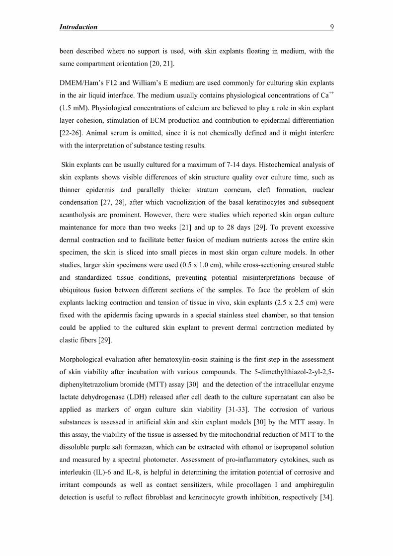
are allowed to differentiate after exposure to ALI (see fig. 1) [9, 13, 43].
Fig. 1: Simplified step-by-step process of in vitro skin reconstruction. Alternatively,
fibroblasts can be seeded directly as a mixture with the components of the scaffold (mainly
bovine or rat tail collagen I), instead of gradually populating the scaffold, as shown in the
picture. ALI: Air Liquid Interface
Dermal skin equivalents were among the first developed after seeding fibroblasts on a bovine
collagen type I lattice [44, 45]. The substitute, after the addition of keratinocytes, was
Introduction 11
introduced commercially for the treatment of chronic wounds and for testing the risk of
potential irritants and corrosive substances [46, 47].
Induction of terminal keratinocyte differentiation through air exposure seems to be the
cardinal factor for multilayered stratified epidermal development [19, 48]. The cornified
envelope of terminally differentiated keratinocytes (corneocytes) is a protein-lipid layer,
which forms a hydrophobic barrier between the environment and the body [49]. The proteins,
which mainly take part in its creation, are involucrin and loricrin, which are cross-linked by
enzymes called transglutaminases (TGs) for the formation of the cornified envelope [50-53].
The lipids, which are contained in the lamellar granules of the keratinocytes of stratum
granulosum, are the mortar between corneocyte proteins that repels water and various
substances. The production of growth factors by fibroblasts is the key to appropriate
epidermal differentiation, stimulating the basement membrane protein synthesis and
keratinocyte differentiation through the secretion of cytokines, such as transforming growth
factor (TGF)-β and keratinocyte growth factor (KGF) [54-56]. The reconstructed epidermis
models can remain viable for up to two months, but absence of desquamation results in a
gradual thickening of stratum corneum, which might influence the results of substance testing
[57]. The expression of proteins of the cornified envelope [mainly involucrin and loricrin [49]
and secondary small proline rich proteins (SPRP) and S100)], as well as the enzymes which
catalyze their cross-linking (TGs), ensure terminal keratinocyte differentiation and
stratification of the artificial epidermis and can be detected with immunohistochemistry
and/or immunofluorescence techniques [58]. The epidermal layer of expression gives
valuable information about how effectively the epidermal differentiation represents the in-
vivo conditions. Keratin (K)10 is an early differentiation marker, detected in all suprabasal
keratinocyte layers, indicating normal differentiation process [59, 60]. K6 and K16 are
referred to as markers of hyperproliferation and wound healing [60]. For the basal membrane
antibodies against collagen IV, VI, laminin and α6 integrin are representative proteins for
structural integrity, which imply an effective functional consistency [58, 61-64]. Adherent
structures such as desmoglein 1 and 3, desmocollin and plakophilin 1 were also identified as
crucial factors of epidermal integrity [64].
Extracellular matrix (ECM) protein expression also plays a major role in reflecting the
molecular cross-talk of different cell types [56, 65]. Newly composed ECM components from
fibroblasts such as collagen type I and III, fibronectin, fibrillin 1 and elastin were detected
with immunohistochemical methods in an engineered human skin equivalent. [58, 66]
The cell origin plays an important role in the reconstruction of skin equivalents. Cell lines
were considered to be a good solution to donor-to-donor variability problems. However, the
Introduction 12
use of the immortalized cell line of HaCaT keratinocytes led to impaired epidermal
differentiation, since the early differentiation markers K1 and K10 were expressed irregularly
across all cell layers, including the stratum basale. Ceramide composition was also impaired,
thus highlighting an impaired barrier function [60].
The scaffold used is important for artificial skin generation. Porous filters were the first
scaffolds used, allowing the integrin-mediated attachment of keratinocytes to their surface,
followed by air exposure-induced differentiation [67, 68]. For generation of full thickness-
skin models, synthetic de-epidermized dermis (DED) [69] or biological human or animal
derived scaffolds [70] and sponges from various materials were used, such as a hydrated gel
of collagen I [71], fibrin gel [72], collagen/chitosan chondroitin-4-6 sulfates [73], synthetic
human elastin [74], polycaprolactone [75, 76], polylactic-co-glycolic gel, [77] their mixture
with collagen, [76, 78, 79] and a collagen vitrigel membrane [80]. These synthetic scaffolds
are valuable for preventing excessive fibroblast contraction of the pseudodermis during long-
term culture, with the disadvantage of not representing the in vivo ECM variable and the lack
of adhesion molecules [9]. These problems can be solved by generating fibroblast-derived
matrices with the self-assembly method: fibroblasts are seeded in high densities on polyester
permeable supports and cultured for three weeks, thus creating a fibroblast-derived human
ECM, with ascorbic acid being the main stimulating supplement of processing pro-collagen to
collagen α-chains [81, 82].
1.4 In vitro engineered skin models: the rationale for integration of
cutaneous appendages
Many skin models have been developed commercially over the last years in order to assess
the properties of agents with a corrosive, irritant or beneficial potential. The reconstructed
epidermis models can remain viable for up to two months, but the gradual thickening of
stratum corneum, because of the absence of desquamation, might influence the results of
substance testing [57].
On the other hand, for the assessment of results of various substances the involvement of
cutaneous appendages, as well as other cell types in an in vivo skin microenvironment should
also be taken into consideration. For this reason, there has been a systematic effort to
introduce of other cell types and cutaneous annexes to in vitro reconstructed skin according to
the future application purpose.
Phototoxicity tests using artificial skin necessitate the integration of melanocytes to skin
equivalents [83-85]. Lee et al. [86] constructed an epidermis equivalent with keratinocytes
and melanocytes and measured the release of the inflammatory cytokines IL-1α, IL-1β and IL-
Introduction 13
6 after topical application of three known phototoxic agents. Cells from individuals with light
and dark phototypes were used for the study of impaired photoprotection [87], while chimeric
substitutes from Caucasian and Negroid donors were applied to clarify the roles of
melanocytes and keratinocytes in photoprotection via skin pigmentation and anti-oxidant
protection, respectively [88].
The study of human hair growth and the epithelial-mesenchymal interactions occurring in hair
follicles led to the development of various organotypic skin structures in investigating hair
growth occurring ex vivo or in integrating hair follicles in reconstructed skin in vitro. Michel
et al. [89] managed to insert complete pilosebaceous units in a fibroblast – keratinocyte
organotypic model. The pilosebaceous units have been obtained with thermolysin digestion of
hairy human skin in order to highlight the importance of hair follicles for percutaneous
substance absorption, without focusing on the role of the sebaceous gland. Havlickova et al.
[90] co-cultured outer root sheath keratinocytes with follicular dermal papilla fibroblasts
under different culture medium and culture design conditions. Krugluger et al. [91]
established a method of a skin organ culture, where hair-follicle formation is induced by
follicle-derived cells.
The introduction of Langerhans cells (LCs) into HSEs was proposed for testing cutaneous
immunological reactions of the skin to potential allergens [92]. The task has proved arduous,
since the first attempts resulted in the integration of round pyknotic cells in the epidermis [9,
93]. To adress this problem, researchers integrated CD34+ hematopoietic progenitor cells,
which were differentiated in a second step to mature LCs with the help of various cytokine
cocktails, including granulocyte macrophage growth factor (GM-CSF), tumor necrosis factor-
α (TNF-α) and TGF-β1. Mature LCs were identified by the expression of specific markers and
the intracellular presence of the characteristic Birbeck granules [93, 94].
Vascularization of 3D skin models is lacking. Angiogenesis models have been developed with
the formation of capillary-like structures from the co-culture of human fibroblasts with human
umbilical vein endothelial cells, in a scaffold composed of chitosan, cross-linked collagen and
glycosaminoglycans [95]. Endothelial cells were integrated directly or in microspheres into
reconstructed skin dermis, forming capillary-like structures but not true capillaries, since the
critical factor of shear stress, caused by blood flow, was missing [95-98].
In another approach, sweat gland cells were used to form a functional epidermis. The fact that
all three TGs were identified in sweat gland-derived epidermis, when only TG5 is detectable
in sweat gland cells in vivo, suggests the possibility of a re-programming of the sweat gland
cells, or the existence of stem cells of the sweat gland capable of forming an epidermis close
to the in vivo anatomy [49].
Introduction 14
Lately, the co-culture of human adipose-derived stromal cells with skin explants highlighted
the paracrine effects on the expression of Wnt1 mRNA in the latter [99]. Wnt signaling is a
pathway involved in driving stem cells to the hair follicle lineage [100].
Since both skin explants and in vitro reconstructed skin lack innervation, Lebonvallet et al
[101] developed an organotypic co-culture model of skin explants with primary sensory
neurons from the dorsal root ganglia of rats. The co-culture lasted for 10 days and a dense
network of nerve fibers was formed, which was reported to affect the apoptosis rate of
epidermal keratinocytes, without modifying their proliferation rate [13].
1.5 Advantages and disadvantages of 3D skin model types
There are major advantages of the skin explant culture, in comparison to skin equivalents
(summarized in fig. 2):
a) the process is much easier and less expensive than the multi-step process of isolating cells
and creating 3D structures,
b) this model is immediately available for application in contrast to the time-consuming
preparation of sufficient number of cells to support a full-thickness reconstructed skin model
(two to three weeks),
c) involvement of ethical issues,
d) the expensive maintenance of cell banks essential for the models, the price of scaffolds
and sponges which are used as temporary matrices,
e) the fact that only a few of the cultures will be useable for further experiments is limiting
the widespread use of reconstructed skin models,
f) skin explants usually include all types of cells, whereas in reconstructed skin mostly
fibroblasts, keratinocytes and occasionally melanocytes have been integrated,
g) the integration of cutaneous annexes is lacking in reconstructed skin, or it proves to be
difficult and problematic,
h) in reconstructed skin the cells are only affected by the age of the donor and do not seem to
depict extracellular matrix changes caused by ageing [102].
However, there are also disadvantages, such as:
a) explants can be used for investigations for a maximum of 14 days of culture depending on
culture conditions, whilst reconstructed skin can be used for up to several weeks,
b) the process followed to create reconstructed skin varies according to its future use for
further investigations, flexibility which the skin explants cannot offer,
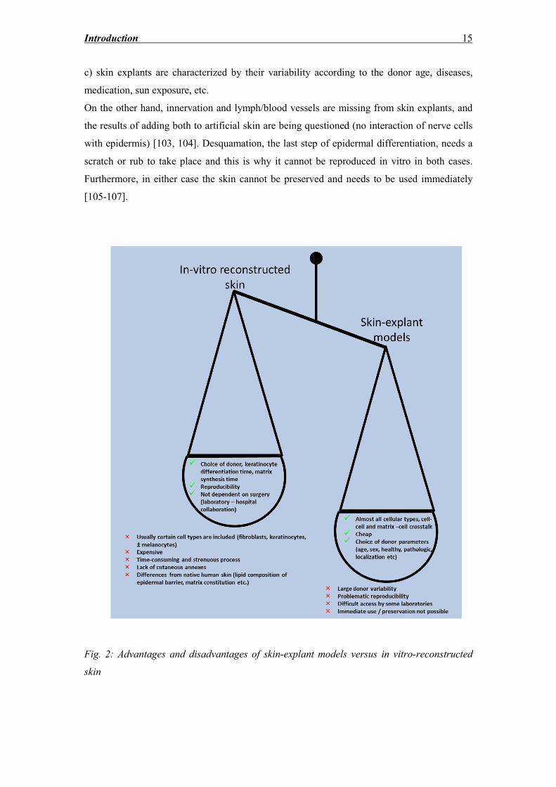
Introduction 15
c) skin explants are characterized by their variability according to the donor age, diseases,
medication, sun exposure, etc.
On the other hand, innervation and lymph/blood vessels are missing from skin explants, and
the results of adding both to artificial skin are being questioned (no interaction of nerve cells
with epidermis) [103, 104]. Desquamation, the last step of epidermal differentiation, needs a
scratch or rub to take place and this is why it cannot be reproduced in vitro in both cases.
Furthermore, in either case the skin cannot be preserved and needs to be used immediately
[105-107].
Fig. 2: Advantages and disadvantages of skin-explant models versus in vitro-reconstructed
skin
Introduction 16
1.6 Methods of culturing primary sebocytes
The culture of sebaceous gland (SG) cells has been a field of experimental interest, since it is
extremely vital for the understanding of the pathophysiology of diseases such as acne
vulgaris, seborrhea or sebostasis. The animal models developed, such as sebocyte-like
differentiating preputial cells [108, 109] would not suffice, since these diseases are
exclusively human. For this, the need for human sebocytes in vitro models was prominent.
Karasek [110] was the first to describe the culture of human sebocytes in a 3T3 feeder layer
after enzymatic dissociation of SG deriving from SG-rich dermal regions, without managing
to retain their characteristics in vivo. Doran et al. [111] altered his method, by immersing 0.4
mm dermal sections in 10 mg/ml dispase, diluted in DMEM containing 10% fetal calf serum
(FCS) and antibiotics for 30 min at 37oC and subsequently in 0.3% trypsin/1% EDTA for 15
min at 37oC. After washing with phosphate buffered saline (PBS), the specimens were
scrapped in serum-containing medium, so that a sufficient number of sebocytes could be
obtained. The suspension was cultured in supplemented Iscove’s medium on a feeder layer of
mitomycin-C-inactivated 3T3 fibroblasts. Xia et al. [112] conceived the idea of SG
microdissection in his approach, based on the fact that trypsinization affects the proliferating
sebocytes and should be avoided, as well as the idea that germinative cells of the SG are
found in the peripheral walls, which need to remain intact in order to provide sufficient
outgrowths of sebocytes later. Small pieces of full-thickness skin were washed with PBS,
treated in 2.4 U/ml dispase for 20 h at 4oC, to separate the epidermis from the dermis, and the
latter was treated with 0.02% desoxyribonuclease at 37oC for 15 min. SG were then
microdissected, their ducts were removed and the globules were seeded on a mitomycin-C-
inactivated 3T3 fibroblast layer in DMEM/Ham’s F12 medium, containing 10ng/ml human
EGF (hEGF), 10% FCS, 0.4μg/ml hydrocortisone, 10-9
M cholera toxin, 3.4 mM L-glutamine,
antibiotics at 37oC with 5% CO2. The sebocytes isolated with this method were subcultured
for a total of three passages. Lee used collagenase to treat the isolated sebaceous glands
before culturing them in William’s E medium supplemented with ITS complex (10 μg/ml
insulin, 10 μg/ml transferrin, 10 ng/ml sodium selenite), 2 mmol/L L-glutamine and
antibiotics [113]. Zouboulis avoided the addition of hydrocortisone in the medium and used a
composition of both human serum and FCS [114]. In order to achieve a better yield of
primary sebocytes, Abdel-Naser proposed the covering of SG lobules with a sterile cover-
glass during the first 72 h of culture, thus providing an excellent contact with the culture plate
and subsequently making it possible to obtain significantly higher numbers of them [115].
Fujie et al. [116] omitted the 3T3 feeder layer but dispersed sebaceous lobules to single cell
solution by trypsinization. Finally, Seltmann et al [117] modified the technique introduced by
Introduction 17
Xia et al. [112], omitted the feeder layer and added to the medium of the microdissected SG
outgrowths 4-10 ng/ml KGF and 1 mg/ml bovine serum albumin (BSA).
1.7 Difficulties of maintaining sebaceous glands and sebocytes in culture
One of the first models developed for studying SG was the maintenance of SG lobules ex
vivo [118]. In this approach, SG were isolated by shearing [119] from normal midline chest
skin of male patients. The specimens were washed in Earle’s balanced salt solution, sliced in
5 mm pieces and repeatedly cut until reaching a porridge-like consistency. The glands were
recognized with a dissecting microscope, surrounding collagen was removed and SG were
maintained floating on polycarbonate filters in 5 ml William’s E medium at 37oC,
supplemented with antibiotics, 2 mM L-glutamine, 10 μg/ml insulin, 10 ng/ml EGF, 10 μg/ml
transferrin, 10 ng/ml hydrocortisone, 10 ng/ml sodium selenite, 3 nM triiodothyronine, 1%
(vol/vol) trace elements mix, 10 μg/ml bovine pituitary extract and buffered in a humidified
chamber with 5% CO2. Phenol-red was not added to the maintenance medium, since its weak
estrogen action affected the lipogenesis rates of the dissected SG. The addition of 10% FCS to
the medium also inhibited lipogenesis. Removing EGF increased the production of sebaceous
lipids over 7 days. In the absence of the former three substances, SG were reported to retain
the in vivo rates of cell proliferation and lipid accumulation, maintaining their in situ
morphology, with peripherally undifferentiated, partially differentiating and mature
sebocytes. The model successfully showed a reduction of lipogenesis through 17β-estradiol
and 13-cis retinoic acid, although the addition of testosterone and 5α-dihydrotestosterone
(DHT) had no stimulatory effect on lipogenesis rates, probably because of absence of the
peroxisome proliferator-activated receptor (PPAR) ligand linoleic acid in the medium [120].
The addition of bovine pituitary extract in the culture medium is also not chemically defined.
Lu et al. [21] developed an organ culture skin model for the long-term study of hair
elongation for over two weeks. It is supported that SG morphology is not altered until day 5.
A careful examination of the photos provided in the publication shows that the sebocytes in
proximity to the germinative layer of the periphery exhibit considerable vacuolization, which
is a sign of potential differentiation. Moreover, no photos showing staining of the SG with
antibodies raised against Ki67 are provided as immunohistochemical evidence that basal
sebocytes still have a proliferative potential. In addition, the epidermis appears completely
degenerated with just a few viable layers from day 12 of organ culture.
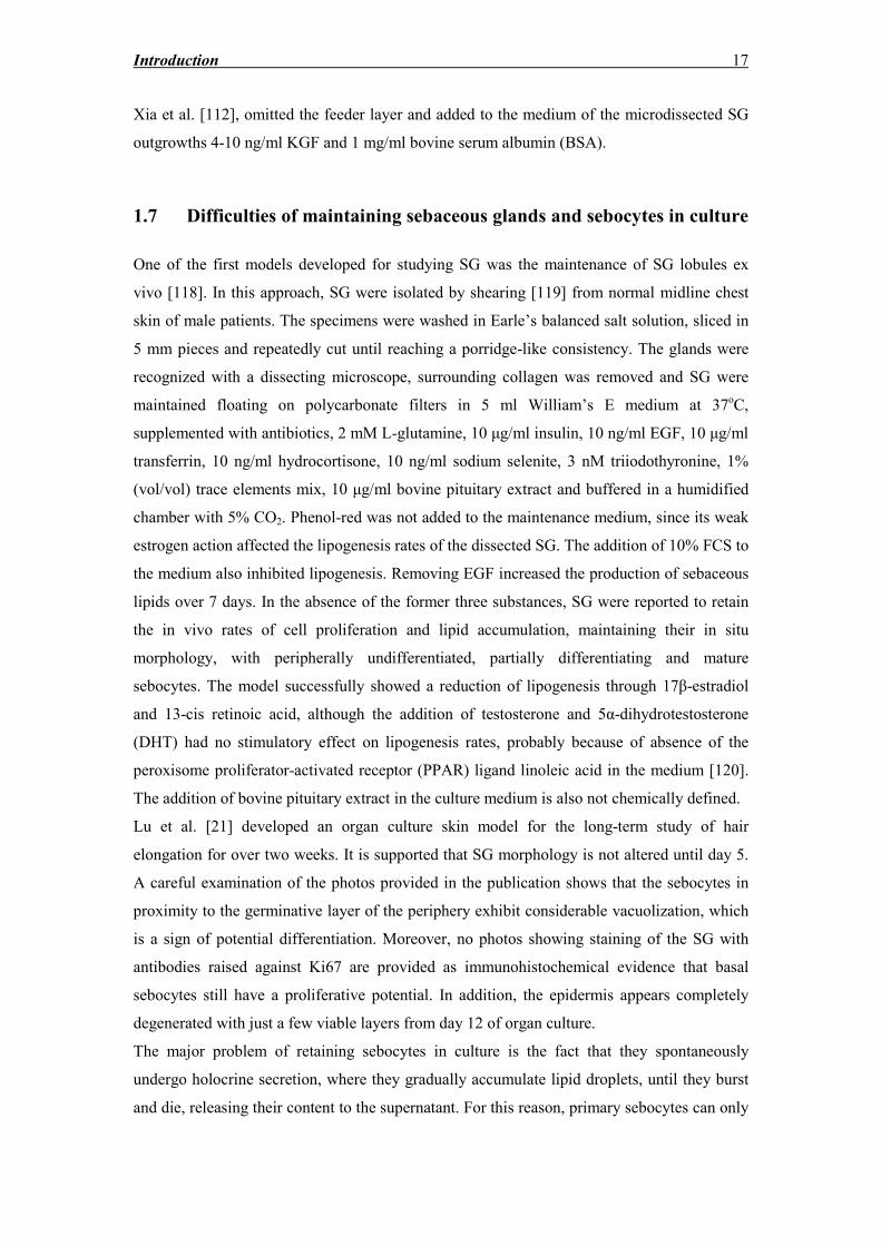
The major problem of retaining sebocytes in culture is the fact that they spontaneously
undergo holocrine secretion, where they gradually accumulate lipid droplets, until they burst
and die, releasing their content to the supernatant. For this reason, primary sebocytes can only
Introduction 18
be subcultured for a maximum of three to six passages. On the other hand, the immortalized
SG cell line SZ95 [121] was shown to present the characteristics of normal sebocytes in vitro.
DNA fragmentation of SZ95 sebocytes was detected already after 6 h, while cell lysis,
assessed by LDH release, could only be detected after 24 h [122]. These data confirm that
sebocytes undergo apoptosis spontaneously (see fig. 4), which could explain their natural
elimination in vitro.
Fig. 3: Sebocytes undergoing holocrine secretion in vivo: Overlay image of TUNEL
(green) staining of a SG to detect cell apoptosis, with DAPI (blue) counterstaining for nuclear
visualization. As it has been shown, DNA fragmentation, as one of the concluding events of
apoptosis, is prominent for differentiating sebocytes, while sebocytes of the basal layer are
not stained.
1.8 The solution of sebocyte cell lines
In order to address the problems of maintaining a model mimicking the SG functions in vitro,
Zouboulis et al. [121] were the first to conceive the idea of an immortalized SG cell line,
which would provide cells able to be subcultured for a sufficient number of passages, while
retaining their fundamental characteristics, namely lipid synthesis and accumulation parallel
to their differentiation. For this, facial SG cells deriving from a 87-year old female patient
were transfected with aPBR-322-based plasmid, which contained the coding region for the
Simian virus-40 large T antigen. The resulting clones, (mainly the clones K7 and K6) were
characterized and were shown to retain their cell type characteristics even after 50
subcultures, without exhibiting any signs of senescence:
Introduction 19
1. Polymorphous appearance with cells of different sizes and abundant lipid
droplets observed in the cytoplasm.
2. Expression of K7, epithelial membrane antigen (EMA), sebaceous gland
antigen (SGA) and other sebocyte markers as determined by
immunocytochemistry and western blotting.
3. Lipid synthesis and composition matching sebum.
4. Inhibition of proliferation induced by treatment with retinoids.
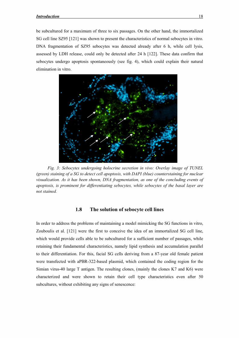
Fig. 4: SZ95 sebocytes in 50% confluency cultured in FCS-containing medium, stained with
antibodies raised against phosphohistone H3 (green) and cleaved PARP (red), indicating
SZ95 sebocytes in the M phase of their cycle (labeled green) and undergoing caspase-
mediated apoptosis (labeled red), respectively. SZ95 undergo apoptosis spontaneously, which
ends with cell rupture and release of the cellular lipid content in the supernatant.
Thiboutot et al. [123], copied the method of Zouboulis et al. [124] to develop a second
immortalized SG cell line from sebocytes isolated from the preauricular area of a 55-year old
male, termed SEB-1. This SG cell line also expresses characteristic sebocyte proteins and
accumulates lipid droplets, which could be detected with Oil red staining.
A third SG cell line was created by Lo Celso et al. [125] from sebocytes isolated from the
face of an adult male after a facelift procedure. This cell line is not fully characterized, since it
was only developed for tumorigenesis studies. The cells were immortalized by transduction
with a retroviral vector containing HPV16/E6E7 genes, packaged in PA317 cells. The
100 μm 100 μm
25 μm
Introduction 20
sebocytes, prior to transduction, were plated on mitomycin C-treated 3T3-J2 cells in
keratinocyte medium. Sebocytes were transduced by co-culture with mitomycin C-treated
packaging cells in the presence of 3T3-J2 cells. Six days later the PA317 and 3T3-J2 cells
were substituted with mitomycin-treated 3T3-J2 NHP cells. The cell line, named Seb-E6E7,
was subcultured for 30 passages, expressing K7, a marker of early sebaceous differentiation.
Surprisingly, both SZ95 and Seb-E6E7 sebocytes were expressing involucrin, which is a
marker of keratinocyte differentiation of interfollicular epidermis. Moreover, after exposure
of SZ95 cells in the air-liquid interface, the basal layers expressed K7, while patches of SZ95
at the suprabasal layers were Nile-Red positive, indicating lipid accumulation. These patches
were involucrin- and cornifin-negative, thus introducing an, at least, bipotent character of
SZ95 differentiation, to either mature sebocytes or interfollicular keratinocytes.
Lastly, a fourth sebocyte cell line, named SEBO662, was generated by copying the method of
Lo Celso et al. [125]. Sebocytes were immortalized with a retroviral vector, containing
HPV16/E6E7 genes, by using a 293T cell-based system [126]. The SEBO662 cells were
cultured for 10 days in the air liquid-interface and expressed the differentiation marker EMA
in suprabasal layers, while it was not abundantly expressed in monolayer culture. On the other
hand, the proliferation marker Ki67 and the early differentiation marker K7 were expressed
abundantly across the reconstituted sebaceous-like epithelium. The SEBO662 cell line is the
less characterized one among four cell lines up to now. The relevant report indicates low
loricrin and filaggrin mRNA levels of SEBO662 cells detected by RT-PCR [127], but no
involucrin studies were performed, which would confirm the data of Lo Celso et al. [125] and
would exclude the possibility of SEBO662 differentiating to an interfollicular keratinocyte
direction [127].
Of the existing cell lines, only the SZ95 SG cell line has been fully characterized, has been
and is being currently used in various laboratory studies worldwide and is internationally
patented, allowing its commercial use after a licensing procedure.
1.9 Common functions of the sebaceous gland
The human SG is a small branched type of multiacinar gland, which can be found in all body
areas except of palms, sole and dorsum of the feet. The SG is an integral part of the
pilosebaceous unit (including also the hair follicle and the arrector pili muscle) and consists of
secretory lobules formed from its epithelial cells, called sebocytes and a short tubular
infundibulum, composed of sebaceous duct cells. Although their number appears to remain
relatively constant with age, their size tends to increase. Numerous dermatological conditions
are correlated to SG disorders, including seborrhea, acne, sebaceous hyperplasia, sebaceous
Introduction 21
adenoma and carcinoma [128, 129]. The multipotent stem cells, which give rise to the
sebaceous cell lineage, reside in the bulge region of the hair follicle. These cells have the
ability to transform to epidermal or follicular keratinocytes or sebocytes [130]. The signal
molecules involved in this process are β-catenin and lymphoid enhancer factor-1 [131, 132].
High levels of β-catenin stimulate the formation of the hair follicle, whilst low levels that of
the SG and epidermis. Overexpression of Lef-1 gene can lead to development of sebaceous
tumors by blocking the β-catenin signaling pathway. Sonic hedgehog is a signaling pathway,
which modulates the terminal differentiation of the hair follicle and inhibition of Wnt genes
through negative dominant Lef-1 results in sebocyte development [133-135].
According to morphological criteria defined by Tosti [136], sebocytes can be classified into 5
differentiation stages: 1. undifferentiated, including the germinative, flattened or cuboidal,
cells with highly basophilic nuclei, 2. early differentiated type, 3. advanced differentiated
type, 4. fully differentiated type, 5. mature sebocytes.
The cardinal role of the SG is the production and secretion of sebum, which is a species-
specific mixture of lipids (see fig. 5) [137]. Its uniqueness among species is probably due to
the functions that sebum has to absolve for every species, which in the case of humans are its
antimicrobial activity, photoprotection, anti-oxidant delivery to the skin surface and
participation in inflammatory processes, through specific lipids. However, all the functions of
human sebum are far from elucidated [135, 138, 139].
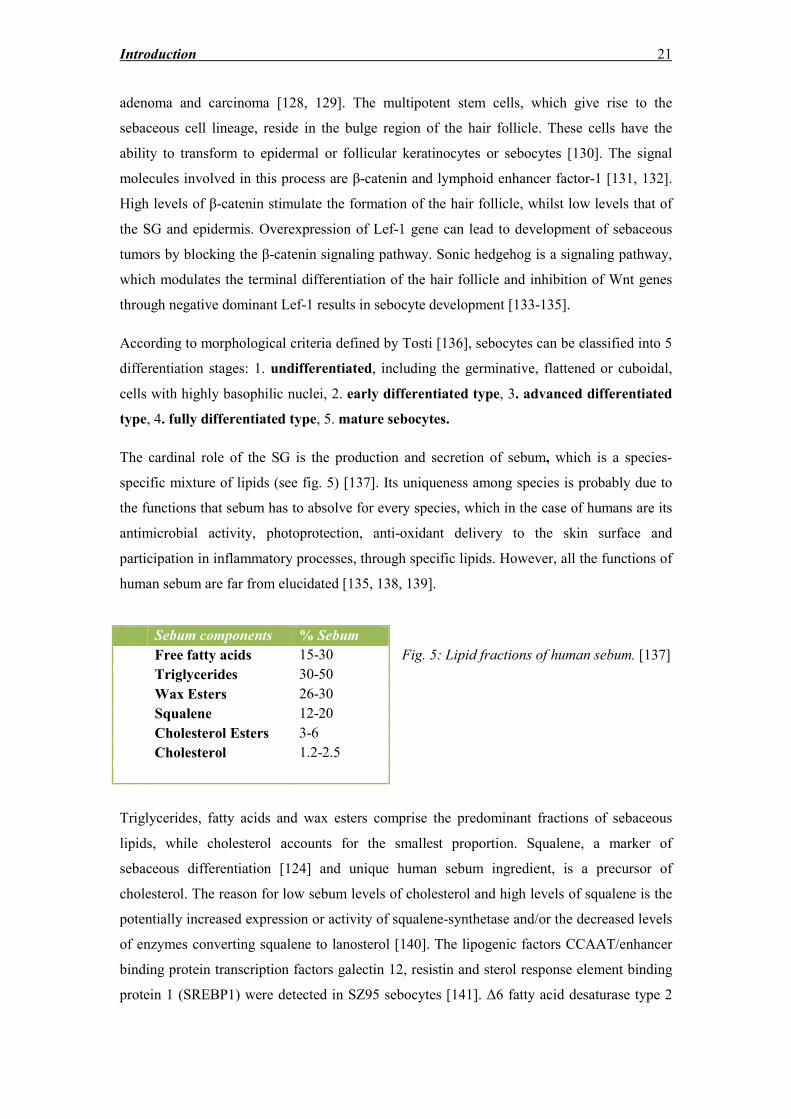
Fig. 5: Lipid fractions of human sebum. [137]
Triglycerides, fatty acids and wax esters comprise the predominant fractions of sebaceous
lipids, while cholesterol accounts for the smallest proportion. Squalene, a marker of
sebaceous differentiation [124] and unique human sebum ingredient, is a precursor of
cholesterol. The reason for low sebum levels of cholesterol and high levels of squalene is the
potentially increased expression or activity of squalene-synthetase and/or the decreased levels
of enzymes converting squalene to lanosterol [140]. The lipogenic factors CCAAT/enhancer
binding protein transcription factors galectin 12, resistin and sterol response element binding
protein 1 (SREBP1) were detected in SZ95 sebocytes [141]. Δ6 fatty acid desaturase type 2
Sebum components % Sebum
Free fatty acids 15-30
Triglycerides 30-50
Wax Esters 26-30
Squalene 12-20
Cholesterol Esters 3-6
Cholesterol 1.2-2.5
Introduction 22
(FADS2), which catalyzes among others the conversion of palmitic acid to the human-
specific sapienic acid, is detected predominantly in differentiated sebocytes [142]. Elongation
of sapienic acid by two carbon atoms leads to another unique derivative of human sebum,
sebaleic acid [143]. Linoleic acid is an essential fatty acid, which undergoes peroxisomal β-
oxidation to arachidonic acid and other fatty acids, which are sebocyte-specific and correlate
to their differentiation level [144].
1.10 Sebaceous gland as a target of circulating hormones and as a site of
steroid hormone synthesis
The experimental field of dermatoendocrinology managed to establish the concept of skin as
an endocrine organ per se, which is more than a passive target of sex hormones. Skin is a
“factory” with all the necessary enzymatic equipment to utilize sex steroid precursors in order
to synthesize more potent sex hormones in an intracrine manner, as well as to facilitate their
de novo synthesis [139, 145, 146]. In this process, the pilosebaceous unit has a fundamental
role [146, 147].
Testosterone and DHT were shown to promote sebocyte proliferation in vitro at
concentrations higher than physiological levels [121, 148], while its synergistic effect with
the PPAR ligand linoleic acid resulted in increased lipogenesis [120]. Interestingly, sebocytes
are capable of synthesis of cholesterol from acetate [149, 150], which is used for the
formation of the cell membrane, the epidermal barrier, sebum and cutaneous steroids [148].
The synthesis of dehydroepiandrosterone (DHEA), the main substrate for the more potent
androgens testosterone and DHT is formed by cholesterol in the skin through the four
upstream enzymes: StAR, P450scc, p450c17 and 3β-hydroxysteroid dehydrogenase (3β-
HSD).
The androgen receptor is expressed in SZ95 sebocytes [151]. 3β-HSD converts DHEA to
androstenedione [69], which is converted in a further step to testosterone by the enzyme 17β-
HSD. 3β-HSD2 mRNA is expressed primarily in the SG [152]. Interestingly, 5 isozymes of
17β-HSD were identified, functioning like an “on-off switch” mechanism for the production
of more potent sex steroids: Isozymes 3 and 5 catalyze the formation of T from
androstenedione, in contrast to isozymes 2 and 4, which oxidize the inactivation of T to its
weaker precursor [153-156]. 17β-HSD3 synthesizes T from androstenedione in Leydig cells
of the testis, while in skin and other peripheral tissues the reaction is catalyzed by 17β-HSD
type 5 [157]. Greater activity of the 17β-HSD types 3 and 5 was detected from sebaceous
Introduction 23
glands of facial skin than other, non-acne prone skin areas, suggesting the in situ more potent
androgen formation in these areas [156, 158], while 17β-HSD2, which can inactivate potent
androgens, was found mostly in sebaceous glands of non-acne prone areas in comparison to
facial skin [154]. This indicates that sebaceous gland utilizes special steroidogenic enzymes to
fine tune the expression of potent androgens of skin in situ.
5α-reductase (5αR) catalyzes the conversion of testosterone to DHT. Of the three isoforms of
5αR that have been described [159], type 1 is the predominant one in the skin [160, 161] and
more abundantly expressed in SG and sweat glands [162], keratinocytes and dermal
fibroblasts [163]. The newly found 5αR3 is detected in prostate cancer and SZ95 sebocytes
[164].
On the other hand, aromatase, the rate-limiting enzyme of estrogen synthesis [169], was
reported to be expressed in anagen and terminal HFs, cultured keratinocytes, melanocytes, SG
and adipose fibroblasts [170]. Both estrogen intracellular receptors (ERα and ERβ) were
immunohistochemically detected in human sebocytes in situ, but ERα was restricted in basal
sebocytes [171].
SG are also involved in glucocorticoidogenesis, since they express the enzyme 11β-HSD,
which catalyzes the formation of active cortisol from deoxycorticosterone and 11-
deoxycortisol and modulates sebum production and Toll-like receptor (TLR)-2 expression
[172].
1.11 Sebocytes as target of various other hormones
Sebocytes express a variety of receptors for other peptide hormones, growth factors,
neurotransmitters such as:
Peptide hormone receptors
Insulin-like growth factor (IGF)-1 receptor which can be activated by IGF-1 secreted from
fibroblasts or high concentrations of insulin [173]. IGF-1 induces lipogenesis and sebaceous
differentiation in sebocyte cell lines [174] and rat preputial gland cells, combined with growth
hormone (GH). GH receptor activation in human sebocytes augments lipid accumulation
induced by the potent androgen DHT [175].
Corticotropin-releasing hormone (CRH) receptors 1 and 2, with CRH receptor 1 being the
predominant receptor in the sebaceous gland. CRH inhibits proliferation of sebocytes in vitro
and promotes lipogenesis and IL-6 and IL-8 secretion [176, 177].
Introduction 24
Melanocortin-1 and melanocortin-5 receptors were detected on the membrane of human
sebocytes. Through binding of α-melanocyte stimulating hormone (MSH), they regulate
inflammation cascades in human sebocytes. More specifically, α-MSH was found to suppress
both basal and IL-1β-induced secretion of IL-8 in SZ95 sebocytes [178]. Melanocortin -5R is
weakly expressed but is considered a marker of terminal sebocyte differentiation [179].
Cannabinoid receptors 1 and 2 are found in differentiated and basal sebocytes, respectively,
and bind endocannabinoids, which affect sebaceous differentiation [180]. Histamine receptor
activation affects endogenous squalene levels [181]. M-opioid receptors are also present and
bind β-endorphin, which stimulates lipogenesis and increases certain fractions of sebaceous
fatty acids [182]. VPAC receptors bind neuropeptide Y, vasoactive intestinal polypeptide
(VIP) and calcitonin gene-related peptide (CGRP) [183].
Nuclear receptors
Apart from the androgen receptor and ERα and ERβ, other nuclear receptors, which are
expressed in human sebocytes, are:
Progesterone receptors: expressed in the nucleus of undifferentiated sebocytes [184].
Retinoic acid receptors and retinoid X receptors, with all-trans retinoic acid (atRA) and 9-cis
retinoic acid as natural ligands respectively [185, 186]. Isotretinoin inhibits the SZ95 sebocyte
proliferation rate through its intracellular transformation to atRA.
PPAR: PPAR subtypes (α, γ, δ) are present in human sebaceous glands and sebocyte cell
lines in mRNA and protein level [187-189]. The predominant PPAR in human sebaceous
glands are the subtypes α and γ1. PPARα has as natural ligands arachidonic acid and
leukotriene B4 (LTB4), regulates sebaceous differentiation, lipid accumulation and
inflammation [190]. PPARγ promotes lipogenesis and differentiation of sebocytes [189],
while regulating inflammation pathways through upregulation of cyclooxygenase (COX) 2
expression and therefore prostaglandin (PG) E2 production [191]. Contradictory data were
provided by Downie et al. [188], which have shown that treatment of whole human sebaceous
gland cultures with PPARα and PPARγ agonists results in an inhibition of sebaceous
lipogenesis. Contradictory results were also obtained from in vivo data, underlining the need
of different experimental models to elucidate their action in sebocyte function [192].
Activation of PPARγ by its agonists, such as troglitazone, results in upregulation of COX2
and PGE2 in mRNA and protein level [191]. PPARδ is involved in terminal sebocyte
differentiation [193]. Treatment of SZ95 sebocytes with a PPARδ agonist resulted in
suppression of basal and staurosporine-induced apoptosis, providing a potential explanation
for the sebostasis induced by these substances [194]. PPAR negatively regulate the
transcription of inflammatory response genes by antagonizing the AP-1, and by promoting the
catabolism of proinflammatory eicosanoids [195].
Introduction 25
Vitamin D receptor: SZ95 sebocytes express all the enzymatic machinery for the synthesis of
vitamin D (D-25-hydroxylase, 25 hydroxyvitamin D-1α-hydroxylase and 1,25-
dihydroxyvitamin D-24-hydroxylase). Vitamin D inhibits SZ95 proliferation of rapidly
growing sebocytes, promotes their proliferation in slow growing culture and modulates lipid
accumulation and secretion of IL-6 and IL-8 [196]. Moreover, the culture of primary
sebocytes with vitamin D promotes the expression of cathelicidin, one of the antimicrobial
peptides related to cutaneous non-specific immunity [197].
Liver X receptors (LXR, α and β isotypes): SZ95 sebocytes express both receptors at the
mRNA and protein levels [182].
Receptors which do not belong to the aforementioned categories:
The vanilloid receptor (VR) belongs to the transient ion channels and is expressed in early
differentiated sebocytes. Its ligand, capsaicin, was shown to inhibit SZ95 sebocyte
proliferation [198]. Fibroblast growth factor receptors, EGF receptor, the proto-oncogene c-
met product (c-MET) are also expressed in sebocytes, placing the sebaceous glands in a
prominent position of systemic and cutaneous molecular cross-talk due to the variety of
molecular signals, which can modulate its function.
1.12 Sebaceous gland and its role in endogenous and adaptive immunity
Sebum fatty acids play an important role to the initiation of the inflammatory process induced
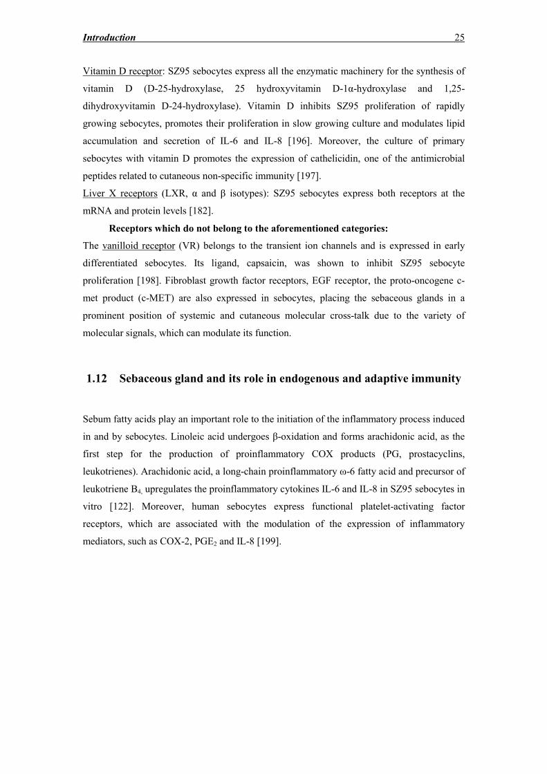
in and by sebocytes. Linoleic acid undergoes β-oxidation and forms arachidonic acid, as the
first step for the production of proinflammatory COX products (PG, prostacyclins,
leukotrienes). Arachidonic acid, a long-chain proinflammatory ω-6 fatty acid and precursor of
leukotriene B4, upregulates the proinflammatory cytokines IL-6 and IL-8 in SZ95 sebocytes in
vitro [122]. Moreover, human sebocytes express functional platelet-activating factor
receptors, which are associated with the modulation of the expression of inflammatory
mediators, such as COX-2, PGE2 and IL-8 [199].
Introduction 26
Fig. 6: Fatty acid metabolism, modified from: Angres S. Thesis, Freie Universität Berlin,
2009
Expression of tumor necrosis factor (TNF)-α in sebocytes is stimulated by Propionibacterium
acnes (P.acnes) [200]. TNF-α treatment resulted in promotion of lipid accumulation of SZ95
sebocytes, activation of the lipogenesis transcription factor sterol-regulatory element binding
protein (SREBP)-1 and the death receptor FAS, which is involved in apoptosis. Moreover,
both phosphatidyl-inositol-3-kinase (PI3K), Akt and c-Jun N-terminal kinase (JNK) pathways
are involved in this process [201].
TLR are transmembrane proteins belonging to the family of pattern-recognition receptors,
which can initiate responses to specific pathogen molecules relatively conserved among
various microbe species [202]. These invariant molecular structures are characterized as
pathogen-associated molecular patterns [203]. TLRs can be activated through
lipopolysaccharide (LPS – gram negative bacteria), lipoteichoic acid (LTA – gram positive
bacteria) or peptidoglycan and induce the cytokine release from SZ95 sebocytes [200]. TLR4
is a receptor highly specific for LPS, while TLR2 is a co-receptor for LPS and is involved in
the recognition of a variety of other molecules deriving from gram-positive bacteria [204].
LPS was shown to stimulate IL-8 expression of human sebocytes, without alteration of IL-1α
expression at protein and mRNA level [204]. The signal cascade after activation of TLR2 or
TLR4 results in the activation of the NF-κB complex through the myeloid differentiation
protein (MyD88) and the IL-1 receptor-associated kinase in a number of cell types [204, 205].
The pilosebaceous unit is also implicated in cutaneous immunological activity, involving
Introduction 27
functions such as MHC class I expression, and expression of CD14, TLR2 and TLR4 [206].
Human sebocytes express TLR2, TLR4, TLR6 and CD14 [206, 207]. TLR2, which is activated
by P. acnes, triggers adaptive immunity mechanisms [206]. In acne lesions, follicular
occlusion promotes proliferation of P. acnes, through the development of anaerobic
conditions. P .acnes virulence factors and pattern recognition ligands stimulate skin immune
response resulting in inflammation [208]. Soluble factors released from hyperproliferating P.
acnes populations diffuse through sebum and reach the SG, allowing the enhancement of
sebaceous lipogenesis and the promotion of inflammatory reactions mediated by infiltrated
immune cells [199]. The major components, which induce immune responses in
keratinocytes and sebocytes, are peptidoglycan and lipoteichoic acid (LTA). The latter has
been shown to suppress the expression of both TLR2 and TLR4 in sebocytes [204]. Moreover,
P. acnes stimulates the production of proinflammatory cytokines, including IL-1β, IL-8, IL-
12 and TNF-α [202]. Bacteria-derived macrophage-activating lipopeptide-2, which is a TLR2
ligand, upregulates both stearoyl coenzyme A desaturase (SCD) and fatty acid Δ6 desaturase
2 (FADS2) in SZ95 sebocytes on the mRNA level [207]. Antibodies raised against inactive
vaccines of P. acnes attenuated IL-8 production from SZ95 sebocytes [209]. Although
lipogenesis and inflammation are indisputably augmented by P. acnes, sebocytes can produce
free fatty acids and cytokines in a basal level, even in the absence of bacteria [199].
Sebocytes are known to produce antimicrobial peptides, a heterogenous group of proteins
with antimicrobial properties against a variety of microbial pathogens [210]. Human beta
defensins (hBD-1 and hBD-2) were detected in pilosebaceous units by immunohistochemistry
and in situ hybridization [211]. Another antimicrobial peptide, psoriasin (S100A7), was also
expressed in the SG [212]. Moreover, P. acnes and lipopolysaccharides (LPS) stimulate the
production of antimicrobial peptides, such as cathelicidin and hBD-2 in SZ95 sebocytes [200,
213]. hBD-1 has no direct bactericidal effects on P. acnes, but it can act synergistically with
cathelicidin [213].
Moreover, apart from the antimicrobial peptides produced by sebocytes, it was recently
shown that histone H4 was exhibiting substantial antimicrobial activity after being isolated
from acid-soluble protein extracts of a sebaceous cell-line [214]. Histone H4 was identified as
one of the predominant peptides exerting antimicrobial effects against Staphylococcus aureus
(S. aureus) and P.acnes. Histones are known as major components of the nucleosome
structure in eukaryotic cells, but their fragments can also have antimicrobial activity. Histone
H4 as well as H2A, H2B and H3, are able to bind to LPS [215]. Interestingly, histone H4
exhibited synergistic effect with antimicrobial fatty acids against S. aureus. The utilization of
histone H4 as an immune defense system against opportunist pathogens provides an attractive
theory in the sebaceous gland function setting, where mature sebocytes undergo holocrine
Introduction 28
secretion and release their intracellular content (including histones) onto the surface of the
skin and exert their antimicrobial properties.
Apart from the antimicrobial peptides in the human SG, sebum lipids were shown to exert
innate antimicrobial properties [207, 216]. The SCD, which is responsible for the synthesis of
monounsaturated fatty acids, is expressed by human sebocytes in vitro and in vivo [141, 199].
Sebum consists of monounsaturated fatty acids, predominantly the ω-9 fatty acid palmitic
acid and oleic acid, which have bactericidal properties against Gram-positive bacteria [207].
Furthermore, the sebocyte-specific fatty acid sapienic acid surprisingly exhibits antibacterial
activity against gram positive-bacteria, such as P. acnes [217]. Oleic acid predominates in
human sebum and lauric acid, although a minor fatty acid, is one of the most potent
antimicrobial peptides against Gram-positive bacteria [217]. Sebocyte vesicles containing
squalene have a protective effect on the skin surface [130].
Aim of Study 29
Aim of Study
Aim of this study was primarily:
1. to develop a simple, robust, reproducible, three-dimensional human skin model
including human sebocytes for studying the pathophysiology of skin diseases,
especially sebostasis, seborrhea and acne,
2. to explore various experimental settings in order to elucidate the potential effects of
the co-culture of ex vivo skin with SZ95 sebocytes in skin viability and structural
integrity,
3. to explore various experimental settings in order to detect the effects of the SZ95
sebocyte-skin explant molecular cross talk on SZ95 sebocyte morphology and
function.
Materials and Methods 30
Materials and Methods
2.1 Materials, media, solutions and equipment
Reagents, solutions and salts
Dulbecco’s PBS w/o Ca++
, Mg++
(Pan Biotech, Aidenbach, DE)
Calcium chloride dehydrate (Roth, Karlsruhe, DE)
h-EGF (Sigma, Munich, DE)
ROL (Biochrom, Berlin, DE)
LA (Sigma, Munich, DE)
Gm 50 mg/ml (Roth, Karlsruhe, DE)
Panexin – NTA (Pan Biotech, Aidenbach, DE)
Fetal bovine serum (FBS) Superior (Biochrom AG, Berlin, DE)
BSA 7,5% (Sigma, Munich, DE)
Amphotericin B 250 μg/ml (Biochrom AG, Berlin, DE)
Trypsin/ EDTA 0.05/0.02% in PBS (Biochrom AG, Berlin, DE)
DMSO (Sigma, Munich, DE)
Isopropanol 100% (Roth, Karlsruhe, DE)
Triton X-100 (Roth, Karlsruhe, DE)
Ethanol 70% and 100% (Roth, Karlsruhe, DE)
Tween® 20 (Roth, Karlsruhe, DE)
Dispase (Invitrogen, Darmstadt, DE)
Oil red (Sigma, Munich, DE)
Formaldehyde solution 37% (Roth, Karlsruhe, DE)
Xylol (Roth, Karlsruhe, DE)
Isopropanol 100% (Roth, Karlsruhe, DE)
Parafilm (Roth, Karlsruhe, DE)
Entellan® mounting medium (Merck, Darmstadt, DE)
Mayer’s Hemalaun (Merck, Darmstadt, DE)
Eosin (Thermofischer Scientific, Bremen, DE)
Materials and Methods 31
Roti®-Mount Aqua mounting medium (Roth, Karlsruhe, DE)
Detection Systems for IHC & ICC
DAB detection system EnVisionTM
FLEX Detection System
(Dako Deutschland GmbH, Hamburg, DE)
Alkaline Phosphatase Detection system REALTM
Detection system, Alkaline
Phosphatase/RED, Rabbit/Mouse (Dako
Deutschland GmbH, Hamburg, DE)
Heat-induced epitope Retrieval Solution pH=6,1 Target Retrieval Solution 10x Concentrate
(Dako Deutschland GmbH, Hamburg, DE)
Proteinase K (ready to use) (Dako Deutschland GmbH, Hamburg, DE)
Primary Antibody Diluent Antibody Diluent with Background
Reducing Components (Dako Deutschland
GmbH, Hamburg, DE)
Media and solutions:
SH-Med: Sebomed® basal Medium (Biochrom AG, Berlin, DE)
+ 10% FBS
+ 50 μg/ml Gm
+ 10 ng/ml h-EGF
+ 1 mM CaCl2
SF-Med: Sebomed® basal Medium (Biochrom AG, Berlin, DE)
+ 0.1 % BSA
+ 5 ng/ml h-EGF
+ 50 μg/ml Gm
+ 1.5 mM CaCl2
+ 1.5 x 10⁻⁷
M LA
+ 10-6
M ROL
SS-Med: SF - Med
+ 10% Panexin-NTA
F-Med: DMEM/F12 (Invitrogen, Darmstadt, DE)
+ 10% FBS
+ 50 μg/ml Gm
Freezing Medium: SH-Med
+ 10% FBS
+ 10% DMSO
Materials and Methods 32
Disinfection solution: PBS
+ 50 μg/ml Gm
+ 2.5 μg/ml Amphotericin B
Plasticware:
25 cm2, 75 cm
2 cell culture flasks (TPP, Trasadingen, CH)
50 ml centrifuge tubes (TPP, Trasadingen, CH)
24 and 96 Well-plates (TPP, Trasadingen, CH)
Millicell®-PCF Culture 24 Well-plate Inserts (Merck Millipore, MA, US)
Serological Pipettes 1, 2, 5, 10, 20, 50 ml (Corning B.V. Life Sciences, Amsterdam,
NL)
Pipette tips (Roth, Karlsruhe, DE)
0.2 μm filters (TPP, Trasadingen, CH)
Cryotubes (Nalgene® cryogenic vials, Rochester, USA)
100 mm BD Falcon Petri dishes (BD Biosciences, Heidelberg, DE)
Eppendorf Lo-Bind tubes 1,5 ml, 2 ml (Eppendorf, Hamburg, DE)
Rotilabo® Staining Chamber (Roth, Karlsruhe, DE)
Equipment:
Centrifuges Beckman GS-15R (Beckman Coulter, Krefeld, DE)
Eppendorf 5415C (Eppendorf, Hamburg, DE)
Microcentrifuge DW-41 Qualitron (Qualitron Systems, CN)
Spectrophotometer/ELISA reader VERSAMax (Molecular Devices, Biberach, DE)
Fluorescence reader SpectraMax Gemini (Molecular Devices, Biberach, DE)
Vortexer MSI Minishaker (IKA®, Works Inc, Wilmington NC,
USA)
Magnetic Stirrer IKA RH Basic 2 (IKA®, Works Inc, Wilmington NC,
USA)
Vibrating Platform Shaker Heidolph Titramax 100 (Heidolph Instruments GmbH &
Co, Schwabach, DE)
Incubators APT lineTM
C150 CO2-Incubator (Binder GmbH,
Tuttlingen, DE)
Materials and Methods 33
Heraeus HeraCell (Heraeus Instruments, Kendro
Laboratory Products GmbH, Hanau, DE)
Microscope Olympus CK40 (Olympus, Tokyo, JP)
Microscope Camera JVC TK-C1381 Color video camera (JVC Deutschland,
Friedberg, DE)
Fluorescence microscope DM2000 (Leica GmbH, Wetzlar, DE)
Laminar flow bench HeraSafe HS (Heraeus Instruments, Kendro Laboratory
Products GmbH, Hanau, DE)
Western Blot Power Supply PowerPac Basic Power Supply (BioRad Laboratories
GmbH, Munich, DE)
Minigel electrophoresis System (BioRad Laboratories GmbH, Munich, DE)
Paraffin Embedding Center Leica EG 1160 (Leica Microsystems GmbH, Nussloch,
DE)
Tissue Processor Shandon CitadelTM
1000 (Thermofischer Scientific,
Bremen, DE)
IHC Autostainer DAKO IHC Autostainer (Dako, Hamburg, DE)
Processing / Embedding cassettes Shandon (Thermofischer Scientific, Bremen, DE)
Histology slides and cover glasses (Roth, Karlsruhe, DE)
IHC slides FLEX IHC Microscope Slides (Dako, Hamburg, DE)
2.2 Cell culture methods
2.2.1 Cell culture basics
All work concerning cell culture and maintenance took place under sterile conditions
provided by a laminar flow. The cells were maintained in 25 cm2 and 75 cm
2 flasks, according
to the amount of cells needed for every experiment. The flasks were kept in the incubator at
37oC and 5% CO2 in a humidified atmosphere. The reason for this is the strict pH
maintenance, which is assured by the use of sodium bicarbonate/carbonic acid as a buffer for
the medium. Medium was changed every other day.
2.2.2 Cell lines
Human immortalized SZ95 sebocytes
SZ95 sebocytes [121] derive from human sebocytes obtained from the facial region of a 87-
year old woman, which were immortalized after transfection with a PBR-322-based plasmid,
carrying the coding region for the Simian Virus 40 large T-antigen. The resulting proliferating
cell cultures were passaged up to over 50 times, while retaining their basic morphological and
functional characteristics, without showing any type of senescence [114, 122, 218]. For this
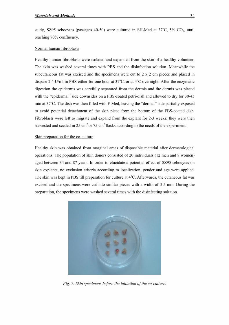
Materials and Methods 34
study, SZ95 sebocytes (passages 40-50) were cultured in SH-Med at 37oC, 5% CO2, until
reaching 70% confluency.
Normal human fibroblasts
Healthy human fibroblasts were isolated and expanded from the skin of a healthy volunteer.
The skin was washed several times with PBS and the disinfection solution. Meanwhile the
subcutaneous fat was excised and the specimens were cut to 2 x 2 cm pieces and placed in
dispase 2.4 U/ml in PBS either for one hour at 37oC, or at 4
oC overnight. After the enzymatic
digestion the epidermis was carefully separated from the dermis and the dermis was placed
with the “epidermal” side downsides on a FBS-coated petri-dish and allowed to dry for 30-45
min at 37oC. The dish was then filled with F-Med, leaving the “dermal” side partially exposed
to avoid potential detachment of the skin piece from the bottom of the FBS-coated dish.
Fibroblasts were left to migrate and expand from the explant for 2-3 weeks; they were then
harvested and seeded in 25 cm2 or 75 cm
2 flasks according to the needs of the experiment.
Skin preparation for the co-culture
Healthy skin was obtained from marginal areas of disposable material after dermatological
operations. The population of skin donors consisted of 20 individuals (12 men and 8 women)
aged between 34 and 87 years. In order to elucidate a potential effect of SZ95 sebocytes on
skin explants, no exclusion criteria according to localization, gender and age were applied.
The skin was kept in PBS till preparation for culture at 4oC. Afterwards, the cutaneous fat was
excised and the specimens were cut into similar pieces with a width of 3-5 mm. During the
preparation, the specimens were washed several times with the disinfecting solution.
Fig. 7: Skin specimens before the initiation of the co-culture.
Materials and Methods 35
After disinfection the specimens were washed with PBS several times and were left floating
in SF-Med with the epidermis facing upwards overnight at 37oC, 5% CO2.
The next day, skin pieces were placed in direct or humoral contact of the dermis with
confluent SZ95 sebocytes or NHFs seeded the previous day.
Skin/SZ95 sebocytes in direct contact OR direct contact co-cultures: For the case of direct
contact, 400 μl was estimated to be the ideal volume of medium, so that the epidermis would
remain exposed to the air: (i.e. culture at the air-liquid interface). The co-culture was
performed for a period of 6 days. In certain experiments, normal human fibroblasts were used
as controls (Skin/fibroblasts in direct contact).
Skin/SZ95 sebocytes in humoral contact OR humoral contact co-cultures: For the case of
humoral contact, 600 μl was estimated to be a sufficient medium volume so that the uplifted
skin explant could be nourished during the co-culture. Standing inserts with a pore size of 0.4
µm were used to facilitate the contact exclusively through the culture medium. Once again, in
certain experiments, normal human fibroblasts were used as controls (Skin/fibroblasts in
direct contact).
A schematic presentation of the co-culture is shown in Fig. 8.
2.2.3 Freezing, thawing, subculturing and counting of cells
Freezing of cells:
Quick freezing of a cell population may result in cell death, because of the ice crystals, which
form from H2O and electrolyte and pH disturbances. In order to avoid cell death, usually
special solutions containing special cryoprotective agents, such as dimethyl sulphoxide
(DMSO), are used, which lower the freezing point of the solution. DMSO is toxic in
concentrations higher than 0.5%, which is the reason that after the cells are suspended in the
cryo-solution, they need to be placed in -70oC as quickly as possible. Moreover, isopropanol
is used to lead to a steady and gradual temperature drop of the cell suspension (about 1oC per
min) from room temperature to -70oC. In this manner, enough time is given for the water
inside the cells to move osmotically outside the cell and the fatal effects of intracellular
freezing are avoided [219]. In addition, it is generally advised to change the medium 24 h
before freezing the cells and to use cells before they reach the stationary phase.
Materials and Methods 36
Process:
Subconfluent cells were washed with PBS twice, trypsinised and counted as described below.
The suspension was centrifuged at 200 g for 6 min and the supernatant was carefully
aspirated. The pellet was re-suspended in freezing medium to a concentration of 3 x 10⁶
cells/ml and 1 ml of cryo-suspension was added to every cryo-tube. The cryo-tubes were
placed in a box containing 200 ml isopropanol and stored for 24 to 72h at -70oC. Then the
cryo-tubes were transferred to the liquid nitrogen storage tank (-130oC) to further suppress the
water crystal formation.
Thawing of cells:
The principle of thawing the cells is exactly opposite to that of freezing them and consists in
bringing them rapidly up to room temperature, since slow thawing can exacerbate the toxic
effects of DMSO on living cells.
Process:
The vials were removed from the liquid nitrogen storage tank and the cap was twisted to a
quarter to relieve the internal pressure and is then retightened. The vial was then thawed by
being kept in the palm for some minutes or by being immersed in a 37oC water bath. The
content of the vial was then immediately diluted 1:30, so that the new concentration of
DMSO would not surpass 0.5%. The cells were seeded to culture flasks after thawing and
were not immediately used for experiments.
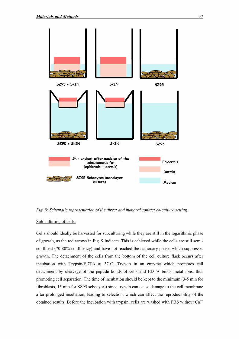
Materials and Methods 37
Fig. 8: Schematic representation of the direct and humoral contact co-culture setting
Sub-culturing of cells:
Cells should ideally be harvested for subculturing while they are still in the logarithmic phase
of growth, as the red arrows in Fig. 9 indicate. This is achieved while the cells are still semi-
confluent (70-80% confluency) and have not reached the stationary phase, which suppresses
growth. The detachment of the cells from the bottom of the cell culture flask occurs after
incubation with Trypsin/EDTA at 37oC. Trypsin in an enzyme which promotes cell
detachment by cleavage of the peptide bonds of cells and EDTA binds metal ions, thus
promoting cell separation. The time of incubation should be kept to the minimum (3-5 min for
fibroblasts, 15 min for SZ95 sebocytes) since trypsin can cause damage to the cell membrane
after prolonged incubation, leading to selection, which can affect the reproducibility of the
obtained results. Before the incubation with trypsin, cells are washed with PBS without Ca++
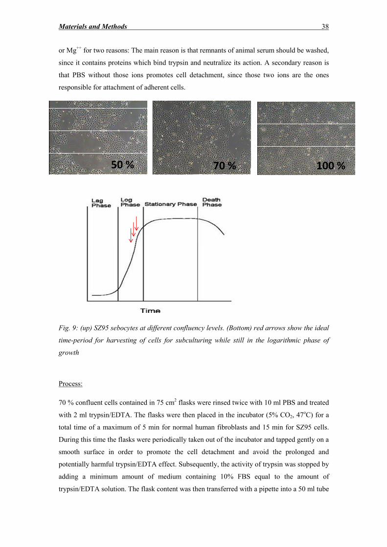
Materials and Methods 38
or Mg++
for two reasons: The main reason is that remnants of animal serum should be washed,
since it contains proteins which bind trypsin and neutralize its action. A secondary reason is
that PBS without those ions promotes cell detachment, since those two ions are the ones
responsible for attachment of adherent cells.
Fig. 9: (up) SZ95 sebocytes at different confluency levels. (Bottom) red arrows show the ideal
time-period for harvesting of cells for subculturing while still in the logarithmic phase of
growth
Process:
70 % confluent cells contained in 75 cm2 flasks were rinsed twice with 10 ml PBS and treated
with 2 ml trypsin/EDTA. The flasks were then placed in the incubator (5% CO2, 47oC) for a
total time of a maximum of 5 min for normal human fibroblasts and 15 min for SZ95 cells.
During this time the flasks were periodically taken out of the incubator and tapped gently on a
smooth surface in order to promote the cell detachment and avoid the prolonged and
potentially harmful trypsin/EDTA effect. Subsequently, the activity of trypsin was stopped by
adding a minimum amount of medium containing 10% FBS equal to the amount of
trypsin/EDTA solution. The flask content was then transferred with a pipette into a 50 ml tube
50 % 70 % 100 %
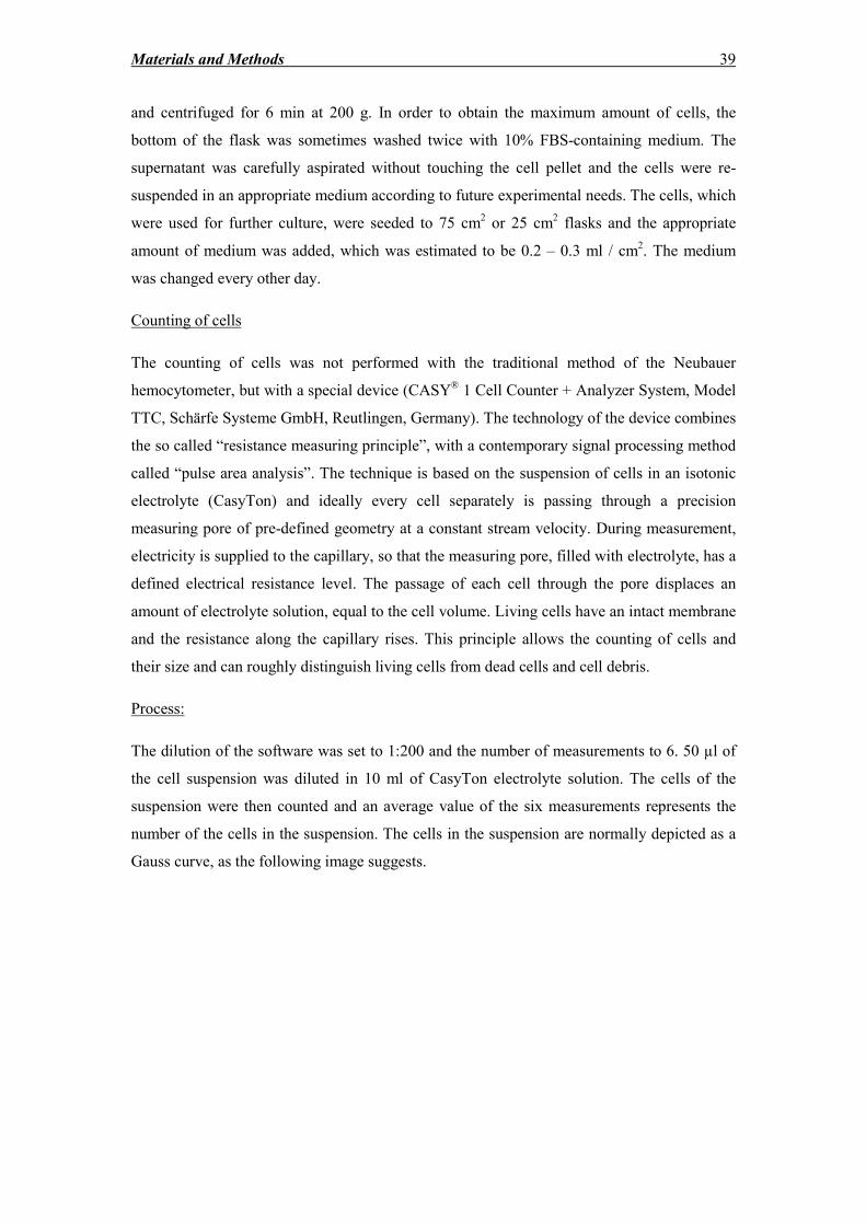
Materials and Methods 39
and centrifuged for 6 min at 200 g. In order to obtain the maximum amount of cells, the
bottom of the flask was sometimes washed twice with 10% FBS-containing medium. The
supernatant was carefully aspirated without touching the cell pellet and the cells were re-
suspended in an appropriate medium according to future experimental needs. The cells, which
were used for further culture, were seeded to 75 cm2 or 25 cm
2 flasks and the appropriate
amount of medium was added, which was estimated to be 0.2 – 0.3 ml / cm2. The medium
was changed every other day.
Counting of cells
The counting of cells was not performed with the traditional method of the Neubauer
hemocytometer, but with a special device (CASY® 1 Cell Counter + Analyzer System, Model
TTC, Schärfe Systeme GmbH, Reutlingen, Germany). The technology of the device combines
the so called “resistance measuring principle”, with a contemporary signal processing method
called “pulse area analysis”. The technique is based on the suspension of cells in an isotonic
electrolyte (CasyTon) and ideally every cell separately is passing through a precision
measuring pore of pre-defined geometry at a constant stream velocity. During measurement,
electricity is supplied to the capillary, so that the measuring pore, filled with electrolyte, has a
defined electrical resistance level. The passage of each cell through the pore displaces an
amount of electrolyte solution, equal to the cell volume. Living cells have an intact membrane
and the resistance along the capillary rises. This principle allows the counting of cells and
their size and can roughly distinguish living cells from dead cells and cell debris.
Process:
The dilution of the software was set to 1:200 and the number of measurements to 6. 50 µl of
the cell suspension was diluted in 10 ml of CasyTon electrolyte solution. The cells of the
suspension were then counted and an average value of the six measurements represents the
number of the cells in the suspension. The cells in the suspension are normally depicted as a
Gauss curve, as the following image suggests.
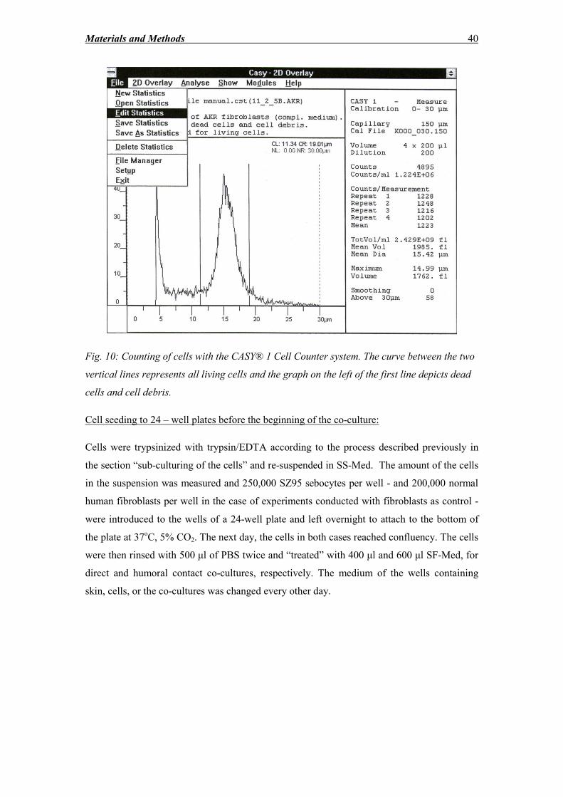
Materials and Methods 40
Fig. 10: Counting of cells with the CASY® 1 Cell Counter system. The curve between the two
vertical lines represents all living cells and the graph on the left of the first line depicts dead
cells and cell debris.
Cell seeding to 24 – well plates before the beginning of the co-culture:
Cells were trypsinized with trypsin/EDTA according to the process described previously in
the section “sub-culturing of the cells” and re-suspended in SS-Med. The amount of the cells
in the suspension was measured and 250,000 SZ95 sebocytes per well - and 200,000 normal
human fibroblasts per well in the case of experiments conducted with fibroblasts as control -
were introduced to the wells of a 24-well plate and left overnight to attach to the bottom of
the plate at 37oC, 5% CO2. The next day, the cells in both cases reached confluency. The cells
were then rinsed with 500 μl of PBS twice and “treated” with 400 μl and 600 μl SF-Med, for
direct and humoral contact co-cultures, respectively. The medium of the wells containing
skin, cells, or the co-cultures was changed every other day.
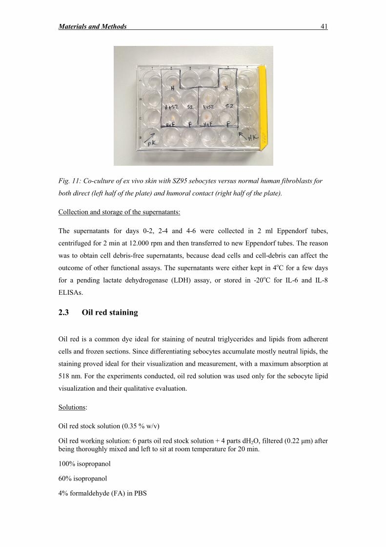
Materials and Methods 41
Fig. 11: Co-culture of ex vivo skin with SZ95 sebocytes versus normal human fibroblasts for
both direct (left half of the plate) and humoral contact (right half of the plate).
Collection and storage of the supernatants:
The supernatants for days 0-2, 2-4 and 4-6 were collected in 2 ml Eppendorf tubes,
centrifuged for 2 min at 12.000 rpm and then transferred to new Eppendorf tubes. The reason
was to obtain cell debris-free supernatants, because dead cells and cell-debris can affect the
outcome of other functional assays. The supernatants were either kept in 4oC for a few days
for a pending lactate dehydrogenase (LDH) assay, or stored in -20oC for IL-6 and IL-8
ELISAs.
2.3 Oil red staining
Oil red is a common dye ideal for staining of neutral triglycerides and lipids from adherent
cells and frozen sections. Since differentiating sebocytes accumulate mostly neutral lipids, the
staining proved ideal for their visualization and measurement, with a maximum absorption at
518 nm. For the experiments conducted, oil red solution was used only for the sebocyte lipid
visualization and their qualitative evaluation.
Solutions:
Oil red stock solution (0.35 % w/v)
Oil red working solution: 6 parts oil red stock solution + 4 parts dH2O, filtered (0.22 μm) after
being thoroughly mixed and left to sit at room temperature for 20 min.
100% isopropanol
60% isopropanol
4% formaldehyde (FA) in PBS
Materials and Methods 42
Process:
After the end of the co-culture, the cells were fixed for 15 min with 4% FA in PBS and then
washed twice with PBS. The staining was performed either immediately or the 24-well-plates
were wrapped with parafilm and kept at 4oC to prevent the wells from drying. Wells were
washed with 60% isopropanol and were allowed to dry completely. 200 μl/well of oil red
working solution was subsequently added for 10 min. Afterwards, the solution is removed and
the wells were washed 4 times with dH2O. After the fourth washing step, the cells were
observed under the microscope and photos were taken.
2.4 Measurement of free LDH release
The LDH assay is a colorimetric assay commonly used for measuring the cytotoxicity of a
certain substance in cells, based on the quantification of LDH enzyme activity, which is
released into the supernatant by damaged cells, which undergo necrosis. In contrast to the
“programmed cell death” called apoptosis, the cell death in necrosis is induced by disruption
of the cellular structure induced by chemicals, radiation, virulent factors and insufficient
nutrition. The procedure eventually leads to cell membrane rupture and release of the stable
enzyme LDH, which is present almost in every cell, since it catalyzes the formation of
pyruvate from lactate. In the first step of the reaction of this colorimetric assay, NAD+ is
reduced to NADH/H+ by the free LDH-mediated formation of pyruvate from lactate. In the
second phase, a diaphorase transfers H/H+ from NADH/H
+ to the pale yellow tetrazolium salt
2-[4-iodophenyl]-3-[4-nitrophenyl]-5-phenyltetrazolium chloride (INT), so that the red
colored formazan salt is produced. The absorbance is then measured with the help of a
spectral photometer at 490 nm, with the reference wavelength set at 690 nm.
For these experiments, LDH release expresses all events (necrosis, necroptosis, apoptosis)
included in skin explant degeneration ex vivo as well as in SZ95 and fibroblast cell death. The
role of the LDH release in detecting cell death for skin explants and cells in monolayer
culture is discussed below.
Materials:
Cytotoxicity Detection Kit (Roche, Mannheim, DE)
Solutions used: (per 96-well plate)
Reaction solution:
Materials and Methods 43
Catalyst solution (ready-to-use): 0.25 μl
Dye solution (ready-to-use): 11.25 μl
Process:
The supernatants were diluted in SF-Med in a dilution 1:5 and 100 μl of every diluted sample
were pipetted to each well of a total of three wells per sample. In the second step, 100 μl of
reaction solution was pipetted to each well. The samples were incubated for 15 min at room
temperature while protected from light and the absorption was measured at 490 nm with a
spectral photometer.
The adjusted results were expressed as optical density (OD) units. The results of LDH release
from supernatants of cells and skin specimens cultured separately were added and compared
with the results obtained from the co-cultures.
2.5 Apoptosis detection – TUNEL reaction
The TUNEL reaction is a method for detecting the cleavage of genomic DNA as one of the
terminal processes of apoptosis. DNA fragmentation is a hallmark of apoptosis, induced
through various pathways of different apoptotic stimuli.
The result of DNA fragmentation is the production of double-stranded DNA fragments of low
molecular weight and single strand breaks of high molecular weight. The method detects the
latter by labeling the so called DNA “nicks” with the enzyme TdT. TdT catalyses the
polymerization of the fluorescein labeled nucleotide dUTP to free -OH DNA ends. The
incorporation of the fluorochrome-conjugated nucleotide allows the detection of the strand
breaks by fluorescence microscopy.
Materials:
In Situ Cell Death detection Kit, Fluorescein (Roche, Mannheim, DE)
Proteinase-K, ready to use (Dako, Hamburg, DE)
DAPI-containing mounting medium: Roti®-Mount FluorCare DAPI (Roth, Karlsruhe, DE)
DNase I recombinant, RNase-free 10.000 U (Roche, Mannheim, DE)
Materials and Methods 44
Process:
1. The slides were pre-heated for 30 min at 59oC and
2. were deparaffinized by immersion in xylol twice for 10 min each and then rehydrated
by immersion in 100% ethanol twice for 10 min each, 95% ethanol twice for 10 min
each and lastly distilled water twice for 5 min each.
3. The specimens were then treated with proteinase-K for 30 minutes at 37oC.
4. The slides were then rinsed three times with PBS,
5. were placed in a humid chamber and then 50 μl of TUNEL reaction mixture (50 μl of
enzyme solution + 450 μl of labeling solution) was applied pro slide for 60 min at
37oC.
6. The slides were then rinsed twice with PBS and then covered with coverslips, using
DAPI-containing medium for mounting.
For the negative control, the labeling solution without the enzyme was used and for the
positive control a sample was treated with 1500 U/ml recombinant DNase I for 10 min at
room temperature, in order to induce DNA strand breaks prior to the labeling procedure.
Samples were directly analyzed under a fluorescence microscope using an excitation
wavelength in the range of 450 – 500 nm, which results in an emission in the wavelength
range of 515 – 565 nm (green). DAPI has an absorption maximum of 358 nm (ultraviolet) and
emits at a maximum of 451 nm (blue).
2.6 IL-6 and IL-8 enzyme-linked immunosorbent assay (ELISA)
Sandwich ELISA for IL-6 and IL-8 is a common biochemistry assay used to determine and
quantify the cytokines in supernatants. Firstly, the primary (monoclonal) antibody, specific
for each one of the two cytokines, is coated to a microplate. Samples and standards are
pipetted to each well and bind specifically to the immobilized antibody, while any unbound
substances are washed away by washing cycles after every step of the procedure. The next
step involves incubation with a polyclonal enzyme-linked antibody, followed by the pipetting
of the substrate solution. The enzyme catalyzes the reaction, leading to color development
which depends on the amount of the substance bound at the first step of the procedure. The
reaction is stopped by adding the stop solution after a given time and the color intensity is
measured with a spectrometer.
Materials and Methods 45
Materials and solutions:
Elisa IL-6 and IL -8 Duo Set (R&D Systems, Minneapolis, USA) for 15 plates each
Clear 96-well microplates (Corning Life Sciences, Amsterdam, N)
ELISA plate sealers (Roth, Karlsruhe, DE)
Reagent Diluent Concentrate (10X) (R&D Systems, Minneapolis, USA)
Substrate Reagent Pack (R&D Systems, Minneapolis, USA)
Streptavidin-HRP (R&D Systems, Minneapolis, USA)
IL-6 and IL-8 standards (diluted appropriately in SF-Med)
IL-6 and IL-8 capture antibody solutions (diluted in PBS)
(Dilution 1:180 → 65 μl of the Capture antibody in 11.7 ml PBS)
IL-6 and IL-8 detection antibody solutions (diluted in Reagent Diluent)
(Dilution 1:180 → 65 μl of the detection antibody in 11.7 ml PBS)
Wash buffer: PBS + 0.05% Tween 20
Reagent Diluent = blocking buffer: 1% BSA in PBS with 0.05% NaN3
[add 9 ml H2O to 1 ml reagent diluent concentrate(10X)]
Substrate solution: 50% substrate reagent A and 50% substrate reagent B
Streptavidin-HRP solution: streptavidin-HRP diluted in reagent diluent (dilution 1:200)
Stop solution – Sulfuric acid 2N (Roth, Karlsruhe, Germany)
Process:
1. The plates were pre-coated with 100 µl/well of the capture antibody solution and
incubated overnight in room temperature.
2. The plates were then washed twice with 200 µl/well and once with 300 µl/well wash
buffer, using a multi-canal pipette.
3. In the next step, 300 µl/well of blocking buffer were added for a minimum of 1.5
hour in room temperature and then the wash step was repeated.
4. The supernatants of SZ95 cells, fibroblasts, skin explants, co-culture with SZ95 cells,
co-culture with fibroblasts as well as the standards were all thawed and pre-diluted in
SF-Med. The dilution was determined from preliminary experiments, so that the
amount of IL-6 and IL-8 would be in the method’s detection range.
Materials and Methods 46
5. 100 µl of diluted samples and standards were added to each well and incubated for 2
hours at room temperature.
6. The wash step was repeated
7. 100 µl of the detection antibody solution was inserted to each well for an incubation
time of 2 h at room temperature.
8. The wash step was repeated
9. The streptavidin-HRP-solution (100 µl/well) was added for an incubation time of 20
min at room temperature while the plate was protected from light.
10. The wash step was repeated
11. 100 µl/well of the Substrate solution were added for an incubation time of 20 min,
while the plate was protected from light.
12. The last step included the pipetting of 50 µl/well stop solution.
13. The plate was tapped gently to ensure thorough mixing and the optical density of
each well was immediately measured at 450 nm, with a wavelength correction of 540
nm.
2.7 Skin preparation for histology
Skin specimens in the beginning of the co-cultures (controls) and after the end of the
experiments (day 6) were fixed in FA 4% in PBS for a minimum of 24 h. The specimens were
then gradually dehydrated and embedded in paraffin with the help of a tissue processor
according to the following protocol:
1. 2 – propanol 75 % - 1 h
2. 2 – propanol 96 % - 1 h
3. 2 – propanol 96 % - 1,5 h
4. 2 – propanol 100 % - 1 h
5. 2 – propanol 100 % - 1 h
6. Xylol 100% - 1 h
Materials and Methods 47
7. Xylol 100% - 1,5 h
8. Xylol 100% - 2 h
9. Liquid paraffin – 1,5 h
10. Liquid paraffin with vacuum – 1 ¾ h
The specimens were then transported to the paraffin embedding center, where they were
inserted to embedding cassettes filled with the appropriate amount of liquid paraffin. The
formed blocks were ready for sectioning after cooling. 3 μm sections were mounted on
special positively charged slides for immunohistochemistry and 5 μm sections were mounted
on normal slides for haematoxylin eosin staining. The sections for immunohistochemistry
were allowed to dry overnight at room temperature, while the ones for hematoxylin eosin
staining were dried at 80oC for 20 – 30 min.
2.8 Hematoxylin eosin staining
Hematoxylin and eosin (H&E) is a very popular staining method, which has been used for the
last century for medical diagnosis, since it has a crucial role in recognizing various tissue
types and their morphologic alterations by cancer and various other diseases. The staining has
the advantage of working exceptionally well with a variety of fixatives and it depicts a broad
spectrum of features of the nucleus, cytoplasm and extracellular matrix. Hematoxylin, a
natural dye obtained from the logwood haematoxylon campechianum, has a deep-purple color
and stains the nucleus by a complex, not fully elucidated reaction. The staining method
involves the application of hemalum, a complex, which consists of oxidized hematoxylin and
aluminum ions. Although it was considered that hematoxylin binds mainly to the DNA of the
cell nuclei, the recent opinion is that the dye-metal complex binds to arginine-rich basic
nucleoproteins, such as histones, thus making the DNA presence not essential for the staining.
On the other hand, eosin is pink and stains intracellular and extracellular proteins non-
specifically. Most of the cytoplasm is eosinophilic. Nuclei show different patterns of
heterochromatin condensation (through hematoxylin staining) and very important for cancer
diagnostics, nucleoli are stained with eosin. The Golgi apparatus is identified by the absence
of staining in a region next to the nucleus. Other colors can also appear in the samples, mainly
caused by the presence of intrinsic pigments, such as melanin in the case of melanocytes.
Hematoxylin alone is also used very often as counterstain for many immunohistochemical
procedures, which use colorimetric substrates, such as peroxidase or alkaline phosphatase
[220].
Materials and Methods 48
Process:
The slides, after drying in 80oC for 20 – 30 min, were placed in
1. xylol twice for 5 min and then immersed 5-6 times in
2. 2-propanol 100%
3. 2-propanol 96%
4. 2-propanol 75%
5. dH2O
6. Then they were left in Mayer’s Hemalaun for 5 min.
7. The slides were washed in warm tap water until the water is not colored purple and
then left in warm tap water for 5 min.
8. The slides were washed with dH2O
9. Slides were put in Eosin solution for 5 min and then
10. Immersed for 5-6 times in dH20,
11. 2-propanol 75%
12. 2-propanol 96%
13. 2-propanol 100% twice
14. Xylol twice
15. The slides were then mounted with Entellan® mounting solution and covered with
coverslips
2.9 Immunocytochemistry and immunohistochemistry
Basics
Immunostaining is a procedure which allows the specific localization of antigen epitopes in
cells (immunocytochemistry) and tissues (immunohistochemistry), by the use of labeled
antibodies as specific reagents, through antigen – antibody interactions, which are
subsequently visualized with the help of a marker, such as a fluorescent dye, a radioactive
element or colloidal gold. The underlying principle is based on the primary antibody – antigen
reaction, which is followed by the application of a secondary antibody raised from a different
species than the primary conjugated with a protein (link antibody or labeled polymer), an
enzyme complex and a chromogenic substrate. The enzymatic activation of the chromogen
results in a visible reaction product at the antigen site. Specimens are then counterstained and
coverslipped. In order to avoid unspecific staining, titration of the antibody, as well as a
blocking step are necessary to avoid non-specific reaction of the secondary antibody with
Materials and Methods 49
tissue epitopes. Ideally, the serum used for the blocking buffer comes from the same species
as that from which the secondary antibody is raised.
Proteolytic digestion and antigen retrieval
The fixation of specimens in FA for a prolonged period of time may result in the masking and
sometimes impairment and non-reversible destruction of antigens, which results in reduced
immunostaining. The procedure of formaldehyde fixation and paraffin embedding is known
to induce conformational changes in antigen molecules through the formation of
intermolecular cross-linkages. Although this is usually not a problem for cells or frozen
sections, it may lead to very weak or false negative staining for paraffin-embedded tissue
sections. The masked sites can be revealed prior to staining mainly with two methods:
1. Proteolytic digestion, which involves the pretreatment of deparaffinized and
rehydrated tissue sections with various enzymes such as proteinase K and pepsin.
2. Heat-induced epitope retrieval method (HIER), which involves the immersion of
the slides in buffers (such as 10 mM Sodium Citrate Buffer) with a defined pH. The
buffer is maintained at 95 – 99oC in a water bath for a maximum of 30 min. These
buffers are designed to break the protein cross-links, thereby unmasking the antigen
epitopes and enhancing the staining intensity of the antibodies.
For the antibodies tested the optimal epitope retrieval method was determined from
preliminary experiments.
Detection systems
Solutions and reagents provided in the two detection systems used:
DAKO REALTM
Detection system, Alkaline Phosphatase/RED, Rabbit/Mouse
DAKO REALTM
Link, Biotinylated Secondary Antibodies
DAKO REALTM
Streptavidin Alkaline Phosphatase
DAKO REALTM
Chromogen Red 1
DAKO REALTM
Chromogen Red 2
DAKO REALTM
Chromogen Red 3
DAKO REALTM
Substrate Buffer
DAKO REALTM
Levamisole (since not serious endogenous alkaline phosphatase activity was
suspected, levamisole was not added to the substrate working solution)
DAKO REALTM
substrate working solution (750 μl Substrate Buffer plus 30 μl of
Chromogen Red 1, 30 μl Chromogen Red 2 and 30 μl Chromogen Red 3)
Materials and Methods 50
DAKO EnVisionTM
FLEX detection System
EnVisionTM
FLEX peroxidase-blocking reagent
EnVisionTM
FLEX/HRP (goat secondary antibodies raised against rabbit/mouse
immunoglobulins coupled with dextran and HRP molecules)
EnVisionTM
FLEX DAB+ chromogen
EnVisionTM
FLEX substrate buffer
EnVisionTM
FLEX wash buffer (20x) (used after being diluted in dH2O)
EnVisionTM
FLEX substrate working solution (30 μl of DAB+ chromogen per 1 ml substrate
buffer)
The detection systems used for IHC were the DAKO EnVisionTM
FLEX detection system and
the DAKO REALTM
Detection system, Alkaline Phosphatase/RED, Rabbit/Mouse. Both
systems are based on the principle of the LSAB method (labeled streptavidin – biotin). The
differences are the enzymes used for catalyzing the substrate – chromogen reaction (horse
radish peroxidase (HRP) for the first case, alkaline phosphatase (AP) for the second case) and
the development of the color following the enzymatic reaction, which are a crispy brown
product for the FLEX system and a red product for the REAL system. Both detection systems
are suitable of the detection of both rabbit and mouse primary antibodies, since they utilize a
mixture of biotinylated anti-rabbit and anti-mouse antibodies. Due to the presence of
stabilizing proteins of DAKO reagents, extra blocking steps to reduce non-specific
background are unnecessary. In order to reduce non-specific background from endogenous
enzyme reactions, cells or tissue can be pretreated with hydrogen peroxide (H2O2) when HRP
is used, or levamisole can be added to the reaction mixture, when endogenous alkaline
phosphatase reaction is suspected.
Table of primary antibodies used for immunocyto- and –histochemistry
Primary
antibody Manufacturer Species Dilution
Incubation
time &
Temperature
Retrieval Method
Anti-Epithelial
Membrane Antigen (EMA)
���� ���� 1:500 1 ℎ @�� 0,2 % ��� 20 in PBS
Monoclonal Anti- Human
Cytokeratin 7,
Clone OV-TL 12/30
DAKO Mouse 1:200 1 hr @RT 0,2 % ��� 20 �� ���
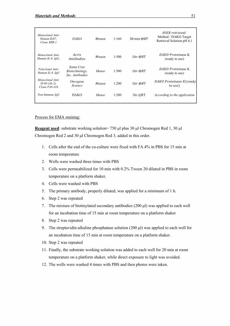
Materials and Methods 51
Monoclonal Anti-
Human Ki67, Clone MIB-1
DAKO ���� 1:160 30 ��� @��
���� �!"# Method / DAKO Target
Retrieval Solution pH 6,1
Monoclonal Anti-Human IL-6, IgG1
�$ ��
�� �%�&�� ���� 1:500 1ℎ @��
���� � � ��"se K
(ready to use)
Polyclonal Anti-
Human IL-8, IgG
Santa Cruz
Biotechnology,
Inc. Antibodies
Mouse 1:500 1ℎ @�� ���� � � ��"se K
(ready to use)
Monoclonal Anti-
SV40 (Ab-2), Clone PAb 416
Oncogene
Science ���� 1:200 1ℎ @��
���� � � ��"� �( "&(
� ��)
Non-Immune IgG
`
DAKO
Mouse
1:200
1hr @RT
According to the application
Process for EMA staining:
Reagent used: substrate working solution= 750 μl plus 30 μl Chromogen Red 1, 30 μl
Chromogen Red 2 and 30 μl Chromogen Red 3, added in this order.
1. Cells after the end of the co-culture were fixed with FA 4% in PBS for 15 min at
room temperature
2. Wells were washed three times with PBS
3. Cells were permeabilized for 10 min with 0.2% Tween 20 diluted in PBS in room
temperature on a platform shaker.
4. Cells were washed with PBS
5. The primary antibody, properly diluted, was applied for a minimum of 1 h.
6. Step 2 was repeated
7. The mixture of biotinylated secondary antibodies (200 μl) was applied to each well
for an incubation time of 15 min at room temperature on a platform shaker
8. Step 2 was repeated
9. The streptavidin-alkaline phosphatase solution (200 μl) was applied to each well for
an incubation time of 15 min at room temperature on a platform shaker.
10. Step 2 was repeated
11. Finally, the substrate working solution was added to each well for 20 min at room
temperature on a platform shaker, while direct exposure to light was avoided.
12. The wells were washed 4 times with PBS and then photos were taken.
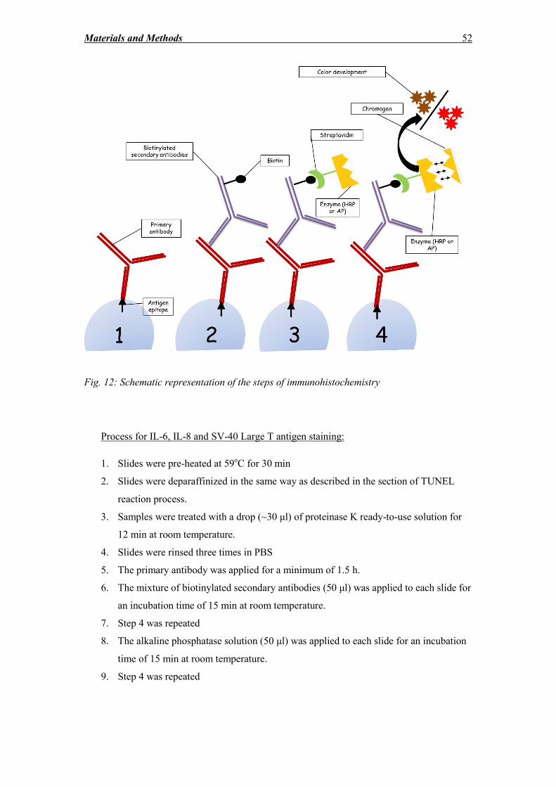
Materials and Methods 52
Fig. 12: Schematic representation of the steps of immunohistochemistry
Process for IL-6, IL-8 and SV-40 Large T antigen staining:
1. Slides were pre-heated at 59oC for 30 min
2. Slides were deparaffinized in the same way as described in the section of TUNEL
reaction process.
3. Samples were treated with a drop (~30 μl) of proteinase K ready-to-use solution for
12 min at room temperature.
4. Slides were rinsed three times in PBS
5. The primary antibody was applied for a minimum of 1.5 h.
6. The mixture of biotinylated secondary antibodies (50 μl) was applied to each slide for
an incubation time of 15 min at room temperature.
7. Step 4 was repeated
8. The alkaline phosphatase solution (50 μl) was applied to each slide for an incubation
time of 15 min at room temperature.
9. Step 4 was repeated
Materials and Methods 53
10. Finally, the substrate working solution was applied to each slide for 20 min at room
temperature, while the slides were placed in a dark chamber, to avoid direct light
exposure.
11. Step 4 was repeated
12. The slides were immersed for 5 min in DAKO hematoxylin and then rinsed with
running tap water until it was no longer colored purple. Afterwards the slides were
left in tap water for 5 min.
13. Finally, the slides were mounted with aqueous mounting medium, covered with cover
glasses and observed under the microscope.
Process for Ki67 staining (with DAKO Autostainer):
Reagents used: DAB working solution: 20 μl of DAB Chromogen Concentrate in 1 ml of
substrate buffer
1. Slides were pre-heated at 59oC for 30 min
2. Slides were deparaffinized in the same way as described in the section of TUNEL
reaction process.
3. Slides were pre-washed with wash buffer.
4. The slides were placed in heat-induced epitope retrieval solution, preheated to 95-
99oC, for 25 min and then are left to cool off for 15 min.
5. Step 3 was repeated
6. Slides were treated with 3% H2O2 (200 μl pro Slide) for 5 min.
7. The primary antibody was then applied (200 μl pro Slide) for 30 min at room
temperature.
8. Step 3 was repeated
9. The slides were then treated with 200 μl of “Dako EnVisionTM
FlexHRP” enzyme
solution for 20 min.
10. Step 3 was repeated
11. Then, the DAB Chromogen concentrate, properly diluted in Substrate Buffer, was
applied twice to each section (200 μl pro Slide) for 5 min at room temperature.
12. Steps 10 and 11 were repeated
13. After a final wash step, the sections were immersed in DAKO FLEXTM
Hematoxylin
for 5 min at room temperature.
14. Step 3 was repeated
15. The specimens were mounted in aqueous mounting medium, covered with coverslips
and observed under the microscope.
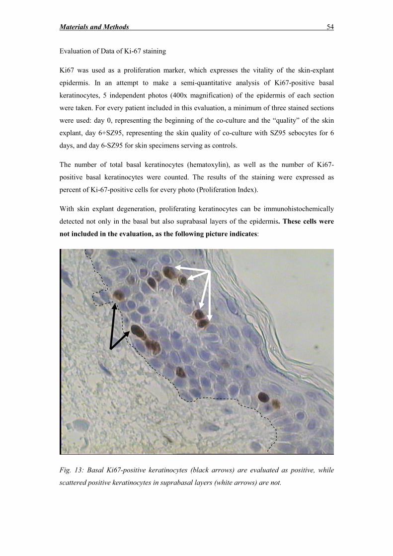
Materials and Methods 54
Evaluation of Data of Ki-67 staining
Ki67 was used as a proliferation marker, which expresses the vitality of the skin-explant
epidermis. In an attempt to make a semi-quantitative analysis of Ki67-positive basal
keratinocytes, 5 independent photos (400x magnification) of the epidermis of each section
were taken. For every patient included in this evaluation, a minimum of three stained sections
were used: day 0, representing the beginning of the co-culture and the “quality” of the skin
explant, day 6+SZ95, representing the skin quality of co-culture with SZ95 sebocytes for 6
days, and day 6-SZ95 for skin specimens serving as controls.
The number of total basal keratinocytes (hematoxylin), as well as the number of Ki67-
positive basal keratinocytes were counted. The results of the staining were expressed as
percent of Ki-67-positive cells for every photo (Proliferation Index).
With skin explant degeneration, proliferating keratinocytes can be immunohistochemically
detected not only in the basal but also suprabasal layers of the epidermis. These cells were
not included in the evaluation, as the following picture indicates:
Fig. 13: Basal Ki67-positive keratinocytes (black arrows) are evaluated as positive, while
scattered positive keratinocytes in suprabasal layers (white arrows) are not.
Materials and Methods 55
Mean values of Ki67-positive basal epidermal cells of 5 photos were calculated per skin
explant section. Care was taken not to overlap fields of view to ensure that cells would not be
counted twice.
2.10 DNA fragmentation evaluation
Similarly to Ki67 evaluation, fluorescence photos in 200x magnification were analyzed with
the help of the ImageJ software (NIH, Bethesda, USA). TUNEL-positive and DAPI-positive
cells of the epidermis were counted, and the former were expressed as a ratio of the total
nucleated cells of the epidermis (DNA fragmentation Index).
2.11 Skin explant stratum corneum and vital epidermis thickness
In this study, thickness of vital epidermis is considered the thickness of all epidermal layers
starting from the basal membrane, without including stratum corneum. The measurements of
the interfollicular epidermis and the stratum corneum were conducted with the help of the
scale of an Olympus CK40 microscope, using 400x magnification.
2.12 Statistical analysis
For the comparison of the populations “percentage of Ki67-positive basal epidermal
keratinocytes”, “percentage of TUNEL-positive epidermal cells”, “stratum corneum
thickness”, “vital non-cornified epidermal thickness”, “LDH release” referring to skin
specimens cultured with and without the presence of SZ95 sebocytes, the two-tailed Student’s
t-test in Microsoft Office Excel 2010 was used. On the other hand, for assessing statistically
the morphological features of skin explant degeneration, namely parakeratosis, basal and
suprabasal keratinocyte vacuolization, flattening of basal keratinocytes, reduction of vital
epidermis layers, cleft formation, intraepidermal and subepidermal bulla formation,
acantholysis and complete separation of the epidermis from the dermis, the non-parametric
chi squared test with one degree of freedom was used. In both cases, the differences were
considered significant, if the P value was estimated to be below 0.05.
2.13 Protein analysis methods
Materials and Methods 56
2.13.1 Skin protein isolation
Protein isolation is performed usually on ice or 4oC, to avoid protein degradation occurring at
high temperatures. To avoid degradation, serine protease inhibitors against trypsin,
chymotrypsin, papain, thrombin etc. are added in the lysis buffer. Since the inhibitors are
usually unstable substances when added to water, their addition to the medium should happen
exactly prior to use. The same goes for phosphatase inhibitors.
Materials and Solutions:
10x RIPA buffer: (Cell Signaling, Frankfurt am Main, Germany)
Mini EDTA-free protease inhibitor: (Roche, Mannheim, Germany)
PhosphoSTOP phosphatase inhibitor cocktail: (Roche, Mannheim, Germany)
Protease inhibitor stock solution: one tablet in 2 ml 1x RIPA buffer
Phosphatase inhibitor stock solution: one tablet in 1 ml 1x RIPA buffer
Lysis working solution: 1x RIPA buffer diluted in ddH2O, with protease inhibitor (dilution
1:25) and phosphatase inhibitor (dilution 1:10).
Process:
The whole procedure took place on ice. Skin specimens were removed from liquid nitrogen,
cut into small pieces with a lancet and manually homogenized in 250 μl lysis working
solution with the help of a tissue homogenizer. The homogenates were centrifuged at 13,000
rpm for 20 min. The supernatants were used immediately for protein quantitation and aliquots
were stored at -70oC.
2.13.2 Protein quantitation
For the detection and quantitation of total protein of skin samples, the BCA™ Protein Assay
(Pierce, Weiskirchen, DE) was used. The method is based on the biuret reaction, namely the
reduction of Cu+2
to Cu+1
by proteins in a medium of alkaline pH, utilizing a reagent
containing bicinchoninic acid [221]. The reaction product of the chelation of two BCA
molecules with one Cu+1
ion has a purple color, which exhibits an absorbance at 562 nm.
Protein concentrations are determined in reference to series of standards of BSA, diluted in
the same diluent used for the samples (0-2000 μg/ml).
Solutions used:
Materials and Methods 57
Working solution (200 μl/well): 50 parts of reagent A + 1 part of reagent B
Process:
25 μl of each sample lysate and standard was transferred to each well of a microplate 96-well
plate in triplicates. 200 μl of the working solution was added to each well. The plate was
placed in a plate reader and was incubated for 30 min at 37oC. The absorbance was then
measured at 562 nm. The total protein concentration of each lysate was calculated with the
help of the standard curve.
2.13.3 Western blot
Reagents and Chemicals:
Acrylamide/bisacrylamide (BioRad Laboratories GmbH, Munich, DE)
Agarose (BioRad Laboratories GmbH, Munich, DE)
Ammonium persulfate (BioRad Laboratories GmbH, Munich, DE)
Bromophenol blue (BioRad Laboratories GmbH, Munich, DE)
EDTA (Sigma, Munich, DE)
Acetic acid (Roth, Karlsruhe, DE)
Glycine (Roth, Karlsruhe, DE)
Methanol (Roth, Karlsruhe, DE)
Sodium acetate (Roth, Karlsruhe, DE)
Sodium chloride (Roth, Karlsruhe, DE)
Sodium citrate (Roth, Karlsruhe, DE)
Sodium dodecyl sulfate (SDS) (Roth, Karlsruhe, DE)
Non-fat dry milk (BioRad Laboratories GmbH, Munich, DE)
Ponceau solution (Sigma, Munich, DE)
Tween-20 (Roth, Karlsruhe, DE)
Tetramethylethylenediamine (TEMED) (Roth, Karlsruhe, DE)
Tris-HCL (Roth, Karlsruhe, DE)
Tris-Base (Roth, Karlsruhe, DE)
β-mercaptoethanol (Roth, Karlsruhe, DE)
Materials and Methods 58
In the first phase of Western blotting, polyacrylamide gel electrophoresis (PAGE) was used to
separate proteins. Polyacrylamide is a polymer that forms a gel with pores that are small
enough to separate proteins. From the available PAGE types, sodium dodecyl sulfate (SDS)
PAGE was used in these series of experiments. SDS is a negatively charged anionic
surfactant, able to coat individual proteins and disrupt their non-covalent bonds, thus leading
to their denaturation and subsequently protein separation based exclusively on their molecular
weight. Proteins travel only in one dimension from the negatively to the positively charged
electrode, through the acrylamide mesh of the gel. Two gels were used: the stacking gel is
acidic (pH 6.8) and has a low (5%) acrylamide concentration to form a porous gel, which
forces proteins to form thin, sharply defined bands. The resolving gel, is more basic (pH 8.8),
and has a higher polyacrylamide content (12%), which causes the gel to have narrower
channels or pores. As a protein, concentrated into sharp bands by the stacking gel, travels
through the resolving gel, the narrower pores allow smaller proteins to travel easily and more
rapidly than larger proteins. The concentration of acrylamide determines the resolution of the
gel, with higher concentrations being suitable for separation of proteins of low molecular
weight and lower concentrations for separation of proteins of high molecular weight. Special
markers of known molecular weight were used as references for the detection of the protein
size. In the second phase, proteins from the gel were transferred onto a membrane using
electrophoresis and finally, specific antibodies were used in order to detect the desired
proteins. The bands were detected with a chemiluminescent reagent, which provides visible
bands after excitation with UV-radiation.
Gel preparation
Reagents and solutions used:
Laemmli running buffer Sample buffer (4x)
192 mM glycine 40% glycerin
25 mM Tris 12% β-mercaptoethanol
0.1% SDS 4% bromophenol blue
0.8% SDS
Resolving-gel buffer (500 ml) Stacking-gel buffer (500 ml)
640 mM Tris-Base 330 mM Tris-Base
120 mM Tris-HCL 0,2% SDS
Materials and Methods 59
0,2% SDS pH= 6.8
pH= 8.8
5% stacking gel (5 ml) 12% resolving gel (10 ml)
30% Acryl-/bisacrylamide 37.5:1 800 μl 4 ml
Stacking-gel buffer 2.5 ml -
Resolving-gel buffer - 5 ml
dH20 1.7 ml 890 μl
10% APS (added last) 100 μl 100 μl
TEMED (added last) 10 μl 10 μl
Standards
8 μl Bio-Rad marker (Precision Plus ProteinTM
Dual Color Standards, BioRad Laboratories
GmbH, Munich, DE) + 2 μl sample buffer
Process:
The skin lysates and the standards were placed in sample buffer for 5 min at 95oC. In the
meantime the gels were ready for electrophoresis; they were placed in the electrophoresis
device, covered with Laemmli buffer. Samples were loaded into the slots and were allowed to
run at 80-200V, until the tracking dye reached the gel bottom.
Western blotting (second phase)
Blotting buffer (1 L)
500 ml volume Laemmli running buffer
200 ml volume methanol
300 ml dH2O
The blot is constructed as indicated below:
White side – positive pole
Sponge
2x gel-blotting-paper
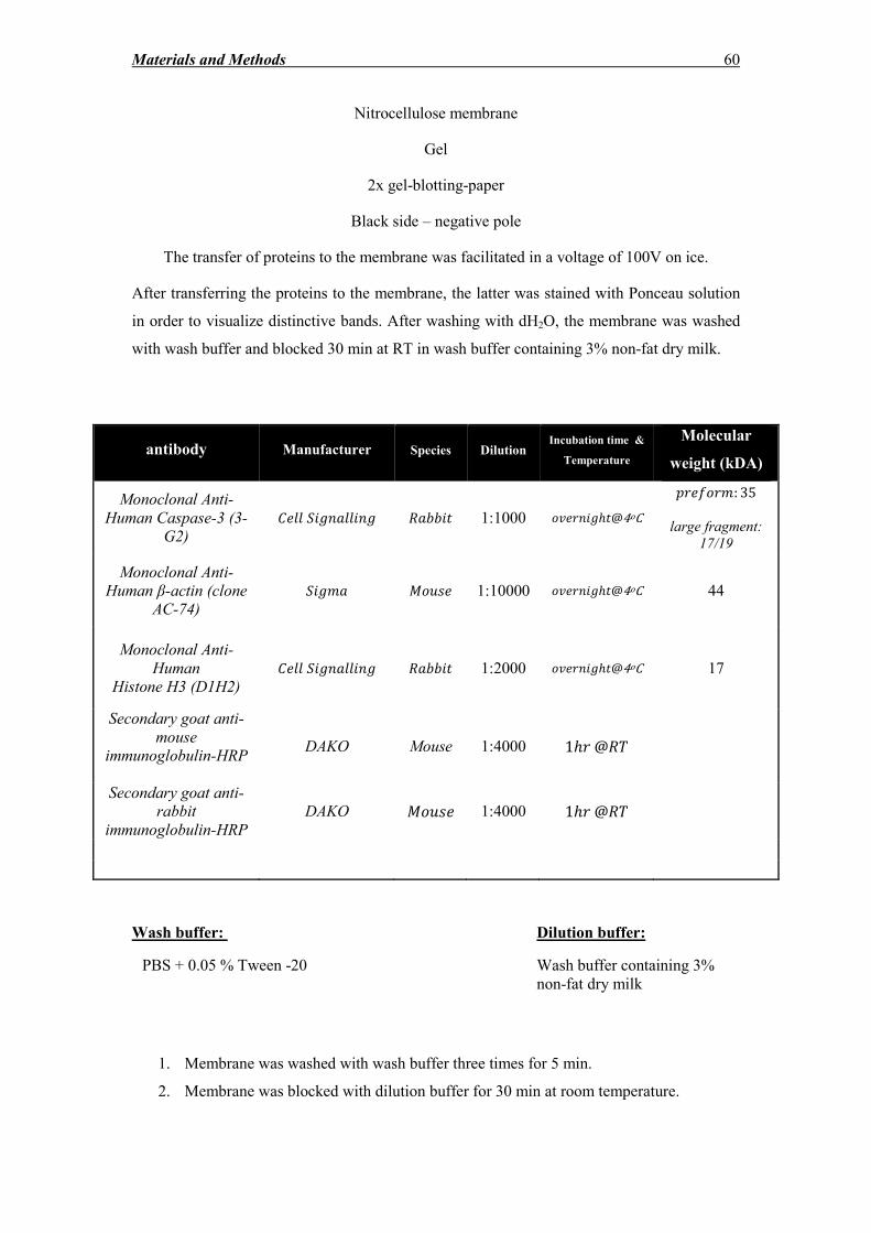
Materials and Methods 60
Nitrocellulose membrane
Gel
2x gel-blotting-paper
Black side – negative pole
The transfer of proteins to the membrane was facilitated in a voltage of 100V on ice.
After transferring the proteins to the membrane, the latter was stained with Ponceau solution
in order to visualize distinctive bands. After washing with dH2O, the membrane was washed
with wash buffer and blocked 30 min at RT in wash buffer containing 3% non-fat dry milk.
antibody Manufacturer Species Dilution Incubation time &
Temperature
Molecular
weight (kDA)
Monoclonal Anti-
Human Caspase-3 (3-
G2)
*## ��+�"##��+ �"%%� 1:1000 �! ��+ℎ @4oC
/ 0� �: 35
large fragment:
17/19
Monoclonal Anti-
Human β-actin (clone
AC-74)
��+�" ���� 1:10000 �! ��+ℎ @4oC 44
Monoclonal Anti-
Human
Histone H3 (D1H2)
*## ��+�"##��+ �"%%� 1:2000 �! ��+ℎ @4oC 17
Secondary goat anti-
mouse
immunoglobulin-HRP
DAKO Mouse 1:4000 1ℎ @��
Secondary goat anti-
rabbit
immunoglobulin-HRP
DAKO ���� 1:4000 1ℎ @��
Wash buffer: Dilution buffer:
PBS + 0.05 % Tween -20 Wash buffer containing 3%
non-fat dry milk
1. Membrane was washed with wash buffer three times for 5 min.
2. Membrane was blocked with dilution buffer for 30 min at room temperature.
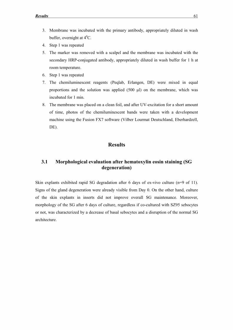
Results 61
3. Membrane was incubated with the primary antibody, appropriately diluted in wash
buffer, overnight at 40C.
4. Step 1 was repeated
5. The marker was removed with a scalpel and the membrane was incubated with the
secondary HRP-conjugated antibody, appropriately diluted in wash buffer for 1 h at
room temperature.
6. Step 1 was repeated
7. The chemiluminescent reagents (Peqlab, Erlangen, DE) were mixed in equal
proportions and the solution was applied (500 μl) on the membrane, which was
incubated for 1 min.
8. The membrane was placed on a clean foil, and after UV-excitation for a short amount
of time, photos of the chemiluminescent bands were taken with a development
machine using the Fusion FX7 software (Vilber Lourmat Deutschland, Eberhardzell,
DE).
Results
3.1 Morphological evaluation after hematoxylin eosin staining (SG
degeneration)
Skin explants exhibited rapid SG degradation after 6 days of ex-vivo culture (n=9 of 11).
Signs of the gland degeneration were already visible from Day 0. On the other hand, culture
of the skin explants in inserts did not improve overall SG maintenance. Moreover,
morphology of the SG after 6 days of culture, regardless if co-cultured with SZ95 sebocytes
or not, was characterized by a decrease of basal sebocytes and a disruption of the normal SG
architecture.
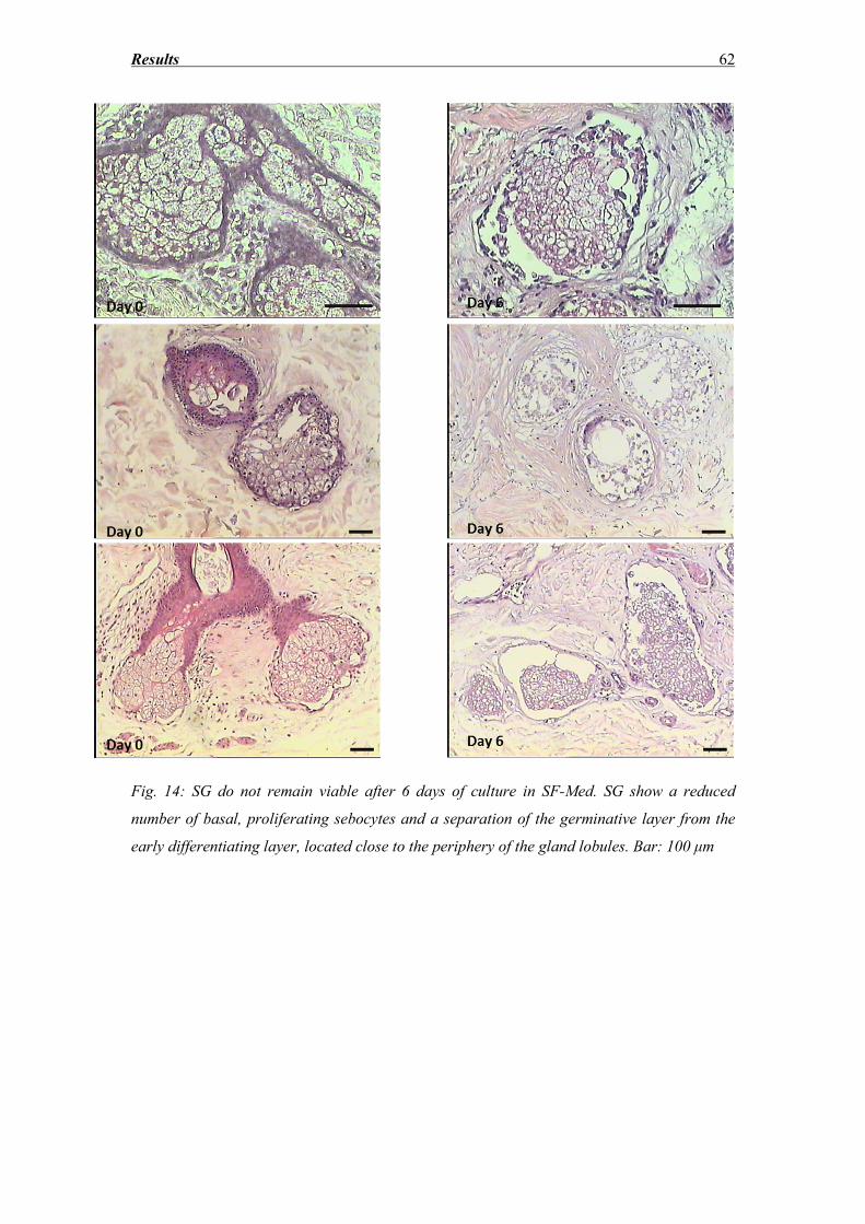
Results 62
Fig. 14: SG do not remain viable after 6 days of culture in SF-Med. SG show a reduced
number of basal, proliferating sebocytes and a separation of the germinative layer from the
early differentiating layer, located close to the periphery of the gland lobules. Bar: 100 μm
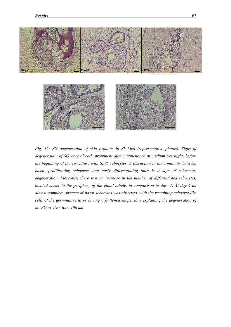
Results 63
Fig. 15: SG degeneration of skin explants in SF-Med (representative photos). Signs of
degeneration of SG were already prominent after maintenance in medium overnight, before
the beginning of the co-culture with SZ95 sebocytes. A disruption in the continuity between
basal, proliferating sebocytes and early differentiating ones is a sign of sebaceous
degeneration. Moreover, there was an increase in the number of differentiated sebocytes,
located closer to the periphery of the gland lobule, in comparison to day -1. At day 6 an
almost complete absence of basal sebocytes was observed, with the remaining sebocyte-like
cells of the germinative layer having a flattened shape, thus explaining the degeneration of
the SG ex vivo. Bar: 100 μm
Results 64
3.2 Biochemical evidence of sebaceous gland degeneration
Fig. 16: The hematoxylin/eosin data were confirmed by TUNEL staining. SG before the
beginning of ex vivo culture exhibited TUNEL-positive cells (green) exclusively towards the
center of the gland, since differentiating sebocytes undergo spontaneous apoptosis and
release their content into the sebaceous duct. On the other hand, after 6 days of culture,
strong staining was detected also in the basal layer of the gland, thus providing biochemical
evidence of SG degeneration ex vivo (n=3). Since basal sebocytes undergo apoptosis, they
cannot further promote the gland’s regeneration. Counterstaining with DAPI (blue). Bar 100
μm
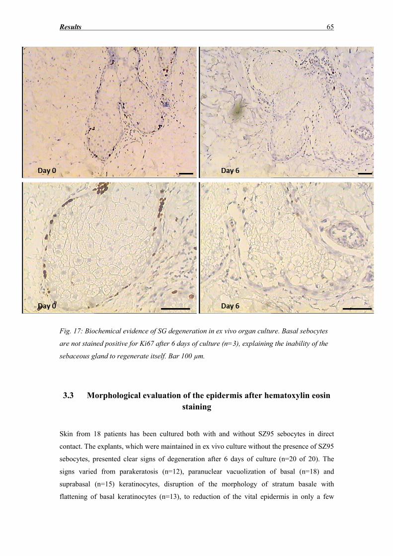
Results 65
Fig. 17: Biochemical evidence of SG degeneration in ex vivo organ culture. Basal sebocytes
are not stained positive for Ki67 after 6 days of culture (n=3), explaining the inability of the
sebaceous gland to regenerate itself. Bar 100 µm.
3.3 Morphological evaluation of the epidermis after hematoxylin eosin
staining
Skin from 18 patients has been cultured both with and without SZ95 sebocytes in direct
contact. The explants, which were maintained in ex vivo culture without the presence of SZ95
sebocytes, presented clear signs of degeneration after 6 days of culture (n=20 of 20). The
signs varied from parakeratosis (n=12), paranuclear vacuolization of basal (n=18) and
suprabasal (n=15) keratinocytes, disruption of the morphology of stratum basale with
flattening of basal keratinocytes (n=13), to reduction of the vital epidermis in only a few
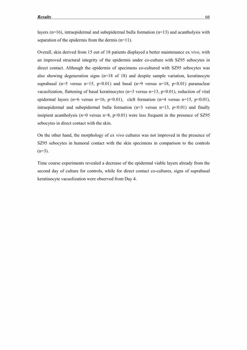
Results 66
layers (n=16), intraepidermal and subepidermal bulla formation (n=13) and acantholysis with
separation of the epidermis from the dermis (n=11).
Overall, skin derived from 15 out of 18 patients displayed a better maintenance ex vivo, with
an improved structural integrity of the epidermis under co-culture with SZ95 sebocytes in
direct contact. Although the epidermis of specimens co-cultured with SZ95 sebocytes was
also showing degeneration signs (n=18 of 18) and despite sample variation, keratinocyte
suprabasal (n=5 versus n=15, p<0.01) and basal (n=9 versus n=18, p<0.01) paranuclear
vacuolization, flattening of basal keratinocytes (n=3 versus n=13, p<0.01), reduction of vital
epidermal layers (n=6 versus n=16, p<0.01), cleft formation (n=4 versus n=15, p<0.01),
intraepidermal and subepidermal bulla formation (n=3 versus n=13, p<0.01) and finally
insipient acantholysis (n=0 versus n=8, p<0.01) were less frequent in the presence of SZ95
sebocytes in direct contact with the skin.
On the other hand, the morphology of ex vivo cultures was not improved in the presence of
SZ95 sebocytes in humoral contact with the skin specimens in comparison to the controls
(n=3).
Time course experiments revealed a decrease of the epidermal viable layers already from the
second day of culture for controls, while for direct contact co-cultures, signs of suprabasal
keratinocyte vacuolization were observed from Day 4.
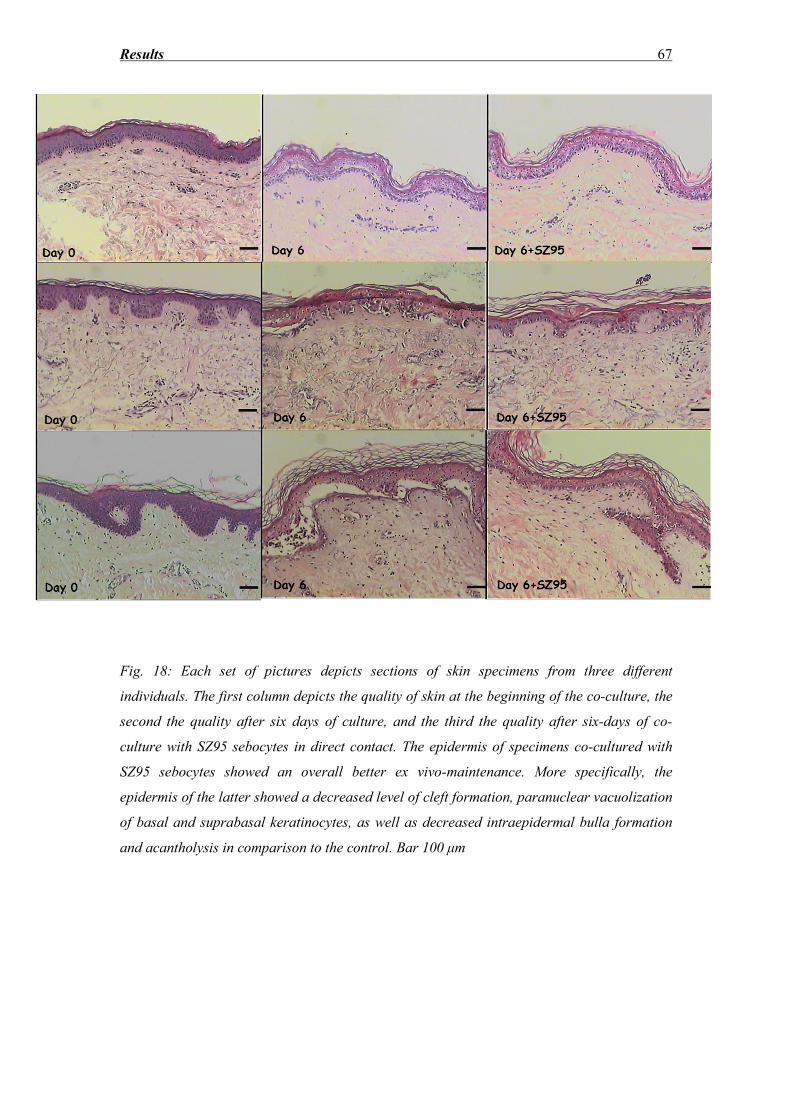
Results 67
Fig. 18: Each set of pictures depicts sections of skin specimens from three different
individuals. The first column depicts the quality of skin at the beginning of the co-culture, the
second the quality after six days of culture, and the third the quality after six-days of co-
culture with SZ95 sebocytes in direct contact. The epidermis of specimens co-cultured with
SZ95 sebocytes showed an overall better ex vivo-maintenance. More specifically, the
epidermis of the latter showed a decreased level of cleft formation, paranuclear vacuolization
of basal and suprabasal keratinocytes, as well as decreased intraepidermal bulla formation
and acantholysis in comparison to the control. Bar 100 μm
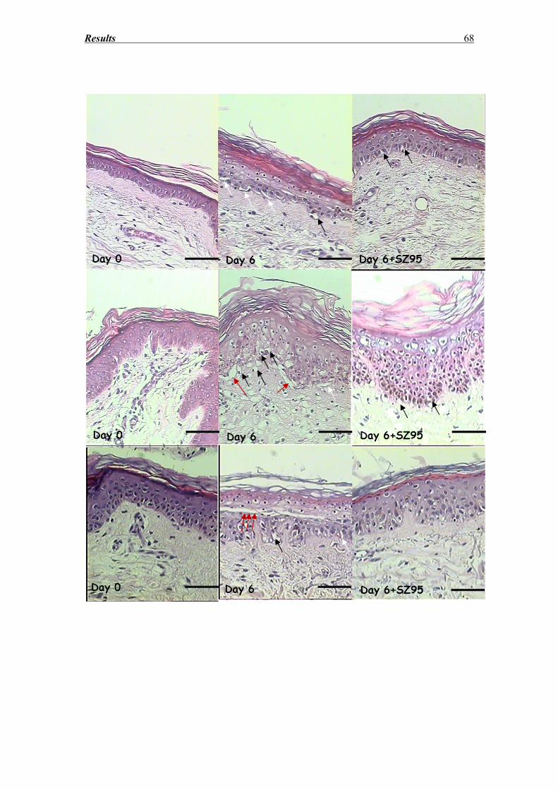
Results 68
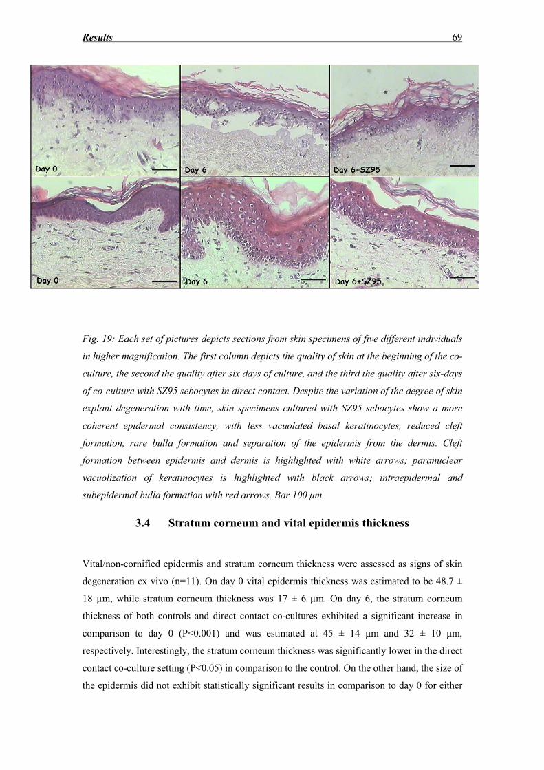
Results 69
Fig. 19: Each set of pictures depicts sections from skin specimens of five different individuals
in higher magnification. The first column depicts the quality of skin at the beginning of the co-
culture, the second the quality after six days of culture, and the third the quality after six-days
of co-culture with SZ95 sebocytes in direct contact. Despite the variation of the degree of skin
explant degeneration with time, skin specimens cultured with SZ95 sebocytes show a more
coherent epidermal consistency, with less vacuolated basal keratinocytes, reduced cleft
formation, rare bulla formation and separation of the epidermis from the dermis. Cleft
formation between epidermis and dermis is highlighted with white arrows; paranuclear
vacuolization of keratinocytes is highlighted with black arrows; intraepidermal and
subepidermal bulla formation with red arrows. Bar 100 μm
3.4 Stratum corneum and vital epidermis thickness
Vital/non-cornified epidermis and stratum corneum thickness were assessed as signs of skin
degeneration ex vivo (n=11). On day 0 vital epidermis thickness was estimated to be 48.7 ±
18 µm, while stratum corneum thickness was 17 ± 6 µm. On day 6, the stratum corneum
thickness of both controls and direct contact co-cultures exhibited a significant increase in
comparison to day 0 (P<0.001) and was estimated at 45 ± 14 μm and 32 ± 10 μm,
respectively. Interestingly, the stratum corneum thickness was significantly lower in the direct
contact co-culture setting (P<0.05) in comparison to the control. On the other hand, the size of
the epidermis did not exhibit statistically significant results in comparison to day 0 for either
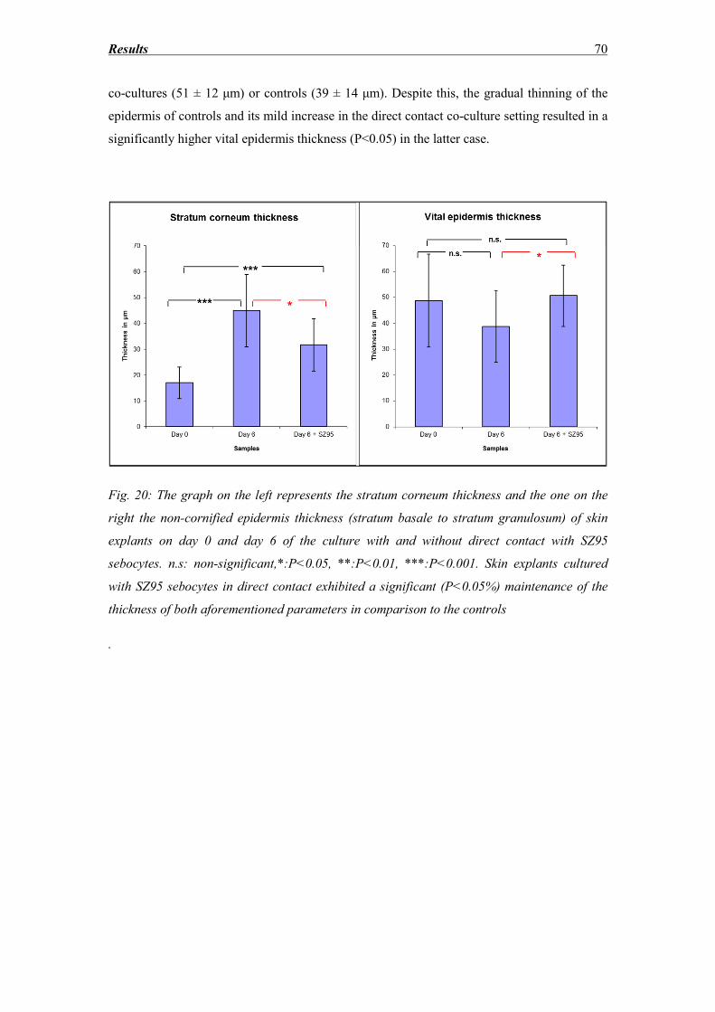
Results 70
co-cultures (51 ± 12 μm) or controls (39 ± 14 μm). Despite this, the gradual thinning of the
epidermis of controls and its mild increase in the direct contact co-culture setting resulted in a
significantly higher vital epidermis thickness (P<0.05) in the latter case.
Fig. 20: The graph on the left represents the stratum corneum thickness and the one on the
right the non-cornified epidermis thickness (stratum basale to stratum granulosum) of skin
explants on day 0 and day 6 of the culture with and without direct contact with SZ95
sebocytes. n.s: non-significant,*:P<0.05, **:P<0.01, ***:P<0.001. Skin explants cultured
with SZ95 sebocytes in direct contact exhibited a significant (P<0.05%) maintenance of the
thickness of both aforementioned parameters in comparison to the controls
.
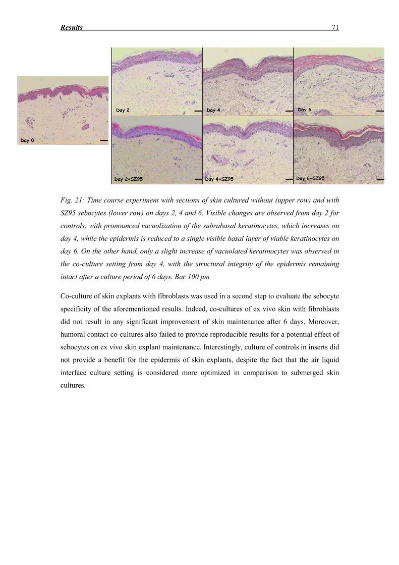
Results 71
Fig. 21: Time course experiment with sections of skin cultured without (upper row) and with
SZ95 sebocytes (lower row) on days 2, 4 and 6. Visible changes are observed from day 2 for
controls, with pronounced vacuolization of the subrabasal keratinocytes, which increases on
day 4, while the epidermis is reduced to a single visible basal layer of viable keratinocytes on
day 6. On the other hand, only a slight increase of vacuolated keratinocytes was observed in
the co-culture setting from day 4, with the structural integrity of the epidermis remaining
intact after a culture period of 6 days. Bar 100 μm
Co-culture of skin explants with fibroblasts was used in a second step to evaluate the sebocyte
specificity of the aforementioned results. Indeed, co-cultures of ex vivo skin with fibroblasts
did not result in any significant improvement of skin maintenance after 6 days. Moreover,
humoral contact co-cultures also failed to provide reproducible results for a potential effect of
sebocytes on ex vivo skin explant maintenance. Interestingly, culture of controls in inserts did
not provide a benefit for the epidermis of skin explants, despite the fact that the air liquid
interface culture setting is considered more optimized in comparison to submerged skin
cultures.
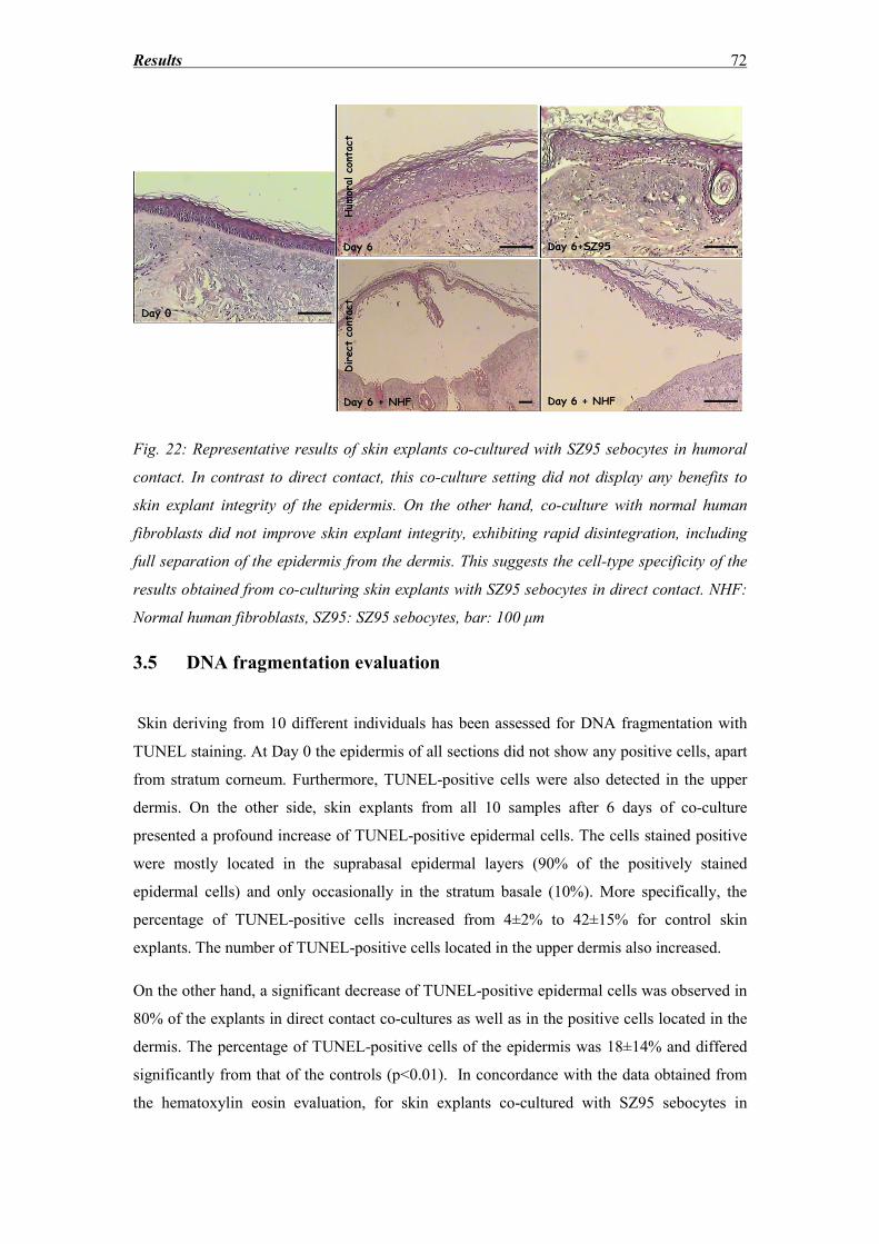
Results 72
Fig. 22: Representative results of skin explants co-cultured with SZ95 sebocytes in humoral
contact. In contrast to direct contact, this co-culture setting did not display any benefits to
skin explant integrity of the epidermis. On the other hand, co-culture with normal human
fibroblasts did not improve skin explant integrity, exhibiting rapid disintegration, including
full separation of the epidermis from the dermis. This suggests the cell-type specificity of the
results obtained from co-culturing skin explants with SZ95 sebocytes in direct contact. NHF:
Normal human fibroblasts, SZ95: SZ95 sebocytes, bar: 100 μm
3.5 DNA fragmentation evaluation
Skin deriving from 10 different individuals has been assessed for DNA fragmentation with
TUNEL staining. At Day 0 the epidermis of all sections did not show any positive cells, apart
from stratum corneum. Furthermore, TUNEL-positive cells were also detected in the upper
dermis. On the other side, skin explants from all 10 samples after 6 days of co-culture
presented a profound increase of TUNEL-positive epidermal cells. The cells stained positive
were mostly located in the suprabasal epidermal layers (90% of the positively stained
epidermal cells) and only occasionally in the stratum basale (10%). More specifically, the
percentage of TUNEL-positive cells increased from 4±2% to 42±15% for control skin
explants. The number of TUNEL-positive cells located in the upper dermis also increased.
On the other hand, a significant decrease of TUNEL-positive epidermal cells was observed in
80% of the explants in direct contact co-cultures as well as in the positive cells located in the
dermis. The percentage of TUNEL-positive cells of the epidermis was 18±14% and differed
significantly from that of the controls (p<0.01). In concordance with the data obtained from
the hematoxylin eosin evaluation, for skin explants co-cultured with SZ95 sebocytes in
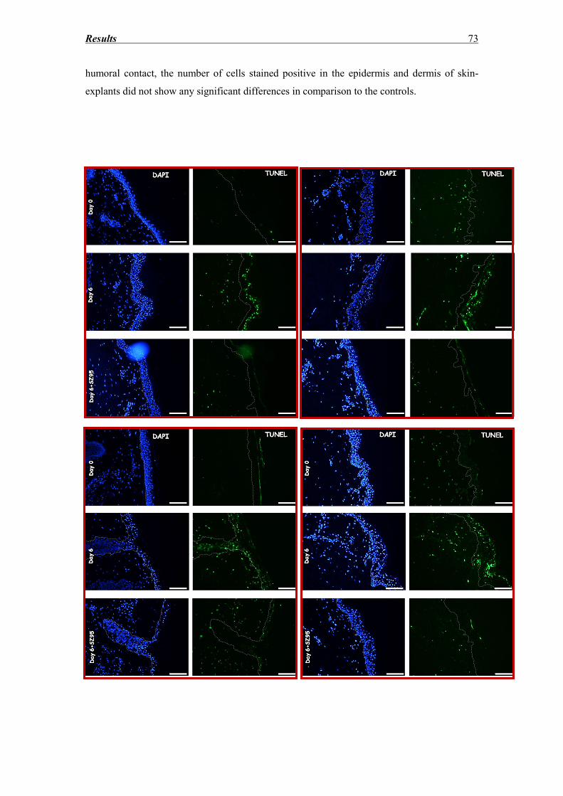
Results 73
humoral contact, the number of cells stained positive in the epidermis and dermis of skin-
explants did not show any significant differences in comparison to the controls.
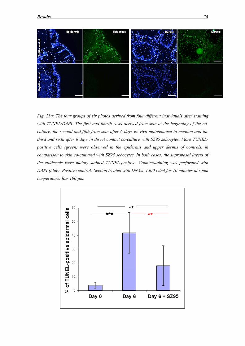
Results 74
Fig. 23a: The four groups of six photos derived from four different individuals after staining
with TUNEL/DAPI. The first and fourth rows derived from skin at the beginning of the co-
culture, the second and fifth from skin after 6 days ex vivo maintenance in medium and the
third and sixth after 6 days in direct contact co-culture with SZ95 sebocytes. More TUNEL-
positive cells (green) were observed in the epidermis and upper dermis of controls, in
comparison to skin co-cultured with SZ95 sebocytes. In both cases, the suprabasal layers of
the epidermis were mainly stained TUNEL-positive. Counterstaining was performed with
DAPI (blue). Positive control: Section treated with DNAse 1500 U/ml for 10 minutes at room
temperature. Bar 100 µm.
Results 75
Fig. 23b: SZ95 sebocytes affect DNA-fragmentation rates of skin explant epidermis: The
graph depicts the results of the statistical analysis after TUNEL-DAPI staining of sections
representative for 10 individuals before (day 0) and after 6 days of co-culture without (day 6)
and with (day 6 + SZ95) the presence of SZ95 sebocytes. Skin explants cultured for 6 days in
serum-free medium present higher rates of TUNEL-positive cells both without (p<0.001) and
with (p<0.01) the presence of SZ95 sebocytes (1). Nonetheless, Skin explants co-cultured with
sebocytes displayed significantly lower DNA-fragmentation rates in comparison to the
controls (p<0.01). n.s: non-significant,*:P<0.05, **:P<0.01, ***:P<0.001.
3.6 LDH release
LDH release is a marker of measuring the level of cell necrosis within 24 h and, therefore, it
can be used to assess the cytotoxic effect of substances in comparison to controls. For longer
time periods, the spontaneous LDH release of skin explants has been used as a measure of all
cellular events leading to cell death. For sebocytes, on the other hand, the interpretation of an
increase of LDH release in the supernatant is controversial, since it could be also interpreted
as an increase in the differentiation and apoptosis of the cells. SZ95 sebocytes are known to
undergo spontaneous holocrine secretion in serum-containing medium, with DNA-
fragmentation observed after 6 h and LDH release after 24 h of culture [122].
Supernatants collected from direct contact co-cultures of 8 different individuals were assessed
for spontaneous LDH release. Because of sample variation (skin deriving from different
localization, age and gender groups), it was difficult to obtain reproducible results which
could allow a quantitative analysis. Despite this fact, patterns could be recognized as
mentioned below:
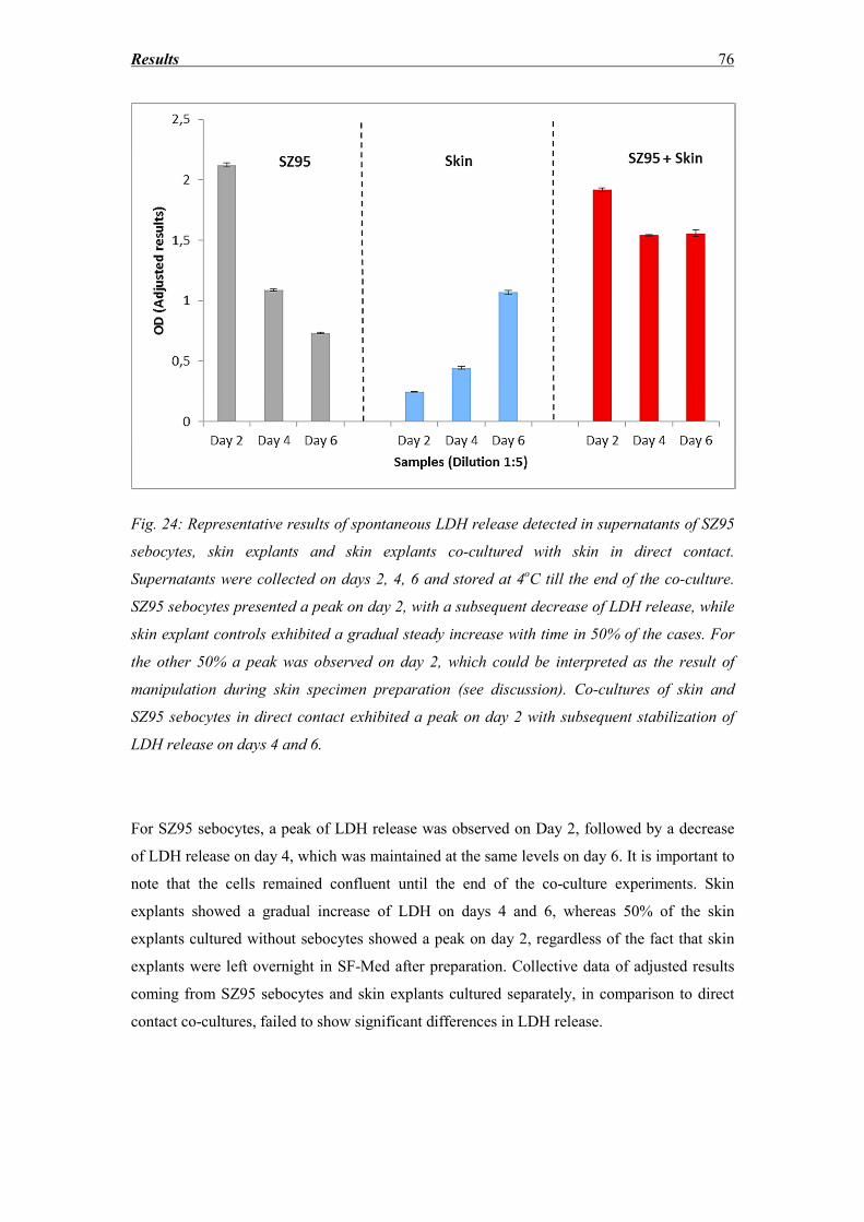
Results 76
Fig. 24: Representative results of spontaneous LDH release detected in supernatants of SZ95
sebocytes, skin explants and skin explants co-cultured with skin in direct contact.
Supernatants were collected on days 2, 4, 6 and stored at 4oC till the end of the co-culture.
SZ95 sebocytes presented a peak on day 2, with a subsequent decrease of LDH release, while
skin explant controls exhibited a gradual steady increase with time in 50% of the cases. For
the other 50% a peak was observed on day 2, which could be interpreted as the result of
manipulation during skin specimen preparation (see discussion). Co-cultures of skin and
SZ95 sebocytes in direct contact exhibited a peak on day 2 with subsequent stabilization of
LDH release on days 4 and 6.
For SZ95 sebocytes, a peak of LDH release was observed on Day 2, followed by a decrease
of LDH release on day 4, which was maintained at the same levels on day 6. It is important to
note that the cells remained confluent until the end of the co-culture experiments. Skin
explants showed a gradual increase of LDH on days 4 and 6, whereas 50% of the skin
explants cultured without sebocytes showed a peak on day 2, regardless of the fact that skin
explants were left overnight in SF-Med after preparation. Collective data of adjusted results
coming from SZ95 sebocytes and skin explants cultured separately, in comparison to direct
contact co-cultures, failed to show significant differences in LDH release.
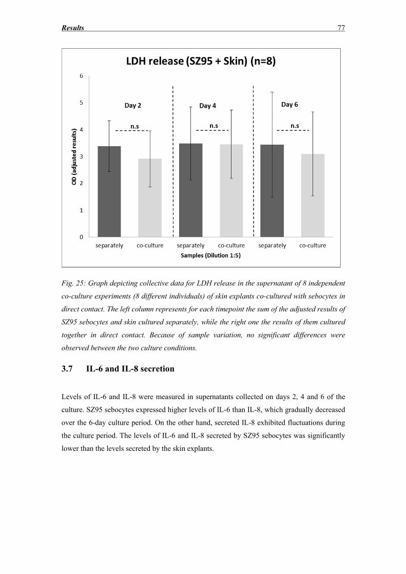
Results 77
Fig. 25: Graph depicting collective data for LDH release in the supernatant of 8 independent
co-culture experiments (8 different individuals) of skin explants co-cultured with sebocytes in
direct contact. The left column represents for each timepoint the sum of the adjusted results of
SZ95 sebocytes and skin cultured separately, while the right one the results of them cultured
together in direct contact. Because of sample variation, no significant differences were
observed between the two culture conditions.
3.7 IL-6 and IL-8 secretion
Levels of IL-6 and IL-8 were measured in supernatants collected on days 2, 4 and 6 of the
culture. SZ95 sebocytes expressed higher levels of IL-6 than IL-8, which gradually decreased
over the 6-day culture period. On the other hand, secreted IL-8 exhibited fluctuations during
the culture period. The levels of IL-6 and IL-8 secreted by SZ95 sebocytes was significantly
lower than the levels secreted by the skin explants.
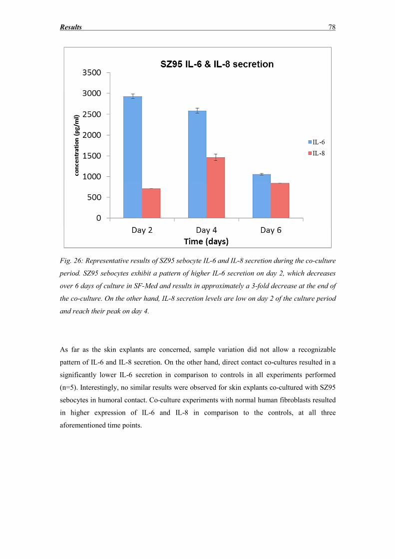
Results 78
Fig. 26: Representative results of SZ95 sebocyte IL-6 and IL-8 secretion during the co-culture
period. SZ95 sebocytes exhibit a pattern of higher IL-6 secretion on day 2, which decreases
over 6 days of culture in SF-Med and results in approximately a 3-fold decrease at the end of
the co-culture. On the other hand, IL-8 secretion levels are low on day 2 of the culture period
and reach their peak on day 4.
As far as the skin explants are concerned, sample variation did not allow a recognizable
pattern of IL-6 and IL-8 secretion. On the other hand, direct contact co-cultures resulted in a
significantly lower IL-6 secretion in comparison to controls in all experiments performed
(n=5). Interestingly, no similar results were observed for skin explants co-cultured with SZ95
sebocytes in humoral contact. Co-culture experiments with normal human fibroblasts resulted
in higher expression of IL-6 and IL-8 in comparison to the controls, at all three
aforementioned time points.
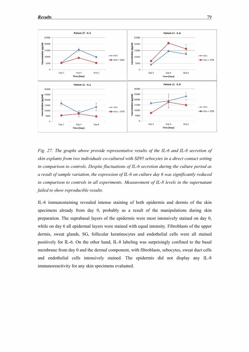
Results 79
Fig. 27: The graphs above provide representative results of the IL-6 and IL-8 secretion of
skin explants from two individuals co-cultured with SZ95 sebocytes in a direct contact setting
in comparison to controls. Despite fluctuations of IL-6 secretion during the culture period as
a result of sample variation, the expression of IL-6 on culture day 6 was significantly reduced
in comparison to controls in all experiments. Measurement of IL-8 levels in the supernatant
failed to show reproducible results.
IL-6 immunostaining revealed intense staining of both epidermis and dermis of the skin
specimens already from day 0, probably as a result of the manipulations during skin
preparation. The suprabasal layers of the epidermis were most intensively stained on day 0,
while on day 6 all epidermal layers were stained with equal intensity. Fibroblasts of the upper
dermis, sweat glands, SG, follicular keratinocytes and endothelial cells were all stained
positively for IL-6. On the other hand, IL-8 labeling was surprisingly confined to the basal
membrane from day 0 and the dermal component, with fibroblasts, sebocytes, sweat duct cells
and endothelial cells intensively stained. The epidermis did not display any IL-8
immunoreactivity for any skin specimens evaluated.
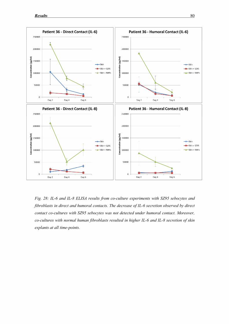
Results 80
Fig. 28: IL-6 and IL-8 ELISA results from co-culture experiments with SZ95 sebocytes and
fibroblasts in direct and humoral contacts. The decrease of IL-6 secretion observed by direct
contact co-cultures with SZ95 sebocytes was not detected under humoral contact. Moreover,
co-cultures with normal human fibroblasts resulted in higher IL-6 and IL-8 secretion of skin
explants at all time-points.
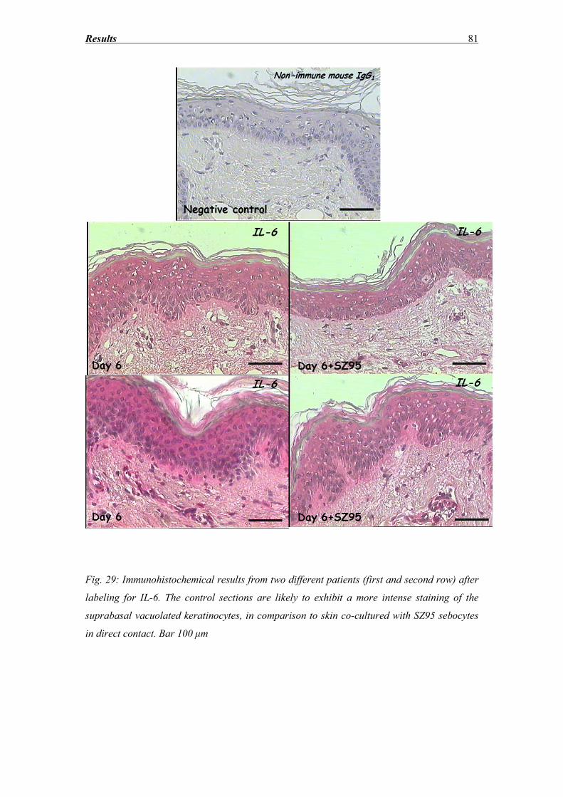
Results 81
Fig. 29: Immunohistochemical results from two different patients (first and second row) after
labeling for IL-6. The control sections are likely to exhibit a more intense staining of the
suprabasal vacuolated keratinocytes, in comparison to skin co-cultured with SZ95 sebocytes
in direct contact. Bar 100 μm
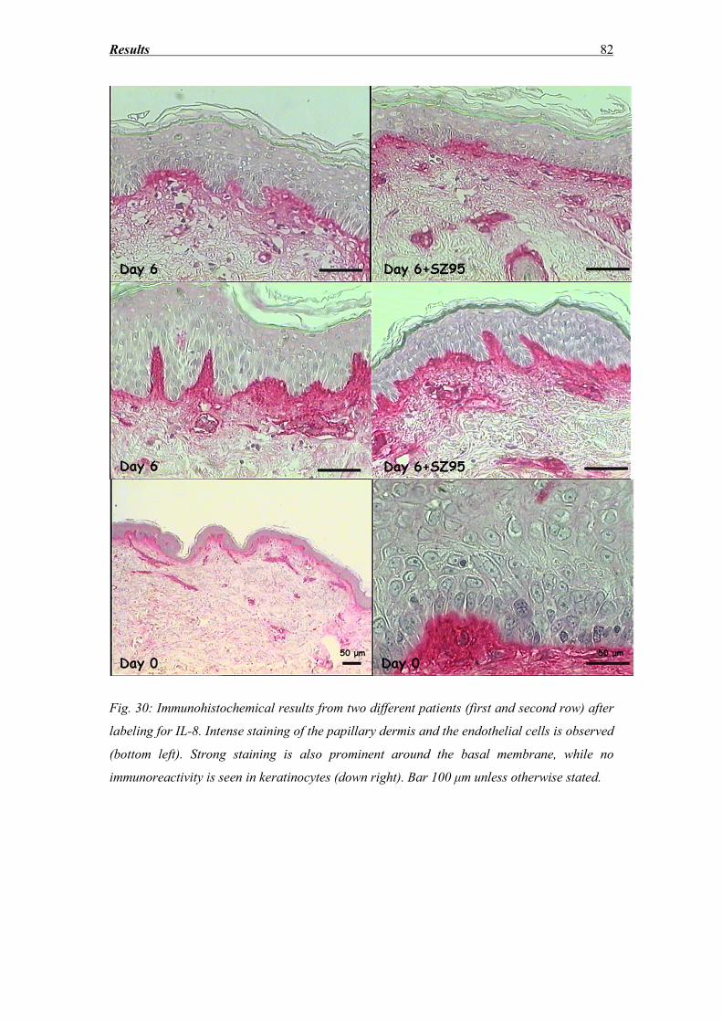
Results 82
Fig. 30: Immunohistochemical results from two different patients (first and second row) after
labeling for IL-8. Intense staining of the papillary dermis and the endothelial cells is observed
(bottom left). Strong staining is also prominent around the basal membrane, while no
immunoreactivity is seen in keratinocytes (down right). Bar 100 μm unless otherwise stated.
Results 83
3.8 SZ95 Sebocytes – Morphology, Oil red and immunocytochemistry
findings
SZ95 sebocytes cultured over 6 days in SF-Med presented an overall monomorphic
morphology with no notable lipid synthesis throughout the culture period. Moreover,
formation of three-dimensional structures growing vertically to the initial monolayer culture
was observed from day 4. Co-culture of the cells with skin in direct contact resulted in a more
polymorphic population of SZ95 sebocytes, with many cells increasing in size, exhibiting the
characteristic abundant, refractile cytoplasm already from the second day of the co-culture. In
addition, many fragmented nuclei were observed, which is a sign of apoptosis. The
accumulation of cytoplasmic lipid droplets was prominent on day 4 and increased further on
Day 6. Moreover, an increased number of fully differentiated sebocytes according to the Tosti
classification [136] was observed already from day 4, with most of the accumulated lipid
droplets located in the perinuclear area.
Surprisingly, part of the cells which were in direct contact with the skin were absent and the
ones in proximity to them were full of lipid droplets. On the other hand, SZ95 sebocytes
located near the periphery of the culture well did not exhibit significant differences regarding
lipid accumulation in comparison to the control. Subsequent staining of the skin sections with
an antibody raised against the SV40 Large Antigen revealed many SZ95 sebocytes attached
on the lower side of the dermis (see Fig. 31).
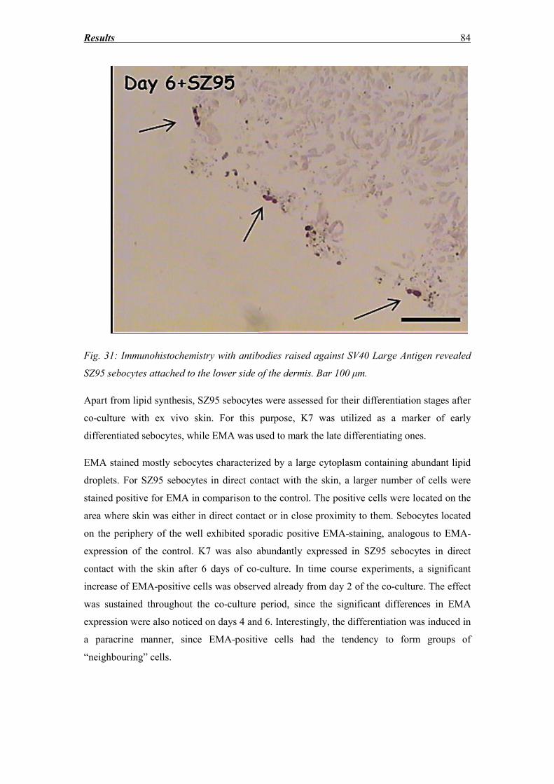
Results 84
Fig. 31: Immunohistochemistry with antibodies raised against SV40 Large Antigen revealed
SZ95 sebocytes attached to the lower side of the dermis. Bar 100 μm.
Apart from lipid synthesis, SZ95 sebocytes were assessed for their differentiation stages after
co-culture with ex vivo skin. For this purpose, K7 was utilized as a marker of early
differentiated sebocytes, while EMA was used to mark the late differentiating ones.
EMA stained mostly sebocytes characterized by a large cytoplasm containing abundant lipid
droplets. For SZ95 sebocytes in direct contact with the skin, a larger number of cells were
stained positive for EMA in comparison to the control. The positive cells were located on the
area where skin was either in direct contact or in close proximity to them. Sebocytes located
on the periphery of the well exhibited sporadic positive EMA-staining, analogous to EMA-
expression of the control. K7 was also abundantly expressed in SZ95 sebocytes in direct
contact with the skin after 6 days of co-culture. In time course experiments, a significant
increase of EMA-positive cells was observed already from day 2 of the co-culture. The effect
was sustained throughout the co-culture period, since the significant differences in EMA
expression were also noticed on days 4 and 6. Interestingly, the differentiation was induced in
a paracrine manner, since EMA-positive cells had the tendency to form groups of
“neighbouring” cells.
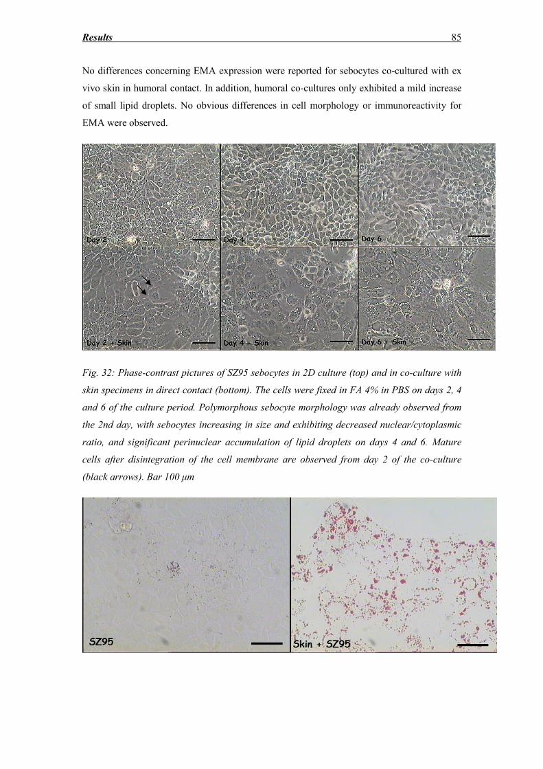
Results 85
No differences concerning EMA expression were reported for sebocytes co-cultured with ex
vivo skin in humoral contact. In addition, humoral co-cultures only exhibited a mild increase
of small lipid droplets. No obvious differences in cell morphology or immunoreactivity for
EMA were observed.
Fig. 32: Phase-contrast pictures of SZ95 sebocytes in 2D culture (top) and in co-culture with
skin specimens in direct contact (bottom). The cells were fixed in FA 4% in PBS on days 2, 4
and 6 of the culture period. Polymorphous sebocyte morphology was already observed from
the 2nd day, with sebocytes increasing in size and exhibiting decreased nuclear/cytoplasmic
ratio, and significant perinuclear accumulation of lipid droplets on days 4 and 6. Mature
cells after disintegration of the cell membrane are observed from day 2 of the co-culture
(black arrows). Bar 100 μm
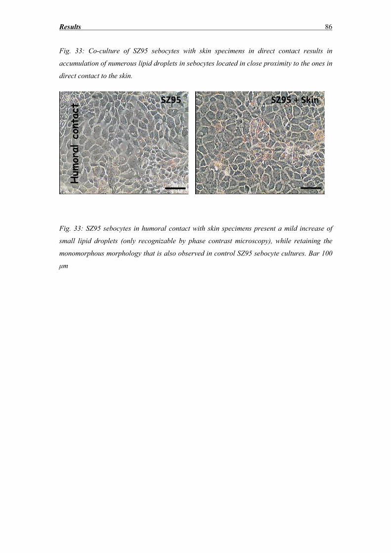
Results 86
Fig. 33: Co-culture of SZ95 sebocytes with skin specimens in direct contact results in
accumulation of numerous lipid droplets in sebocytes located in close proximity to the ones in
direct contact to the skin.
Fig. 33: SZ95 sebocytes in humoral contact with skin specimens present a mild increase of
small lipid droplets (only recognizable by phase contrast microscopy), while retaining the
monomorphous morphology that is also observed in control SZ95 sebocyte cultures. Bar 100
μm
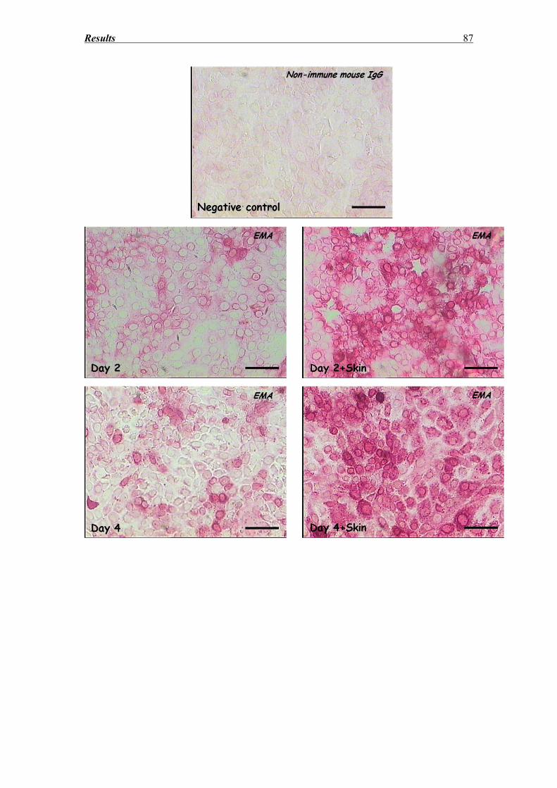
Results 87
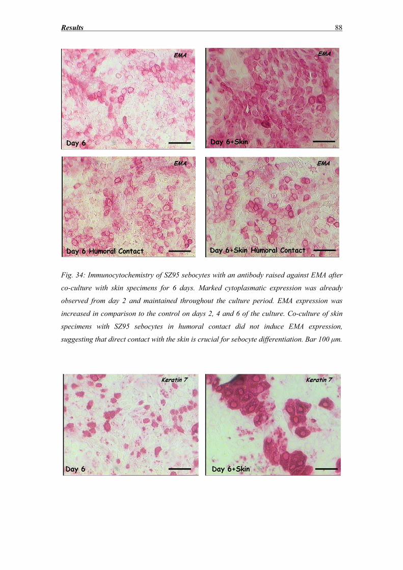
Results 88
Fig. 34: Immunocytochemistry of SZ95 sebocytes with an antibody raised against EMA after
co-culture with skin specimens for 6 days. Marked cytoplasmatic expression was already
observed from day 2 and maintained throughout the culture period. EMA expression was
increased in comparison to the control on days 2, 4 and 6 of the culture. Co-culture of skin
specimens with SZ95 sebocytes in humoral contact did not induce EMA expression,
suggesting that direct contact with the skin is crucial for sebocyte differentiation. Bar 100 μm.
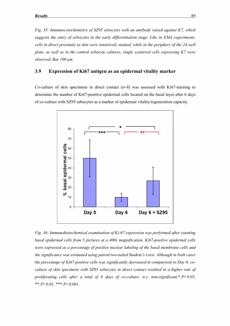
Results 89
Fig. 35: Immunocytochemistry of SZ95 sebocytes with an antibody raised against K7, which
suggests the entry of sebocytes in the early differentiation stage. Like in EMA experiments,
cells in direct proximity to skin were intensively stained, while in the periphery of the 24-well
plate, as well as in the control sebocyte cultures, single scattered cells expressing K7 were
observed. Bar 100 μm
3.9 Expression of Ki67 antigen as an epidermal vitality marker
Co-culture of skin specimens in direct contact (n=8) was assessed with Ki67-staining to
determine the number of Ki67-positive epidermal cells located on the basal layer after 6 days
of co-culture with SZ95 sebocytes as a marker of epidermal vitality/regeneration capacity.
Fig. 36: Immunohistochemical examination of Ki-67 expression was performed after counting
basal epidermal cells from 5 pictures at a 400x magnification. Ki67-positive epidermal cells
were expressed as a percentage of positive nuclear labeling of the basal membrane cells and
the significance was estimated using paired two-tailed Student’s t-test. Although in both cases
the percentage of Ki67-positive cells was significantly decreased in comparison to Day 0, co-
culture of skin specimens with SZ95 sebocytes in direct contact resulted in a higher rate of
proliferating cells after a total of 6 days of co-culture. n.s: non-significant,*:P<0.05,
**:P<0.01, ***:P<0.001.
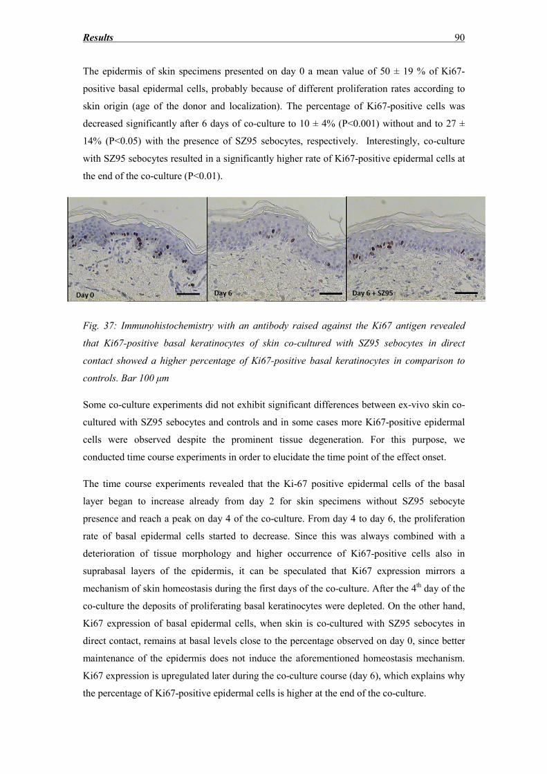
Results 90
The epidermis of skin specimens presented on day 0 a mean value of 50 ± 19 % of Ki67-
positive basal epidermal cells, probably because of different proliferation rates according to
skin origin (age of the donor and localization). The percentage of Ki67-positive cells was
decreased significantly after 6 days of co-culture to 10 ± 4% (P<0.001) without and to 27 ±
14% (P<0.05) with the presence of SZ95 sebocytes, respectively. Interestingly, co-culture
with SZ95 sebocytes resulted in a significantly higher rate of Ki67-positive epidermal cells at
the end of the co-culture (P<0.01).
Fig. 37: Immunohistochemistry with an antibody raised against the Ki67 antigen revealed
that Ki67-positive basal keratinocytes of skin co-cultured with SZ95 sebocytes in direct
contact showed a higher percentage of Ki67-positive basal keratinocytes in comparison to
controls. Bar 100 μm
Some co-culture experiments did not exhibit significant differences between ex-vivo skin co-
cultured with SZ95 sebocytes and controls and in some cases more Ki67-positive epidermal
cells were observed despite the prominent tissue degeneration. For this purpose, we
conducted time course experiments in order to elucidate the time point of the effect onset.
The time course experiments revealed that the Ki-67 positive epidermal cells of the basal
layer began to increase already from day 2 for skin specimens without SZ95 sebocyte
presence and reach a peak on day 4 of the co-culture. From day 4 to day 6, the proliferation
rate of basal epidermal cells started to decrease. Since this was always combined with a
deterioration of tissue morphology and higher occurrence of Ki67-positive cells also in
suprabasal layers of the epidermis, it can be speculated that Ki67 expression mirrors a
mechanism of skin homeostasis during the first days of the co-culture. After the 4th day of the
co-culture the deposits of proliferating basal keratinocytes were depleted. On the other hand,
Ki67 expression of basal epidermal cells, when skin is co-cultured with SZ95 sebocytes in
direct contact, remains at basal levels close to the percentage observed on day 0, since better
maintenance of the epidermis does not induce the aforementioned homeostasis mechanism.
Ki67 expression is upregulated later during the co-culture course (day 6), which explains why
the percentage of Ki67-positive epidermal cells is higher at the end of the co-culture.
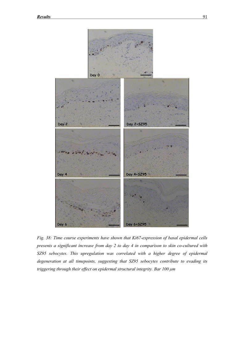
Results 91
Fig. 38: Time course experiments have shown that Ki67-expression of basal epidermal cells
presents a significant increase from day 2 to day 4 in comparison to skin co-cultured with
SZ95 sebocytes. This upregulation was correlated with a higher degree of epidermal
degeneration at all timepoints, suggesting that SZ95 sebocytes contribute to evading its
triggering through their effect on epidermal structural integrity. Bar 100 μm
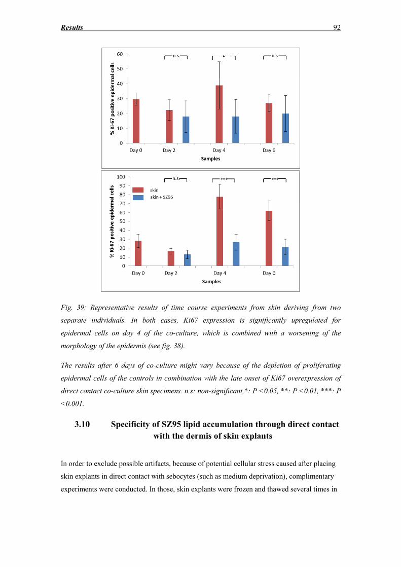
Results 92
Fig. 39: Representative results of time course experiments from skin deriving from two
separate individuals. In both cases, Ki67 expression is significantly upregulated for
epidermal cells on day 4 of the co-culture, which is combined with a worsening of the
morphology of the epidermis (see fig. 38).
The results after 6 days of co-culture might vary because of the depletion of proliferating
epidermal cells of the controls in combination with the late onset of Ki67 overexpression of
direct contact co-culture skin specimens. n.s: non-significant,*: P <0.05, **: P <0.01, ***: P
<0.001.
3.10 Specificity of SZ95 lipid accumulation through direct contact
with the dermis of skin explants
In order to exclude possible artifacts, because of potential cellular stress caused after placing
skin explants in direct contact with sebocytes (such as medium deprivation), complimentary
experiments were conducted. In those, skin explants were frozen and thawed several times in
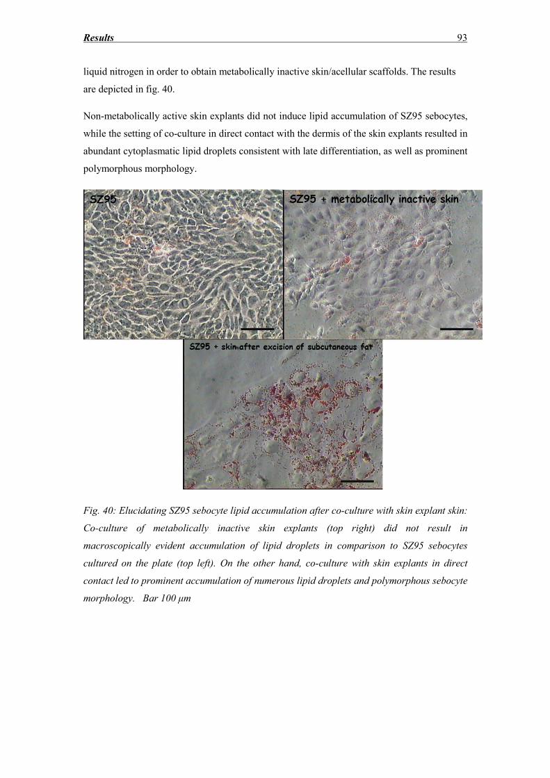
Results 93
liquid nitrogen in order to obtain metabolically inactive skin/acellular scaffolds. The results
are depicted in fig. 40.
Non-metabolically active skin explants did not induce lipid accumulation of SZ95 sebocytes,
while the setting of co-culture in direct contact with the dermis of the skin explants resulted in
abundant cytoplasmatic lipid droplets consistent with late differentiation, as well as prominent
polymorphous morphology.
Fig. 40: Elucidating SZ95 sebocyte lipid accumulation after co-culture with skin explant skin:
Co-culture of metabolically inactive skin explants (top right) did not result in
macroscopically evident accumulation of lipid droplets in comparison to SZ95 sebocytes
cultured on the plate (top left). On the other hand, co-culture with skin explants in direct
contact led to prominent accumulation of numerous lipid droplets and polymorphous sebocyte
morphology. Bar 100 μm
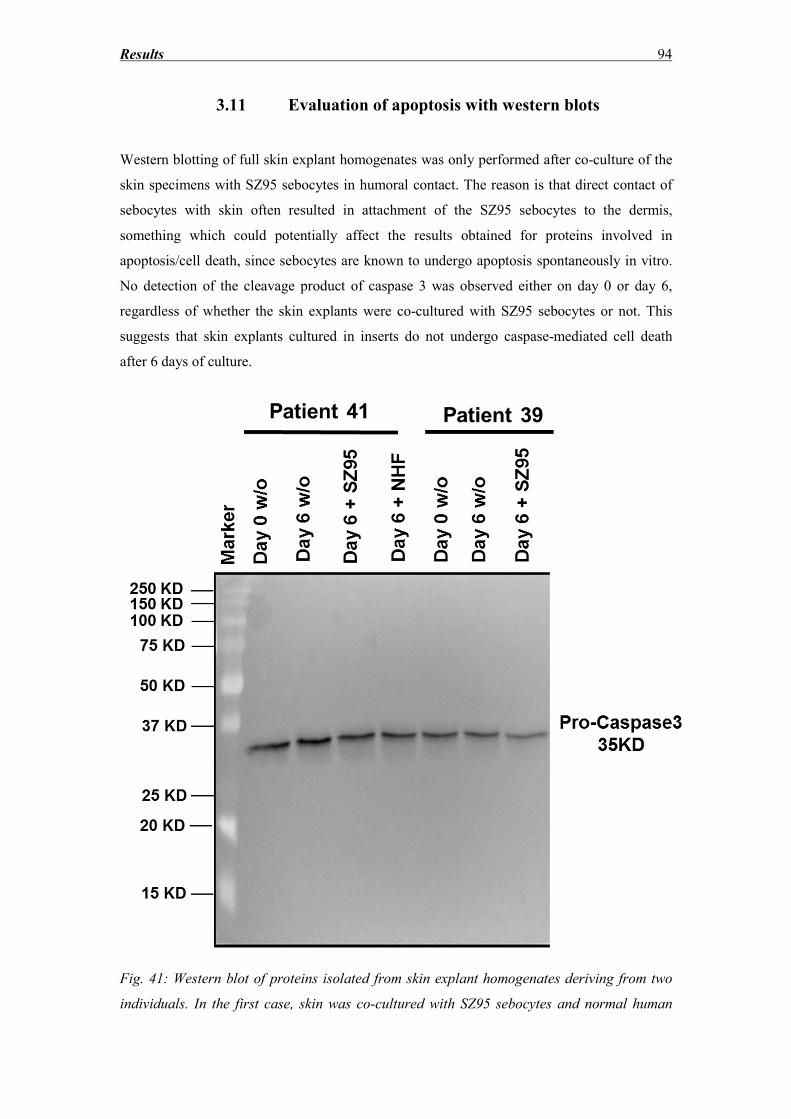
Results 94
3.11 Evaluation of apoptosis with western blots
Western blotting of full skin explant homogenates was only performed after co-culture of the
skin specimens with SZ95 sebocytes in humoral contact. The reason is that direct contact of
sebocytes with skin often resulted in attachment of the SZ95 sebocytes to the dermis,
something which could potentially affect the results obtained for proteins involved in
apoptosis/cell death, since sebocytes are known to undergo apoptosis spontaneously in vitro.
No detection of the cleavage product of caspase 3 was observed either on day 0 or day 6,
regardless of whether the skin explants were co-cultured with SZ95 sebocytes or not. This
suggests that skin explants cultured in inserts do not undergo caspase-mediated cell death
after 6 days of culture.
Fig. 41: Western blot of proteins isolated from skin explant homogenates deriving from two
individuals. In the first case, skin was co-cultured with SZ95 sebocytes and normal human
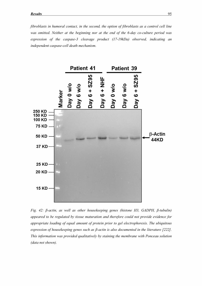
Results 95
fibroblasts in humoral contact, in the second, the option of fibroblasts as a control cell line
was omitted. Neither at the beginning nor at the end of the 6-day co-culture period was
expression of the caspase-3 cleavage product (17-19kDa) observed, indicating an
independent caspase-cell death mechanism.
Fig. 42: β-actin, as well as other housekeeping genes (histone H3, GADPH, β-tubulin)
appeared to be regulated by tissue maturation and therefore could not provide evidence for
appropriate loading of equal amount of protein prior to gel electrophoresis. The ubiquitous
expression of housekeeping genes such as β-actin is also documented in the literature [222].
This information was provided qualitatively by staining the membrane with Ponceau solution
(data not shown).
Discussion 96
Discussion
4.1 Integration of the variable “sebaceous gland” in a new skin explant
model
The aim of this study was to provide a short-term, reproducible, skin explant co-culture
model, where the variable “sebaceous gland (SG)” and its effects on skin explant homeostasis
and maintenance are elucidated. As mentioned before, the immortalized SG cell line SZ95
provides a convenient solution for the problem of retaining SG cells, both in vitro and ex
vivo, without them undergoing rapidly holocrine secretion, leading to programmed cell death.
For this purpose, SZ95 sebocytes were cultured with skin in direct contact as well as humoral
contact (contact only through the culture medium with the help of culture well inserts). In
both cases, the epidermis was air-exposed, since this is considered helpful to maintain the
structural integrity of the skin and to allow complete differentiation and stratification of the
epidermis [13, 19]. This was also confirmed in our study, since the submerged edges of the
skin specimens cultured in the direct contact setting showed morphological signs of
degeneration of the epidermis, as well as increased rates of TUNEL-positive epidermal cells.
4.2 Choice of medium
The choice of the medium in any co-culture setting is a compromise of essential ingredients
for both cells and the skin. High calcium concentrations are known to be crucial for the
epidermis, since skin tissue quickly degenerates under conditions of low extracellular Ca++
[223]. Hashimoto was among the first to observe acantholysis-like suprabasal cleft-formation
induced already within 12 h of skin organ culture in medium containing less than 0.1 mM
Ca++
[224]. William’s E medium, which is often used for skin organ culture, contains
physiologic concentrations of calcium [21, 27]. Varani et al [3] cultured skin explants in
keratinocyte basal medium supplemented with calcium to a total of 1.4 mM. A potential
explanation for the beneficial role of Ca++
in the culture medium was given by the skin
explant model of Tavakkol et al [23]. Lower levels of mRNA of the IGF-1 receptor were
detected in skin explant homogenates cultured in low Ca++
concentrations. IGF-1 is known to
promote keratinocyte proliferation in vitro [23]. On the other hand, physiologic
concentrations of Ca++
favor sebocyte proliferation in contrast to differentiation. Retinol was
also included in the medium, because it comprises the precursor of sebaceous lipids. Culture
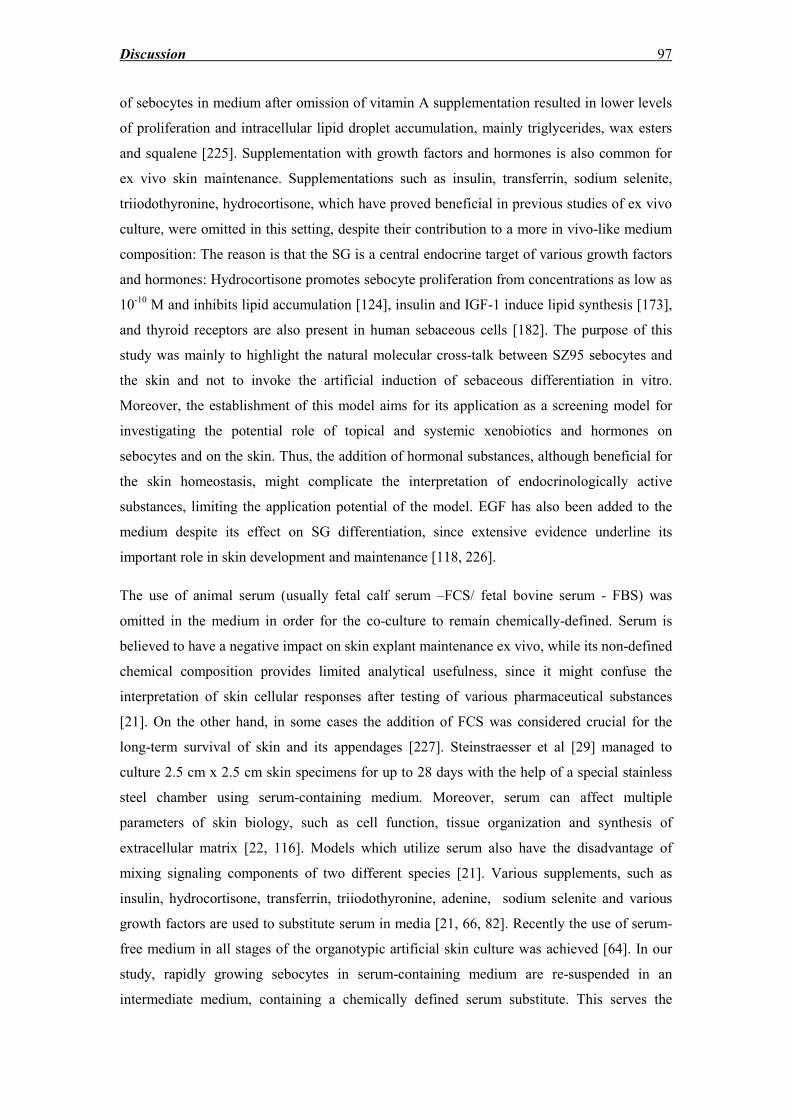
Discussion 97
of sebocytes in medium after omission of vitamin A supplementation resulted in lower levels
of proliferation and intracellular lipid droplet accumulation, mainly triglycerides, wax esters
and squalene [225]. Supplementation with growth factors and hormones is also common for
ex vivo skin maintenance. Supplementations such as insulin, transferrin, sodium selenite,
triiodothyronine, hydrocortisone, which have proved beneficial in previous studies of ex vivo
culture, were omitted in this setting, despite their contribution to a more in vivo-like medium
composition: The reason is that the SG is a central endocrine target of various growth factors
and hormones: Hydrocortisone promotes sebocyte proliferation from concentrations as low as
10-10
M and inhibits lipid accumulation [124], insulin and IGF-1 induce lipid synthesis [173],
and thyroid receptors are also present in human sebaceous cells [182]. The purpose of this
study was mainly to highlight the natural molecular cross-talk between SZ95 sebocytes and
the skin and not to invoke the artificial induction of sebaceous differentiation in vitro.
Moreover, the establishment of this model aims for its application as a screening model for
investigating the potential role of topical and systemic xenobiotics and hormones on
sebocytes and on the skin. Thus, the addition of hormonal substances, although beneficial for
the skin homeostasis, might complicate the interpretation of endocrinologically active
substances, limiting the application potential of the model. EGF has also been added to the
medium despite its effect on SG differentiation, since extensive evidence underline its
important role in skin development and maintenance [118, 226].
The use of animal serum (usually fetal calf serum –FCS/ fetal bovine serum - FBS) was
omitted in the medium in order for the co-culture to remain chemically-defined. Serum is
believed to have a negative impact on skin explant maintenance ex vivo, while its non-defined
chemical composition provides limited analytical usefulness, since it might confuse the
interpretation of skin cellular responses after testing of various pharmaceutical substances
[21]. On the other hand, in some cases the addition of FCS was considered crucial for the
long-term survival of skin and its appendages [227]. Steinstraesser et al [29] managed to
culture 2.5 cm x 2.5 cm skin specimens for up to 28 days with the help of a special stainless
steel chamber using serum-containing medium. Moreover, serum can affect multiple
parameters of skin biology, such as cell function, tissue organization and synthesis of
extracellular matrix [22, 116]. Models which utilize serum also have the disadvantage of
mixing signaling components of two different species [21]. Various supplements, such as
insulin, hydrocortisone, transferrin, triiodothyronine, adenine, sodium selenite and various
growth factors are used to substitute serum in media [21, 66, 82]. Recently the use of serum-
free medium in all stages of the organotypic artificial skin culture was achieved [64]. In our
study, rapidly growing sebocytes in serum-containing medium are re-suspended in an
intermediate medium, containing a chemically defined serum substitute. This serves the
Discussion 98
purpose of minimizing the cellular stress of the medium switch to serum-free, which could
have a negative potential effect on ex vivo skin during the co-culture. The SZ95 sebocytes
remain in this medium overnight and then the medium is switched to serum-free with the
beginning of the co-culture.
Fig. 41: Summary of cellular events occurring during culture of skin ex vivo. The signs of
degeneration of the epidermis are presented according to their chronological appearance,
based on previous literature and the present study [21, 27, 29, 101].
4.3 Sebaceous gland maintenance ex vivo
In our study, SG ex vivo have exhibited signs of degeneration already on Day 0, with
morphological signs of sebocyte progressive differentiation of cells proximal to the stratum
germinativum of the SG, while prominent disruption of the continuity between basal
sebocytes and early differentiated ones affected the SG architecture. The detachment of the
cells from the basal lamina indicates that one of the first stages of sebaceous degeneration ex
vivo is the defect or loss of hemidesmosomes, connecting stratum germinativum with the
basal membrane [228]. Results from Ki67 and TUNEL staining have revealed the complete
absence of Ki67-positive cells after 6 days of culture, while the basal layer was stained
positive for TUNEL, suggesting that the proliferating sebocytes ex vivo, which should be
responsible for the regeneration of the gland, undergo apoptosis and are not in a position to
retain the morphology and function of the appendage ex vivo. Interestingly, co-culture of the
Discussion 99
skin with SZ95 sebocytes did not influence ex vivo SG survival. A potential explanation for
the quick SG degeneration in comparison to epidermis could be the difference between
sebaceous gland and epidermis regeneration. Sebocyte renewal rate is estimated to be
approximately 21-25 days, while the keratinocyte rate is shorter (28 days) [228, 229].
Therefore, the higher fraction of proliferating sebocytes, in comparison to the epidermis,
makes the SG more susceptible to losing its regeneration capacity after a major stress
stimulus, such as the extraction of the appendage from the in vivo microenvironment and its
subsequent culture ex vivo.
Lu et al. [21] reported unaltered SG morphology for 5 days in his hair shaft elongation model
in vitro. From the illustrations provided, early differentiated sebocytes appear to undergo
rapid differentiation. Since SG were not the focus of that study, there is no biochemical
evidence (Ki67/TUNEL) to support that the SG also remained functional. The model of ex
vivo isolation of SG of Guy et al. [118] describes the maintenance of glands as full organs ex
vivo with the ability to retain in situ rates of cell proliferation and lipogenesis, as well as in
vivo-like responses after treatment with DHT, EGF, 13-cis RA and other substances. Despite
the obvious advantage of imitating ideally the in vivo-conditions, the model includes the
arduous process of SG micro-dissection of the perilobular collagen. Moreover, the activity of
the SG and the sebum composition differ greatly between individuals, which appear to have a
considerable age-related variation [230-232]: decreased androgen levels of elderly individuals
affect the rate of sebaceous differentiation and secretory output of the gland, resulting in
glandular hyperplasia [233, 234]. Estrogens suppress lipid synthesis of the SG directly, while
low estrogen levels after menopause are correlated with reduced SG activity [129, 235]. The
effects of hormones on SG regulation were also confirmed in vitro at the molecular level,
since incubation of SZ95 sebocytes with age-specific growth factor and hormonal mixtures
regulates the expression of many genes at the mRNA level involved in DNA repair and
stability, mitochondrial function, oxidative stress, apoptosis and tumourigenesis [236].
Because of the aforementioned data, it is obvious that by obtaining SG of different individuals
in order to test hormones, sample variation might lead to considerable misinterpretations. The
main advantage of the model proposed in this study is that the effect of sebocytes on skin
remains constant and reproducible, since the SG of the skin degenerate quickly ex vivo and
are substituted by the immortalized SG cell line SZ95.
4.4 Direct contact of SZ95 sebocytes with skin explant dermis promotes
skin explant epidermal integrity
Discussion 100
Since the epidermis turnover rate is 4 weeks, a maximum of 14 days is usually selected by
most research teams as the last time-point of skin culture in order to keep the skin explant
epidermal stratification intact. The morphological features, which were observed in the
epidermis of our model, namely keratinocyte vacuolization, parakeratosis, cleft formation,
suprabasal, and intrabasal bulla formation with acantholysis and lastly separation of the
epidermis from the dermis, have already been reported in previous experimental settings [23,
27, 29, 101, 224]. In this study the endpoint of 7 days was chosen (skin explants remain
overnight in serum-free medium before the beginning of the co-culture), since separation of
the epidermis from the dermis was often observed after the 8th day of culture. Signs of
epidermal degradation in this setting were observed already from day 2, with significant
reduction of the viable keratinocyte layers, keratinocyte vacuolization and parakeratosis. Skin
cultured with SZ95 sebocytes in direct contact exhibited an overall improvement in
maintenance in more than 80% of the skin samples used. Skin specimens cultured with SZ95
sebocytes in direct contact exhibited only early events of epidermal degeneration (mainly
partial nuclear vacuolization and parakeratosis), indicating that the latter provide the essential
factors to sustain the epidermal skin explant vitality. Since these effects were not detected
when the cells were co-cultured in humoral contact with skin specimens, it seems that the
molecular cross-talk takes place in a paracrine manner. Moreover, since sebocytes are in
direct contact with the dermis after excision of subcutaneous fat, it can be postulated that the
effect of sebocytes on epidermis is indirectly mediated through the dermal component of the
skin. Interestingly, presence of SZ95 sebocytes resulted in a better maintenance of the vital
epidermis thickness and prevented excessive thickening of stratum corneum, which is a
common problem in both in vitro reconstructed skin as well as ex vivo organ culture because
of lack of desquamation [105]. These data suggest a normalizing effect of sebocytes in
epidermal stratification in vitro, which could be controlled in the future through the
expression of special markers of different stages of keratinocyte differentiation (early
differentiation markers K1 and K10 [35, 67, 237], late differentiation markers loricrin,
involucrin, transglutaminases [50-53]).
The effect of SZ95 sebocytes on skin explant vitality, regeneration and structural integrity of
the epidermis can be mediated through a wide variety of growth factors and hormones. The
SG has a cardinal role in dermatoendocrinology, since it is a target and source of numerous
hormones, growth factors and chemokines. Sebocytes can synthesize de novo cholesterol
from acetate [149, 150] and possess all the essential enzymatic machinery to produce sex
steroids as well as glucocorticoids [123] by promoting the expression of cyclin D2, which
facilitates G1 to S phase transition [238]. They increase acid mucopolysaccharides,
Discussion 101
hyaluronic acid, collagen I and III in the dermis [239, 240], promote wound healing through
acceleration of keratinocyte GM-CSF secretion [241], protect from photoageing [242] and
prevent wrinkle formation and skin dryness of post-menopausal women [243]. Procollagen 1
and 3 mRNA levels, androgen receptor (AR) and heat shock protein 47 (HSP47) expression
of postmenopausal woman skin were upregulated after DHEA treatment, suggesting a role of
androgens in skin tissue homeostasis and ageing [244]. A recent work of Haag et al. [245]
showed that only confluent SZ95 sebocytes and not fibroblasts or keratinocytes express the
enzyme 3β-HSD 1 in vitro, which is responsible for the catalyzation of the formation of
androstenedione from DHEA, as the first step for in situ testosterone formation. Moreover,
sebocytes are a site of antioxidant production [228], such as the antioxidant repair enzymes
methionine-S-sulfoxide reductase A (MSRA) and three isotypes of methionine-R-sulfoxide
reductase B (MSRB) [246].
4.5 Explanation for IL-6 downregulation after co-culture with SZ95
sebocytes
The proinflammatory cytokines IL-6 and IL-8 were used for the detection of inflammation as
a sign of disturbed homeostasis of cells and skin explants. IL-6, together with TNF-α and IL-
1, is responsible for the control of the acute inflammatory response, fever, the acute response
in the liver and the transition from acute inflammation to either acquired immunity or chronic
inflammatory disease [247-249]. Skin cell types, which are known to express IL-6, are CD8+
T cells, adipocytes, fibroblasts, endothelial cells, mast cells, keratinocytes, sebocytes,
Langerhans cells, neutrophils, monocytes and eosinophils [248, 250, 251]. IL-6 induces
signaling by forming a heterodimeric receptor complex of the molecule with its receptor (IL-6
R), which is solely produced by hepatocytes, monocytes and resting lymphocytes. On the
other hand, IL-8 is a member of the CXC subfamily of chemokines and a potent neutrophil
chemotactic and activating factor. It is one of the primary inflammatory cytokines produced
by keratinocytes, sebocytes, fibroblasts, endothelial cells, neutrophils, T-cells etc. [252-255]
IL-6 and IL-8 are both released in the supernatant of unstressed sebocyte culture [190, 256],
together with other cytokines such as IL-1α, IL-1β, and TNF-α [200], while their expression
increases significantly in a stressed environment, for example in the presence of LPS and P.
acnes. A mild IL-6 expression was detected in SG of uninvolved skin of acne patients and a
stronger one in SG of acne skin lesions. Moreover, IL-6 was barely detectable in the skin of
healthy volunteers. In our study, we observed a strong immunoreactivity of the epidermal and
dermal component, including cells of cutaneous appendages, such as follicular keratinocytes,
endothelial cells and sebaceous duct cells as early as on Day 0, indicating upregulation of the
Discussion 102
cytokine after skin explant manipulation. IL-8 exhibited a stronger expression in SG of acne
patients than in healthy volunteers [256]. In this study, the expression was limited mostly
around the basal membrane and the dermis, including fibroblasts and cutaneous appendages,
but surprisingly, epidermal keratinocytes were negative. The pattern of IL-8 expression
differs from the induction of IL-8 expression in keratinocytes of acne lesions, after P. acnes
interaction with TLR-2 on their surface [257, 258].
Downregulation of IL-6 expression in skin explants after co-culture with SZ95 sebocytes in
direct contact could be explained by the strong involvement of sebocytes in immunological
processes. Sebocytes comprise an integral part of the innate immune system. As mentioned
before, sebum contains lipids which exhibit antimicrobial properties and which are
particularly effective mainly against Gram-positive but also Gram-negative bacteria, such as
methicillin resistant S. aureus, Streptococcus salivarius, P. acnes, Fusobacterium nucleatum,
Pseudomonas aeruginosa and E. Coli [217, 259, 260]. Lipids with an antimicrobial role can
be both saturated, such as lauric acid and palmitic acid, as well as unsaturated such as
sapienic acid. Saturated lipids can easily form monomers in aqueous solutions, to which their
antimicrobial activity is attributed. Short chained lipids exhibit in general greater
antimicrobial activities than the long chained ones. Furthermore, the production of
antimicrobial peptides in the form of triglyceride esters leads to their release and action after
bacterial hydrolysis or their chemical process by epidermal acid lipase [259, 260]. Histone H4
and the antimicrobial peptides cathelicidin and β-defensin are produced by human sebocytes
in vitro [200, 213, 214]. Human β-defensin 2 was upregulated after incubation of human
sebocytes with lauric, palmitic or oleic acid, thus introducing the idea of a positive feedback
mechanism of the sebaceous gland antimicrobial activity through sebum free fatty acid
release [209]. Interestingly, the decrease of IL-6 expression seemed to be cell-type specific,
since co-culture with normal human fibroblasts deriving from a young female resulted in
higher IL-6 expression in comparison to the control. Furthermore, antimicrobial peptides are
believed to possess a broader palette of functions, apart from the obvious central role in the
host immune defense: hCAP18, the C-terminal fragment of the antimicrobial peptide
cathelicidin (LL-37), is detected at high levels in the skin in vivo after wounding. Moreover,
treatment with antibodies against cathelicidin resulted in considerable impairment of wound
healing, as shown by a significant decrease of keratinocyte proliferation. These findings
suggest a role of cathelicidin in re-epithelialization and the potential contribution of sebocytes
in retaining the ex vivo skin keratinocyte proliferation rate [261].
Discussion 103
4.6 The insufficiency of LDH release as a marker of cell death for co-
cultures
Since late apoptosis can convert into necrosis [262] and in order to avoid possible
misinterpretations of LDH release already mentioned in the section on materials and methods,
we propose the term of skin “spontaneous LDH release” as a marker for cell death,
programmed or not. The skin explant culture model of Lu et al. [21] and Kleszczynski and
Fischer [27] observed a spike of LDH release after the establishment of the co-culture and
more specifically after 48 h and 24 h, respectively, which was explained as cell death
following manipulations during skin preparation. We also confirmed this peak by many co-
culture experiments, although skin specimens were left overnight in SF-Med before the
beginning of the co-culture to avoid this bias. After this time point, spontaneous LDH release
by SZ95 sebocytes also exhibited a peak after 24 hours, probably because of an increase of
sebaceous differentiation induced by the switch from SS-Med to SF-Med.
LDH release of SZ95 sebocytes and skin cultured separately failed to show significant
differences compared with LDH-release from the co-culture setting. Despite this, any
obtained result could be controversial, since SZ95 sebocytes of the co-culture setting were
more differentiated and underwent holocrine secretion more often, which also lead to a
release of LDH in the supernatant. Apart from sample variation, higher rates of sebaceous
differentiation increased the LDH levels of the supernatant, while reduced cell death of skin
explants would have decreased it. For this purpose, complimentary methods are essential to
elucidate how SZ95 sebocytes can affect skin explant vitality.
4.7 Sebaceous differentiation and lipid accumulation through skin
explant molecular interaction offers a more in-vivo like phenotype
One of the major handicaps of monolayer sebocyte culture is the fact that they only undergo
terminal differentiation in vitro after specific stimulation, thus failing to accurately represent
their in vivo behavior under normal culture conditions. The rates of initial stage and late
differentiated sebocytes are 40% and 60% respectively, which are close to the rates observed
in vivo and many differentiation markers expressed in vivo are also expressed in vitro [263].
On the other hand, sebocytes exhibited a much lower rate of mature sebocytes, approximately
4% to 8% in comparison to the percentage observed in the “necrosis” zone of the SG, which
is estimated to be 20% [264]. Moreover, cultured sebocytes synthetize decreased amounts of
squalene and wax esters in comparison to freshly isolated SG [124]. In our experiments,
direct contact with the skin induced the progressive differentiation of SZ95 sebocytes with a
higher percentage of cells with a big refractile cytoplasm, fragmented nuclei and abundant
Discussion 104
lipid droplets after oil red staining. Visible differences were already observed from the second
day of co-culture and became more conspicuous over the 6-day culture period.
Furthermore, SZ95 sebocytes in direct contact with the skin showed strong immunoreactivity
for the differentiation markers K7 and EMA. EMA is a glycoprotein of high molecular
weight, belonging to the group of human fat globulins. EMA has been shown to have a
specific immunoreactivity for differentiated sebocytes, staining sebaceous cells also positive
for the SGA [265]. EMA is considered a late differentiation marker, while K7 is considered
an early one [127, 266]. Moreover, K7 is expressed exclusively in sebocytes and not
keratinocytes, thus comprising a marker of sebaceous lineage [121, 130]. The problem of the
incomplete terminal sebaceous differentiation was addressed in the study of Barrault et al.
[127], who reported significant differences in EMA expression and lipid synthesis between an
immortalized SG line cultured in vitro and in the air-liquid interface, proposing the latter as a
model, which can better represent the in vivo conditions. However, the air liquid-interface-
induced differentiation raises the question about the reason for this phenomenon. A
postulation would be that Ca++
deprivation after the culture is lifted from a medium containing
physiologic concentration of Ca++
in the air-liquid interface leads to their differentiation.
Furthermore, a small percentage of the SG cell population express involucrin [125], a typical
marker for keratinocyte terminal differentiation. For this reason, upregulation of cornified
envelope proteins needs to be examined and excluded to ensure a possible differentiation of
sebocytes into a more keratinocyte-like phenotype. In our study, sebocytes remained
submerged in a Ca++ concentration of 1.5 mM and underwent sebaceous differentiation with
concomitant lipid accumulation only after direct contact with the dermis, suggesting a
molecular cross-talk with the dermal component of the skin. This setting is closer to in vivo
conditions, since the differentiating sebocytes are in direct contact with the dermis.
4.8 The crucial role of molecular interaction of the epithelial and
mesenchymal component of the skin
Since induction of sebaceous differentiation was mediated through direct but not through
humoral contact with the skin, paracrine molecular cross-talk of SZ95 sebocytes with the
dermal component could be the decisive factor for sebaceous lipogenesis and differentiation.
IGF-1, which is produced by human fibroblasts, was shown to induce lipogenesis in primary
sebocytes [164], SZ95 sebocytes [173] and the SEB-1 sebaceous cell line. Interestingly, the
promotion of lipid accumulation was mediated through increased expression of sterol
Discussion 105
response binding protein 1 (SREBP-1), a cardinal transcription factor for the regulation of
many genes involved in lipogenesis. In this process, Akt activation/phosphorylation is
involved, since the addition of a phosphoinositide 3-Kinase inhibitor (PI3-K) blocked the
expression of SREBP-1 [174]. Maintenance of the co-culture system with an IGF-1-specific
neutralizing antibody might provide evidence of whether IGF-1 could be a major factor
involved in the sebaceous differentiation detected. Interestingly, IGF-1 receptor is abundantly
expressed in all epithelial cells with proliferative potential, including the follicular outer root
sheath keratinocytes, basal sebocytes and the hair matrix and is downregulated in highly
differentiated cells of epithelial origin [267].
Based on the data presented in this work, it can be hypothesized that the mediation of a better
epidermal integrity after co-culture of skin explants with SZ95 sebocytes is due to paracrine
interaction of fibroblasts and sebocytes. The former then affect epidermal keratinocyte
vitality, differentiation and proliferation rate indirectly. This postulation derived from the fact
that no overall improvement of the epidermal status was observed in the humoral co-culture
setting, thus suggesting that holocrine secretion and release of sebaceous content in the
supernatant is not the major factor affecting the epidermis during ex vivo skin maintenance.
Interaction between cells residing in the dermis and epidermis is shown to be crucial for
epidermal regeneration and function: Experimental murine grafts and clinical grafts showed a
significant decrease of epidermal keratinocyte clonogenic capacity, despite the initial
reconstitution of a stratified epithelium after grafting on the lumbo-dermal fascia of mice or a
granulation tissue bed. Surprisingly, the presence of dermis on the grafts increased the self-
renewal capacity of keratinocyte stem cells [268] (see below). A prime example of
keratinocyte-fibroblast interaction is the upregulation of keratinocyte growth factor (KGF) by
the release of the cytokine IL-1β. KGF is a part of the fibroblast growth family and ligand of
the KFGR, which is a tyrosine kinase receptor commonly expressed in epithelial cells [269,
270]. It is known for its paracrine mechanism of action, promoting keratinocyte proliferation
and migration, as well as exhibiting cytoprotective effects after cellular stress induced by
reactive oxygen species or γ-irradiation [271]. Sebocytes are also known to express KGF
receptor [272] and IL-1β [176, 206], thus proposing another molecular interaction loop
favoring epidermal homeostasis.
Moreover, fibroblasts are able to degrade growth inhibitors of keratinocytes, such as TGF-β,
and retain the clonogenic capacity of the latter in vitro [273]. On the other hand, TGF-β
appears to be crucial for the regulation of normal keratinocyte differentiation and apoptosis,
through the activation of TGF-β-activated kinase 1 (TAK1), which is an upstream signaling
molecule of NF-κB. TAK1-deficient mice exhibited an increase in keratinocyte proliferation
and apoptosis, expression of the basal keratinocyte markers K5 and K14 throughout the whole
Discussion 106
epidermis [274]. Expression of IL-1α and GM-CSF from UVB-induced keratinocytes
promotes the expression of fibroblast-derived elastase [275] and neutral endopeptidase [276],
which is implicated in the mechanism of UVB-induced wrinkling. The work of Aoki et al.
[277] stressed the importance of mesenchymal cells for the support of epidermal regeneration
through the utilization of a reconstruction model, although not essentially skin-derived (heart,
spleen, lung, liver, kidney). These mesenchymal cell types affected epidermis homeostasis, as
reflected by the more in-vivo like pattern of involucrin, CK10, CK14 and the epidermal
progenitor marker p63.
4.9 Role of the culture setting in the SZ95 sebocyte-mediated skin
explant vitality
In order to elucidate the molecular mechanisms of wound repair, Wand et al. [278] cultured
keratinocytes and fibroblasts in direct contact and humoral contact and studied the molecular
interactions which affected proliferation and migration of keratinocytes. Interestingly, TGF-
β1, heparin-binding EGF-like growth factor (HB-EGF) and IL-1α mediated their effects to
keratinocyte homeostasis only in the direct co-culture setting [278]. These data suggested the
crucial effect of direct contact between different cell types for providing the essential
microenvironment with the essential chemokine and cytokine levels needed. Surprisingly,
when those three factors were added exogenously, they induced similar effects with direct
contact co-culture but in a 10-fold higher concentration. Taking this data into consideration,
we also observed in our study that only direct contact of the dermis with SZ95 sebocytes
improved ex vivo skin viability. Sebocyte – fibroblast cross talk could lead to release of
signaling molecules which are beneficiary for skin homeostasis. Furthermore, experiments
with humoral contact co-cultures were conducted with a bigger quantity of medium,
suggesting that the further dilution of the growth factors released from SZ95 sebocytes or
fibroblasts did not reach the concentrations needed to affect epidermal regeneration.
Discussion 107
4.10 Skin explant culture to the next level: hypotheses and future
perspectives
In normal skin, both keratinocyte proliferation and differentiation are tightly coupled in order
to retain a normal architecture of a constantly regenerating tissue. The regeneration capacity
of the skin is maintained due to the existence of stem cells, both in the basal layer of the
interfollicular epidermis, the bulge region of the hair follicle and potentially the sebaceous
gland [279]. Self-renewal of skin is maintained through asymmetric division of these cells,
which account for the 10% of the keratinocyte population [280] and have a low tendency to
divide [281], giving rise to the stem cells remaining in the niche and the transient amplifying
cells (TA cells) [282]. The latter are lineage-restricted and may undergo only a certain
number of divisions (2-6) before they differentiate and give rise to the other layers of the
epidermis. The normally slow cycling keratinocyte stem cells can be stimulated to proliferate
in response to growth factors or wounding [283, 284]. Bulge stem cells give rise to the hair
follicle and to all components of the epidermis, in the case of injury. Despite this, the role of
this stem population in the homeostasis of the epidermis under normal conditions is
controversial: Ito et al. [285] selectively ablated the bulge cells after selectively targeting
them with a herpes simplex virus thymidine kinase gene and proved that those cells do not
contribute to epidermal regeneration. Similarly, although bulge cells can contribute to the
regeneration of the sebaceous gland after wounding, they do not take part in sebaceous gland
formation under homeostatic conditions [286]. This underlines the necessity of a progenitor
population of sebaceous cells, which can proliferate, differentiate and undergo holocrine
secretion. Such a population has been identified in murine skin [287]. SZ95 sebocytes were
suggested to have a bipotent stem cell manner, since they can express involucrin and cornifin,
which typically characterize epithelial cells of the hair follicle and interfollicular epidermis.
These proteins of the cornified envelope did not co-localize with markers of sebaceous
differentiation, such as EMA and lipid accumulation, detected with Nile Red staining [125].
The aforementioned literature data suggest that SZ95 sebocytes might provide a substitution
to the sebocyte progenitor population, which is shown to be depleted, as shown from the
results of Ki67 and TUNEL staining.
Ki67 is a widely known cell cycle-associated antigen, which is known to be expressed in all
cell cycle stages, with the exception of G0 and early G1. For this reason, Ki67 is normally
confined to the basal layers of human epidermis [288, 289]. Therefore, Ki67 labels all
proliferating cells and cannot distinguish between stem cells of the epidermis and transient
amplifying keratinocytes [290]. Proliferating cell nuclear antigen (PCNA), another
Discussion 108
proliferation marker, is expressed in the nucleus of skin stem cells from the mid-G1 phase. On
the other hand, cyclin D is a cell-type specific protein expressed as the cell leaves the G0
phase and re-enters the cell-cycle and precedes the expression of PCNA [280, 291, 292].
Since Ki67 is expressed from the early G1 phase, it is preferred to PCNA for detecting all
cells undergoing cell division. Ki67 is also used to assess the proliferation potential of the
epidermis of ex vivo skin culture, since its gradual downregulation of expression is one of the
first documented biochemical events during epidermal degeneration [13, 21, 27, 293]. It was
correctly reported that, during skin explant maturation, Ki67 positive cells are not restricted to
the basal layers of the epidermis but are gradually distributed in a randomized fashion within
all epidermal layers [29]. This is the reason why we included only the Ki67-positive
epidermal cells confined to the basal layers for the evaluation of sections on day 6.
The long-term hair shaft elongation model of Lu depicted a gradual decrease of Ki-67
positive cells starting with first time point 5 days of culture, without presenting data for the
first five days of skin explant culture [21]. On the other hand, Ki67 positive cells in the cross-
section model of Kleszczynski and Fischer depicted a gradual decrease of KI67-positive cells
on 24 h and 48 h, while culture from 96 h led to complete absence of KI67-positive cells [27].
Here one should underline that skin explant size was bigger than in our study, enhancing the
potential effects of skin explant contraction to the optimal delivery of nutrients to the tissue
[29]. By contrast, in the present study, time course experiments have shown an increase of the
Ki67-positive basal epidermal cells already from the second day of culture throughout day 4,
with a subsequent decrease observed on day 6. Surprisingly, this increase was not observed
for days 2 and 4 in comparison to the control, when skin explants were co-cultured with SZ95
sebocytes in direct contact. Ki67 expression is significantly upregulated for epidermal cells
from the second to the fourth day of culture, suggesting that Ki67 upregulation could be
considered as a part of a homeostatic mechanism, trying to regenerate the impaired skin
explant epidermis. The subsequent depletion of Ki67 epidermal cells might be explained by
the abnormal differentiation of keratinocytes without the presence of sebocytes, as depicted
from the deteriorating skin explant morphology. Impaired differentiation and stratification
without the presence of SZ95 sebocytes could be further investigated by utilization of early
(keratin 1, keratin 10) and late (involucrin) differentiation markers. The absence of sebocytes
impairs skin homeostasis, potentially leads to mass recruitment of epidermal stem cells from
the reserve stem cell pool to transient amplifying cells and subsequently their quick depletion,
probably due to lack of inhibition signals [268, 294].
Discussion 109
4.11 Potential effects of SZ95 sebocytes on skin explant keratinocyte
apoptosis
There are many ways to detect the manner of keratinocyte differentiation and/or apoptosis in
skin explant culture. [295]. DNA fragmentation is one of the late events of apoptosis, often
mediated by caspases, a family of cysteinyl-aspartate specific proteases [296]. Caspases are
classified into two major groups: the initiator caspases (including caspases -2, -8,-9, 10) and
the executioner caspases, which consist of the caspases -3, 6 and 7 [297, 298]. Of these the
caspase-3 is considered the dominant executioner caspase, the activation of which ultimately
leads to programmed cell death [299]. The cleaved product of the caspase 3 proform has been
used in many studies to determine the apoptosis ratio of the skin explant epidermis [27, 29].
Activation of caspases is triggered by two distinct pathways: the receptor pathway and the
mitochondrial pathway. The first is triggered by “death” stimuli at certain receptors, such as
Fas and tumor necrosis factor receptor, transmitting signals to the cell interior and activating
the initiator caspases [300, 301]. The reason why DNA fragmentation is considered the
hallmark of apoptosis is the fact that there are caspase-independent pathways of apoptosis
[(namely caspase-independent cell death- (CICD)], which lead to DNA fragmentation [302].
These processes are related to the mitochondrial proteins EndoG and AIF. The release of the
mitochondrial DNAse EndoG is dependent on Bcl-2 family proteins. Although these proteins
normally require active caspases for their activation [303], both EndoG and AIF are caspase-
independent [304]. Moreover, the accumulation of the high temperature requirement protein
A2 (HtrA2)/Omi in the nucleus activates the p73 protein, which upregulates pro-apoptotic
genes and contributes to the caspase-independent mitochondrial apoptosis [305, 306].
According to the previous data and since the mechanisms of skin explant cell death are not
fully elucidated, we chose DNA fragmentation as the endpoint of both programmed cell death
processes, including apoptosis or CICD occurring ex vivo. Skin explants exhibited staining
only in stratum corneum on day 0. DNA fragmentation was observed abundantly in the
suprabasal layers of the epidermis and the upper dermis of skin explants after 6 days of
culture. Surprisingly, the increase of TUNEL-positive cells was not observed when they were
co-cultured with SZ95 sebocytes in direct contact. On the other hand, no significant,
reproducible differences were observed when the co-culture was conducted with SZ95
sebocytes and skin explants in humoral contact. This is another indication that SZ95
sebocytes prevent keratinocytes of the skin explant epidermis from undergoing apoptosis, as a
sign of skin explant degeneration.
In accordance with these results, western blots of skin explants deriving from humoral contact
co-cultures did not exhibit any cleavage of the preform of caspase-3, thus indicating that the
Conclusion 110
dominant executioner caspase was not involved in the cellular events comprising the pattern
of skin explant degeneration of the epidermis in vitro.
Conclusion
This study provides the rationale for the development of a simple, robust, short-term, skin
explant model for the testing of topical and systemic pharmaceutical and/or cosmetical
products, which necessitate the integration of the variable “sebaceous gland”. This part of the
pilosebaceous unit is difficult to be maintained ex vivo and the isolation of sebocytes in
monolayer culture is a particularly arduous task, limited by the predetermined fate of those
cells to undergo holocrine secretion and die. For this reason we substituted the quickly
degenerating skin explant sebaceous glands with a monolayer culture of the immortalized
sebaceous gland cell line SZ95, in direct contact with the dermis, and managed to elucidate
parts of a biochemical – probably paracrine – interaction. Moreover, through the utilization of
the well-known, standardized sebaceous gland cell line SZ95, this variable remains
reproducibly constant, despite skin explant donor variation.
The molecular cross-talk of SZ95 sebocytes with the skin explants resulted in an improved
maintenance of the skin specimen epidermis, exhibiting less events of paranuclear suprabasal
and basal keratinocyte vacuolization, cleft formation, intraepidermal and subepidermal bulla
formation, thickening of stratum corneum and complete separation of the epidermis from the
dermis in comparison to the control. On the other hand, skin explants co-cultured in inserts,
maintaining humoral contact with SZ95 sebocytes, namely only through the culture medium,
did not show any improvement in skin explant quality of the epidermis. On the other hand,
co-culture with another cell type (normal human fibroblasts) did not provide similar skin
explant maintenance, indicating the cell type specificity of the results. Moreover, a higher
DNA-fragmentation ratio of control skin explants was detected through TUNEL staining,
suggesting a role of SZ95 sebocytes in keratinocyte differentiation ex vivo. Moreover, co-
culture of skin specimens with SZ95 sebocytes in direct contact led to a higher percentage of
Ki67-positive basal epidermal cells, indicating a potential role of sebocytes in maintaining the
keratinocyte stem cell population. In addition, skin explant IL-6 secretion was found to be
downregulated after co-culture with SZ95 sebocytes. This anti-inflammatory action might be
explained by the production of antimicrobial peptides of the latter.
On the other hand, direct contact of the skin with sebocytes mediated their lipid induction and
differentiation, as shown from upregulation of the differentiation markers K7 and EMA, as
well as by oil red staining.
Conclusion 111
The aforementioned data prove a molecular interaction which combines sebaceous
differentiation with an improved structural integrity of the epidermis. Future experimental
work might provide further insight into the responsible paracrine molecules involved. Apart
from their role in adaptive immunity and the production of lipids, which might have a
nutritious role for the skin explant, sebocytes possess the enzymatic machinery for the
synthesis and metabolism of a vast variety of hormones, which could favor keratinocyte
proliferation. Other growth factors commonly produced from cells of epithelial origin might
also be involved.
The established molecular interaction supports the inclusion of sebocytes in human skin
models and offers the possibility of substance testing both locally (by applying the substance
on the top of the air-exposed epidermis) and systematically (by dissolving the substance
directly in the co-culture medium). Lastly, molecular interactions of sebocytes with the skin
might also be investigated after pretreatment of SZ95 sebocytes with the substance and its
indirect effect on skin explant morphology and viability throughout the culture period.
Abbreviation list 112
Abbreviation list
ΑR: androgen receptor
BSA: bovine serum albumin
COX2: cyclooxygenase 2
CRH: corticotrophin-releasing hormone
DAB: diaminobenzidine
DAPI: 4',6-diamidino-2-phenylindole
DHEA: dehydroepiandrosterone
DHT: dihydrotestosterone
DMEM: Dulbecco’s modified Eagle’s medium
DMSO: dimethyl sulfoxide
dUTP: deoxyuridine triphosphate
EDTA: ethylenediaminetetraacetic acid
EMA: epithelial membrane antigen
ER: estrogen receptor
FA: formaldehyde
FBS: fetal bovine serum
GH: growth hormone
Gm: gentamycin
h-EGF: human epidermal growth factor
HBD: human β-defensin
HRP: horse radish peroxidase
HSE: human skin equivalent
H&E: hematoxylin and eosin
IGF-1: insulin-growth factor-1
IL: interleukin
KGF: keratinocyte growth factor
LA: linoleic acid
NHFs: normal human fibroblasts
113
P450scc: P450 side-chain cleavage enzyme
P450c17: P450 17α-hydroxylase/17, 20-lyase
PARP: poly ADP ribose polymerase
PBS: phosphate Buffer saline
PGE2: prostaglandin E2
PI3K: phosphatidyl-inositol-3-kinase
PR: progesterone
LTB4 : leukotriene B4
ROL: retinol
RT: room temperature
SREBP-1: sterol-regulatory element binding protein-1
StAR: steroidogenic acute regulatory protein
T: testosterone
TdT: terminal deoxynucleotidyl transferase
TLR: Toll-like receptor
TUNEL: terminal deoxynucleotidyl transferase dUTP nick end labeling
VIP: vasoactive intestinal peptide
3β-HSD: 3β-hydroxysteroid dehydrogenase
5αR: 5α-reductase
17β-HSD: 17β-hydroxysteroid dehydrogenase
References 114
References
1. Paus R., Exploring the "Thyroid-Skin Connection": Concepts, Questions, and Clinical
Relevance. J Invest Dermatol, 2010. 130(1): p. 7-10.
2. Schneider M. R., Schmidt-Ullrich R., Paus R., The Hair Follicle as a Dynamic
Miniorgan. Current biology : CB, 2009. 19(3): p. R132-R142.
3. Varani J., Perone P., Spahlinger D. M., et al., Human skin in organ culture and
human skin cells (keratinocytes and fibroblasts) in monolayer culture for assessment
of chemically induced skin damage. Toxicol Pathol, 2007. 35(5): p. 693-701.
4. Mazzoleni G., Di Lorenzo D., Steimberg N., Modelling tissues in 3D: the next future
of pharmaco-toxicology and food research? Genes Nutr, 2009. 4(1): p. 13-22.
5. Sun T., Jackson S., Haycock J. W., MacNeil S., Culture of skin cells in 3D rather
than 2D improves their ability to survive exposure to cytotoxic agents. J Biotechnol,
2006. 122(3): p. 372-81.
6. Draize J. H., Woodard G., Calvery H. O., Methods for the study of irritation and
toxicity of substances applied topically to the skin and mucous membranes. Journal of
Pharmacology and Experimental Therapeutics, 1944. 82(3): p. 377-390.
7. Auxenfans C., Fradette J., Lequeux C., et al., Evolution of three dimensional skin
equivalent models reconstructed in vitro by tissue engineering. Eur J Dermatol, 2009.
19(2): p. 107-13.
8. Phillips L., 2nd, Steinberg M., Maibach H. I., Akers W. A., A comparison of rabbit
and human skin response to certain irritants. Toxicol Appl Pharmacol, 1972. 21(3):
p. 369-82.
9. Groeber F., Holeiter M., Hampel M., Hinderer S., Schenke-Layland K., Skin tissue
engineering - In vivo and in vitro applications. Adv Drug Deliv Rev, 2011. 63(4-5):
p. 352-66.
10. Kao J., Patterson F. K., Hall J., Skin penetration and metabolism of topically applied
chemicals in six mammalian species, including man: an in vitro study with
benzo[a]pyrene and testosterone. Toxicol Appl Pharmacol, 1985. 81(3 Pt 1): p. 502-
16.
11. Donahue B. A.McArthur J. G.Spratt S. K., et al., Selective uptake and sustained
expression of AAV vectors following subcutaneous delivery. J Gene Med, 1999. 1(1):
p. 31-42.
12. Becker R. A.Borgert C. J.Webb S., et al., Report of an ISRTP workshop: progress
and barriers to incorporating alternative toxicological methods in the U.S. Regul
Toxicol Pharmacol, 2006. 46(1): p. 18-22.
References 115
13. Lebonvallet N., Jeanmaire C., Danoux L., Sibille P., Pauly G., Misery L., The
evolution and use of skin explants: potential and limitations for dermatological
research. Eur J Dermatol, 2010. 20(6): p. 671-84.
14. Ameglio F.Bonifati C.Fazio M., et al., Interleukin-11 production is increased in
organ cultures of lesional skin of patients with active plaque-type psoriasis as
compared with nonlesional and normal skin. Similarity to interleukin-1 beta,
interleukin-6 and interleukin-8. Arch Dermatol Res, 1997. 289(7): p. 399-403.
15. Bonifati C., Mussi A., D'Auria L., et al., Spontaneous release of leukemia inhibitory
factor and oncostatin-M is increased in supernatants of short-term organ cultures
from lesional psoriatic skin. Arch Dermatol Res, 1998. 290(1-2): p. 9-13.
16. Trowell O. A., The culture of mature organs in a synthetic medium. Exp Cell Res,
1959. 16(1): p. 118-47.
17. Jensen F. C., Gwatkin R. B., Biggers J. D., A Simple Organ Culture Method Which
Allows Simultaneous Isolation of Specific Types of Cells. Exp Cell Res, 1964. 34: p.
440-7.
18. Companjen A. R., van der Wel L. I., Wei L., Laman J. D., Prens E. P., A modified ex
vivo skin organ culture system for functional studies. Arch Dermatol Res, 2001.
293(4): p. 184-90.
19. Prunieras M., Regnier M., Woodley D., Methods for cultivation of keratinocytes with
an air-liquid interface. J Invest Dermatol, 1983. 81(1 Suppl): p. 28s-33s.
20. Le Poole I. C., Van den Wijngaard R. M., Westerhof W., et al., Organotypic culture
of human skin to study melanocyte migration. Pigment Cell Res, 1994. 7(1): p. 33-43.
21. Lu Z., Hasse S., Bodo E., Rose C., Funk W., Paus R., Towards the development of a
simplified long-term organ culture method for human scalp skin and its appendages
under serum-free conditions. Exp Dermatol, 2007. 16(1): p. 37-44.
22. Fukamizu H., Grinnell F., Spatial organization of extracellular matrix and fibroblast
activity: effects of serum, transforming growth factor beta, and fibronectin. Exp Cell
Res, 1990. 190(2): p. 276-82.
23. Tavakkol A., Varani J., Elder J. T., Zouboulis C. C., Maintenance of human skin in
organ culture: role for insulin-like growth factor-1 receptor and epidermal growth
factor receptor. Arch Dermatol Res, 1999. 291(12): p. 643-51.
24. Varani J., Preservation of human skin structure and function in organ culture. Histol
Histopathol, 1998. 13(3): p. 775-83.
25. Varani J., Perone P., Fligiel S. E., Inman D. R., Voorhees J. J., all-trans-retinoic acid
preserves viability of fibroblasts and keratinocytes in full-thickness human skin and
fibroblasts in isolated dermis in organ culture. Arch Dermatol Res, 1994. 286(8): p.
443-7.
References 116
26. Garvin S., Nilsson U. W., Huss F. R., Kratz G., Dabrosin C., Estradiol increases
VEGF in human breast studied by whole-tissue culture. Cell Tissue Res, 2006.
325(2): p. 245-51.
27. Kleszczynski K., Fischer T. W., Development of a short-term human full-thickness
skin organ culture model in vitro under serum-free conditions. Arch Dermatol Res,
2012. 304(7): p. 579-87.
28. Mol M. A., van den Berg R. M., Benschop H. P., Involvement of caspases and
transmembrane metalloproteases in sulphur mustard-induced microvesication in
adult human skin in organ culture: directions for therapy. Toxicology, 2009. 258(1):
p. 39-46.
29. Steinstraesser L.Rittig A.Gevers K., et al., A human full-skin culture system for
interventional studies. Eplasty, 2009. 9: p. e5.
30. van de Sandt J. J. M., Rutten A. A. J. J. L., Koëter H. B. W. M., Cutaneous toxicity
testing in organ culture: Neutral red uptake and reduction of tetrazolium salt (MTT).
Toxicology in Vitro, 1993. 7(1): p. 81-86.
31. Rutten A. A., van de Sandt J. J., In vitro dermal toxicology using skin organ cultures.
Toxicol In Vitro, 1994. 8(4): p. 703-5.
32. Perkins M. A., Osborne R., Rana F. R., Ghassemi A., Robinson M. K., Comparison
of in vitro and in vivo human skin responses to consumer products and ingredients
with a range of irritancy potential. Toxicol Sci, 1999. 48(2): p. 218-29.
33. Vrba M., Kamaryt J., Lactic dehydrogenase (LDH) isoenzymes in cell cultures from
different human tissues and organs. Folia Biol (Praha), 1971. 17(2): p. 128-33.
34. Varani J., Perone P., Spahlinger D. M., et al., Human Skin in Organ Culture and
Human Skin Cells (Keratinocytes and Fibroblasts) in Monolayer Culture for
Assessment of Chemically Induced Skin Damage. Toxicologic Pathology, 2007.
35(5): p. 693-701.
35. Pittelkow M. R., Cook P. W., Shipley G. D., Derynck R., Coffey R. J., Jr.,
Autonomous growth of human keratinocytes requires epidermal growth factor
receptor occupancy. Cell Growth Differ, 1993. 4(6): p. 513-21.
36. Cook P. W., Pittelkow M. R., Shipley G. D., Growth factor-independent proliferation
of normal human neonatal keratinocytes: production of autocrine- and paracrine-
acting mitogenic factors. J Cell Physiol, 1991. 146(2): p. 277-89.
37. Cook P. W., Mattox P. A., Keeble W. W., et al., A heparin sulfate-regulated human
keratinocyte autocrine factor is similar or identical to amphiregulin. Mol Cell Biol,
1991. 11(5): p. 2547-57.
References 117
38. Nakano R., Tsuyama S., Murata F., Novel method to investigate kinetics of rat skin
cells by means of an occlusive dressing method using bromodeoxyuridine. J Dermatol
Sci, 1997. 14(1): p. 54-62.
39. Garcia R. L., Coltrera M. D., Gown A. M., Analysis of proliferative grade using anti-
PCNA/cyclin monoclonal antibodies in fixed, embedded tissues. Comparison with
flow cytometric analysis. Am J Pathol, 1989. 134(4): p. 733-9.
40. Zuliani T., Denis V., Noblesse E., et al., Hydrogen peroxide-induced cell death in
normal human keratinocytes is differentiation dependent. Free Radic Biol Med, 2005.
38(3): p. 307-16.
41. Gavrieli Y., Sherman Y., Ben-Sasson S. A., Identification of programmed cell death
in situ via specific labeling of nuclear DNA fragmentation. J Cell Biol, 1992. 119(3):
p. 493-501.
42. Soma T., Ogo M., Suzuki J., Takahashi T., Hibino T., Analysis of apoptotic cell death
in human hair follicles in vivo and in vitro. J Invest Dermatol, 1998. 111(6): p. 948-
54.
43. Mertsching H., Weimer M., Kersen S., Brunner H., Human skin equivalent as an
alternative to animal testing. GMS Krankenhhyg Interdiszip, 2008. 3(1): p. Doc11.
44. Bell E., Ivarsson B., Merrill C., Production of a tissue-like structure by contraction of
collagen lattices by human fibroblasts of different proliferative potential in vitro.
Proc Natl Acad Sci U S A, 1979. 76(3): p. 1274-8.
45. Dallon J. C., Ehrlich H. P., A review of fibroblast-populated collagen lattices. Wound
Repair Regen, 2008. 16(4): p. 472-9.
46. Bell E., Ehrlich H. P., Buttle D. J., Nakatsuji T., Living tissue formed in vitro and
accepted as skin-equivalent tissue of full thickness. Science, 1981. 211(4486): p.
1052-4.
47. Bell E.Sher S.Hull B., et al., The reconstitution of living skin. J Invest Dermatol,
1983. 81(1 Suppl): p. 2s-10s.
48. Regnier M., Duval C., Schmidt R., Potential cosmetic applications of reconstructed
epidermis. Int J Cosmet Sci, 1999. 21(1): p. 51-8.
49. Tharakan S., Pontiggia L., Biedermann T., et al., Transglutaminases, involucrin, and
loricrin as markers of epidermal differentiation in skin substitutes derived from
human sweat gland cells. Pediatric Surgery International, 2010. 26(1): p. 71-77.
50. Raghunath M., Höpfner B., Aeschlimann D., et al., Cross-linking of the dermo-
epidermal junction of skin regenerating from keratinocyte autografts. Anchoring
fibrils are a target for tissue transglutaminase. The Journal of Clinical Investigation,
1996. 98(5): p. 1174-1184.

References 118
51. Candi E., Schmidt R., Melino G., The cornified envelope: a model of cell death in the
skin. Nat Rev Mol Cell Biol, 2005. 6(4): p. 328-340.
52. Nemes Z., Steinert P. M., Bricks and mortar of the epidermal barrier. Exp Mol Med,
1999. 31(1): p. 5-19.
53. Candi E., Oddi S., Terrinoni A., et al., Transglutaminase 5 Cross-links Loricrin,
Involucrin, and Small Proline-rich Proteins in Vitro. Journal of Biological Chemistry,
2001. 276(37): p. 35014-35023.
54. Wong T., McGrath J. A., Navsaria H., The role of fibroblasts in tissue engineering
and regeneration. Br J Dermatol, 2007. 156(6): p. 1149-55.
55. Nolte S. V., Xu W., Rennekampff H. O., Rodemann H. P., Diversity of fibroblasts--a
review on implications for skin tissue engineering. Cells Tissues Organs, 2008.
187(3): p. 165-76.
56. Cooper M. L., Hansbrough J. F., Spielvogel R. L., Cohen R., Bartel R. L., Naughton
G., In vivo optimization of a living dermal substitute employing cultured human
fibroblasts on a biodegradable polyglycolic acid or polyglactin mesh. Biomaterials,
1991. 12(2): p. 243-8.
57. Ponec M., Skin constructs for replacement of skin tissues for in vitro testing. Adv
Drug Deliv Rev, 2002. 54 Suppl 1: p. S19-30.
58. Black A. F., Bouez C., Perrier E., Schlotmann K., Chapuis F., Damour O.,
Optimization and characterization of an engineered human skin equivalent. Tissue
Eng, 2005. 11(5-6): p. 723-33.
59. El Ghalbzouri A., Commandeur S., Rietveld M. H., Mulder A. A., Willemze R.,
Replacement of animal-derived collagen matrix by human fibroblast-derived dermal
matrix for human skin equivalent products. Biomaterials, 2009. 30(1): p. 71-8.
60. Boelsma E., Verhoeven M. C., Ponec M., Reconstruction of a human skin equivalent
using a spontaneously transformed keratinocyte cell line (HaCaT). J Invest Dermatol,
1999. 112(4): p. 489-98.
61. Forsberg S., Saarialho-Kere U., Rollman O., Comparison of growth-inhibitory agents
by fluorescence imaging of human skin re-epithelialization in vitro. Acta Derm
Venereol, 2006. 86(4): p. 292-9.
62. Harriger M. D., Hull B. E., Cornification and basement membrane formation in a
bilayered human skin equivalent maintained at an air-liquid interface. J Burn Care
Rehabil, 1992. 13(2 Pt 1): p. 187-93.
63. Casasco A., Casasco M., Zerbinati N., Icaro Cornaglia A., Calligaro A., Cell
proliferation and differentiation in a model of human skin equivalent. Anat Rec,
2001. 264(3): p. 261-72.
References 119
64. Ng W., Ikeda S., Standardized, Defined Serum-free Culture of a Human Skin
Equivalent on Fibroblast-populated Collagen Scaffold. Acta Derm Venereol, 2011.
91(4): p. 387-91.
65. Phillips T. J., New skin for old: developments in biological skin substitutes. Arch
Dermatol, 1998. 134(3): p. 344-9.
66. Mewes K. R., Raus M., Bernd A., Zoller N. N., Sattler A., Graf R., Elastin expression
in a newly developed full-thickness skin equivalent. Skin Pharmacol Physiol, 2007.
20(2): p. 85-95.
67. Poumay Y., Coquette A., Modelling the human epidermis in vitro: tools for basic and
applied research. Arch Dermatol Res, 2007. 298(8): p. 361-9.
68. Rosdy M., Clauss L. C., Terminal epidermal differentiation of human keratinocytes
grown in chemically defined medium on inert filter substrates at the air-liquid
interface. J Invest Dermatol, 1990. 95(4): p. 409-14.
69. MacNeil S., Shepherd J., Smith L., Production of tissue-engineered skin and oral
mucosa for clinical and experimental use. Methods Mol Biol, 2011. 695: p. 129-53.
70. Kinsner A., Lesiak-Cyganowska E., Sladowski D., In vitro reconstruction of full
thickness human skin on a composite collagen material. Cell Tissue Bank, 2001. 2(3):
p. 165-71.
71. Parenteau N. L., Bilbo P., Nolte C. J., Mason V. S., Rosenberg M., The organotypic
culture of human skin keratinocytes and fibroblasts to achieve form and function.
Cytotechnology, 1992. 9(1-3): p. 163-71.
72. Stark H. J., Boehnke K., Mirancea N., et al., Epidermal homeostasis in long-term
scaffold-enforced skin equivalents. J Investig Dermatol Symp Proc, 2006. 11(1): p.
93-105.
73. Sahuc F., Nakazawa K., Berthod F., Collombel C., Damour O., Mesenchymal-
epithelial interactions regulate gene expression of type VII collagen and kalinin in
keratinocytes and dermal-epidermal junction formation in a skin equivalent model.
Wound Repair Regen, 1996. 4(1): p. 93-102.
74. Rnjak J., Li Z., Maitz P. K., Wise S. G., Weiss A. S., Primary human dermal
fibroblast interactions with open weave three-dimensional scaffolds prepared from
synthetic human elastin. Biomaterials, 2009. 30(32): p. 6469-77.
75. Bruin P., Smedinga J., Pennings A. J., Jonkman M. F., Biodegradable lysine
diisocyanate-based poly(glycolide-co-epsilon-caprolactone)-urethane network in
artificial skin. Biomaterials, 1990. 11(4): p. 291-5.
76. Ng K. W., Khor H. L., Hutmacher D. W., In vitro characterization of natural and
synthetic dermal matrices cultured with human dermal fibroblasts. Biomaterials,
2004. 25(14): p. 2807-18.
References 120
77. Ng K. W., Hutmacher D. W., Reduced contraction of skin equivalent engineered
using cell sheets cultured in 3D matrices. Biomaterials, 2006. 27(26): p. 4591-8.
78. Chen G., Sato T., Ohgushi H., Ushida T., Tateishi T., Tanaka J., Culturing of skin
fibroblasts in a thin PLGA-collagen hybrid mesh. Biomaterials, 2005. 26(15): p.
2559-66.
79. Dai N. T.Yeh M. K.Chiang C. H., et al., Human single-donor composite skin
substitutes based on collagen and polycaprolactone copolymer. Biochem Biophys
Res Commun, 2009. 386(1): p. 21-5.
80. Uchino T., Takezawa T., Ikarashi Y., Reconstruction of three-dimensional human
skin model composed of dendritic cells, keratinocytes and fibroblasts utilizing a
handy scaffold of collagen vitrigel membrane. Toxicology in Vitro, 2009. 23(2): p.
333-337.
81. Oikarinen A., Kylmaniemi M., Autio-Harmainen H., Autio P., Salo T.,
Demonstration of 72-kDa and 92-kDa forms of type IV collagenase in human skin:
variable expression in various blistering diseases, induction during re-
epithelialization, and decrease by topical glucocorticoids. J Invest Dermatol, 1993.
101(2): p. 205-10.
82. Cvetkovska B., Islam N., Goulet F., Germain L., Identification of functional markers
in a self-assembled skin substitute in vitro. In Vitro Cellular & Developmental
Biology - Animal, 2008. 44(10): p. 444-450.
83. Bertaux B., Morliere P., Moreno G., Courtalon A., Masse J. M., Dubertret L., Growth
of melanocytes in a skin equivalent model in vitro. Br J Dermatol, 1988. 119(4): p.
503-12.
84. Bessou S., Surleve-Bazeille J. E., Sorbier E., Taieb A., Ex vivo reconstruction of the
epidermis with melanocytes and the influence of UVB. Pigment Cell Res, 1995. 8(5):
p. 241-9.
85. Topol B. M., Haimes H. B., Dubertret L., Bell E., Transfer of melanosomes in a skin
equivalent model in vitro. J Invest Dermatol, 1986. 87(5): p. 642-7.
86. Lee J. H., Kim J. E., Kim B. J., Cho K. H., In vitro phototoxicity test using artificial
skin with melanocytes. Photodermatol Photoimmunol Photomed, 2007. 23(2-3): p.
73-80.
87. Maresca V., Flori E., Briganti S., et al., UVA-induced modification of catalase charge
properties in the epidermis is correlated with the skin phototype. J Invest Dermatol,
2006. 126(1): p. 182-90.
88. Garibyan L., Fisher D. E., How sunlight causes melanoma. Curr Oncol Rep, 2010.
12(5): p. 319-26.
References 121
89. Michel M., L'Heureux N., Pouliot R., Xu W., Auger F. A., Germain L.,
Characterization of a new tissue-engineered human skin equivalent with hair. In
Vitro Cell Dev Biol Anim, 1999. 35(6): p. 318-26.
90. Havlickova B., Biro T., Mescalchin A., Arenberger P., Paus R., Towards optimization
of an organotypic assay system that imitates human hair follicle-like epithelial-
mesenchymal interactions. Br J Dermatol, 2004. 151(4): p. 753-65.
91. Krugluger W., Rohrbacher W., Laciak K., Moser K., Moser C., Hugeneck J.,
Reorganization of hair follicles in human skin organ culture induced by cultured
human follicle-derived cells. Exp Dermatol, 2005. 14(8): p. 580-5.
92. Williams I. R., Kupper T. S., Immunity at the surface: homeostatic mechanisms of the
skin immune system. Life Sci, 1996. 58(18): p. 1485-507.
93. Regnier M., Staquet M. J., Schmitt D., Schmidt R., Integration of Langerhans cells
into a pigmented reconstructed human epidermis. J Invest Dermatol, 1997. 109(4): p.
510-2.
94. Laubach V., Zoller N., Rossberg M., et al., Integration of Langerhans-like cells into a
human skin equivalent. Arch Dermatol Res, 2011. 303(2): p. 135-9.
95. Black A. F., Hudon V., Damour O., Germain L., Auger F. A., A novel approach for
studying angiogenesis: a human skin equivalent with a capillary-like network. Cell
Biol Toxicol, 1999. 15(2): p. 81-90.
96. Donovan D., Brown N. J., Bishop E. T., Lewis C. E., Comparison of three in vitro
human 'angiogenesis' assays with capillaries formed in vivo. Angiogenesis, 2001.
4(2): p. 113-21.
97. Ponec M., El Ghalbzouri A., Dijkman R., Kempenaar J., van der Pluijm G., Koolwijk
P., Endothelial network formed with human dermal microvascular endothelial cells in
autologous multicellular skin substitutes. Angiogenesis, 2004. 7(4): p. 295-305.
98. Dewey C. F., Jr., Bussolari S. R., Gimbrone M. A., Jr., Davies P. F., The dynamic
response of vascular endothelial cells to fluid shear stress. J Biomech Eng, 1981.
103(3): p. 177-85.
99. Kim D.-W., Lee J.-S., Yoon E.-S., Lee B.-I., Park S.-H., Dhong E.-S., Influence of
Human Adipose-Derived Stromal Cells on Wnt Signaling in Organotypic Skin
Culture. Journal of Craniofacial Surgery, 2011. 22(2): p. 694-698.
100. Huelsken J., Vogel R., Erdmann B., Cotsarelis G., Birchmeier W., beta-Catenin
controls hair follicle morphogenesis and stem cell differentiation in the skin. Cell,
2001. 105(4): p. 533-45.
101. Lebonvallet N.Boulais N.Le Gall C., et al., Effects of the re-innervation of
organotypic skin explants on the epidermis. Exp Dermatol, 2012. 21(2): p. 156-8.
References 122
102. Pageon H., Techer M. P., Asselineau D., Reconstructed skin modified by glycation of
the dermal equivalent as a model for skin aging and its potential use to evaluate anti-
glycation molecules. Exp Gerontol, 2008. 43(6): p. 584-8.
103. Gingras M., Beaulieu M. M., Gagnon V., Durham H. D., Berthod F., In vitro study of
axonal migration and myelination of motor neurons in a three-dimensional tissue-
engineered model. Glia, 2008. 56(3): p. 354-64.
104. Gingras M., Bergeron J., Dery J., Durham H. D., Berthod F., In vitro development of
a tissue-engineered model of peripheral nerve regeneration to study neurite growth.
FASEB J, 2003. 17(14): p. 2124-6.
105. Ponec M., Kempenaar J., Weerheim A., Lack of desquamation - the Achilles heel of
the reconstructed epidermis. Int J Cosmet Sci, 2002. 24(5): p. 263-72.
106. Vicanova J., Mommaas A. M., Mulder A. A., Koerten H. K., Ponec M., Impaired
desquamation in the in vitro reconstructed human epidermis. Cell Tissue Res, 1996.
286(1): p. 115-22.
107. Mackenzie I., Howlett J., Ranasinghe A., An organ culture model for examining
epidermal desquamation. J Invest Dermatol, 1985. 85(4): p. 314-8.
108. Geiger J. M., Retinoids and sebaceous gland activity. Dermatology, 1995. 191(4): p.
305-10.
109. Xia L., Zouboulis C. C., Ju Q., Culture of human sebocytes in vitro.
Dermatoendocrinol, 2009. 1(2): p. 92-5.
110. Karasek M., Isolation and chacterization of cells from the human sebaceous gland. In
Vitro, 1986. 22(22).
111. Doran T. I., Baff R., Jacobs P., Pacia E., Characterization of human sebaceous cells
in vitro. J Invest Dermatol, 1991. 96(3): p. 341-8.
112. Xia L. Q., Zouboulis C., Detmar M., Mayer-da-Silva A., Stadler R., Orfanos C. E.,
Isolation of human sebaceous glands and cultivation of sebaceous gland-derived cells
as an in vitro model. J Invest Dermatol, 1989. 93(3): p. 315-21.
113. Lee C. M., Epithelia: Advances in Cell Physiology and Cell Culture, C.J. Jones,
Editor. 1990, Kluwer. p. 333-350.
114. Zouboulis C. C., Xia L. Q., Detmar M., et al., Culture of human sebocytes and
markers of sebocytic differentiation in vitro. Skin Pharmacol, 1991. 4(2): p. 74-83.
115. Abdel-Naser M. B., Selective cultivation of normal human sebocytes in vitro; a
simple modified technique for a better cell yield. Exp Dermatol, 2004. 13(9): p. 562-
6.
116. Fujie T., Shikiji T., Uchida N., Urano Y., Nagae H., Arase S., Culture of cells derived
from the human sebaceous gland under serum-free conditions without a biological
feeder layer or specific matrices. Arch Dermatol Res, 1996. 288(11): p. 703-8.
References 123
117. Seltmann H., Rühl R., Seiffert K., Plum C., Orfanos C., Nau H., Isotretinoin
treatment of human sebocytes in vitro results in low isotretinoin, but considerably
elevated tretinoin intracellular levels and its effect is not affected by the presence of
retinol. J Invest Dermatol, 1997. 108(374).
118. Guy R., Ridden C., Kealey T., The improved organ maintenance of the human
sebaceous gland: modeling in vitro the effects of epidermal growth factor, androgens,
estrogens, 13-cis retinoic acid, and phenol red. J Invest Dermatol, 1996. 106(3): p.
454-60.
119. Kealey T., Effects of retinoids on human sebaceous glands isolated by shearing.
Methods Enzymol, 1990. 190: p. 338-46.
120. Makrantonaki E., Zouboulis C. C., Testosterone metabolism to 5alpha-
dihydrotestosterone and synthesis of sebaceous lipids is regulated by the peroxisome
proliferator-activated receptor ligand linoleic acid in human sebocytes. Br J
Dermatol, 2007. 156(3): p. 428-32.
121. Zouboulis C. C., Seltmann H., Neitzel H., Orfanos C. E., Establishment and
characterization of an immortalized human sebaceous gland cell line (SZ95). J Invest
Dermatol, 1999. 113(6): p. 1011-20.
122. Wrobel A.Seltmann H.Fimmel S., et al., Differentiation and apoptosis in human
immortalized sebocytes. J Invest Dermatol, 2003. 120(2): p. 175-81.
123. Thiboutot D., Jabara S., McAllister J. M., et al., Human skin is a steroidogenic tissue:
steroidogenic enzymes and cofactors are expressed in epidermis, normal sebocytes,
and an immortalized sebocyte cell line (SEB-1). J Invest Dermatol, 2003. 120(6): p.
905-14.
124. Zouboulis C. C.Xia L.Akamatsu H., et al., The human sebocyte culture model
provides new insights into development and management of seborrhoea and acne.
Dermatology, 1998. 196(1): p. 21-31.
125. Lo Celso C., Berta M. A., Braun K. M., et al., Characterization of Bipotential
Epidermal Progenitors Derived from Human Sebaceous Gland: Contrasting Roles of
c-Myc and β-Catenin. Stem Cells, 2008. 26(5): p. 1241-1252.
126. Swift S., Lorens J., Achacoso P., Nolan G. P., Rapid production of retroviruses for
efficient gene delivery to mammalian cells using 293T cell-based systems. Curr
Protoc Immunol, 2001. Chapter 10: p. Unit 10 17C.
127. Barrault C., Dichamp I., Garnier J., et al., Immortalized sebocytes can spontaneously
differentiate into a sebaceous-like phenotype when cultured as a 3D epithelium.
Experimental Dermatology, 2012. 21(4): p. 314-316.
128. Zouboulis C. C., Acne and sebaceous gland function. Clin Dermatol, 2004. 22(5): p.
360-6.
References 124
129. Zampeli V. A., Makrantonaki E., Tzellos T., Zouboulis C. C., New pharmaceutical
concepts for sebaceous gland diseases: implementing today's pre-clinical data into
tomorrow's daily clinical practice. Curr Pharm Biotechnol, 2012. 13(10): p. 1898-
913.
130. Zouboulis C. C., Schagen S., Alestas T., The sebocyte culture: a model to study the
pathophysiology of the sebaceous gland in sebostasis, seborrhoea and acne. Arch
Dermatol Res, 2008. 300(8): p. 397-413.
131. Niemann C., Unden A. B., Lyle S., Zouboulis Ch C., Toftgard R., Watt F. M., Indian
hedgehog and beta-catenin signaling: role in the sebaceous lineage of normal and
neoplastic mammalian epidermis. Proc Natl Acad Sci U S A, 2003. 100 Suppl 1: p.
11873-80.
132. Takeda H., Lyle S., Lazar A. J., Zouboulis C. C., Smyth I., Watt F. M., Human
sebaceous tumors harbor inactivating mutations in LEF1. Nat Med, 2006. 12(4): p.
395-7.
133. Merrill B. J., Gat U., DasGupta R., Fuchs E., Tcf3 and Lef1 regulate lineage
differentiation of multipotent stem cells in skin. Genes Dev, 2001. 15(13): p. 1688-
705.
134. Niemann C., Owens D. M., Hulsken J., Birchmeier W., Watt F. M., Expression of
DeltaNLef1 in mouse epidermis results in differentiation of hair follicles into
squamous epidermal cysts and formation of skin tumours. Development, 2002.
129(1): p. 95-109.
135. Zouboulis C. C., Adjaye J., Akamatsu H., Moe-Behrens G., Niemann C., Human skin
stem cells and the ageing process. Exp Gerontol, 2008. 43(11): p. 986-97.
136. Tosti A., A comparison of the histodynamics of sebaceous glands and epidermis in
man: a microanatomic and morphometric study. J Invest Dermatol, 1974. 62(3): p.
147-52.
137. Picardo M., Ottaviani M., Camera E., Mastrofrancesco A., Sebaceous gland lipids.
Dermatoendocrinol, 2009. 1(2): p. 68-71.
138. Zouboulis C. C., [The sebaceous gland]. Hautarzt, 2010. 61(6): p. 467-8, 4704, 476-
7.
139. Zouboulis C. C., The human skin as a hormone target and an endocrine gland.
Hormones (Athens), 2004. 3(1): p. 9-26.
140. Smith K. R., Thiboutot D. M., Thematic review series: skin lipids. Sebaceous gland
lipids: friend or foe? J Lipid Res, 2008. 49(2): p. 271-81.
141. Harrison W. J., Bull J. J., Seltmann H., Zouboulis C. C., Philpott M. P., Expression of
lipogenic factors galectin-12, resistin, SREBP-1, and SCD in human sebaceous
glands and cultured sebocytes. J Invest Dermatol, 2007. 127(6): p. 1309-17.
References 125
142. Ge L., Gordon J. S., Hsuan C., Stenn K., Prouty S. M., Identification of the delta-6
desaturase of human sebaceous glands: expression and enzyme activity. J Invest
Dermatol, 2003. 120(5): p. 707-14.
143. Zheng Y., Eilertsen K. J., Ge L., et al., Scd1 is expressed in sebaceous glands and is
disrupted in the asebia mouse. Nat Genet, 1999. 23(3): p. 268-70.
144. Pappas A., Anthonavage M., Gordon J. S., Metabolic fate and selective utilization of
major fatty acids in human sebaceous gland. J Invest Dermatol, 2002. 118(1): p. 164-
71.
145. Zouboulis C. C., The skin as an endocrine organ. Dermatoendocrinol, 2009. 1(5): p.
250-2.
146. Zouboulis C. C., Human skin: an independent peripheral endocrine organ. Horm
Res, 2000. 54(5-6): p. 230-42.
147. Chen W. C., Zouboulis C. C., Hormones and the pilosebaceous unit.
Dermatoendocrinol, 2009. 1(2): p. 81-6.
148. Thiboutot D., Jabara S., McAllister J. M., et al., Human Skin is a Steroidogenic
Tissue: Steroidogenic Enzymes and Cofactors Are Expressed in Epidermis, Normal
Sebocytes, and an Immortalized Sebocyte Cell Line (SEB-1). J Investig Dermatol,
2003. 120(6): p. 905-914.
149. Proksch E., Feingold K. R., Elias P. M., Epidermal HMG CoA reductase activity in
essential fatty acid deficiency: barrier requirements rather than eicosanoid
generation regulate cholesterol synthesis. J Invest Dermatol, 1992. 99(2): p. 216-20.
150. Cassidy D. M., Lee C. M., Laker M. F., Kealey T., Lipogenesis in isolated human
sebaceous glands. FEBS Lett, 1986. 200(1): p. 173-6.
151. Milewich L., Sontheimer R. D., Herndon J. H., Jr., Steroid sulfatase activity in
epidermis of acne-prone and non-acne-prone skin of patients with acne vulgaris.
Arch Dermatol, 1990. 126(10): p. 1312-4.
152. Hoffmann R., Rot A., Niiyama S., Billich A., Steroid sulfatase in the human hair
follicle concentrates in the dermal papilla. J Invest Dermatol, 2001. 117(6): p. 1342-
8.
153. Fimmel S., Saborowski A., Terouanne B., Sultan C., Zouboulis C. C., Inhibition of
the androgen receptor by antisense oligonucleotides regulates the biological activity
of androgens in SZ95 sebocytes. Horm Metab Res, 2007. 39(2): p. 149-56.
154. Labrie F., Luu-The V., Labrie C., Pelletier G., El-Alfy M., Intracrinology and the
skin. Horm Res, 2000. 54(5-6): p. 218-29.
155. Chen W., Thiboutot D., Zouboulis C. C., Cutaneous androgen metabolism: basic
research and clinical perspectives. J Invest Dermatol, 2002. 119(5): p. 992-1007.
References 126
156. Thiboutot D., Martin P., Volikos L., Gilliland K., Oxidative activity of the type 2
isozyme of 17beta-hydroxysteroid dehydrogenase (17beta-HSD) predominates in
human sebaceous glands. J Invest Dermatol, 1998. 111(3): p. 390-5.
157. Sakurai N.Miki Y.Suzuki T., et al., Systemic distribution and tissue localizations of
human 17beta-hydroxysteroid dehydrogenase type 12. J Steroid Biochem Mol Biol,
2006. 99(4-5): p. 174-81.
158. Fritsch M., Orfanos C. E., Zouboulis C. C., Sebocytes are the key regulators of
androgen homeostasis in human skin. J Invest Dermatol, 2001. 116(5): p. 793-800.
159. Dufort I., Rheault P., Huang X. F., Soucy P., Luu-The V., Characteristics of a highly
labile human type 5 17beta-hydroxysteroid dehydrogenase. Endocrinology, 1999.
140(2): p. 568-74.
160. Zouboulis C. C., Chen W. C., Thornton M. J., Qin K., Rosenfield R., Sexual
hormones in human skin. Horm Metab Res, 2007. 39(2): p. 85-95.
161. Andersson S., Russell D. W., Structural and biochemical properties of cloned and
expressed human and rat steroid 5 alpha-reductases. Proc Natl Acad Sci U S A,
1990. 87(10): p. 3640-4.
162. Grino P. B., Griffin J. E., Wilson J. D., Testosterone at high concentrations interacts
with the human androgen receptor similarly to dihydrotestosterone. Endocrinology,
1990. 126(2): p. 1165-72.
163. Chen W., Zouboulis C. C., Fritsch M., et al., Evidence of heterogeneity and
quantitative differences of the type 1 5alpha-reductase expression in cultured human
skin cells--evidence of its presence in melanocytes. J Invest Dermatol, 1998. 110(1):
p. 84-9.
164. Deplewski D., Rosenfield R. L., Role of hormones in pilosebaceous unit development.
Endocr Rev, 2000. 21(4): p. 363-92.
165. Lai J. J., Chang P., Lai K. P., Chen L., Chang C., The role of androgen and androgen
receptor in skin-related disorders. Arch Dermatol Res, 2012. 304(7): p. 499-510.
166. Uemura M., Tamura K., Chung S., et al., Novel 5 alpha-steroid reductase (SRD5A3,
type-3) is overexpressed in hormone-refractory prostate cancer. Cancer Sci, 2008.
99(1): p. 81-6.
167. Seiffert K., Seltmann H., Fritsch M., Zouboulis C. C., Inhibition of 5alpha-reductase
activity in SZ95 sebocytes and HaCaT keratinocytes in vitro. Horm Metab Res, 2007.
39(2): p. 141-8.
168. Samson M., Labrie F., Zouboulis C. C., Luu-The V., Biosynthesis of
dihydrotestosterone by a pathway that does not require testosterone as an
intermediate in the SZ95 sebaceous gland cell line. J Invest Dermatol, 2010. 130(2):
p. 602-4.
References 127
169. Simpson E. R.Mahendroo M. S.Means G. D., et al., Aromatase cytochrome P450, the
enzyme responsible for estrogen biosynthesis. Endocr Rev, 1994. 15(3): p. 342-55.
170. Conrad F., Paus R., Estrogens and the hair follicle. J Dtsch Dermatol Ges, 2004. 2(6):
p. 412-23.
171. Kariya Y., Moriya T., Suzuki T., et al., Sex steroid hormone receptors in human skin
appendage and its neoplasms. Endocr J, 2005. 52(3): p. 317-25.
172. Fu G., Gao Q. G., Lian X. H., Yu J., Xiang M. M., Yang T., Committed
differentiation of hair follicle bulge cells into sebocytes: an in vitro study. Int J
Dermatol, 2010. 49(2): p. 135-40.
173. Makrantonaki E., Vogel K., Fimmel S., Oeff M., Seltmann H., Zouboulis C. C.,
Interplay of IGF-I and 17beta-estradiol at age-specific levels in human sebocytes and
fibroblasts in vitro. Exp Gerontol, 2008. 43(10): p. 939-46.
174. Smith T. M., Cong Z., Gilliland K. L., Clawson G. A., Thiboutot D. M., Insulin-like
growth factor-1 induces lipid production in human SEB-1 sebocytes via sterol
response element-binding protein-1. J Invest Dermatol, 2006. 126(6): p. 1226-32.
175. Deplewski D., Rosenfield R. L., Growth hormone and insulin-like growth factors
have different effects on sebaceous cell growth and differentiation. Endocrinology,
1999. 140(9): p. 4089-94.
176. Krause K., Schnitger A., Fimmel S., Glass E., Zouboulis C. C., Corticotropin-
releasing hormone skin signaling is receptor-mediated and is predominant in the
sebaceous glands. Horm Metab Res, 2007. 39(2): p. 166-70.
177. Zouboulis C. C.Seltmann H.Hiroi N., et al., Corticotropin-releasing hormone: an
autocrine hormone that promotes lipogenesis in human sebocytes. Proc Natl Acad Sci
U S A, 2002. 99(10): p. 7148-53.
178. Bohm M.Schiller M.Stander S., et al., Evidence for expression of melanocortin-1
receptor in human sebocytes in vitro and in situ. J Invest Dermatol, 2002. 118(3): p.
533-9.
179. Zhang L., Li W. H., Anthonavage M., Eisinger M., Melanocortin-5 receptor: a
marker of human sebocyte differentiation. Peptides, 2006. 27(2): p. 413-20.
180. Dobrosi N.Toth B. I.Nagy G., et al., Endocannabinoids enhance lipid synthesis and
apoptosis of human sebocytes via cannabinoid receptor-2-mediated signaling.
FASEB J, 2008. 22(10): p. 3685-95.
181. Pelle E., McCarthy J., Seltmann H., et al., Identification of histamine receptors and
reduction of squalene levels by an antihistamine in sebocytes. J Invest Dermatol,
2008. 128(5): p. 1280-5.
182. Zouboulis C. C., Sebaceous gland receptors. Dermatoendocrinol, 2009. 1(2): p. 77-
80.
References 128
183. Abstracts of the 3rd Teupitzer Colloquium 2000. Basic Research in Endocrine
Dermatology. Berlin, September 17ndash;20, 2000. Hormone Research in
Paediatrics, 2000. 53(2): p. 89-103.
184. Pelletier G., Ren L., Localization of sex steroid receptors in human skin. Histol
Histopathol, 2004. 19(2): p. 629-36.
185. Reichrath J., Mittmann M., Kamradt J., Muller S. M., Expression of retinoid-X
receptors (-alpha,-beta,-gamma) and retinoic acid receptors (-alpha,-beta,-gamma)
in normal human skin: an immunohistological evaluation. Histochem J, 1997. 29(2):
p. 127-33.
186. Tsukada M., Schroder M., Roos T. C., et al., 13-cis retinoic acid exerts its specific
activity on human sebocytes through selective intracellular isomerization to all-trans
retinoic acid and binding to retinoid acid receptors. J Invest Dermatol, 2000. 115(2):
p. 321-7.
187. Chen W., Yang C. C., Sheu H. M., Seltmann H., Zouboulis C. C., Expression of
peroxisome proliferator-activated receptor and CCAAT/enhancer binding protein
transcription factors in cultured human sebocytes. J Invest Dermatol, 2003. 121(3): p.
441-7.
188. Downie M. M., Sanders D. A., Maier L. M., Stock D. M., Kealey T., Peroxisome
proliferator-activated receptor and farnesoid X receptor ligands differentially
regulate sebaceous differentiation in human sebaceous gland organ cultures in vitro.
Br J Dermatol, 2004. 151(4): p. 766-75.
189. Trivedi N. R.Cong Z.Nelson A. M., et al., Peroxisome proliferator-activated
receptors increase human sebum production. J Invest Dermatol, 2006. 126(9): p.
2002-9.
190. Alestas T., Ganceviciene R., Fimmel S., Muller-Decker K., Zouboulis C. C., Enzymes
involved in the biosynthesis of leukotriene B4 and prostaglandin E2 are active in
sebaceous glands. J Mol Med (Berl), 2006. 84(1): p. 75-87.
191. Zhang Q., Seltmann H., Zouboulis C. C., Konger R. L., Involvement of PPARgamma
in oxidative stress-mediated prostaglandin E(2) production in SZ95 human sebaceous
gland cells. J Invest Dermatol, 2006. 126(1): p. 42-8.
192. Michalik L., Wahli W., Peroxisome proliferator-activated receptors (PPARs) in skin
health, repair and disease. Biochim Biophys Acta, 2007. 1771(8): p. 991-8.
193. Di-Poi N., Michalik L., Desvergne B., Wahli W., Functions of peroxisome
proliferator-activated receptors (PPAR) in skin homeostasis. Lipids, 2004. 39(11): p.
1093-9.
References 129
194. Schuster M., Zouboulis C. C., Ochsendorf F., et al., Peroxisome proliferator-
activated receptor activators protect sebocytes from apoptosis: a new treatment
modality for acne? Br J Dermatol, 2011. 164(1): p. 182-6.
195. Weindl G., Schafer-Korting M., Schaller M., Korting H. C., Peroxisome proliferator-
activated receptors and their ligands: entry into the post-glucocorticoid era of skin
treatment? Drugs, 2005. 65(14): p. 1919-34.
196. Kramer C., Seltmann H., Seifert M., Tilgen W., Zouboulis C. C., Reichrath J.,
Characterization of the vitamin D endocrine system in human sebocytes in vitro. J
Steroid Biochem Mol Biol, 2009. 113(1-2): p. 9-16.
197. Lee W. J., Cha H. W., Sohn M. Y., Lee S. J., Kim do W., Vitamin D increases
expression of cathelicidin in cultured sebocytes. Arch Dermatol Res, 2012. 304(8): p.
627-32.
198. Toth B. I.Geczy T.Griger Z., et al., Transient receptor potential vanilloid-1 signaling
as a regulator of human sebocyte biology. J Invest Dermatol, 2009. 129(2): p. 329-
39.
199. Zouboulis C. C., Propionibacterium acnes and sebaceous lipogenesis: a love-hate
relationship? J Invest Dermatol, 2009. 129(9): p. 2093-6.
200. Nagy I.Pivarcsi A.Kis K., et al., Propionibacterium acnes and lipopolysaccharide
induce the expression of antimicrobial peptides and proinflammatory
cytokines/chemokines in human sebocytes. Microbes Infect, 2006. 8(8): p. 2195-205.
201. Choi J. J., Park M. Y., Lee H. J., et al., TNF-alpha increases lipogenesis via JNK and
PI3K/Akt pathways in SZ95 human sebocytes. J Dermatol Sci, 2012. 65(3): p. 179-88.
202. Kim J., Review of the innate immune response in acne vulgaris: activation of Toll-like
receptor 2 in acne triggers inflammatory cytokine responses. Dermatology, 2005.
211(3): p. 193-8.
203. Takeuchi O., Hoshino K., Kawai T., et al., Differential roles of TLR2 and TLR4 in
recognition of gram-negative and gram-positive bacterial cell wall components.
Immunity, 1999. 11(4): p. 443-51.
204. Koreck A., Pivarcsi A., Dobozy A., Kemeny L., The role of innate immunity in the
pathogenesis of acne. Dermatology, 2003. 206(2): p. 96-105.
205. Medzhitov R., Janeway C., Jr., The Toll receptor family and microbial recognition.
Trends Microbiol, 2000. 8(10): p. 452-6.
206. Oeff M. K., Seltmann H., Hiroi N., et al., Differential regulation of Toll-like receptor
and CD14 pathways by retinoids and corticosteroids in human sebocytes.
Dermatology, 2006. 213(3): p. 266.
References 130
207. Georgel P.Crozat K.Lauth X., et al., A toll-like receptor 2-responsive lipid effector
pathway protects mammals against skin infections with gram-positive bacteria. Infect
Immun, 2005. 73(8): p. 4512-21.
208. Bojar R. A., Holland K. T., Acne and Propionibacterium acnes. Clin Dermatol, 2004.
22(5): p. 375-9.
209. Nakatsuji T., Liu Y. T., Huang C. P., Zoubouis C. C., Gallo R. L., Huang C. M.,
Antibodies elicited by inactivated propionibacterium acnes-based vaccines exert
protective immunity and attenuate the IL-8 production in human sebocytes: relevance
to therapy for acne vulgaris. J Invest Dermatol, 2008. 128(10): p. 2451-7.
210. Bals R., Epithelial antimicrobial peptides in host defense against infection. Respir
Res, 2000. 1(3): p. 141-50.
211. Chronnell C. M.Ghali L. R.Ali R. S., et al., Human beta defensin-1 and -2 expression
in human pilosebaceous units: upregulation in acne vulgaris lesions. J Invest
Dermatol, 2001. 117(5): p. 1120-5.
212. Glaser R., Harder J., Lange H., Bartels J., Christophers E., Schroder J. M.,
Antimicrobial psoriasin (S100A7) protects human skin from Escherichia coli
infection. Nat Immunol, 2005. 6(1): p. 57-64.
213. Lee D. Y., Yamasaki K., Rudsil J., et al., Sebocytes express functional cathelicidin
antimicrobial peptides and can act to kill propionibacterium acnes. J Invest
Dermatol, 2008. 128(7): p. 1863-6.
214. Lee D. Y., Huang C. M., Nakatsuji T., et al., Histone H4 is a major component of the
antimicrobial action of human sebocytes. J Invest Dermatol, 2009. 129(10): p. 2489-
96.
215. Augusto L. A., Decottignies P., Synguelakis M., Nicaise M., Le Marechal P., Chaby
R., Histones: a novel class of lipopolysaccharide-binding molecules. Biochemistry,
2003. 42(13): p. 3929-38.
216. Clarke S. R., Mohamed R., Bian L., et al., The Staphylococcus aureus surface protein
IsdA mediates resistance to innate defenses of human skin. Cell Host Microbe, 2007.
1(3): p. 199-212.
217. Wille J. J., Kydonieus A., Palmitoleic acid isomer (C16:1delta6) in human skin
sebum is effective against gram-positive bacteria. Skin Pharmacol Appl Skin Physiol,
2003. 16(3): p. 176-87.
218. Zouboulis C. C., Sebaceous cells in monolayer culture. In Vitro Cell Dev Biol, 1992.
28A(11-12): p. 699.
219. Mazur P., Freezing of living cells: mechanisms and implications. Am J Physiol, 1984.
247(3 Pt 1): p. C125-42.
References 131
220. Fischer A. H., Jacobson K. A., Rose J., Zeller R., Hematoxylin and eosin staining of
tissue and cell sections. CSH Protoc, 2008. 2008: p. pdb prot4986.
221. Smith P. K.Krohn R. I.Hermanson G. T., et al., Measurement of protein using
bicinchoninic acid. Anal Biochem, 1985. 150(1): p. 76-85.
222. Kouadjo K. E., Nishida Y., Cadrin-Girard J. F., Yoshioka M., St-Amand J.,
Housekeeping and tissue-specific genes in mouse tissues. BMC Genomics, 2007. 8: p.
127.
223. Varani J., Larson B. K., Perone P., Inman D. R., Fligiel S. E., Voorhees J. J., All-
trans retinoic acid and extracellular Ca2+ differentially influence extracellular
matrix production by human skin in organ culture. Am J Pathol, 1993. 142(6): p.
1813-22.
224. Hashimoto T., Experimental suprabasal bulla formation in organ cultured human
skin with low calcium medium. J Invest Dermatol, 1988. 90(4): p. 501-4.
225. Zouboulis C. C., Korge B. P., Mischke D., Orfanos C. E., Altered proliferation,
synthetic activity, and differentiation of cultured human sebocytes in the absence of
vitamin A and their modulation by synthetic retinoids. J Invest Dermatol, 1993.
101(4): p. 628-33.
226. Philpott M. P., Kealey T., Effects of EGF on the morphology and patterns of DNA
synthesis in isolated human hair follicles. J Invest Dermatol, 1994. 102(2): p. 186-91.
227. Rabinovitch P. S., Regulation of human fibroblast growth rate by both noncycling
cell fraction transition probability is shown by growth in 5-bromodeoxyuridine
followed by Hoechst 33258 flow cytometry. Proc Natl Acad Sci U S A, 1983. 80(10):
p. 2951-5.
228. Schneider M. R., Paus R., Sebocytes, multifaceted epithelial cells: lipid production
and holocrine secretion. Int J Biochem Cell Biol, 2010. 42(2): p. 181-5.
229. Plewig G., Christophers E., Renewal rate of human sebaceous glands. Acta Derm
Venereol, 1974. 54(3): p. 177-82.
230. Pochi P. E., Strauss J. S., Downing D. T., Age-related changes in sebaceous gland
activity. J Invest Dermatol, 1979. 73(1): p. 108-11.
231. Yamamoto A., Serizawa S., Ito M., Sato Y., Effect of aging on sebaceous gland
activity and on the fatty acid composition of wax esters. J Invest Dermatol, 1987.
89(5): p. 507-12.
232. Stewart M. E., Steele W. A., Downing D. T., Changes in the relative amounts of
endogenous and exogenous fatty acids in sebaceous lipids during early adolescence.
J Invest Dermatol, 1989. 92(3): p. 371-8.
233. Fenske N. A., Lober C. W., Structural and functional changes of normal aging skin. J
Am Acad Dermatol, 1986. 15(4 Pt 1): p. 571-85.
References 132
234. Plewig G., Kligman A. M., Proliferative activity of the sebaceous glands of the aged.
J Invest Dermatol, 1978. 70(6): p. 314-7.
235. Zouboulis C. C., Boschnakow A., Chronological ageing and photoageing of the
human sebaceous gland. Clin Exp Dermatol, 2001. 26(7): p. 600-7.
236. Makrantonaki E., Adjaye J., Herwig R., et al., Age-specific hormonal decline is
accompanied by transcriptional changes in human sebocytes in vitro. Aging Cell,
2006. 5(4): p. 331-44.
237. Poumay Y., Herphelin F., Smits P., De Potter I. Y., Pittelkow M. R., High-cell-
density phorbol ester and retinoic acid upregulate involucrin and downregulate
suprabasal keratin 10 in autocrine cultures of human epidermal keratinocytes. Mol
Cell Biol Res Commun, 1999. 2(2): p. 138-44.
238. Kanda N., Watanabe S., 17beta-estradiol stimulates the growth of human
keratinocytes by inducing cyclin D2 expression. J Invest Dermatol, 2004. 123(2): p.
319-28.
239. Raine-Fenning N. J., Brincat M. P., Muscat-Baron Y., Skin aging and menopause :
implications for treatment. Am J Clin Dermatol, 2003. 4(6): p. 371-8.
240. Bentley J. P., Brenner R. M., Linstedt A. D., et al., Increased hyaluronate and
collagen biosynthesis and fibroblast estrogen receptors in macaque sex skin. J Invest
Dermatol, 1986. 87(5): p. 668-73.
241. Kanda N., Watanabe S., 17beta-estradiol enhances the production of granulocyte-
macrophage colony-stimulating factor in human keratinocytes. J Invest Dermatol,
2004. 123(2): p. 329-37.
242. Thornton M. J., Oestrogen functions in skin and skin appendages. Expert Opin Ther
Targets, 2005. 9(3): p. 617-29.
243. Kanda N., Watanabe S., Regulatory roles of sex hormones in cutaneous biology and
immunology. J Dermatol Sci, 2005. 38(1): p. 1-7.
244. El-Alfy M.Deloche C.Azzi L., et al., Skin responses to topical
dehydroepiandrosterone: implications in antiageing treatment? Br J Dermatol, 2010.
163(5): p. 968-76.
245. Haag M., Hamann T., Kulle A. E., et al., Age and skin site related differences in
steroid metabolism in male skin point to a key role of sebocytes in cutaneous
hormone metabolism. Dermatoendocrinol, 2012. 4(1): p. 58-64.
246. Taungjaruwinai W. M., Bhawan J., Keady M., Thiele J. J., Differential expression of
the antioxidant repair enzyme methionine sulfoxide reductase (MSRA and MSRB) in
human skin. Am J Dermatopathol, 2009. 31(5): p. 427-31.
247. Naugler W. E., Karin M., The wolf in sheep's clothing: the role of interleukin-6 in
immunity, inflammation and cancer. Trends Mol Med, 2008. 14(3): p. 109-19.
References 133
248. Schuett H., Luchtefeld M., Grothusen C., Grote K., Schieffer B., How much is too
much? Interleukin-6 and its signalling in atherosclerosis. Thromb Haemost, 2009.
102(2): p. 215-22.
249. Hodge D. R., Hurt E. M., Farrar W. L., The role of IL-6 and STAT3 in inflammation
and cancer. Eur J Cancer, 2005. 41(16): p. 2502-12.
250. Kestler D. P., Agarwal S., Cobb J., Goldstein K. M., Hall R. E., Detection and
analysis of an alternatively spliced isoform of interleukin-6 mRNA in peripheral
blood mononuclear cells. Blood, 1995. 86(12): p. 4559-67.
251. Alberti L., Bachelot T., Duc A., Biota C., Blay J. Y., A spliced isoform of interleukin
6 mRNA produced by renal cell carcinoma encodes for an interleukin 6 inhibitor.
Cancer Res, 2005. 65(1): p. 2-5.
252. Oppenheim J. J., Zachariae C. O., Mukaida N., Matsushima K., Properties of the
novel proinflammatory supergene "intercrine" cytokine family. Annu Rev Immunol,
1991. 9: p. 617-48.
253. Miller M. D., Krangel M. S., Biology and biochemistry of the chemokines: a family of
chemotactic and inflammatory cytokines. Crit Rev Immunol, 1992. 12(1-2): p. 17-46.
254. Matsushima K., Baldwin E. T., Mukaida N., Interleukin-8 and MCAF: novel
leukocyte recruitment and activating cytokines. Chem Immunol, 1992. 51: p. 236-65.
255. Taub D. D., Oppenheim J. J., Review of the chemokine meeting the Third
International Symposium of Chemotactic Cytokines. Cytokine, 1993. 5(3): p. 175-9.
256. Kurokawa I.Danby F. W.Ju Q., et al., New developments in our understanding of
acne pathogenesis and treatment. Exp Dermatol, 2009. 18(10): p. 821-32.
257. Grange P. A., Raingeaud J., Calvez V., Dupin N., Nicotinamide inhibits
Propionibacterium acnes-induced IL-8 production in keratinocytes through the NF-
kappaB and MAPK pathways. J Dermatol Sci, 2009. 56(2): p. 106-12.
258. Abd El All H. S., Shoukry N. S., El Maged R. A., Ayada M. M.,
Immunohistochemical expression of interleukin 8 in skin biopsies from patients with
inflammatory acne vulgaris. Diagn Pathol, 2007. 2: p. 4.
259. Drake D. R., Brogden K. A., Dawson D. V., Wertz P. W., Thematic review series:
skin lipids. Antimicrobial lipids at the skin surface. J Lipid Res, 2008. 49(1): p. 4-11.
260. Toth B. I., Olah A., Szollosi A. G., Czifra G., Biro T., "Sebocytes' makeup": novel
mechanisms and concepts in the physiology of the human sebaceous glands. Pflugers
Arch, 2011. 461(6): p. 593-606.
261. Heilborn J. D., Nilsson M. F., Kratz G., et al., The cathelicidin anti-microbial peptide
LL-37 is involved in re-epithelialization of human skin wounds and is lacking in
chronic ulcer epithelium. J Invest Dermatol, 2003. 120(3): p. 379-89.
References 134
262. Parhamifar L., Andersen H., Moghimi S. M., Lactate dehydrogenase assay for
assessment of polycation cytotoxicity. Methods Mol Biol, 2013. 948: p. 13-22.
263. Zouboulis C. C., Krieter A., Gollnick H., Mischke D., Orfanos C. E., Progressive
differentiation of human sebocytes in vitro is characterized by increasing cell size
and altering antigen expression and is regulated by culture duration and retinoids.
Exp Dermatol, 1994. 3(4): p. 151-60.
264. Jenkinson D. M., Elder H. Y., Montgomery I., Moss V. A., Comparative studies of
the ultrastructure of the sebaceous gland. Tissue Cell, 1985. 17(5): p. 683-98.
265. Latham J. A., Redfern C. P., Thody A. J., De Kretser T. A., Immunohistochemical
markers of human sebaceous gland differentiation. J Histochem Cytochem, 1989.
37(5): p. 729-34.
266. Ju Q., Fimmel S., Hinz N., Stahlmann R., Xia L., Zouboulis C. C., 2,3,7,8-
Tetrachlorodibenzo-p-dioxin alters sebaceous gland cell differentiation in vitro. Exp
Dermatol, 2011. 20(4): p. 320-5.
267. Hodak E., Gottlieb A. B., Anzilotti M., Krueger J. G., The insulin-like growth factor
1 receptor is expressed by epithelial cells with proliferative potential in human
epidermis and skin appendages: correlation of increased expression with epidermal
hyperplasia. J Invest Dermatol, 1996. 106(3): p. 564-70.
268. Leary T., Jones P. L., Appleby M., Blight A., Parkinson K., Stanley M., Epidermal
keratinocyte self-renewal is dependent upon dermal integrity. J Invest Dermatol,
1992. 99(4): p. 422-30.
269. Werner S., Keratinocyte growth factor: a unique player in epithelial repair
processes. Cytokine Growth Factor Rev, 1998. 9(2): p. 153-65.
270. Beer H. D., Gassmann M. G., Munz B., et al., Expression and function of
keratinocyte growth factor and activin in skin morphogenesis and cutaneous wound
repair. J Investig Dermatol Symp Proc, 2000. 5(1): p. 34-9.
271. Michelson P. H., Tigue M., Panos R. J., Sporn P. H., Keratinocyte growth factor
stimulates bronchial epithelial cell proliferation in vitro and in vivo. Am J Physiol,
1999. 277(4 Pt 1): p. L737-42.
272. Akimoto N., Sato T., Sakiguchi T., Kitamura K., Kohno Y., Ito A., Cell proliferation
and lipid formation in hamster sebaceous gland cells. Dermatology, 2002. 204(2): p.
118-23.
273. Dexter T. M., Allen T. D., The regulation of growth and development of normal and
leukaemic cells. J Pathol, 1983. 141(3): p. 415-33.
274. Sayama K.Hanakawa Y.Nagai H., et al., Transforming growth factor-beta-activated
kinase 1 is essential for differentiation and the prevention of apoptosis in epidermis. J
Biol Chem, 2006. 281(31): p. 22013-20.
References 135
275. Nakajima H., Yoshioka R., Ezaki Y., Nagai T., Imokawa G., Paracrine cytokine
interaction between UVB-exposed epidermal keratinocytes and dermal fibroblasts in
stimulating expression of skin fibroblast-derived elastase. Cytokine, 2012. 59(1): p.
166-75.
276. Nakajima H., Ezaki Y., Nagai T., Yoshioka R., Imokawa G., Epithelial-mesenchymal
interaction during UVB-induced up-regulation of neutral endopeptidase. Biochem J,
2012. 443(1): p. 297-305.
277. Aoki S., Takezawa T., Uchihashi K., Sugihara H., Toda S., Non-skin mesenchymal
cell types support epidermal regeneration in a mesenchymal stem cell or
myofibroblast phenotype-independent manner. Pathol Int, 2009. 59(6): p. 368-75.
278. Wang Z., Wang Y., Farhangfar F., Zimmer M., Zhang Y., Enhanced keratinocyte
proliferation and migration in co-culture with fibroblasts. PLoS One, 2012. 7(7): p.
e40951.
279. Abbas O., Mahalingam M., Epidermal stem cells: practical perspectives and
potential uses. Br J Dermatol, 2009. 161(2): p. 228-36.
280. Sun X., Fu X., Sheng Z., Cutaneous stem cells: something new and something
borrowed. Wound Repair Regen, 2007. 15(6): p. 775-85.
281. Tiede S., Kloepper J. E., Bodo E., Tiwari S., Kruse C., Paus R., Hair follicle stem
cells: walking the maze. Eur J Cell Biol, 2007. 86(7): p. 355-76.
282. Jones P. H., Watt F. M., Separation of human epidermal stem cells from transit
amplifying cells on the basis of differences in integrin function and expression. Cell,
1993. 73(4): p. 713-24.
283. Potten C. S., Loeffler M., Stem cells: attributes, cycles, spirals, pitfalls and
uncertainties. Lessons for and from the crypt. Development, 1990. 110(4): p. 1001-
20.
284. Horsley V., Upward bound: follicular stem cell fate decisions. EMBO J, 2011.
30(15): p. 2986-7.
285. Ito M., Liu Y., Yang Z., et al., Stem cells in the hair follicle bulge contribute to
wound repair but not to homeostasis of the epidermis. Nat Med, 2005. 11(12): p.
1351-4.
286. Cotsarelis G., Epithelial stem cells: a folliculocentric view. J Invest Dermatol, 2006.
126(7): p. 1459-68.
287. Horsley V., O'Carroll D., Tooze R., et al., Blimp1 defines a progenitor population
that governs cellular input to the sebaceous gland. Cell, 2006. 126(3): p. 597-609.
288. Sawhney N., Hall P. A., Ki67-structure, function and new antibodies. J Pathol, 1992.
168: p. 161-162.
References 136
289. McCormick D., Chong H., Hobbs C., Datta C., Hall P. A., Detection of the Ki-67
antigen in fixed and wax-embedded sections with the monoclonal antibody MIB1.
Histopathology, 1993. 22(4): p. 355-60.
290. Caldwell C. J., Hobbs C., McKee P. H., The relationship of Ki67 and involucrin
expression in proliferative, pre-neoplastic and neoplastic skin. Clin Exp Dermatol,
1997. 22(1): p. 11-6.
291. Javier A. F., Bata-Csorgo Z., Ellis C. N., Kang S., Voorhees J. J., Cooper K. D.,
Rapamycin (sirolimus) inhibits proliferating cell nuclear antigen expression and
blocks cell cycle in the G1 phase in human keratinocyte stem cells. J Clin Invest,
1997. 99(9): p. 2094-9.
292. Tam S. W., Theodoras A. M., Shay J. W., Draetta G. F., Pagano M., Differential
expression and regulation of Cyclin D1 protein in normal and tumor human cells:
association with Cdk4 is required for Cyclin D1 function in G1 progression.
Oncogene, 1994. 9(9): p. 2663-74.
293. Peramo A., Marcelo C. L., Visible effects of rapamycin (sirolimus) on human skin
explants in vitro. Arch Dermatol Res, 2012.
294. Hall P. A., Watt F. M., Stem cells: the generation and maintenance of cellular
diversity. Development, 1989. 106(4): p. 619-33.
295. Kitazumi I., Tsukahara M., Regulation of DNA fragmentation: the role of caspases
and phosphorylation. FEBS J, 2011. 278(3): p. 427-41.
296. Li J., Yuan J., Caspases in apoptosis and beyond. Oncogene, 2008. 27(48): p. 6194-
206.
297. Riedl S. J., Shi Y., Molecular mechanisms of caspase regulation during apoptosis.
Nat Rev Mol Cell Biol, 2004. 5(11): p. 897-907.
298. Riedl S. J., Salvesen G. S., The apoptosome: signalling platform of cell death. Nat
Rev Mol Cell Biol, 2007. 8(5): p. 405-13.
299. Slee E. A., Adrain C., Martin S. J., Executioner caspase-3, -6, and -7 perform
distinct, non-redundant roles during the demolition phase of apoptosis. J Biol Chem,
2001. 276(10): p. 7320-6.
300. Micheau O., Tschopp J., Induction of TNF receptor I-mediated apoptosis via two
sequential signaling complexes. Cell, 2003. 114(2): p. 181-90.
301. Wilson N. S., Dixit V., Ashkenazi A., Death receptor signal transducers: nodes of
coordination in immune signaling networks. Nat Immunol, 2009. 10(4): p. 348-55.
302. Tait S. W., Green D. R., Caspase-independent cell death: leaving the set without the
final cut. Oncogene, 2008. 27(50): p. 6452-61.
303. Li L. Y., Luo X., Wang X., Endonuclease G is an apoptotic DNase when released
from mitochondria. Nature, 2001. 412(6842): p. 95-99.

References 137
304. Arnoult D., Gaume B., Karbowski M., Sharpe J. C., Cecconi F., Youle R. J.,
Mitochondrial release of AIF and EndoG requires caspase activation downstream of
Bax/Bak-mediated permeabilization. EMBO J, 2003. 22(17): p. 4385-99.
305. Marabese M., Mazzoletti M., Vikhanskaya F., Broggini M., HtrA2 enhances the
apoptotic functions of p73 on bax. Cell Death Differ, 2008. 15(5): p. 849-58.
306. Blink E., Maianski N. A., Alnemri E. S., Zervos A. S., Roos D., Kuijpers T. W.,
Intramitochondrial serine protease activity of Omi/HtrA2 is required for caspase-
independent cell death of human neutrophils. Cell Death Differ, 2004. 11(8): p. 937-
9.

Curriculum Vitae 138
Curriculum Vitae
My curriculum vitae will not be published in the electronic version of my work for the purpose of
data protection laws.
Mein Lebenslauf wird aus datenschutzrechtlichen Gründen in der elektronischen Version meiner
Arbeit nicht veröffentlicht.
Publication List 142
Publication List
Journal articles:
Nikolakis G, Seltmann H, Hossini AM, Makrantonaki E, Knolle J, Zouboulis CC.
Human skin and SZ95 sebocytes exhibit a homeostatic molecular cross-talk in a novel
three-dimensional co-culture model. J Invest Dermatol 2013, submitted.
Nikolakis G, Zouboulis CC. Three-dimensional skin models and their application in
functional dermatological research. Exp Dermatol 2013, under revision.
Zampeli VA, Lippert U, Nikolakis G, Makrantonaki
E, Tzellos
TG, Krause
U,
Zouboulis CC. Disseminated refractory pyoderma gangraenosum during an ulcerative
colitis flare treated with infliximab. J Dermatol Case Rep 2013, under revision.
Nikolakis G, Makrantonaki E, Zouboulis CC. Skin mirrors human ageing. Horm Mol
Biol Clin Investig 2013, accepted.
Slominski A, Zbytek B, Nikolakis G, Manna
PR, Skobowiat C, Zmijewski M, Li
W,
Janjetovic Z, Postlethwaite
A, Zouboulis
CC, Tuckey RC. Steroidogenesis in the skin:
implications for local immune functions J Biochem Mol Biol 2013; 137: 107-123.
Nikolakis G, El-Haj N, Zouboulis CC. Akne – State of the art. Kompendium
Dermatol 2012; 8:11-13.
Gregoriou S, Rigopoulos D, Charissi C, Agiasofitou E, Nikolakis G,
Kontochristopoulos G. Transgrediens et progrediens palmoplantar keratoderma
(Greither disease) and confluent and reticulated papillomatosis of Gougerot and
Carteaud in the same patient: a coincidental finding? Pediatr Dermatol 2008; 25:405-
406.
Kontochristopoulos G, Gregoriou S, Agiasofitou E, Nikolakis G, Rigopoulos D,
Katsambas A. Regression of relapsing dyshidrotic eczema after treatment of
concomitant hyperhidrosis with botulinum toxin-A. Dermatol Surg 2007; 33:1289.
Publication List 143
Books chapters:
Zouboulis CC, Desinioti C, Nikolakis G. Molecular aspects of sebaceous
differentiation. In: Zouboulis CC, Katsambas AD, Kligman AM (eds) Pathogenesis
and Treatment of Acne and Rosacea. Springer, Berlin, Heidelberg, New York, 2014,
in press.
Nikolakis G, Triantafyllidis J. Overview of the molecular Biology of Esophagus
Cancer. In: Esophagus cancer. Greek Association for Gastrointestinal Oncology,
Diktynna, Athens, 2008
Nikolakis G, Nikolakis D. Differential Diagnosis of Colorectal Cancer. In:
„Colorectal cancer“, Greek Association for Gastrointestinal Oncology, Diktynna,
Athens, 2008, Diktynna, Athen 2008
Posters (international):
Nikolakis G, Seltmann H, Hossini A, Zouboulis CC. Benefits of full skin organ
culture with immortalized sebaceous cells. 4th Luebeck Regenerative Medicine
Symposium, 27-28.06.2013, Luebeck, Germany
Seltmann H, Nikolakis G, Zouboulis CC. Novel pattern of sebaceous differentiation
and lipogenesis induced by the ω-9 fatty acid palmitic acid. 40th Annual Meeting of
the Arbeitsgemeinschaft Dermatologische Forschung (ADF), 14-16.03.2013, Dessau,
Germany
Liakou AI, Nikolakis G, Seltmann H, Bonitsis NG, Zolker-Fischer S, Langner K,
Zouboulis CC. Bacterial colonization in hidradenitis suppurativa/acne inversa: a
prospective study on 50 patients. 40th Annual Meeting of the Arbeitsgemeinschaft
Dermatologische Forschung (ADF), 14-16.03.2013, Dessau, Germany
Publication List 144
Nikolakis G, Seltmann H, Zouboulis CC. Cell-tissue cross talk of SZ95 sebocytes
with skin maintained ex vivo in a 3D co-culture model. 40th Annual Meeting of the
Arbeitsgemeinschaft Dermatologische Forschung (ADF), 14-16.03.2013, Dessau,
Germany
Nikolakis G, Seltmann H, Hossini A, Zouboulis CC. SZ95 sebocytes affect ex vivo
skin homeostasis. 42nd Annual ESDR Meeting, 19-22.09.2012, Venice, Italy
Nikolakis G, Zouboulis CC. Towards the establishment and characterization of a
human skin explant co-culture model with SZ95 sebocytes. 3D Cell Culture:
Advanced Model Systems, Applications & Enabling Technologies, 14-16.03.2012,
Zurich, Switzerland
Seltmann H, Nikolakis G, Dessinioti C, Menon G, Zouboulis CC. Effects of the
combination of vitamin D and calcium in SZ95 sebocyte regulation. 39th Annual
Meeting of the Arbeitsgemeinschaft Dermatologische Forschung (ADF), 1-3.03.2012,
Marburg, Germany
Nikolakis G, Georgopoulos I, Damianos A, Sambatakou H, Papatheodoridis G,
Mariolis I, Archimandritis A. Assessment of liver fibrosis in HIV infected patients:
Association with metabolic syndrome and other risk factors. 16th International
Student Congress of Medical Sciences, 2-5.06.2009, University Medical Center
Groningen, The Netherlands
Piagou M, Tsaraklis A, Panagouli E, Nikolakis G, Piagos G. Abdominal aortic
aneurysms/intraperitoneal tumors: clinical manifestations and differential diagnosis.
3rd International Congress on Gastrointestinal Oncology. 14-16.06.2007, Hersonissos,
Crete
Piagou M, Makri A, Piagos G, Nikolakis G. Anatomical Variations of the Hepatic
Artery. 3rd International Congress on Gastrointestinal Oncology. 14-16.06.2007,
Hersonissos, Crete
Publication List 145
Piagou M, Panagouli E, A. Tsaraklis A, Piagos G, Nikolakis G. Curative Algorithm
of patients with hepatocellular carcinoma (HCC). 3rd International Congress on
Gastrointestinal Oncology. 14-16.06.2007, Hersonissos, Crete
Piagos K, Piagos G, Nikolakis G, Piagou M. Medical malpractice involving colon
and rectal diseases: legal issues. 3rd International Congress on Gastrointestinal
Oncology. 14-16.06.2007, Hersonissos, Crete
Piagos K, Piagos G, Nikolakis G, Piagou M. Percutaneous endoscopic gastrostomy
(PEG) tube placement: legal and ethical principles. 3rd International Congress on
Gastrointestinal Oncology. 14-16.06.2007, Hersonissos, Crete
Piagou M, Panagouli E, Tsaraklis A, Nikolakis G, Piagos G, Douvi G. Hereditary
colorectal cancer syndromes. 3rd International Congress on Gastrointestinal
Oncology. 14-16.06.2007, Hersonissos, Crete
Piagou M, Panagouli E, Tsaraklis A, Piagos G, Nikolakis G. Hereditary pancreatic
cancer. 3rd International Congress on Gastrointestinal Oncology. 14-16.06.2007,
Hersonissos, Crete
Nikolakis G, Piagou M, Piagos G, Douvi G. Oesophageal cancer associated with
human Papillomavirus. 3rd International Congress on Gastrointestinal Oncology. 14-
16.06.2007, Hersonissos, Crete
Piagou M, Makri A, Nikolakis G, Piagos G. Lymphatic drainage of the stomach and
metastatic cancer. 3rd International Congress on Gastrointestinal Oncology. 14-
16.06.2007, Hersonissos, Crete
Piagou M, Piagos G, Nikolakis G, Mazarakis A. Burning mouth syndrome in diabetic
patient: an update on treatment methods. 22th International Symposium of the
Federation of the International Donau- Symposia of Diabetes Mellitus. 7-9.06.2007,
Kos island, Greece
Publication List 146
Piagou M, Makri A, Piagos G, Douvetzemis S, Nikolakis G. Rhinocerebral
mucormycosis: a rare but life- threatening opportunistic infection in diabetic patients
22th International Symposium of the Federation of the International Donau- Symposia
of Diabetes Mellitus. 7-9.06.2007, Kos island, Greece
Piagou M, Nikolakis G, Piagos G. Echinococcosis: clinical manifestations and
treatment of one of the most important zoonotic diseases. 22nd International Congress
of Hydatidology & International Symposium on Zoonoses. 15-16.05.2007, Athens,
Greece
Piagou M, Nikolakis G, Piagos G. Animals, zoonotic diseases and public health.
22nd International Congress of Hydatidology & International Symposium on
Zoonoses. 15-16.05.2007, Athens, Greece
Piagou M, Piagos G, Nikolakis G. Hyoid Bone Fractures and Swallowing
Dysfunction. 1st Practical Symposium on Voice – Swallowing and Their Disorders.
20-22.04.2007, Athens, Greece
Piagou M, Piagos G, Nikolakis G. Eagle Syndrome: An unusual cause of Swallowing
and Voice Disorders. 1st Practical Symposium on Voice – Swallowing and Their
Disorders. 20-22.04.2007, Athens, Greece
Piagou M, Piagos G, Nikolakis G. Peripheral facial palsy and swallowing disorders.
1st Practical Symposium on Voice – Swallowing and Their Disorders. 20-22.04.2007,
Athens, Greece
Selected oral presentations:
Zampeli V, Nikolakis G, Zouboulis CC. Hormonelle Diagnostik und Therapie bei der
Akne - wann sind sie indiziert? 47. DDG-Tagung. 1.-4.05.2013, Dresden, Germany
Nikolakis G, Bonitsis N, Zouboulis CC. [Guideline-based therapy of Hidradenitis
suppurativa by reference to a clinical case]. 18. Frühjahrstagung der Gesellschaft für
Dermatologie und Venerologie Sachsen-Anhalt. 23.03.2013, Quedlinburg, Germany
Publication List 147
Nikolakis G, Seltmann H, Zouboulis CC. Cell-tissue cross talk of SZ95 sebocytes
with skin maintained ex vivo in a 3D co-culture model. 40th Annual Meeting of the
Arbeitsgruppe Dermatoendokrinologie (ADE). 13.03.2013, Dessau, Germany
Slominski A, Zbytek B, Nikolakis G, Manna
PR, Skobowiat C, Zmijewski M, Li
W,
Janjetovic Z, Postlethwaite
A, Zouboulis
CC, Tuckey RC.Steroidogenesis in the skin:
implications for local immune functions. 2nd Congress on Steroid Research, 10-
12.03.2013, Chicago, USA
Seltmann H, Nikolakis G, Dessinioti C, Menon G, Zouboulis CC. Effects of the
combination of vitamin D and calcium in SZ95 sebocyte regulation. 39th Annual
Meeting of the Arbeitsgemeinschaft Dermatologische Forschung (ADF). 1-3.03.2012,
Marburg, Germany
Georgopoulos I, Nikolakis G, Sambatakou H, Vlachopoulos H, Archimandritis A.
The role of HIV-induced sub-clinical inflammation of arteries in pathogenesis of
cardiovascular disease in infected patients. 16th International Student Congress of
Medical Sciences, 2-5.06.2009, University Medical Center Groningen, The
Netherlands
Eidesstattliche Versicherung 148
Eidesstattliche Versicherung
„Ich, Georgios Nikolakis, versichere an Eides statt durch meine eigenhändige Unterschrift,
dass ich die vorgelegte Dissertation mit dem Thema: “Towards the establishment and
characterization of a human skin explant co-culture model with SZ95 sebocytes“, „zur
Etablierung und Charakterisierung eines humanen Ko-Kultur-Hautmodells mit SZ95
Sebozyten“ selbstständig und ohne nicht offengelegte Hilfe Dritter verfasst und keine anderen
als die angegebenen Quellen und Hilfsmittel genutzt habe.
Alle Stellen, die wörtlich oder dem Sinne nach auf Publikationen oder Vorträgen anderer
Autoren beruhen, sind als solche in korrekter Zitierung (siehe „Uniform Requirements for
Manuscripts (URM)“ des ICMJE -www.icmje.org) kenntlich gemacht. Die Abschnitte zu
Methodik (insbesondere praktische Arbeiten, Laborbestimmungen, statistische Aufarbeitung)
und Resultaten (insbesondere Abbildungen, Graphiken und Tabellen) entsprechen den URM
(s.o) und werden von mir verantwortet.
Meine Anteile an etwaigen Publikationen zu dieser Dissertation entsprechen denen, die in der
untenstehenden gemeinsamen Erklärung mit dem/der Betreuer/in, angegeben sind. Sämtliche
Publikationen, die aus dieser Dissertation hervorgegangen sind und bei denen ich Autor bin,
entsprechen den URM (s.o) und werden von mir verantwortet.
Die Bedeutung dieser eidesstattlichen Versicherung und die strafrechtlichen Folgen einer
unwahren eidesstattlichen Versicherung (§156,161 des Strafgesetzbuches) sind mir bekannt
und bewusst.“

Acknowledgements 149
Acknowledgements
I would like to express my gratitude to Prof. Dr. Dr. h.c. Christos C. Zouboulis for trusting me
and giving me the opportunity to broaden my scientific horizons through this piece of
scientific work.
Moreover, I would like to thank my teacher, Mr. Holger Seltmann, for teaching me a totally
new way of thinking.
I would like to thank Ms. Nancy Stolze and Ms. Nicole Nestler for their help with skin
sectioning.
I would like to thank Dr. Dietrich Trebing and the nurses Mrs. Katharina Knüpfer and Mrs.
Doreen Wolf for their excellent cooperation regarding the delivery of skin explants from the
operation room to the laboratory.
Special thanks to Mr. Ottfried Balthasar, for his help, advice and guidance with the operation
of the skin section autostainer