Embed Size (px)
Citation preview
International Journal of
Radiation Oncologybiology physics
www.redjournal.org
Clinical Investigation: Gastrointestinal Cancer
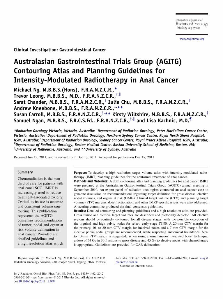
Australasian Gastrointestinal Trials Group (AGITG)Contouring Atlas and Planning Guidelines forIntensity-Modulated Radiotherapy in Anal CancerMichael Ng, M.B.B.S.(Hons), F.R.A.N.Z.C.R.,*Trevor Leong, M.B.B.S., M.D., F.R.A.N.Z.C.R.,y,k
Sarat Chander, M.B.B.S., F.R.A.N.Z.C.R.,y Julie Chu, M.B.B.S., F.R.A.N.Z.C.R.,y
Andrew Kneebone, M.B.B.S., F.R.A.N.Z.C.R.,z,**Susan Carroll, M.B.B.S., F.R.A.N.Z.C.R.,x,** Kirsty Wiltshire, M.B.B.S., F.R.A.N.Z.C.R.,y
Samuel Ngan, M.B.B.S., F.R.C.S.Ed., F.R.A.N.Z.C.R.,y,k and Lisa Kachnic, M.D.{
*Radiation Oncology Victoria, Victoria, Australia; yDepartment of Radiation Oncology, Peter MacCallum Cancer Centre,Victoria, Australia; zDepartment of Radiation Oncology, Northern Sydney Cancer Centre, Royal North Shore Hospital,NSW, Australia; xDepartment of Radiation Oncology, Sydney Cancer Centre, Royal Prince Alfred Hospital, NSW, Australia;{Department of Radiation Oncology, Boston Medical Center, Boston University School of Medicine, Boston, MA;kUniversity of Melbourne, Australia; and **University of Sydney, Australia
Received Jun 19, 2011, and in revised form Dec 13, 2011. Accepted for publication Dec 18, 2011
Summary
Chemoradiation is the stan-dard of care for patients withanal canal SCC. IMRT isincreasingly used to reducetreatment-associated toxicity.Critical to its use is accurateand consistent volume con-touring. This publicationrepresents the AGITGconsensus recommendationsof tumor, nodal and organ atrisk volume delineation inanal cancer. Provided aredetailed guidelines anda high resolution atlas which
Reprint requests to: Michael Ng, M.B.B.
Radiation Oncology Victoria, 230 Cooper Stre
Int J Radiation Oncol Biol Phys, Vol. 83, No. 5
0360-3016/$ - see front matter � 2012 Elsevie
doi:10.1016/j.ijrobp.2011.12.058
Purpose: To develop a high-resolution target volume atlas with intensity-modulated radio-therapy (IMRT) planning guidelines for the conformal treatment of anal cancer.Methods and Materials: A draft contouring atlas and planning guidelines for anal cancer IMRTwere prepared at the Australasian Gastrointestinal Trials Group (AGITG) annual meeting inSeptember 2010. An expert panel of radiation oncologists contoured an anal cancer case togenerate discussion on recommendations regarding target definition for gross disease, electivenodal volumes, and organs at risk (OARs). Clinical target volume (CTV) and planning targetvolume (PTV) margins, dose fractionation, and other IMRT-specific issues were also addressed.A steering committee produced the final consensus guidelines.Results: Detailed contouring and planning guidelines and a high-resolution atlas are provided.Gross tumor and elective target volumes are described and pictorially depicted. All electiveregions should be routinely contoured for all disease stages, with the possible exception ofthe inguinal and high pelvic nodes for select, early-stage T1N0. A 20-mm CTV margin forthe primary, 10- to 20-mm CTV margin for involved nodes and a 7-mm CTV margin for theelective pelvic nodal groups are recommended, while respecting anatomical boundaries. A 5-to 10-mm PTV margin is suggested. When using a simultaneous integrated boost technique,a dose of 54 Gy in 30 fractions to gross disease and 45 Gy to elective nodes with chemotherapyis appropriate. Guidelines are provided for OAR delineation.
S.(Hons), F.R.A.N.Z.C.R.,
et, Epping, 3076, Victoria.
Australia. Tel: þ613-9418-2200; Fax: þ613-9418-2288; E-mail: mng@
radoncvic.com.au
Conflict of interest: none.
, pp. 1455e1462, 2012
r Inc. All rights reserved.
Ng et al. International Journal of Radiation Oncology � Biology � Physics1456
will be useful to the prac-
ticing radiation oncologist.Conclusion: These consensus planning guidelines and high-resolution atlas complement the ex-isting Radiation Therapy Oncology Group (RTOG) elective nodal ano-rectal atlas and provideadditional anatomic, clinical, and technical instructions to guide radiation oncologists in theplanning and delivery of IMRT for anal cancer. � 2012 Elsevier Inc.
Keywords: Anal cancer, IMRT, Chemoradiation, Atlas, Guidelines
Introduction
With the advent of computed tomography (CT) planningand conformal radiation techniques including intensity-modulatedradiotherapy (IMRT), comes the prerequisite for accurate andconsistent contouring of target volumes.
Conformal radiotherapy for anal cancer allows the ability tospare surrounding organs at risk (OAR). Normal tissues such assmall bowel, femoral heads, perineum, and external genitalia oftenreceive high doses of radiation with more conventional tech-niques, which can result in significant acute and late toxicity. Theuse of IMRT provides an opportunity to spare OAR and to reducetoxicity in anal cancer patients.
The implementation of IMRT requires a clear understanding oftarget volume definition for the complex elective nodal regions inanal cancer. During the early conduct of the Radiation TherapyOncology Group (RTOG) 0529 Phase II study investigating IMRTfor anal cancer, it became necessary to create an atlas becausemany initial target volumes submitted for quality assurancerequired contouring revision (1). However, this atlas providedcontouring guidance for elective nodal volumes only and did notprovide instruction in the contouring of gross disease and OAR.
As such, an international workshop was convened at the 2010Australasian Gastrointestinal Trials Group (AGITG) annualmeeting to develop detailed contouring and planning guidelinesfor the IMRT treatment of anal cancer, supplemented by a high-resolution atlas. This article reports the AGITG recommendations.
Methods and Materials
The AGITG is a national cooperative trials group that consists ofradiation oncologists, surgeons, and medical oncologists whoconduct clinical trials in gastrointestinal malignancies. In 2010,the Radiation Oncology Committee of AGITG organized a con-touring workshop with member radiation oncologists to discussradiotherapy target and OAR volume delineation for anal cancer,with the aim of developing consensus guidelines. Dr. Lisa Kachnic(L.K.), principal investigator of the RTOG 0529 study of IMRT inanal cancer, was invited to participate in the workshop.
To facilitate discussion and productivity at the workshop,a draft set of IMRT contouring and planning guidelines was sentto all 19 workshop participants before the meeting. These werebased on anal canal IMRT guidelines previously developed in2009 by the Gastrointestinal Unit at the Peter MacCallum CancerCentre (PMCC). The aim of the PMCC guidelines, building off ofprevious publications describing clinical target volume (CTV)delineation for ano-rectal cancer (2, 3), was to standardize CTVsamong radiation oncologists.
The draft guidelines described seven elective regions to beconsidered when treating anal cancer, including the mesorectum,presacral space, internal iliac lymph nodes, external iliac lymphnodes, obturator lymph nodes, ischiorectal fossa, and inguinal
lymph nodes. Each elective region was described individually,including borders for CTV delineation. The guidelines also con-tained recommendations for contouring target volumes for grossdisease and instructions for standardized contouring of OAR.Planning target volume (PTV) margins, dose fractionation, andother IMRT-specific issues were also addressed.
The AGITG workshop was coordinated by two chairs, A.K. andM.N., and three radiation oncologists (T.L., S.Ca. and L.K.) whowere invited to form a discussion panel. Before the meeting, eachpanelist was sent an anonymized CT dataset of a female patient witha T2N0 anal cancer. The gross tumor volume (GTV) was alreadydefined, and panelists were asked to delineate the above targetvolumes and OAR. These volumes were then displayed, serving asdiscussion points for the development of consensus guidelines and anatlas (Fig. 3e5: Atlas Panels 1,2,3 respectively). Where available,references are provided with these final recommendations.
Results
Elective nodal volumes
Mesorectum
The mesorectum is not well visualized on CT, and if fat-saturatedT2 magnetic resonance imaging cannot be obtained, neighboringstructures can be used to delineate this volume.
Cranial: The level of the recto-sigmoid junction; best identifiedwhere the rectum runs anteriorly to join the sigmoid colon (Atlas4b).
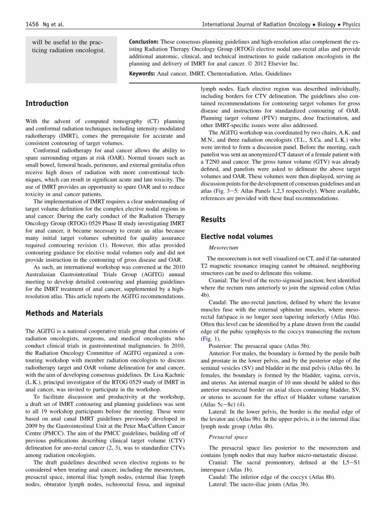
Caudal: The ano-rectal junction, defined by where the levatormuscles fuse with the external sphincter muscles, where meso-rectal fat/space is no longer seen tapering inferiorly (Atlas 10a).Often this level can be identified by a plane drawn from the caudaledge of the pubic symphysis to the coccyx transecting the rectum(Fig. 1).
Posterior: The presacral space (Atlas 5b).Anterior: For males, the boundary is formed by the penile bulb
and prostate in the lower pelvis, and by the posterior edge of theseminal vesicles (SV) and bladder in the mid pelvis (Atlas 6b). Infemales, the boundary is formed by the bladder, vagina, cervix,and uterus. An internal margin of 10 mm should be added to thisanterior mesorectal border on axial slices containing bladder, SV,or uterus to account for the effect of bladder volume variation(Atlas 5ce8c) (4).
Lateral: In the lower pelvis, the border is the medial edge ofthe levator ani (Atlas 9b). In the upper pelvis, it is the internal iliaclymph node group (Atlas 4b).
Presacral space
The presacral space lies posterior to the mesorectum andcontains lymph nodes that may harbor micro-metastatic disease.
Cranial: The sacral promontory, defined at the L5eS1interspace (Atlas 1b).
Caudal: The inferior edge of the coccyx (Atlas 8b).Lateral: The sacro-iliac joints (Atlas 3b).
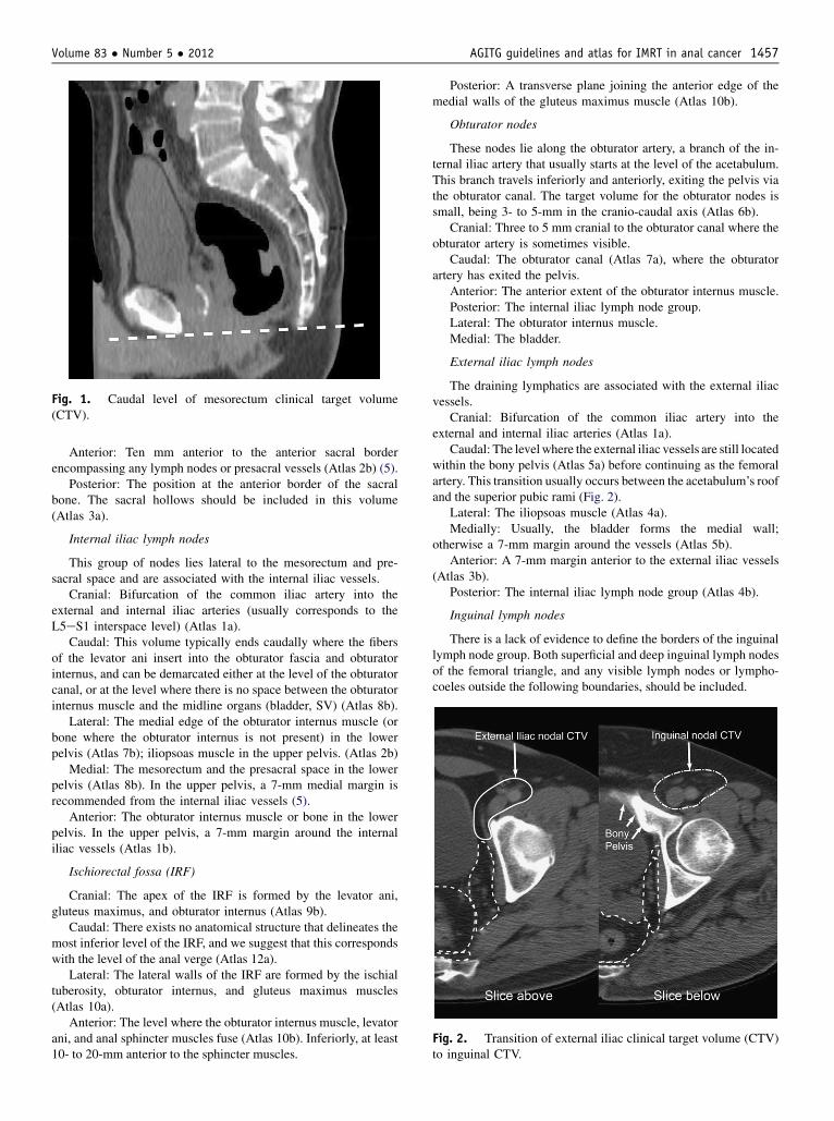
Fig. 1. Caudal level of mesorectum clinical target volume(CTV).
Fig. 2. Transition of external iliac clinical target volume (CTV)to inguinal CTV.
Volume 83 � Number 5 � 2012 AGITG guidelines and atlas for IMRT in anal cancer 1457
Anterior: Ten mm anterior to the anterior sacral borderencompassing any lymph nodes or presacral vessels (Atlas 2b) (5).
Posterior: The position at the anterior border of the sacralbone. The sacral hollows should be included in this volume(Atlas 3a).
Internal iliac lymph nodes
This group of nodes lies lateral to the mesorectum and pre-sacral space and are associated with the internal iliac vessels.
Cranial: Bifurcation of the common iliac artery into theexternal and internal iliac arteries (usually corresponds to theL5eS1 interspace level) (Atlas 1a).
Caudal: This volume typically ends caudally where the fibersof the levator ani insert into the obturator fascia and obturatorinternus, and can be demarcated either at the level of the obturatorcanal, or at the level where there is no space between the obturatorinternus muscle and the midline organs (bladder, SV) (Atlas 8b).
Lateral: The medial edge of the obturator internus muscle (orbone where the obturator internus is not present) in the lowerpelvis (Atlas 7b); iliopsoas muscle in the upper pelvis. (Atlas 2b)
Medial: The mesorectum and the presacral space in the lowerpelvis (Atlas 8b). In the upper pelvis, a 7-mm medial margin isrecommended from the internal iliac vessels (5).
Anterior: The obturator internus muscle or bone in the lowerpelvis. In the upper pelvis, a 7-mm margin around the internaliliac vessels (Atlas 1b).
Ischiorectal fossa (IRF)
Cranial: The apex of the IRF is formed by the levator ani,gluteus maximus, and obturator internus (Atlas 9b).
Caudal: There exists no anatomical structure that delineates themost inferior level of the IRF, and we suggest that this correspondswith the level of the anal verge (Atlas 12a).
Lateral: The lateral walls of the IRF are formed by the ischialtuberosity, obturator internus, and gluteus maximus muscles(Atlas 10a).
Anterior: The level where the obturator internus muscle, levatorani, and anal sphincter muscles fuse (Atlas 10b). Inferiorly, at least10- to 20-mm anterior to the sphincter muscles.
Posterior: A transverse plane joining the anterior edge of themedial walls of the gluteus maximus muscle (Atlas 10b).
Obturator nodes
These nodes lie along the obturator artery, a branch of the in-ternal iliac artery that usually starts at the level of the acetabulum.This branch travels inferiorly and anteriorly, exiting the pelvis viathe obturator canal. The target volume for the obturator nodes issmall, being 3- to 5-mm in the cranio-caudal axis (Atlas 6b).
Cranial: Three to 5 mm cranial to the obturator canal where theobturator artery is sometimes visible.
Caudal: The obturator canal (Atlas 7a), where the obturatorartery has exited the pelvis.
Anterior: The anterior extent of the obturator internus muscle.Posterior: The internal iliac lymph node group.Lateral: The obturator internus muscle.Medial: The bladder.
External iliac lymph nodes
The draining lymphatics are associated with the external iliacvessels.
Cranial: Bifurcation of the common iliac artery into theexternal and internal iliac arteries (Atlas 1a).
Caudal: The levelwhere the external iliac vessels are still locatedwithin the bony pelvis (Atlas 5a) before continuing as the femoralartery. This transition usually occurs between the acetabulum’s roofand the superior pubic rami (Fig. 2).
Lateral: The iliopsoas muscle (Atlas 4a).Medially: Usually, the bladder forms the medial wall;
otherwise a 7-mm margin around the vessels (Atlas 5b).Anterior: A 7-mm margin anterior to the external iliac vessels
(Atlas 3b).Posterior: The internal iliac lymph node group (Atlas 4b).
Inguinal lymph nodes
There is a lack of evidence to define the borders of the inguinallymph node group. Both superficial and deep inguinal lymph nodesof the femoral triangle, and any visible lymph nodes or lympho-coeles outside the following boundaries, should be included.
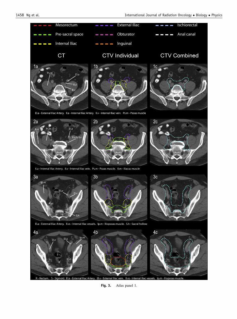
Fig. 3. Atlas panel 1.
Ng et al. International Journal of Radiation Oncology � Biology � Physics1458
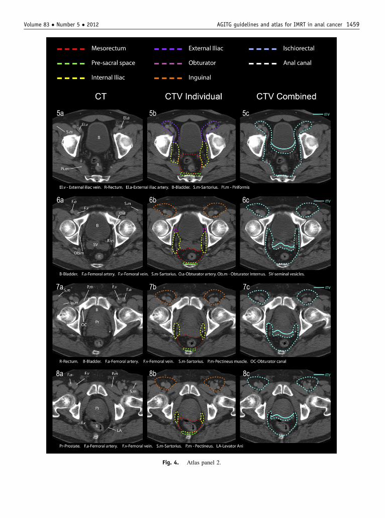
Fig. 4. Atlas panel 2.
Volume 83 � Number 5 � 2012 AGITG guidelines and atlas for IMRT in anal cancer 1459
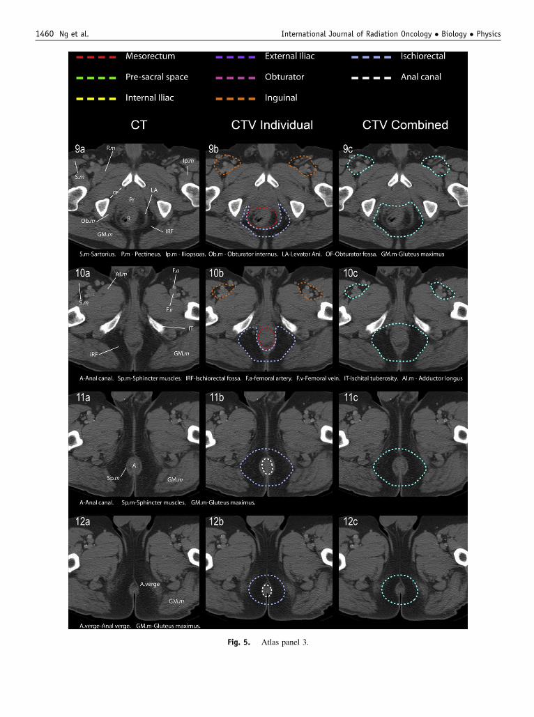
Fig. 5. Atlas panel 3.
Ng et al. International Journal of Radiation Oncology � Biology � Physics1460
Volume 83 � Number 5 � 2012 AGITG guidelines and atlas for IMRT in anal cancer 1461
Cranial: The level where the external iliac artery leaves thebony pelvis to become the femoral artery (Fig. 2).
Caudal: There is no consensus on the definition of the inferiorextent. Some publications have recommended (a) the positionwhere the great saphenous vein enters the femoral vein witha margin (2), or (b) where the muscles of sartorius and adductorlongus cross (6). A compromise is the lower edge of the ischialtuberosities, which lies between (a) and (b) as defined above andis an identifiable landmark (Atlas 10a).
Posterior: The bed of the femoral triangle is formed bythe iliopsoas, pectineus, and adductor longus muscles (Atlas 10a).
Anterior: A minimum 20-mm margin on the inguinal vessels,inclusive of any visible lymph nodes or lymphocoeles (Atlas 7b).
Lateral: The medial edge of sartorius or iliopsoas (Atlas 7a).Medial: A 10- to 20-mm margin around the femoral vessels.
The medial third to half of the pectineus or adductor longusmuscle serves as an approximate border (Atlas 7b).
Elective nodal volumes to be covered per stage ofdisease
All nodal volumes described should be covered for all stages. Inpatients with select early T1N0 cancers, particularly those patientswith major comorbidities, it may be appropriate to omit electivenodal irradiation superior to the caudal edge of the sacro-iliacjoints, and the inguinal nodes (low risk of failure, <5%) (7).
Clinical target volumes for gross disease
Primary tumor
� GTV: The GTV should be delineated as a separate structure basedon all available clinical and imaging information.
� CTV: This volume must encompass (1) the GTV, (2) the entireanal canal from the ano-rectal junction to the anal verge, and (3)the internal and external anal sphincters. A further 20-mmisotropic margin should be added to items (1), (2), and (3)above, to account for microscopic disease, while respectinganatomical boundaries. Attention must be given, especially foranal verge and perianal lesions, that a 20-mm radial and caudalmargin is used to ensure coverage of perianal skin.
Involved nodes
� GTV: The involved node(s).� CTV: The involved node(s) or nodal region(s) with a 10- to 20-mm margin, respecting anatomical boundaries.
Planning target volumes
An isotropic 10-mm expansion is recommended on CTVs togenerate PTVs. Daily image guidance is recommended for IMRT,especially prone patients, which may allow CTVePTV marginreduction to 5- to 7-mm.
Dose and fractionation with IMRT
Doseand fractionation for radical treatment dependson the following:disease stage; whether excisional biopsy has been performed; use ofconcurrent chemotherapy; macroscopic vs. microscopic disease; andthe use of a simultaneous integrated boost (SIB) technique.We prefer
a SIB technique because of the reduced planning complexitycompared with a sequential approach.
In general, gross disease should be treated to 54 Gy over 30fractions when using chemotherapy. However, for T1 and non-bulky T2 tumors, a dose of 50.4 Gy in 28 fractions is appro-priate. Involved nodes/regions should receive 50.4 to 54 Gy,depending on size.
Higher doses to treat elective nodal regions must be consideredwhen using SIB techniques to account for longer treatment duration.For total doses of 54Gy ormore over 30 fractions, the recommendedelective dose is 45 Gy. If using 50.4 Gy in 28 fractions, the rec-ommended elective dose is 42 Gy (8). When using a sequentialtechnique, an initial elective dose of 30 to 36Gy, followed by a boostto macroscopic disease totaling 50.4 to 60 Gy, is appropriate (9).
Organs at risk (OAR)
Femoral head and neck: The entire femoral head and neck shouldbe contoured. The inferior extent is the cranial edge of the lessertrochanter.
Urinary bladder: The entire external outline of the bladder wallshould be contoured.
Bowel: Although many descriptions for contouring the“bowel” exist, only one publication has correlated dose to thebowel with gastrointestinal toxicity in anal cancer patients (10).We have therefore followed the recommendations in this report.Small bowel and large bowel, opacified or nonopacified, shouldbe delineated from 15-mm superior to the cranial aspect of thePTV, extending inferiorly to the recto-sigmoid junction. In theanterioreposterior direction, the bowel will be contoured fromthe anterior abdominal wall to the most posterior extent ofbowel. In the lateral direction, the borders are bowel edge tobowel edge.
External genitalia and perineum: There are no establishedrecommendations for contouring this volume. In males, thisvolume will include the penis, scrotum, and area including skinand fat anterior to the pubic symphysis. In females, this volumewill include the clitoris, labia majora and minora, and areaincluding skin and fat anterior to pubic symphysis. The cranialextent of this volume is the caudal edge of the pubic symphysis.
Bone marrow: Both iliac crests will be used to define “bonemarrow.” Delineation will extend cranially from the top of the iliaccrests to the superior part of the acetabulum caudally. The left andright iliac crests are combined into one volume (8, 11).
No recommended dose constraints are provided, given the lackof data specific to anal cancer and the variation among differentinstitutions. Guidelines on dose constraints may be found per theRTOG 0529 closed study protocol (http://www.rtog.org).
Discussion
The implementation of IMRT in anal cancer requires accurate CTcontouring of clinical target volumes. Roels et al. originallyhighlighted the need for guidelines in contour delineation inradiotherapy for rectal cancer (3). Subsequently, the RTOGreleased an atlas and guidelines for ano-rectal cancer that wasdriven by the need to improve contouring quality in the RTOG0529 trial (2). With the early implementation of IMRT inAustralia, the AGITG meeting provided a unique opportunity forGI radiation oncologists to develop detailed contouring andplanning consensus guidelines for IMRT in anal cancer.
Ng et al. International Journal of Radiation Oncology � Biology � Physics1462
This document aims to address the important points that need tobe considered when implementing IMRT for anal cancer. We haveattempted to provide clear descriptions of individual nodal groupscomplementary to the RTOG ano-rectal IMRT directives, and theseare supplemented with a high-resolution atlas. The determination ofCTV borders for individual nodal groups has been based ona combination of anatomical landmarks (when unambiguous),descriptions available in the published literature, and traditionalfield borders based on bony landmarks. In addition, we haveprovided detailed instruction in the contouring of gross disease andOAR, as well as considerations for planning parameters.
There were specific issues for which we did not automaticallyreach consensus during the meeting. For example, defining thelateral and inferior boundaries of the IRF was difficult. It wasbelieved that microscopic disease was unlikely to be found at theselateral edges; however, without supporting evidence, we conserva-tively recommended treating the entire IRF. The RTOG guidelinesdo not consider the IRF to be an area at risk. However, becausetraditional 2D pelvic fields from previous randomized controlledtrials encompassed the entire IRF (intentionally or unintentionally)(9), we have recommended including the IRF. Patterns of failuredata from RTOG 0529 will help to clarify whether the IRF is at risk.
The inguinal lymph node group was another region whereconsensus was difficult to achieve. Some publications have recom-mended using a uniform radial margin around the pelvic vessels toencompass lymph nodes (5, 12). However, applying a “margin rule”for femoral vessels has its limitations, given that there is no clearanatomical compartment and that variation is seenwith different bodyhabitus. To account for body habitus, our recommendations usea combination of landmark-based boundaries and margins when noanatomical boundaries exist. The inferior edge of the ischial tuber-ositywas selectedas thecaudal level for contouring the inguinal fossa.As there is no anatomic landmark, the tuberosity level providedacceptable caudal coverage and is easily identifiable.
We also provided some recommendations for delineation ofthe CTV for gross disease. The group acknowledged that tradi-tional two-dimensional fields have recommended a 20- to 30-mmmargin for the field edge around gross disease for the “boost”volume. The consensus for this boost volume was to include theentire anal canal and sphincter muscles with a further margin.There was variation in this expansion margin during the meeting,with a range of 10- to 20-mm. With no published pathology datato help clarify the microscopic extent, we conservatively recom-mended a 20-mm margin. Similarly, there was a variation of 10-to 20-mm for the CTV margin around involved nodes to accountfor extracapsular extension (ECE). A minimum of 10-mm wasbased on pathological studies on ECE of metastatic lymph nodesin head-and-neck squamous cell carcinoma (13).
One minor area of contention was how to describe the anteriorCTVof the mesorectum. Nuyytens et al. reported that the anteriorwall of the mesorectum can vary by 10-mm (4). The RTOG atlasaccounts for the 10 mm of motion by contouring into the bladderand describing this as the CTV (2). Roels et al. defined theanterior CTV border of the mesorectum behind the bladder or SV,and did not address potential organ motion (3). We have addressedthis issue by using ICRU63 definitions where we describe aninternal margin of 10-mm to be added to the CTV at the levels ofthe bladder to form an internal target volume.
This report does not discuss other important IMRT issues in analcancer, such as patient setup and immobilization, supine vs. proneposition, use of fiducials, and bolus. These issues need to beconsidered by the clinician, but are beyond the scope of this article.
These anal cancer IMRT guidelines are based on recommen-dations from a consensus working group. When there weredifferences in opinion and lack of supporting evidence, we havegenerally been more conservative in our recommendations. Assuch, these consensus guidelines should be continuously re-evaluated as centers gain more clinical experience and as newevidence is reported.
Conclusion
IMRT for anal cancer has significant potential to benefit patients.These detailed guidelines, supplemented with a high-resolutionatlas, aim to improve the understanding of target volume defini-tion and IMRT planning for anal cancer.
References
1. Kachnic LA, Winter K, Myerson RJ, et al. RTOG 0529: A phase II
evaluation of dose-painted IMRT in combination with 5-fluorouracil
and mitomycin-C for reduction of acute morbidity in carcinoma of
the anal canal. Int J Radiat Oncol Biol Phys 2009;75:S5.
2. Myerson RJ, Garofalo MC, El Naqa I, et al. Elective clinical target
volumes for conformal therapy in anorectal cancer: A Radiation
Therapy Oncology Group consensus panel contouring atlas. Int J
Radiat Oncol Biol Phys 2009;74:824-830.
3. Roels S, Duthoy W, Haustermans K, et al. Definition and delineation
of the clinical target volume for rectal cancer. Int J Radiat Oncol Biol
Phys 2006;65:1129-1142.
4. Nuyttens JJ, Robertson JM, Yan D, et al. The variability of the
clinical target volume for rectal cancer due to internal organ motion
during adjuvant treatment. Int J Radiat Oncol Biol Phys 2002;53:
497-503.
5. Taylor A, Rockall A, Reznek R, et al. Mapping pelvic lymph nodes:
Guidelines for delineation in intensity-modulated radiotherapy. Int J
Radiat Oncol Biol Phys 2005;63:1604-1612.
6. Portaluri M, Bambace S, Perez C, et al. Clinical and anatomical
guidelines in pelvic cancer contouring for radiotherapy treatment
planning. Cancer Radiother 2004;8:222-229.
7. Tomaszewski JM, Link E, Leong T, et al. Twenty-five-year experience
with radical chemoradiation for anal cancer. Int J Radiat Oncol Biol
Phys 2011 Oct 21. [Epub ahead of print]. PMID: 22019078.
8. Kachnic L, Winter K, Myerson R. RTOG 05-29: A phase II evaluation
of dose-painted IMRT in combination with 5-fluorouracil and
mitomycin-C for reduction of acute morbidity in carcinoma of the anal
canal. 2005. Available at www.rtog.org. Accessed Feburary 15, 2011.
9. Ajani JA, Winter KA, Gunderson LL, et al. Fluorouracil, mitomycin,
and radiotherapy vs fluorouracil, cisplatin, and radiotherapy for
carcinoma of the anal canal: A randomized controlled trial. JAMA
2008;299:1914-1921.
10. Devisetty K, Mell LK, Salama JK, et al. A multi-institutional acute
gastrointestinal toxicity analysis of anal cancer patients treated with
concurrent intensity-modulated radiation therapy (IMRT) and
chemotherapy. Radiother Oncol 2009;93:298-301.
11. Menkarios C, Azria D, Laliberte B, et al. Optimal organ-sparing
intensity-modulated radiation therapy (IMRT) regimen for the treat-
ment of locally advanced anal canal carcinoma: A comparison of
conventional and IMRT plans. Radiat Oncol 2007;2:41.
12. Chao KS, Lin M. Lymphangiogram-assisted lymph node target
delineation for patients with gynecologic malignancies. Int J Radiat
Oncol Biol Phys 2002;54:1147-1152.
13. ApisarnthanaraxS,Elliott DD, El-NaggarAK, et al. Determining optimal
clinical target volume margins in head-and-neck cancer based on
microscopic extracapsular extension of metastatic neck nodes. Int J
Radiat Oncol Biol Phys 2006;64:678-683.