Embed Size (px)
Citation preview
Technical Report
Automated Tumor VolumetryUsing Computer-Aided
Image Segmentation
Bilwaj Gaonkar, MS, Luke Macyszyn, MA, MD, Michel Bilello, MD, PhD,Mohammed Salehi Sadaghiani, MD, Hamed Akbari, MD, PhD, Mark A. Atthiah, MD, Zarina S. Ali, MD,Xiao Da, MS, Yiqang Zhan, PhD, Donald O’ Rourke, MD, Sean M. Grady, MD, Christos Davatzikos, PhD
Rationale and Objectives: Accurate segmentation of brain tumors, and quantification of tumor volume, is important for diagnosis, moni-toring, and planning therapeutic intervention. Manual segmentation is not widely used because of time constraints. Previous efforts have
mainly producedmethods that are tailored to a particular type of tumor or acquisition protocol and havemostly failed to produce amethod
that functions on different tumor types and is robust to changes in scanning parameters, resolution, and image quality, thereby limiting their
clinical value. Herein, we present a semiautomatic method for tumor segmentation that is fast, accurate, and robust to a wide variation inimage quality and resolution.
Materials andMethods: A semiautomatic segmentationmethod based on the geodesic distance transformwas developed and validated
by using it to segment 54 brain tumors. Glioblastomas, meningiomas, and brain metastases were segmented. Qualitative validation wasbased on physician ratings provided by three clinical experts. Quantitative validation was based on comparing semiautomatic andmanual
segmentations.
Results: Tumor segmentations obtained using manual and automatic methods were compared quantitatively using the Dice measure ofoverlap. Subjective evaluation was performed by having human experts rate the computerized segmentations on a 0–5 rating scale where
5 indicated perfect segmentation.
Conclusions: The proposedmethod addresses a significant, unmet need in the field of neuro-oncology. Specifically, this method enables
clinicians to obtain accurate and reproducible tumor volumes without the need for manual segmentation.
Key Words: Tumor segmentation; volumetric analysis; geodesic distance.
ªAUR, 2015
Quantification of tumor volume has become increas-
ingly important for diagnosis, staging, assessment
of therapy response, and more recently determina-
tion of eligibility for clinical trial enrollment (1–3).
Currently, assessment of tumor volume is based on two-
dimensional (2D) measurements, using standards such as the
MacDonald criteria (4) for gliomas, Herscovici criteria (5)
for meningiomas, or the RECIST standards for general
oncology (6).
Acad Radiol 2015; -:1–9
From the Department of Radiology, University of Pennsylvania, 3600 MarketSt, Suite 380, Philadelphia, Pennsylvania, 19104 (B.G., M.B., M.S.S., H.A.,X.D., C.D.); Center for Biomedical Image Computing and Analytics (B.G.,L.M., M.B., H.A., X.D., C.D.) and Department of Neurosurgery (L.M., M.A.A.,Z.S.A., D.O.R., S.M.G.), University of Pennsylvania, Philadelphia,Pennsylvania; and Siemens Medical Solutions, Malvern, Pennsylvania (Y.Z.).Received August 26, 2014; accepted January 8, 2015. B.G. and L.M.contributed equally to the study. Conflicts of Interest: None. FundingSources: This work was supported in part by grants R01NS042645 fromNational Institutes of Health (PI: Davatzikos). Address correspondence to:B.G. e-mail: [email protected]
ªAUR, 2015http://dx.doi.org/10.1016/j.acra.2015.01.005
These criteria allow clinicians to obtain a rough estimate of
tumor volume by sacrificing accuracy for speed. An accurate
measurement of tumor volume, however, requires a complete
segmentation of the tumor. This type of segmentation, which
can currently be performed manually, requires a tremendous
amount of time and hence is not widely used. Thus, automa-
tion of tumor segmentation represents an important clinical
need that would be invaluable for treating and monitoring pa-
tients with brain tumors. Furthermore, such automatic seg-
mentations are likely to be more reproducible and therefore
preferable over manual segmentations because of their consis-
tency, which is especially important for longitudinal tumor
monitoring.
The neuroimaging community has attempted to address
the need for automatic tumor segmentation over the past
two decades. The earliest methods included the use of fuzzy
clustering-based approaches (7,8). Direct application of such
methods leads to a large number of false-positive voxels
labeled as tumors. Later methods based on level sets and active
contours (9,10) often fail in the context of aggressive tumors
harboring significant structural complexity. Machine-
learning–based methods have been fairly successful at the
1
GAONKAR ET AL Academic Radiology, Vol -, No -, - 2015
task of tumor segmentation (11–21). However, these methods
are often tumor type specific and very sensitive to changes in
noise and acquisition protocol. Additionally, there is a
constant need for retraining with most learning-based
methods when there is a slight change in the imaging protocol
or if the scanning site changes. Furthermore, many of these
methods are based on complex algorithms that are expensive
to reimplement and difficult to integrate into existing clinical
workflows. Finally, most of these methods have been validated
in a narrow and limited research setting and not necessarily in
a clinical setting. In general, the narrow focus of previously
described techniques has prevented their widespread utiliza-
tion in the clinical arena.
In this work, we present a novel tumor segmentation tech-
nique that is semiautomatic, fast, and based on a relatively sim-
ple learning-free algorithm.We have validated our method on
three different tumor types acquired under a diverse set of
image acquisition protocols and resolutions and drawn from
studies using different preprocessing steps. Qualitative and
quantitative results present the efficacy of the proposed
method in the presence of substantial noise, scanner variation,
processing variation, and tissue (tumor) heterogeneity.
MATERIALS AND METHODS
Institutional review board approval was obtained for this study
with waiver of informed consent for retrospective review of
medical records. All imaging data came from patients treated
at the Hospital of the University of Pennsylvania. In general,
these imaging studies contained differences between cases in
terms of resolution, noise level, and pixel spacing. Overall,
our data set contained images of 24 glioblastomas, 15 menin-
giomas, and 15 metastatic brain tumors. T1 contrast-
enhanced images were available for all tumors and were
used for automatic and manual segmentations.
The data used in this project varied across cases in terms of
acquisition protocol, resolution, and pixel spacing. It was
sequentially chosen. Some of the data came from a 3.0-T
magnetic resonance (MR) imaging scanner systems (Siemens
and GEHealthcare) and some of it came from a 1.5-T systems.
Similarly, the pixel spacing varied from 0.42� 0.42 to 0.97�0.97, and image dimensions varied between 256� 256 to 512
� 512. Slice thicknesses during acquisitions varied between
1 and 5 mm. The echo times and repetition times involved
in computing the T1 images also varied. This was a retrospec-
tive study, and we used a random sample of cases available on
the internal University of Pennsylvania Picture Archiving and
Communication System.
Thus, there was tremendous variation between cases with
respect to noise and inhomogeneity. The segmentation results
presented here are testimony to that the proposed method is
able to successfully segment these brain tumors in spite of
the considerable variation in the underlying data.
We use the adaptive geodesic algorithm described by
Gaonkar and Shu (22) to segment brain tumors. This is a
semiautomatic method that was originally devised to
2
segment the vertebral column on computed tomographic
images using the adaptive geodesic distance (23,24). The
method is fast, easy to use, and robust to noise and bias. A
seed region is placed by the clinician inside a tumor, and
the segmentation algorithm is initiated. The ‘‘adaptive
geodesic distance’’ is a mathematical measure that may be
computed at any voxel within the image. At a given voxel,
this measure provides a joint quantification of 1) the spatial
distance of the voxel from the seed region and 2) the
variation of the image intensity profile between the voxel
and the seed, both of which are important clues for tumor
segmentation The algorithm computes the adaptive
geodesic distance at every voxel in the image to yield an
‘‘adaptive geodesic distance transform image.’’ This
transformed image appears as a geodesic distance–weighted
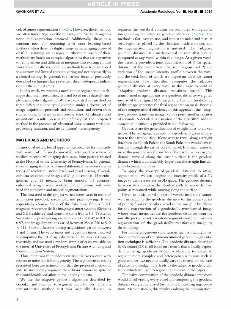
inverse of the original MR image (Fig 1b) and thresholding
of this image generates the final segmentation mask. Because
of the computational efficiency of this approach, the ‘‘adap-
tive geodesic transform image’’ can be performed in a matter
of seconds. A detailed explanation of the algorithm and the
associated intuition is provided in the following.
Geodesics are the generalization of straight lines to curved
spaces. The pedagogic example of a geodesic is given in rela-
tion to the earth’s surface. If one were to travel along a straight
line from the North Pole to the South Pole, onewould have to
burrow through the earth’s core to travel. It is much easier to
make this journeyover the surface of the earth. In this case, the
distance traveled along the earth’s surface is the geodesic
distance which is considerably larger than the straight line dis-
tance between the poles.
To apply the concept of geodesic distances to image
segmentation, we can imagine the intensity profile of a 2D
image to define a surface in 3D space. The geodesic distance
between two points is the shortest path between the two
points as measured while moving along the surface.
Given an initial voxel (or set of voxels) inside the tumor,
we can compute the geodesic distance to this point (or set
of points) from every other voxel in the image. This allows
for the construction of a geodesically transformed image
whose voxel intensities are the geodesic distances from the
initially picked voxel. Geodesic segmentation then involves
segmentation of the geodesically transformed image by
thresholding.
For nonheterogeneous solid tumors such as meningiomas,
direct application of the aforementioned geodesic segmenta-
tion technique is sufficient. The geodesic distance described
by Criminisi (23) is still based on a metric that is locally depen-
dent on image gradients alone. To adapt the technique to
segment more complex and heterogeneous tumors such as
glioblastomas, we need to locally vary the metric on the basis
of prior knowledge. This leads to the adaptive geodesic dis-
tance which we used to segment all tumors in the paper.
The naive computation of the geodesic distance transform
would entail visiting every voxel and computing the geodesic
distance using a discretized form of the Euler–Lagrange equa-
tions. Mathematically, this involves solving the minimization:
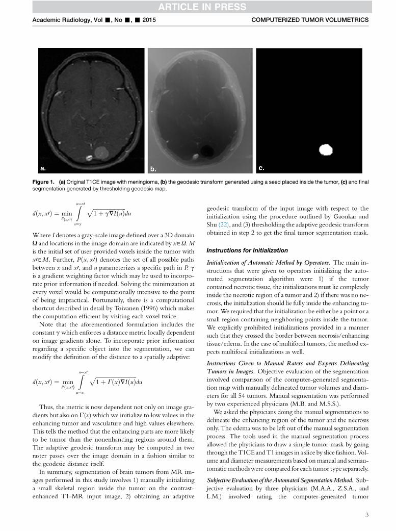
Figure 1. (a) Original T1CE image with meningioma, (b) the geodesic transform generated using a seed placed inside the tumor, (c) and finalsegmentation generated by thresholding geodesic map.
Academic Radiology, Vol -, No -, - 2015 COMPUTERIZED TUMOR VOLUMETRICS
dðx; x0Þ ¼ minPfx;x0g
Zu¼x0
u¼x
ffiffiffiffiffiffiffiffiffiffiffiffiffiffiffiffiffiffiffiffiffiffiffi1þ gVIðuÞ
pdu
Where I denotes a gray-scale image defined over a 3D domain
U and locations in the image domain are indicated by x˛U.Mis the initial set of user provided voxels inside the tumor with
x0˛M . Further, Pðx; x0Þ denotes the set of all possible paths
between x and x0, and u parameterizes a specific path in P. g
is a gradient weighting factor which may be used to incorpo-
rate prior information if needed. Solving the minimization at
every voxel would be computationally intensive to the point
of being impractical. Fortunately, there is a computational
shortcut described in detail by Toivanen (1996) which makes
the computation efficient by visiting each voxel twice.
Note that the aforementioned formulation includes the
constant gwhich enforces a distance metric locally dependent
on image gradients alone. To incorporate prior information
regarding a specific object into the segmentation, we can
modify the definition of the distance to a spatially adaptive:
dðx; x0Þ ¼ minPfx;x0g
Zu¼x0
u¼x
ffiffiffiffiffiffiffiffiffiffiffiffiffiffiffiffiffiffiffiffiffiffiffiffiffiffiffiffiffi1þ GðxÞVIðuÞ
pdu
Thus, the metric is now dependent not only on image gra-
dients but also on G(x) which we initialize to low values in the
enhancing tumor and vasculature and high values elsewhere.
This tells the method that the enhancing parts are more likely
to be tumor than the nonenhancing regions around them.
The adaptive geodesic transform may be computed in two
raster passes over the image domain in a fashion similar to
the geodesic distance itself.
In summary, segmentation of brain tumors from MR im-
ages performed in this study involves 1) manually initializing
a small skeletal region inside the tumor on the contrast-
enhanced T1-MR input image, 2) obtaining an adaptive
geodesic transform of the input image with respect to the
initialization using the procedure outlined by Gaonkar and
Shu (22), and (3) thresholding the adaptive geodesic transform
obtained in step 2 to get the final tumor segmentation mask.
Instructions for Initialization
Initialization of Automatic Method by Operators. The main in-
structions that were given to operators initializing the auto-
mated segmentation algorithm were 1) if the tumor
contained necrotic tissue, the initializations must lie completely
inside the necrotic region of a tumor and 2) if there was no ne-
crosis, the initialization should lie fully inside the enhancing tu-
mor.We required that the initialization be either be a point or a
small region containing neighboring points inside the tumor.
We explicitly prohibited initializations provided in a manner
such that they crossed the border between necrosis/enhancing
tissue/edema. In the case of multifocal tumors, the method ex-
pects multifocal initializations as well.
Instructions Given to Manual Raters and Experts DelineatingTumors in Images. Objective evaluation of the segmentation
involved comparison of the computer-generated segmenta-
tion map with manually delineated tumor volumes and diam-
eters for all 54 tumors. Manual segmentation was performed
by two experienced physicians (M.B. and M.S.S.).
We asked the physicians doing the manual segmentations to
delineate the enhancing region of the tumor and the necrosis
only. The edema was to be left out of the manual segmentation
process. The tools used in the manual segmentation process
allowed the physicians to draw a simple tumor mask by going
through theT1CE andT1 images in a slice by slice fashion. Vol-
ume and diameter measurements based on manual and semiau-
tomaticmethodswere compared for each tumor type separately.
Subjective Evaluation of theAutomated SegmentationMethod. Sub-jective evaluation by three physicians (M.A.A., Z.S.A., and
L.M.) involved rating the computer-generated tumor
3
Figure 2. Segmentations generated byour technique for (a) glioblastomas, (b)me-
ningiomas, and (c) brain metastasis.
TABLE 1. Expert Rating of Tumor Segmentations
Tumor
Average Rating
Rater 1 Rater 2 Rater 3
Glioblastoma 4.3 (�0.9) 4.2 (�1.0) 3.7 (�1.2)
Meningioma 4.6 (�0.6) 4.5 (�0.7) 4.7 (�0.5)
Metastasis 4.4 (�0.8) 4.0 (�1.0) 4.2 (�1.0)
GAONKAR ET AL Academic Radiology, Vol -, No -, - 2015
segmentations on a scale from 1 (unacceptable) to 5 (perfect)
segmentation. A rating of 5 was given by the physician when
the semiautomatic method labeled all or almost the entire tumor
correctly and did not produce any false-positive labeling. When
the segmentation produced overshot the actual tumor signifi-
cantly or when therewas gross undersegmentation of the tumor,
the ratings were subsequently reduced.
Objective Comparison of Automatic Segmentation to ManualSegmentation Using theDice Ratio. TheDice measure of overlap
(25) provides a formal way to evaluate overlap between two
segmentations. Formally, if Sa and Sm are the automatic and
manual segmentations for a given tumor and SaXSm is the
overlap between the two, then the dice ratio is defined as:
Dice ¼ 2V ðSaXSmÞV ðSaÞ þ V ðSmÞ
WhereV ($) defines the volume of the relevant object. The
closer a Dice ratio is to one, the more the overlap between the
manual and automatic segmentation. A Dice ratio of zero
implies no overlap between the automatic and manual seg-
4
mentations. In practice, the Dice ratios usually lie somewhere
in the middle of these two extremes.
RESULTS
A visual representation of the segmentations generated by the
proposed technique is shown in Figure 2. The figures show a
representative section of an axial slice through each tumor on a
T1 contrast-enhanced MR image. All three tumor types
segmented as a part of this study are shown in the figure.
Although the figure shows only one slice, the adaptive

Figure 3. Comparison of volumes of tumors computed using the automatic and manual methods. The size of the green circles represent vol-
umes computed using manual segmentations and size of blue circles represent volumes computed using automatic segmentations.
Academic Radiology, Vol -, No -, - 2015 COMPUTERIZED TUMOR VOLUMETRICS
geodesic distance transform and the segmentation map were
computed over the entire 3D tumor volume. As seen from
Figure 2, all three types of tumors are adequately delineated
by the proposed technique.
As outlined in the methods, qualitative validation was based
on physician ratings (out of 5) provided by three experts.
Table 1 summarizes the ratings provided by each of our three
raters for all three tumor types included in this study. The
overall mean across raters was 4.1 for glioblastomas, 4.6 for
meningiomas, and 4.2 for brain metastasis segmentations.
The slightly less values for glioblastomas are consistent with
the fact that these tumors are heterogeneous and extremely
challenging to segment, manually or semiautomatically. Me-
ningiomas, on the other hand, are relatively homogeneous
and therefore comparatively easier to segment.
Traditionally, the most frequently used and important mea-
sures for characterizing human brain tumors included
maximum diameter and volume, respectively. A quantitative
evaluation comparing tumor diameters and volumes as
measured by human experts versus computer-aided methods
is critical for validation of the proposed methodology. A visual
summary of these comparisons is presented in Figures 3 and 4.
The Dice ratio, a quantitative measure of overlap between the
manual and the automatic segmentations, is shown in
Figures 5–7. The mean Dice ratios for the glioblastoma,
meningioma, and metastases are 0.82, 0.83, and 0.69,
respectively.
DISCUSSION
Automated segmentation of brain tumors is a problem that
has received substantial attention in the medical image
analysis community over the last decade. Despite a sub-
stantial body of literature, a method that can segment
different types of brain tumors in a clinical setting has
remained elusive. A considerable proportion of proposed
segmentation methods require specialized preprocessing.
Additionally, computational time associated with some of
these methods is significant enough that it prevents effi-
cient clinical use. Furthermore, methods that are highly
5
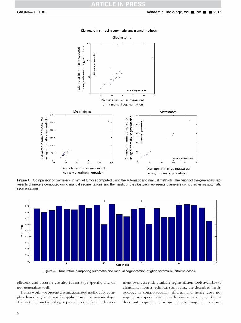
Figure 4. Comparison of diameters (in mm) of tumors computed using the automatic and manual methods. The height of the green bars rep-
resents diameters computed using manual segmentations and the height of the blue bars represents diameters computed using automatic
segmentations.
Figure 5. Dice ratios comparing automatic and manual segmentation of glioblastoma multiforme cases.
GAONKAR ET AL Academic Radiology, Vol -, No -, - 2015
efficient and accurate are also tumor type specific and do
not generalize well.
In this work, we present a semiautomated method for com-
plete lesion segmentation for application in neuro-oncology.
The outlined methodology represents a significant advance-
6
ment over currently available segmentation tools available to
clinicians. From a technical standpoint, the described meth-
odology is computationally efficient and hence does not
require any special computer hardware to run, it likewise
does not require any image preprocessing, and remains
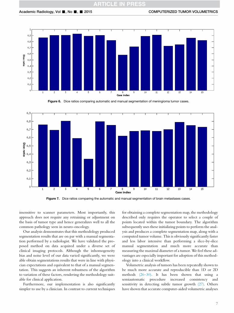
Figure 6. Dice ratios comparing automatic and manual segmentation of meningioma tumor cases.
Figure 7. Dice ratios comparing the automatic and manual segmentation of brain metastases cases.
Academic Radiology, Vol -, No -, - 2015 COMPUTERIZED TUMOR VOLUMETRICS
insensitive to scanner parameters. Most importantly, this
approach does not require any retraining or adjustment on
the basis of tumor type and hence generalizes well to all the
common pathology seen in neuro-oncology.
Our analysis demonstrates that this methodology produced
segmentation results that are on par with a manual segmenta-
tion performed by a radiologist. We have validated the pro-
posed method on data acquired under a diverse set of
clinical imaging protocols. Although the inhomogeneity
bias and noise level of our data varied significantly, we were
able obtain segmentations results that were in line with physi-
cian expectations and equivalent to that of a manual segmen-
tation. This suggests an inherent robustness of the algorithm
to variation of these factors, rendering the methodology suit-
able for clinical application.
Furthermore, our implementation is also significantly
simpler to use by a clinician. In contrast to current techniques
for obtaining a complete segmentation map, the methodology
described only requires the operator to select a couple of
points located within the tumor boundary. The algorithm
subsequently uses these initializing points to perform the anal-
ysis and produces a complete segmentation map, along with a
computed tumor volume. This is obviously significantly faster
and less labor intensive than performing a slice-by-slice
manual segmentation and much more accurate than
measuring the maximal diameter of a tumor.We feel these ad-
vantages are especially important for adoption of this method-
ology into a clinical workflow.
Volumetric analysis of tumors has been repeatedly shown to
be much more accurate and reproducible than 1D or 2D
methods (26–30). It has been shown that using a
semiautomatic procedure increased consistency and
sensitivity in detecting subtle tumor growth (27). Others
have shown that accurate computer-aided volumetric analyses
7
GAONKAR ET AL Academic Radiology, Vol -, No -, - 2015
have a significant impact on assessment of tumor response to
therapy, compared to 2D methods (27,29). Although the
RECIST criteria advocate the use of 1D and 2D
measurements for determining therapy response and
quantifying disease progression, Warren et al. have shown
that there is substantial discordance between these metrics
when 1D or 2D methods are used, compared to a 3D
volumetric assessment.
Hence, although more accurate, sensitive, and reproducible,
the adoption of volumetric criteria in neuro-oncology has been
limited by the availability of a methodology that was efficient
and generalizedwell, independent of tumor type or scanner pa-
rameters. In this work, we present a computer-aided segmenta-
tion method that fulfills these criteria. Although the results
presented here focus primarily on brain tumors, the method
may be extended to and used for the segmentation of other
large solid tumors as well. Likewise, because the algorithm is
extremely fast, one could potentially use this methodology
for real-time tumor tracking during invasive procedures such
as thermal ablation or cryoablation (31).
The chief limitation of this method is the dependence of
the segmentation on a manual initialization. The method de-
pends on an experienced clinician to provide the small set of
initial voxels that drive subsequent computation. If some of
this initialization crosses the tumor boundary, the method
will fail. Furthermore, in case of multifocal tumors, one needs
to provide multiple initializations. Consequently, multifocal
tumors require more clinician time as compared to unifocal
ones. A secondary limitation associated with this method is
that it will not operate in the presence of motion artifacts.
Motion artifacts introduce undulatory imaging patterns that
throw off the computation of the adaptive geodesic distance.
Addressing each of these limitations is outside the scope of
the present work.
Three-dimensional volumetric tumor analysis has always
been clinically desirable, and image segmentation represents
the closest assessment of the actual tumor size and volume. Be-
ing able to accurately assess tumor volume has important im-
plications for both the diagnosis and management of patients
with brain malignancies. The outlined methodology addresses
this unmet clinical need for a segmentation technique that is
robust to variation in image quality and tumor type. Going
forward, we hope that the ease of use of described approach
will lead to rapid clinical adoption. This change, from 1D to
3D calculation of tumor volume, growth, and response to
treatment will have important implications in clinical and
research applications.
MATLAB and C++ codes associated with this method are
freely available at http://cbica.upenn.edu/Bilwaj.Gaonkar/
tumor_segmentation.zip.
REFERENCES
1. Kreisl TN, Kim L, Moore K, et al. Phase II trial of single-agent bevacizumab
followed by bevacizumab plus irinotecan at tumor progression in recurrent
glioblastoma. J ClinOncol 2009; 27(5):740–745.
8
2. Brada M, Hoang-xuan K, Rampling R, et al. Multicenter phase II trial of te-
mozolomide in patients with glioblastoma multiforme at first relapse. Ann
Oncol 2001; 12(2):259–266.
3. Vredenburgh JJ, Desjardins A, Herndon JE, et al. Phase II trial of bevaci-
zumab and irinotecan in recurrent malignant glioma. Clin Cancer Res
2007; 13(4):1253–1259.
4. Wen PY, Macdonald DR, Reardon DA, et al. Updated response assess-
ment criteria for high-grade gliomas: response assessment in neuro-
oncology working group. J ClinOncol 2010; 28(11):1963–1972.
5. Herscovici Z, Rappaport Z, Sulkes J, et al. Natural history of conservatively
treated meningiomas. Neurology 2004; 63(6):1133–1134.
6. Therasse P, Eisenhauer EA, Verweij J. RECIST revisited: a review of
validation studies on tumour assessment. Eur J Cancer 2006; 42(8):
1031–1039.
7. Phillips WE, Velthuizen RP, Phuphanich S, et al. Application of fuzzy
c-means segmentation technique for tissue differentiation in MR images
of a hemorrhagic glioblastoma multiforme. MagnReson Imaging 1995;
13(2):277–290.
8. canClark MC, Hall LO, Goldgof DB, et al. Automatic tumor segmentation
using knowledge-based techniques. IEEE Trans Med Imaging 1998;
17(2):187–201.
9. Lefohn AE, Cates JE,Whitaker RT. Interactive, GPUbased level sets for 3D
segmentation in Medical Image Computing and Computer-Assisted Inter-
vention-MICCAI 2003;564–572.
10. Cobzas D, Birkbeck N, Schmidt M, et al. 3D variational brain tumor
segmentation using a high dimensional feature set. IEEE 11th International
Conference in Computer Vision; 2007; 1–8.
11. Zhang J, Ma KK, ErMeng H, et al. Tumor segmentation from mag-
netic resonance imaging by learning via one-class support vector ma-
chine. International Workshop on Advanced Image Technology
2004;207–211.
12. Verma R, Zacharaki EI, Ou Y, et al. Multiparametric tissue characterization
of brain neoplasms and their recurrence using pattern classification of MR
images. AcadRadiol 2008; 15(8):966–977.
13. Ayachi R, Amor NB. Brain tumor segmentation using support vector
machines. Symbolic and quantitative approaches to reasoning with uncer-
tainty. Springer Lecture Notes in Computer Science, 2009; 736–747.
14. Corso JJ, Sharon E, Dube S, et al. Efficient multilevel brain tumor segmen-
tation with integrated Bayesian model classification. IEEE Trans Med
Imaging 2008; 27(5):629–640.
15. Menze BH, Van leemput K, Lashkari D, et al. A generative model for brain
tumor segmentation in multi-modal images. Med Image ComputComput
Assist Interv 2010; 13(Pt 2):151–159.
16. Gering DT, GrimsonWEL, Kikinis R. Recognizing deviations from normalcy
for brain tumor segmentation. Medical Image Computing and Computer-
Assisted Intervention - MICCAI 2002; 2488:388–395.
17. Prastawa M, Bullitt E, Ho S, et al. A brain tumor segmentation framework
based on outlier detection. Med Image Anal 2004; 8(3):275–283.
18. KausMR,Warfield SK, Nabavi A, et al. Automated segmentation of MR im-
ages of brain tumors. Radiology 2001; 218(2):586–591.
19. Gaonkar B, Erus G, Bryan N, et al. Automated segmentation of brain
lesions by combining intensity and spatial Information. IEEE International
Symposium on Biomedical Imaging 2010;93–96.
20. Gaonkar B, Erus G, Pohl KM, et al. Automated segmentation of cortical
necrosis using a wavelet based abnormality detection system. Proc
IEEE IntSymp Biomed Imaging 2011;1391–1395.
21. Gooya A, Pohl KM, Bilello M, et al. GLISTR: glioma image segmentation
and registration. IEEE Trans Med Imaging 2012; 31(10):1941–1954.
22. Gaonkar B, Shu L, Hermosillo G, et al. Adaptive geodesic transform for
segmentation of vertebrae on CT images. SPIE Medical Imaging 2014.
903516–903516.
23. Antonio C, Toby S, Andrew B. Geos: Geodesic Image Segmentation,
European Conference in Computer Vision 2008;99–112.
24. Toivanen PJ. New geodesic distance transforms for gray-scale images
Pattern Recognition Letters 1996; 17:437–450.
25. Dice LR. Measures of the amount of ecologic association between spe-
cies. Ecology 1945; 26(3):297–302.
26. Hashiba T, Hashimoto N, Izumoto S, et al. Serial volumetric assessment of
the natural history and growth pattern of incidentally discovered meningi-
omas. J Neurosurg 2009; 110(4):675–684.
27. Hopper KD, Kasales CJ, Eggli KD, et al. The impact of 2D versus 3D
quantitation of tumor bulk determination on current methods of as-
sessing response to treatment. J Comput Assist Tomogr 1996;
20(6):930–937.

Academic Radiology, Vol -, No -, - 2015 COMPUTERIZED TUMOR VOLUMETRICS
28. Pohl KM, Konukoglu E, Novellas S, et al. A newmetric for detecting change
in slowly evolving brain tumors: validation in meningioma patients. Neuro-
surgery 2011; 68(1 Suppl Operative):225–233.
29. Sorensen AG, Patel S, Harmath C, et al. Comparison of diameter and
perimeter methods for tumor volume calculation. J Clin Oncol 2001;
19(2):551–557.
30. Warren KE, Patronas N, Aikin AA, et al. Comparison of one-, two-, and
three-dimensional measurements of childhood brain tumors. J Natl Can-
cer Inst 2001; 93(18):1401–1405.
31. Ahrar K, Ahrar JU, Javadi S, et al. Real-time magnetic resonance imaging-
guided cryoablation of small renal tumors at 1.5 T. Invest Radiol 2013;
48(6):437–444.
9


![Guyau’s Idea of Time: A Cognitive Viewjamichon.nl/jam_writings/1988_guyau_idea.pdf[162] Jean-Marie Guyau, in spite of his short life, became and remained a considerable presence](https://img.pdfslide.net/doc/110x75/60bde84e2140375bff212b68/guyauas-idea-of-time-a-cognitive-162-jean-marie-guyau-in-spite-of-his-short.jpg)