Embed Size (px)
Citation preview
1143
Arq Neuropsiquiatr 2009;67(4):1143-1156
View and review
AutosomAl recessive AtAxiAs
20 types, and counting
Emília Katiane Embiruçu1 , Marcília Lima Martyn1, David Schlesinger2, Fernando Kok3
Abstract – More than 140 years after the first description of Friedreich ataxia, autosomal recessive ataxias have become one of the more complex fields in Neurogenetics. Currently this group of diseases contains more than 20 clinical entities and an even larger number of associated genes. Some disorders are very rare, restricted to isolated populations, and others are found worldwide. An expressive number of recessive ataxias are treatable, and responsibility for an accurate diagnosis is high. The purpose of this review is to update the practitioner on clinical and pathophysiological aspects of these disorders and to present an algorithm to guide the diagnosis.
KEY WORDS: autosomal recessive ataxias, cerebellar ataxia, cerebellum, Friedreich ataxia.
Ataxias autossômicas recessivas: 20 tipos e muito mais
resumo – Mais de 140 anos após a primeira descrição da ataxia de Friedreich, as ataxias autossômicas recessivas se transformaram em um dos mais complexos campos da Neurogenética. Atualmente, este grupo de doenças é composto por mais de 20 entidades clínicas e possui um número ainda maior de genes associados. Algumas doenças são muito raras, tendo sido observadas apenas em populações isoladas, enquanto que outras são encontradas no mundo todo. Um número expressivo de ataxias é tratável, e a responsabilidade em se fazer um diagnóstico correto é alta. A finalidade desta revisão é a de atualizar o neurologista a respeito dos principais aspectos clínicos e fisiopatológicos destas doenças e de apresentar um algoritmo para auxiliar a sua investigação e o seu diagnóstico.
PALAVRAS-CHAVE: ataxias autossômicas recessivas, cerebelo, ataxia cerebelar, ataxia de Friedreich.
Pediatric Neurology Service and Outpatient Neurogenetics Clinic, Hospital das Clínicas, University of São Paulo School of Medicine, São Paulo SP, Brazil: 1Neurologista Infantil, Doutoranda em Neurologia pela Faculdade de Medicina da Universidade de São Paulo; 2Neurologista Clínico, Médico Preceptor do Serviço de Neurologia do Hospital das Clínicas da Universidade de São Paulo e Doutorando em Genética do Instituto de Biociências da Universidade de São Paulo; 3Neurologista Infantil, Professor Livre-Docente Médico Assistente do Serviço de Neurologia Infantil do Hospital das Clínicas da Universidade de São Paulo.
Received 11 May 2009, received in final form 3 August 2009. Accepted 22 September 2009.
Dr. Fernando Kok – Serviço de Neurologia Infantil / Hospital das Clínicas da Faculdade de Medicina da USP - Av. Dr. Enéas de Carvalho Aguiar 255 / 5011 - 05403-000 São Paulo SP - Brasil. E-mail: [email protected]
More than 20 different clinical types of autosomal re-cessive ataxias (ARA) are currently recognized. They are clinically characterized by balance abnormalities, incoor-dination, kinetic and postural tremor, and dysarthria1. Typ-ically, symptoms start before 25 years of age, and cerebel-lum, brainstem, and spinocerebellar tracts are involved2. Peripheral neuropathy, ophthalmological abnormalities, and non-neurological signs and symptoms might also be present1. Friedreich ataxia, the most common ARA, was first described in 1863 and is seen worldwide1,2. In the last few years, several other conditions have also been recog-nized and their loci and genes identified.
Pathophysiology is quite variable and the defective gene product might be involved with: (A) Cerebellar and/or brain stem development; (B) Mitochondrial energy gen-eration; (C) Intermediate metabolism; (D) DNA repair; and (E) Cerebellar integrity maintenance. Several classifica-tions have been proposed so far, using clinical, neuroim-aging, genetic and pathophysiologic data2. In this review, a pathophysiological classification is used (Table 1).
We should be particularly aware of treatable forms of ARA, which includes Refsum disease, ataxia with vitamin E deficiency, coenzyme Q10 deficiency, cerebrotendinous xanthomatosis, and abetalipoproteinemia.
Arq Neuropsiquiatr 2009;67(4)
1144
Autosomal recessive ataxiasEmbiruçu et al.
Table 1. Classification and molecular aspects of the autosomal recessive ataxias
Gene (Locus) Protein Protein function
Congenital
Cayman ataxia ATCAY (19p13.3) Caytaxin Synapse between granulates and Purkinje cells (?)
Joubert syndrome AHI1 (16q23.3) Jouberin Cerebellar estruturation; cilia estruturation and functions
NPHP1 (2q13) Nefrocistin-1
CEP290 (12q21.34) Nefrocistin-6
TMEM67 (8q21.1-q22.1) Meckelin
RPGRIP1L (16q12.2) Protein phantom
Cerebellar hypoplasia associated VLDL receptor VLDLR (9p24.2-3) VLDL Receptor Signalling neuroblast migartion
Mitochondrial
Friedreich ataxia FRDA (9q13) Frataxin Mitochondrial iron metabollism
Coenzyme Q10 deficiency with cerebellar ataxia
PDSS1 (10p12.1) and PDSS2 (6q21)
Prenyldiphosphate synthase subunit 1 e 2
Coenzyme Q10 biosynthesis
COQ2 (4q21-q22) OH-benzoate polyiprenyl transferase
Coenzyme Q10 biosynthesis
ADCK3 (CABC1) (1q42.2) ADCK3 (Mitochondrial protein) Coenzyme Q10 biosynthesis
Ataxia with mutation in polymerase gamma POLG (15q22-26) DNA polymerase g Mitochondrial DNA maintenance
Infantile onset spinocerebellar ataxia C10orf2 (10q24) Twinkle Mitochondrial DNA repair and maintenance
Metabolic
Ataxia with vitamin E deficiency a-TTP (8q13.1-13.3) a-tocopherol transfer protein a-tocopherol incorporation in VLDL
Abetalipoproteinemia MTP (4q22-24) Microsomal tryigliceride transfer protein
Lipoprotein metabolism
Refsum disease PHYH (10pter-11.2) Phytanoyl-CoA hydroxylase Fatty acid a-oxidation
PEX7 (6q21-22.2) Peroxisomal biogenesis factor-7 Peroxisomal protein importation
Cerebrotendinous xanthomatosis CYP27 (2q33-ter) Sterol 27-hydroxylase Bile acid synthesis
DNA repair defects
Ataxia telangiectasia ATM (11q22.3) Ataxia telangiectasia mutated DNA double-strand break repair
Ataxia-telangiectasia- like disorder MRE11A (11q21) Meiotic recombination 11 DNA double strand break repair
Ataxia with oculomotor apraxia type 1 APTX (9p13) Aprataxin DNA single strand break repair
Ataxia with oculomotor apraxia type 2 SETX (9q34) Senataxin DNA and RNA repair
Spinocerebellar ataxia with axonal neuropathy TDP1 (14q31-32) Tyrosyl DNAphosphodiesterase I
DNA repair
Degenerative
Spastic ataxia of Charlevoix Saguenay SACS (13q11) Sacsin Chaperone-mediated protein foling
Marinesco-Sjögren syndrome SIL1 (5q31) BiP associated protein Stabilization and folding of newly synthesized polypeptides
Legend: (?), possibly.
Arq Neuropsiquiatr 2009;67(4)
1145
Autosomal recessive ataxiasEmbiruçu et al.
AtAxiA cAused by cerebellAr And/or brAinstem mAlformAtionIn this group, neuroimaging studies are able to identi-
fy cerebellar and/or brainstem malformation and clinical-ly it is characterized by non progressive cerebellar atax-ia. Three conditions are discussed in this section: Cayman ataxia, Joubert syndrome and Cerebellar hypoplasia asso-ciated to VLDL receptor.
Cayman ataxiaCayman ataxia (CA) is a condition characterized by
variable developmental delay, early onset hypotonia and non-progressive axial cerebellar ataxia, associated to ny-stagmus, intention tremor, and dysarthria3. MRI presents with cerebellar hypoplasia3. The CA has only been found in Grand Cayman Island, where heterozygote frequen-cy is supposed to be of 18%3. CA is caused by mutation at ATCAY, which codes for caytaxin, a protein involved with glutamate synthesis and also with synaptogenesis of cerebellar granular neurons and Purkinje cells4. Inter-estingly, ATCAY contains a CRAL-TRIO domain that binds small lipophillic molecules, similar to the alpha-tocoph-erol transport protein that causes ataxia with vitamin E deficiency3.
Joubert syndromeJoubert syndrome (JS) is a rare genetically heteroge-
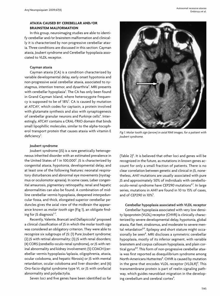
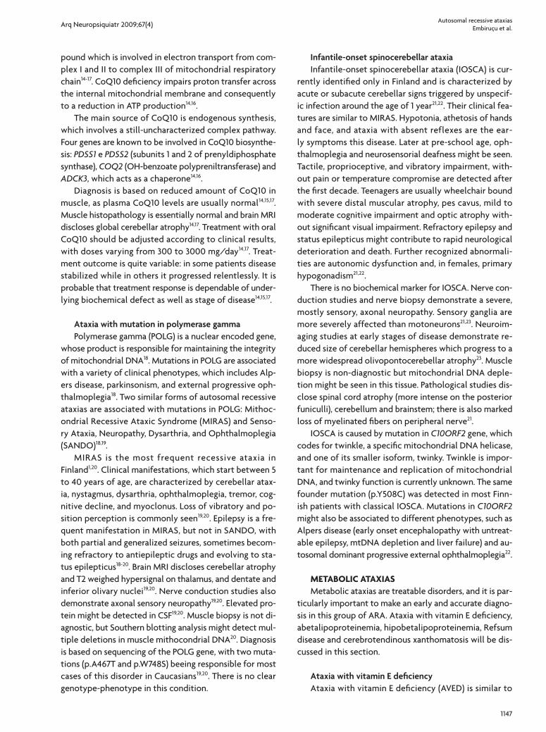
neous inherited disorder with an estimated prevalence in the United States of 1 in 100,0005. JS is characterized by congenital ataxia, hypotonia, developmental delay, and at least one of the following features: neonatal respira-tory disturbances and abnormal eye movements (nystag-mus or oculomotor apraxia). In some cases, Leber congen-ital amaurosis, pigmentary retinopathy, renal and hepatic abnormalities can also be found. A combination of mid-line cerebellar vermis hypoplasia, deepened interpedun-cular fossa, and thick, elongated superior cerebellar pe-duncles gives the axial view of the midbrain the appear-ance known as molar tooth sign (Fig 1), an obligate find-ing for JS diagnosis5-7.
Recently, Valente, Brancati and Dallapiccola6 proposed a clinical classification of JS in which the molar tooth sign was considered an obligatory criterion. They were able to recognize six subgroups of JS: (1) Pure Joubert syndrome; (2) JS with retinal abnormality; (3) JS with renal disorders; (4) CORS (cerebello-oculo-renal syndrome), or JS with ret-inal abnormality and kidney involvement; (5) COACH (cer-ebellar vermis hypoplasia/aplasia, oligophrenia, ataxia, ocular coloboma, and hepatic fibrosis) or JS with mental retardation, ocular coloboma and liver disorder; and (6) Oro-facio-digital syndrome type VI, or JS with orofacial abnormality and polydactylia.
Seven loci and five genes have been identified so far
(Table 2)6. It is believed that other loci and genes will be recognized in the future, as mutations in known genes ac-count for only a small fraction of patients. There is no clear correlation between genetic and clinical in JS, none-theless, AHI1 mutations are usually associated with pure JS and approximately 50% of individuals with cerebello-oculo-renal syndrome have CEP290 mutations6,7. In large series, mutations in AHI1 are found in 10 to 15% of cases, and of CEP290 in 10%5.
Cerebellar hypoplasia associated with VLDL receptor Cerebellar hypoplasia associated with very low densi-
ty lipoprotein (VLDL) receptor (CHVR) is clinically charac-terized by severe developmental delay, hypotonia, global ataxia, flat feet, strabismus, and moderate to severe men-tal retardation8,9. Epilepsy and short stature might occa-sionally be seen8. MRI discloses a symmetric cerebellar hypoplasia, mostly of its inferior segment, with variable brainstem and corpus callosum hypoplasia, and plain cor-tical gyrus8,9. This form of non-progressive cerebellar atax-ia was first reported as disequilibrium syndrome among North-Americans Hutterites8. CHVR is caused by mutation in the gene that encodes VLDL receptor (VLDLR)9. This transmembrane protein is part of reelin signaling path-way, which guides neuroblast migration in the develop-ing cerebellum and cerebral cortex9.
Fig 1. Molar tooth sign (arrow) in axial RMI images, for a patient with Joubert syndrome.
Arq Neuropsiquiatr 2009;67(4)
1146
Autosomal recessive ataxiasEmbiruçu et al.
AtAxiAs cAused by deficiency of mitochondriAl energy generAtionAtaxias caused by deficiency of mitochondrial energy
generation includes Friedreich ataxia, Ataxia with CoQ10 deficiency, Mitochondrial recessive ataxic syndrome (MIRAS) and Infantile-onset spinal cerebellar ataxia (IOSCA).
Friedreich ataxiaFriedreich ataxia (FA) is the most common recessive
ataxia worldwide, with an estimated prevalence in Cau-casian population of 1:30,000 to 1:50,00010 and a carrier frequency of 1:851. Its onset is usually in the second de-cade of life, but can vary from 2 to 25 years of age. Clini-cal manifestations are characterized by a combination of sensory and cerebellar symptoms, and gait instability is usually the first recognized abnormality. Relentlessly pro-gressive ataxia is characteristic, and after 10 to 15 years of onset, patients are usually wheelchair bound. Dysarthria is also an early and incapacitating symptom, leading to an almost incomprehensible speech. Vibratory and posi-tional sense is affected, and Romberg sign is usually posi-tive. Deep tendon reflexes are absent, but extensor plan-tar reflex (Babinski sign) is usually present. Abnormal eye movements and defective fixation are also observed. Cog-nitive function is preserved, but communication abilities can be affected. Systemic abnormalities as hypertrophic cardiomiopathy, cardiac conduction defects and diabetes can occur. As disease progresses, pes cavus and scoliosis are almost always present. Although there are significant variations in the onset and rate of disease progression, the mean age of death has been reported to be approximately 38 years, with a range as wide as 5 to 70 years. Death usu-ally is secondary to progressive cardiomyopathy10.
Brain MRI in FA is normal; using the multigradient echo sequence it is sometimes possible to detect iron depos-its in dentate nuclei of cerebellum. Spinal cord MRI can disclose mild atrophy of its cervical segment, which is ex-plained by the large loss of primary sensory neurons in
the dorsal root ganglia, early in the course of the disease. Nerve conduction studies characteristically show axonal sensory neuropathy10. Atypical forms of FA, as late on-set or with maintained reflexes, have been proposed, but it is now clear that they are also caused by mutations in the same gene.
FA is caused by mutations in the FRDA gene, which en-codes frataxin, a protein involved in mitochondrial iron regulation. Loss of mitochondrial iron-sulfur centers, im-pairment of mitochondrial respiratory chain, increased mitochondrial iron and increased oxidative damage are observed when frataxin is deficient. Almost all patients are homozygotes for a GAA expansion which occurs in intron 1 of FRDA gene. Normal individuals have up to 40 GAA repeats, and in patients this number can vary from 70 or 90 to over 1,700 repeats. Presence of biallelic expansion confirms diagnosis, independent of clinical phenotype10,11. Close to 2% of patients are compound heterozygotes, with a combination of a GAA expansion in one allele and a point mutation in the other1.
Coenzyme Q10 (CoQ10), and its synthetic analog ide-benone, vitamin E and, more recently the iron chelator deferiprone have been used for treating FA, with some promising but still very preliminary results12,13. Deferiprone, an atypical iron chelator, may decrease accumulation of toxic iron in the mitochondria in patients, but the recom-mended dose and the efficiency of this treatment have not yet been determined12.
Ataxia with coenzyme Q10 deficiencyPrimary deficiency of coenzyme Q10 (CoQ10) is a ge-
netically heterogeneous disorder, with a highly variable clinical spectrum, which includes multi-systemic man-ifestations as well as CNS compromise14-16. Five clinical subtypes have been recognized: (1) Encephalomyophat-ic, with mitochondrial myopathy, recurrent myoglobinu-ria and CNS symptoms and signs; (2) Early infantile multi-systemic, with severe visceral and brain manifestations; (3) Leigh syndrome; (4) Pure myopathic; (5) Ataxic14,15,17.
The ataxic subtype is the most common presentation of CoQ10 deficiency14,15. It is characterized by progressive ataxia, cerebellar atrophy and reduced muscle CoQ1014,15. Early symptoms might include developmental delay, hy-potonia and frequent falls. Global, progressive ataxia, and dysarthria start before the adolescence17. Epileptic sei-zures, proximal or distal muscle weakness, dysphagia, oph-thalmoparesis, nystagmus, peripheral axonal neuropathy, pyramidal signs and scoliosis might also be present14,15,17. Mental retardation or cognitive decline is also sometimes seen14,17. The adult onset form of ataxia and CoQ10 de-ficiency is usually associated with hypergonadotrophic hypogonadism14.
CoQ10 (also known as ubiquinone) is a lipophylic com-
Table 2. Joubert syndrome identified loci, genes and their products.
Locus Location Gene Protein
JBTS1 9q34.3 Not known Not known
JBTS2 11p12-q13.3 Not known Not known
JBTS3 6q23.3 AHI1 Jouberin
JBTS4 2q13 NPHP1 Nephrocystin-1
JBTS5 12q21.34 CEP290 (NPHP6)
Nephrocystin-6
JBTS6 8q21.1-q22.1 TMEM67 Meckelin
JBTS7 16q12.2 RPGRIP1L Protein phantom
Arq Neuropsiquiatr 2009;67(4)
1147
Autosomal recessive ataxiasEmbiruçu et al.
pound which is involved in electron transport from com-plex I and II to complex III of mitochondrial respiratory chain14-17. CoQ10 deficiency impairs proton transfer across the internal mitochondrial membrane and consequently to a reduction in ATP production14,16.
The main source of CoQ10 is endogenous synthesis, which involves a still-uncharacterized complex pathway. Four genes are known to be involved in CoQ10 biosynthe-sis: PDSS1 e PDSS2 (subunits 1 and 2 of prenyldiphosphate synthase), COQ2 (OH-benzoate polypreniltransferase) and ADCK3, which acts as a chaperone14,16.
Diagnosis is based on reduced amount of CoQ10 in muscle, as plasma CoQ10 levels are usually normal14,15,17. Muscle histopathology is essentially normal and brain MRI discloses global cerebellar atrophy14,17. Treatment with oral CoQ10 should be adjusted according to clinical results, with doses varying from 300 to 3000 mg/day14,17. Treat-ment outcome is quite variable: in some patients disease stabilized while in others it progressed relentlessly. It is probable that treatment response is dependable of under-lying biochemical defect as well as stage of disease14,15,17.
Ataxia with mutation in polymerase gamma Polymerase gamma (POLG) is a nuclear encoded gene,
whose product is responsible for maintaining the integrity of mitochondrial DNA18. Mutations in POLG are associated with a variety of clinical phenotypes, which includes Alp-ers disease, parkinsonism, and external progressive oph-thalmoplegia18. Two similar forms of autosomal recessive ataxias are associated with mutations in POLG: Mithoc-ondrial Recessive Ataxic Syndrome (MIRAS) and Senso-ry Ataxia, Neuropathy, Dysarthria, and Ophthalmoplegia (SANDO)18,19.
MIRAS is the most frequent recessive ataxia in Finland1,20. Clinical manifestations, which start between 5 to 40 years of age, are characterized by cerebellar atax-ia, nystagmus, dysarthria, ophthalmoplegia, tremor, cog-nitive decline, and myoclonus. Loss of vibratory and po-sition perception is commonly seen19,20. Epilepsy is a fre-quent manifestation in MIRAS, but not in SANDO, with both partial and generalized seizures, sometimes becom-ing refractory to antiepileptic drugs and evolving to sta-tus epilepticus18-20. Brain MRI discloses cerebellar atrophy and T2 weighed hypersignal on thalamus, and dentate and inferior olivary nuclei19,20. Nerve conduction studies also demonstrate axonal sensory neuropathy19,20. Elevated pro-tein might be detected in CSF19,20. Muscle biopsy is not di-agnostic, but Southern blotting analysis might detect mul-tiple deletions in muscle mithocondrial DNA20. Diagnosis is based on sequencing of the POLG gene, with two muta-tions (p.A467T and p.W748S) beeing responsible for most cases of this disorder in Caucasians19,20. There is no clear genotype-phenotype in this condition.
Infantile-onset spinocerebellar ataxia Infantile-onset spinocerebellar ataxia (IOSCA) is cur-
rently identified only in Finland and is characterized by acute or subacute cerebellar signs triggered by unspecif-ic infection around the age of 1 year21,22. Their clinical fea-tures are similar to MIRAS. Hypotonia, athetosis of hands and face, and ataxia with absent reflexes are the ear-ly symptoms this disease. Later at pre-school age, oph-thalmoplegia and neurosensorial deafness might be seen. Tactile, proprioceptive, and vibratory impairment, with-out pain or temperature compromise are detected after the first decade. Teenagers are usually wheelchair bound with severe distal muscular atrophy, pes cavus, mild to moderate cognitive impairment and optic atrophy with-out significant visual impairment. Refractory epilepsy and status epilepticus might contribute to rapid neurological deterioration and death. Further recognized abnormali-ties are autonomic dysfunction and, in females, primary hypogonadism21,22.
There is no biochemical marker for IOSCA. Nerve con-duction studies and nerve biopsy demonstrate a severe, mostly sensory, axonal neuropathy. Sensory ganglia are more severely affected than motoneurons21,23. Neuroim-aging studies at early stages of disease demonstrate re-duced size of cerebellar hemispheres which progress to a more widespread olivopontocerebellar atrophy23. Muscle biopsy is non-diagnostic but mitochondrial DNA deple-tion might be seen in this tissue. Pathological studies dis-close spinal cord atrophy (more intense on the posterior funiculli), cerebellum and brainstem; there is also marked loss of myelinated fibers on peripheral nerve21.
IOSCA is caused by mutation in C10ORF2 gene, which codes for twinkle, a specific mitochondrial DNA helicase, and one of its smaller isoform, twinky. Twinkle is impor-tant for maintenance and replication of mitochondrial DNA, and twinky function is currently unknown. The same founder mutation (p.Y508C) was detected in most Finn-ish patients with classical IOSCA. Mutations in C10ORF2 might also be associated to different phenotypes, such as Alpers disease (early onset encephalopathy with untreat-able epilepsy, mtDNA depletion and liver failure) and au-tosomal dominant progressive external ophthalmoplegia22.
metAbolic AtAxiAsMetabolic ataxias are treatable disorders, and it is par-
ticularly important to make an early and accurate diagno-sis in this group of ARA. Ataxia with vitamin E deficiency, abetalipoproteinemia, hipobetalipoproteinemia, Refsum disease and cerebrotendinous xanthomatosis will be dis-cussed in this section.
Ataxia with vitamin E deficiency Ataxia with vitamin E deficiency (AVED) is similar to
Arq Neuropsiquiatr 2009;67(4)
1148
Autosomal recessive ataxiasEmbiruçu et al.
Friedreich ataxia (FA). Age of onset usually varied from 4 to 20 years, with outliers ranging from 2 to 52 years24,25. Clinical manifestations are characterized by progressive trunk and limbs ataxia, dysarthria, disturbance of position-al and vibratory lower limbs senses, Babinski sign and abol-ished deep tendon reflexes. Scoliosis and pes cavus are commonly seen24-26, yet retinopathy is less frequent25,26. Dystonia (13%) and head titubation (28%) are more com-monly seen in AVED than in FA24,26. Cardiomyopathy and an acute cardiac event might be associated with prema-ture death among AVED cases25,26.
AVED is caused by mutations in the a-tocopherol transfer protein gene (TTPA), which codes a protein re-sponsible for transferring a-tocopherol from chilomicrons to VLDL26,27. TTPA disfunction causes very low level of cir-culating a-tocopherol and tissue deficiency of this vita-min27. Several different pathogenic mutations have been reported so far. Two mutations, associated with a severe phenotype, are particularly frequent in Europe, North Af-rica and North America: c.744delA and c.486delT24,25. On the other hand, the mutation p.H101G was only detected in Japanese families and is associated with later onset and pigmentary retinopathy24. Age of onset, clinical manifesta-tions and progression velocity are quite variable in AVED. It is usually stated that mutations causing profound TTPA protein depletion are responsible for a more severe phe-notype and mutations leading to amino acid substitution are associated to a milder form of disease24-26.
Diagnosis in a symptomatic individual is established with the determination of a-tocopherol (vitamin E) serum level, which is always below 2.5 mg/ml (reference values: 5-15 mg/ml)24,25. Brain MRI is usually normal, but mild cer-ebellar atrophy might also be seen1,26. A pattern of axonal sensitive neuropathy is often observed at nerve conduc-tion studies24.
AVED treatment consists of vitamin E oral adminis-tration at a dose of 600 to 2,400 mg/day. Serum levels might be used as guidance for oral dose adjustment24-26. Differential diagnosis of a-tocopherol primary deficien-cy includes intestinal fat mal-absorption and abetalipo-proteinemia26. As a rule, vitamin E serum levels should be checked in all patients with FA clinical phenotype with-out molecular confirmation for this condition.
Abetalipoproteinemia and hypobetalipoproteinemiaAbetalipoproteinemia (ABL), a multisystem disorder
caused by a defect in lipoprotein metabolism, is charac-terized by acanthocytosis, atypical pigmentary retinopa-thy and spinocerebellar degeneration28,29. In the first year of life, main manifestations are chronic diarrhea and fail-ure to thrive. Neurological features, present after the first decade of life, include absent deep tendon reflexes, su-perficial and deep sensory abnormalities, weakness and
global ataxia29. As disease progresses, atypical pigmentary retinopathy characterized by small, irregularly distributed white spots, and night and color blindness is detected29. Clinical manifestations of ABL are secondary to deficient absorption of the lipid-soluble vitamins A, D, E, and K1,28.
Apolipoprotein B (ApoB) is the main protein of both VLDL and LDL, and their assembly is dependent on mi-crosomal triglyceride transfer protein (MTP)28. Mutations in the gene coding for the large (88 kD) subunit of MTP is responsible for ABL and determine very low levels of LDL and VLDL cholesterol. Decreased levels of vitamins A, K, and E, anemia, very low sedimentation rate, increased pro-thrombin time and elevated creatine kinase are also ob-served. Deficient MTP can also lead to lipid infiltration of small bowel mucosa and hepatic steatosis28,29. Nerve con-duction velocity usually discloses sensory axonal periph-eral neuropathy1,29.
ABL treatment is done with supplementation of vita-min A (100 to 400 IU/Kg/day), vitamin E (2,400 to 14,400 IU/day), and vitamin K (5 mg/day)29. It is also recom-mended a low fat diet combined with essential fatty acid supplementation28,29. Coagulation tests are used to moni-tor vitamin K and serum levels of vitamin A and E to check supplementation adequacy for these vitamins28,29.
Hypobetalipoproteinemia (HBL) is similar to ABL. It is caused by mutations in APOB gene, which encodes apo-lipoprotein B (ApoB)2. APOB heterozygotes have lower levels of ApoB, VLDL- and LDL-cholesterol, while MTP heterozygotes have normal levels of these substances2,29,30.
Refsum disease Refsum disease (RD) is a peroxisomal disorder clini-
cally characterized by pigmentary retinopathy, cerebel-lar ataxia, mixed motor-sensory neuropathy and elevat-ed CSF protein31,32. Its onset usually occurs before 20 years of age, with night blindness secondary to retinopathy, fol-lowed by progressive constriction of visual field and optic atrophy, cataracts, vitreous opacities and nystagmus32,33. Other common clinical manifestations are anosmia, co-chlear deafness, ichthyosis, bone dysplasia and cardiac ab-normalities31-33. Psychiatric disorders are uncommon31,33. If not adequately treated, RD can cause premature cardi-ac death31,32.
Elevation of serum phytanic acid (>200 mM/L; refer-ence value <30 mM/L) is very suggestive, but not specif-ic of RD31,33. Phytanic acid is a branched long chain fat-ty acid present on dairy products and red meat33. It is a by-product of chlorophyll catabolism and is not endog-enously synthesized31-33. Diagnostic confirmation can be made measuring the activity of phytanoyl-CoA hydroxi-lase in fibroblasts or by molecular analysis of the respon-sible gene31.
RD is a genetically heterogeneous disorder. Most cas-
Arq Neuropsiquiatr 2009;67(4)
1149
Autosomal recessive ataxiasEmbiruçu et al.
es are caused by mutations in PHYH (encoding phytanoyl-CoA hydroxylase), a peroxisomal matrix enzyme which catalyses -oxidation of branched chain fatty acids31-33. Deficiency in PEX7 (encoding peroxin-7), a protein in-volved in peroxisomal import of some enzymes, in-cluding phytanoyl-CoA hydroxylase, also results in this phenotype2,31. PEX7 mutations may also cause a severe peroxisomal biogenesis disorder known as rhizomelic chondrodysplasia punctata32.
Treatment is based on phytanic acid dietary restric-tion, combined, if necessary, with plasmapheresis. With reduction of phytanic acid serum levels, RD symptoms stabilize and there may be some improvement of atax-ia and ichthyosis, albeit effects on pigmentary retinopa-thy are uncertain32,33.
Cerebrotendinous xanthomatosisCerebrotendinous xanthomatosis (CTX) is a rare bile
acid synthesis disorder34. Its main clinical manifestations are juvenile cataracts, chronic diarrhea, and tendinous xanthomas34-36. In the neonatal period, a potentially le-thal cholestatic syndrome has been reported34. After the second decade of life, progressive neurological deteriora-tion may occur, characterized by cognitive decline, psychi-atric manifestations, cerebellar ataxia, progressive spastic paraplegia, dysphagia, and less frequently, seizures and pe-ripheral neuropathy34,36,37. Exceptionally, neurological man-ifestations are restricted to the spinal cord35. There is a wide intra- and inter-familial clinical variability34,37. Coro-nary heart disease without elevated cholesterol is an im-portant cause of morbidity and mortality in adults34,35.
CTX is caused by mutations in CYP27A1 gene, which codes for sterol 27- hydroxylase, a protein expressed mostly in liver34,36. This enzyme is essential for bile ac-ids synthesis, including chenodeoxycholic acid36. In the absence of sterol 27- hydroxylase, the substrate of this enzyme is converted to cholestanol by the action of 7 a-hydroxylase34,36. Elevated serum cholestanol is the bio-chemical hallmark of CTX35,37. Increased urinary excretion of bile alcohol glucuronides might also be present37. This disorder can be treated by oral administration of chen-odeoxycholic acid, which inhibits 7 a-hydroxylase and consequentely cholestanol synthesis34-37. Liver transplant is another therapeutic alternative34. Cholestanol determi-nation in asymptomatic siblings of CTX patients is recom-mended to improve clinical outcome34,37.
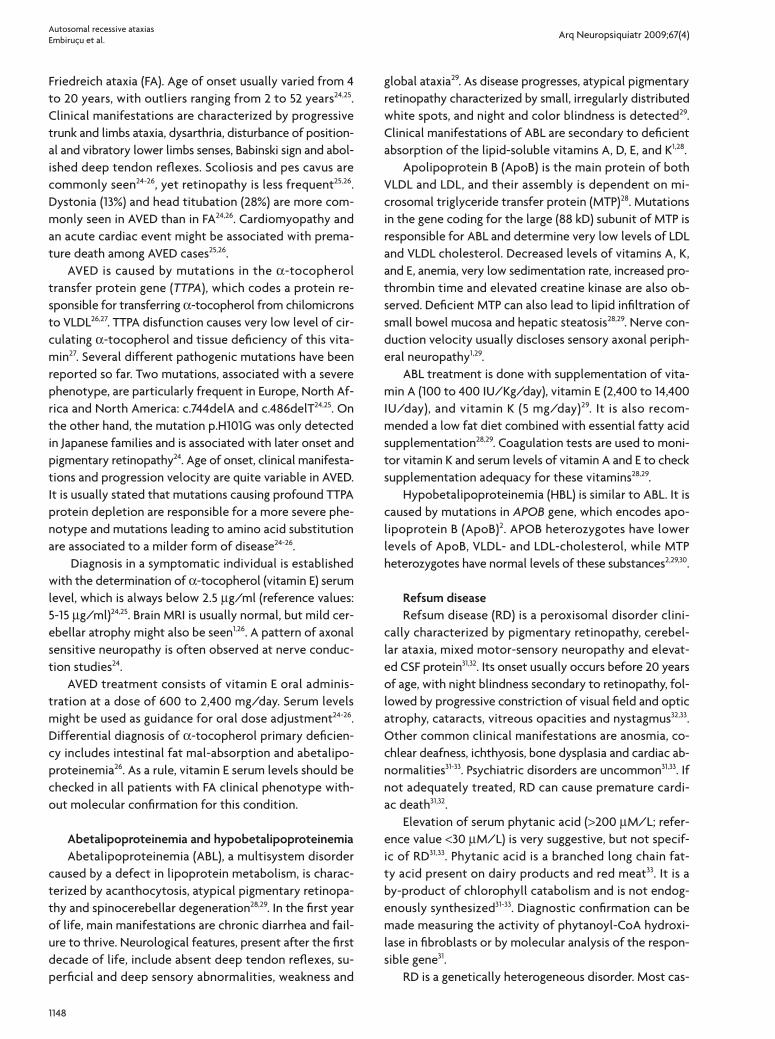
Brain MRI most distinctive abnormalities are detected at T2-weighed and FLAIR sequences, which demonstrate bilateral, heterogeneous and hyperintense sign in den-tate nuclei and adjacent cerebellar white matter (Fig 2)35,36. Other less characteristic abnormalities which might be detected are brain stem, cerebellar and cerebral atrophy and diffuse hyperintense cerebral white matter lesions35,36.
MR spectroscopy (MRS) of CTX patients discloses reduc-tion of N-acetylaspartate and increase in lactate36.
AtAxiAs With dnA rePAir defectsThis group has as a common pathogeny a defect in
double or single strand DNA repair; besides ataxia, extrin-sic ocular movements are frequently affected. Ataxia-te-langiectasia, ataxia telangiectasia-like, apraxia and oculo-motor apraxia types 1, 2 and 3, and spinocerebellar ataxia with axonal neuropathy type 1 belong to this group.
Ataxia-telangiectasiaAtaxia-telangiectasia (AT) has an estimated frequen-
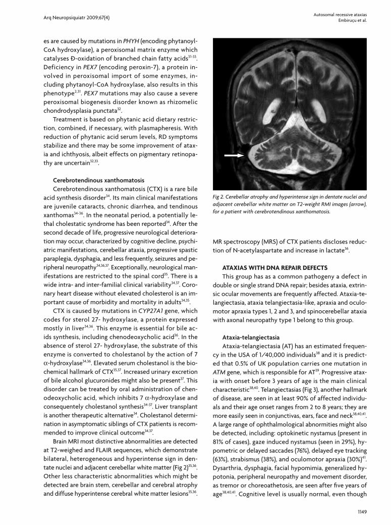
cy in the USA of 1/40,000 individuals38 and it is predict-ed that 0.5% of UK population carries one mutation in ATM gene, which is responsible for AT39. Progressive atax-ia with onset before 3 years of age is the main clinical characteristic38,40. Telangiectasias (Fig 3), another hallmark of disease, are seen in at least 90% of affected individu-als and their age onset ranges from 2 to 8 years; they are more easily seen in conjunctivas, ears, face and neck38,40,41. A large range of ophthalmological abnormities might also be detected, including: optokinetic nystamus (present in 81% of cases), gaze induced nystamus (seen in 29%), hy-pometric or delayed saccades (76%), delayed eye tracking (63%), strabismus (38%), and oculomotor apraxia (30%)41. Dysarthria, dysphagia, facial hypomimia, generalized hy-potonia, peripheral neuropathy and movement disorder, as tremor or choreoathetosis, are seen after five years of age38,40,41. Cognitive level is usually normal, even though
Fig 2. Cerebellar atrophy and hyperintense sign in dentate nuclei and adjacent cerebellar white matter on T2-weight RMI images (arrow), for a patient with cerebrotendinous xanthomatosis.
Arq Neuropsiquiatr 2009;67(4)
1150
Autosomal recessive ataxiasEmbiruçu et al.
severe dysarthria and incoordination might leave an im-pression of mental retardation. Independent gait is lost by the end of first decade38,40. Immunodeficiency (main-ly humoral) leading to chronic sinopulmonary infection and increased susceptibility to cancer are other impor-tant features in AT. Risk of lymphoproliferative disorders are dramatically increased in AT38,40. Treating cancer in AT patients is particularly challenging, because of their in-creased radiosensitivity and adverse side effects to che-motherapy40. Female carriers of one mutated copy of the gene have 3–4 fold increased risk of breast cancer when compared to general population39.
ATM gene encodes for a ATM serine/threonine kinase, a large protein with 3,056 amino acids which is part of the phosphatidyl-inositol-3-kinase (PI3-K) complex, responsi-ble for DNA repair during the cell cycle, avoiding incorpo-ration of deleterious mutations38,39. ATM gene is very large, containing 66 exons, and its sequencing with current tech-nology is still cumbersome, but feasible38,39. Most patients are compound heterozygotes for ATM mutations and a large variety of sequence variants have been recognized, thus making interpretation of results difficult. Pathogen-ic mutations are usually nonsense mutations (85%), and missense mutations are responsible for 10% of detected pathogenic changes38,39,40.
Some laboratory tests might help AT investigation: se-rum alpha-fetoprotein is elevated in more than 95% of cases and low levels of IgA, IgE and reduced T lymphocyte count, with normal or elevated B lymphocytes are usually detected. Karyotype might show translocation between chromosomes 7 and 1438-41 and radiosensivity test might be used to demonstrate chromosomal breakage predis-position.
Brain MRI discloses, except in early stages of disease, cerebellar atrophy, which is initially more evident on the hemispheres and superior vermis and later becomes dif-
fuse38. CT-scan and plain radiography should be avoided because of increased X-ray sensitivity.
Ataxia-telangiectasia like Ataxia-telangiectasia like (ATL) is a very rare clini-
cal condition which is characterized by slowly progres-sive ataxia with onset between 1 and 7 years of age, as-sociated to oculomotor apraxia and dysarthria42. No oc-ular or facial telangiectasias are detected and cognition is preserved39,42,43. Reflexes might be initially brisk and be-came reduced42. At advanced stages of disease, tongue and facial dyskinesia, choreoathetosis, and dystonia, sug-gesting basal ganglia compromise, might be seen42,43. ATL is progressive up to the adolescence, when it stabilizes43. There is no increased risk for infections or neoplasias, as is seen in AT, but occasionally microcephaly is present39,42.
Brain MRI detects cerebellar atrophy, and laboratory tests are non-informative43. Radiosensitivity test is usual-ly present but in a lesser degree than in AT35,42-44.
ATL is caused by mutation in MRE11 gene, located in chromosome 11q21, near the ATM gene. Its product is part of the MRN complex, which recognizes DNA double strand breakage. Both missense and null mutations have been reported. Severity varies according to the type of molecular defect. Most of reported cases were original from Saudi Arabia 39,42-44.
Ataxia with oculomotor apraxia type 1 Ataxia with oculomotor apraxia type 1 (AOA1) is a con-
dition characterized by involuntary movements (chorea and dystonia) and/or progressive global ataxia, with dys-arthria associated with hands and head tremor. Onset can vary between 1 to 20 years of age and developmental de-lay might be seen before clinical symptoms became appar-ent. As disease progresses, movement disorder are atten-uated and peripheral neuropathy signs, as distal atrophy,
Fig 3. Ocular telangiectasia for a patient with ataxia telangiectasia patient.
Arq Neuropsiquiatr 2009;67(4)
1151
Autosomal recessive ataxiasEmbiruçu et al.
pes cavus, superficial and deep sensory impairment, hypo/arreflexia, become apparent. The most distinctive clini-cal signs in AOA1 are related to external eye movements: gaze-evoked nystagmus (found in all patients), oculomo-tor apraxia (seen in 86%), saccadic pursuit, hypometric saccades, fixation instability, and excessive blinking45-47. In advanced stages, oculomotor apraxia might be masked by progressive external ophthalmoparesis, which starts with upward gaze paralysis45. Optic atrophy and retinal exsu-dative lesions have been occasionally reported (Barbot, 2001; Le Ber, 2003). Variable cognitive impairment might be seen and mental retardation is not uncommon46,47.
Laboratory findings include hypoalbuminemia and hypercholesterolemia46,47. Elevated creatine kinase is occa-sionally detected47. Nerve conduction velocity studies dis-close sensory-motor axonal neuropathy. MRI reveals marked cerebellar atrophy, mild brainstem atrophy and, in advanced cases, cortical atrophy45-47. Loss of myelinated fibers with maintenance of amyelinic ones is seen at sural nerve45,47.
AOA1 is caused by mutation in APTX gene, which en-codes aprataxin, a nuclear protein involved in single-strand DNA repair, acting in the same pathway of the ATM protein39,46. Several mutations have been reported so far, most of them in exons 5, 6, and 7 of APTX gene47. This con-dition was originally reported in Japan (where it is the most common cause of ARA) but is found worldwide. In Por-tugal, AOA1 is the second most common cause of ARA46.
Ataxia with oculomotor apraxia type 2Ataxia with oculomotor apraxia type 2 (AOA2) is char-
acterized by global progressive ataxia with onset usually between 8 and 25 years of age48,49, dysarthria, axonal mo-tor sensory neuropathy, and oculomotor apraxia, which is seen in less than 50% of cases48-50. Saccadic pursuit is seen in all patients, gaze evoked nystagmus in 89%, and bilateral limited abduction of the eyes with strabismus in 61% of the patients48. Dystonia, head and postural tremor, chorea, dysphagia, pes cavus, and scoliosis are occasional-ly seen. Cognitive function is usually preserved, but exec-utive dysfunction is sometimes observed48-50. Premature ovarian failure was also reported in some patients48. Pro-gression is slow, and most patients are wheelchair bound 10 years after its onset48,49.
Serum alpha-fetoprotein is mildly to moderately el-evated in all patients with AOA239,48-50. Increased serum creatine kinase, cholesterol, and immunoglobulin IgG and IgA, and reduced serum albumin are inconstantly seen48,50. Brain MRI discloses diffuse cerebellar atrophy, more in-tense in the vermis, occasionally associated with pontine atrophy50. Nerve conduction studies detect sensory-mo-tor axonal neuropathy and nerve biopsy demonstrates that large myelinated fibers are more severely affected than thin ones48,50.
AOA2 is caused by mutation in SETX gene (encod-ing senataxin), a protein with DNA and RNA helicase ac-tivity and which is involved in RNA processing and DNA repair39,49,50. Amyotrophic lateral sclerosis type 4 (ALS4), is caused by dominant mutations in senataxin50.
Ataxia with oculomotor apraxia type 3 Ataxia with oculomotor apraxia type 3 (AOA3) is a re-
cently described ARA with a phenotype similar to ataxia-telangiectasia, but with onset after 8 years of age. Report-ed clinical features are ataxic gait, dysarthria, oculomotor apraxia and cerebral atrophy. No telangiectasia, biochem-ical abnormalities, or nerve conduction impairment was detected. Other forms of ataxia with oculomotor aprax-ia were excluded. Studies performed in fibroblasts dem-onstrated a defect in repairing DNA, making these cells sensitive to agents that cause single strand breaks in DNA. Nevertheless, locus for AOA3 remains elusive51.
Spinocerebellar ataxia with axonal neuropathy type 1 Spinocerebellar ataxia with axonal neuropathy type
1 (SCAN1) is a rare disorder recognized in 2002 in a large consanguineous family of Saudi Arabia52. Age of onset is around 14 years, characterized by moderate ataxia, dysar-thria, muscular weakness, distal atrophy, pes cavus and re-duction of vibratory and postural sense. Epilepsy may oc-cur, but there is no cognitive decline or oculomotor ab-normality. Nerve conduction studies disclose motor-sen-sory axonal neuropathy and biochemical tests are non di-agnostic, but low serum albumin and elevated cholesterol are occasionally seen52. Mild cerebellar and cerebral atro-phy might be present on MRI studies. SCAN1 is caused by mutation in TDP1 gene, which codes for tyrosil DNA phos-phodiesterase 1 (TDP1), a protein involved in single strand DNA repair39,52,53. SCAN1 is an additional example of ner-vous system vulnerability to impaired DNA repair, as oc-curs in AOA1, AOA2 and AT52,53.
degenerAtive AtAxiAsDegenerative ataxias have as a common feature the
compromise of a protein that acts as a chaperone, help-ing protein folding. Two conditions belong to this group: autossomal recessive ataxia of Charlevoix-Saguenay and Marinesco-Sjögren syndrome.
Autosomal recessive spastic ataxia of Charlevoix Saguenay Spastic ataxia of Charlevoix-Saguenay (SACS) was first
reported in Charlevoix and Saguenay region of northeast Quebec province, Canada1,54. In this area, its incidence was estimated in 1/1,932 newborns, and 1 in every 22 of its in-habitants are supposed to be carrier of the mutation re-sponsible for SACS2,55. This condition has now been re-
Arq Neuropsiquiatr 2009;67(4)
1152
Autosomal recessive ataxiasEmbiruçu et al.
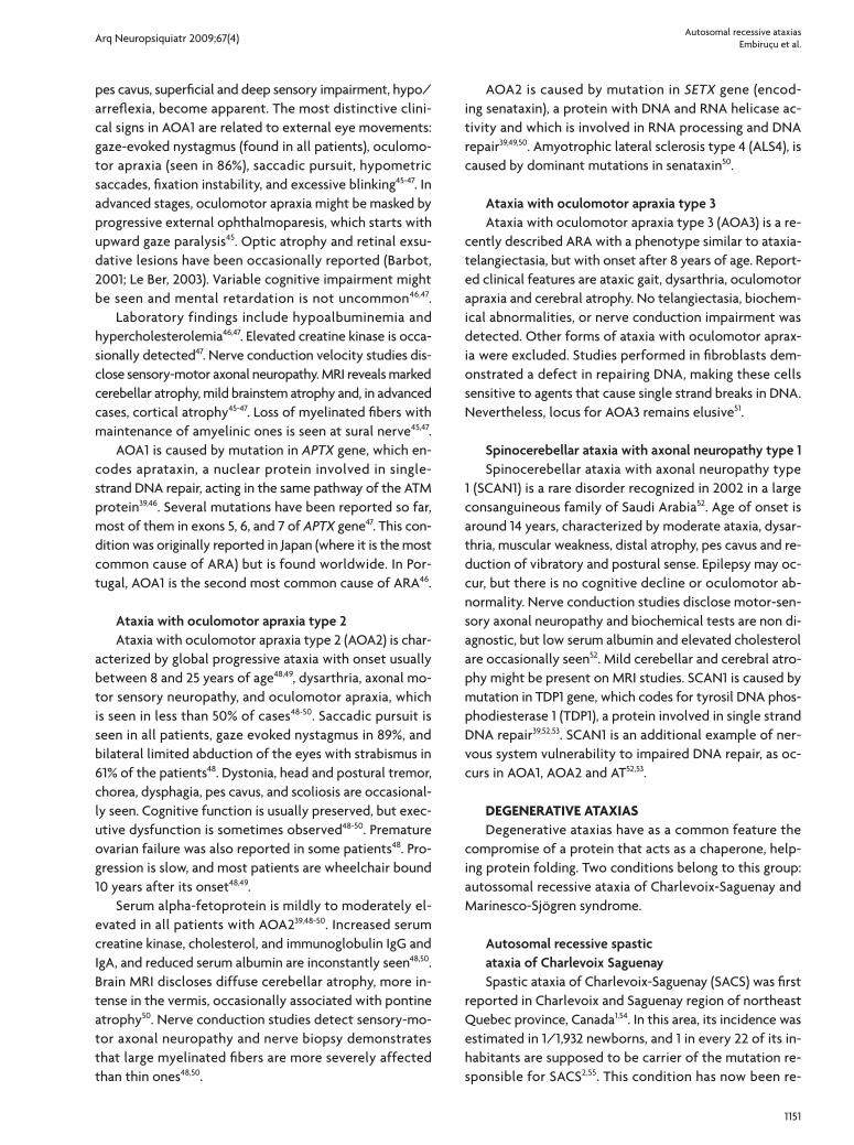
Tabl
e 3.
Clin
ical
cha
ract
eriz
atio
n an
d di
agno
stic
exa
ms
of th
e au
toso
mal
rece
ssiv
e at
axia
s.
CA
JSC
HVR
FAA
DCQ
APG
MIO
SCA
AVED
ABL
RDC
TXAT
AT li
keA
OA
1A
OA
2SC
AN
1SA
CS
MSS
Age
at
onse
t† <
1<
1<
1>
5<
20>
5<
22–
52<
20<
201–
36<
51–
7<
208–
2213
–15
1–10
< 20
Psyc
omot
or d
elay
++
+–
+–
––
––
––
–+
––
++
Hyp
oton
ia+
++
–+
–+
––
––
+–
+–
––
+
Hea
d ti
tuba
tion
––
––
––
–+
––
––
–+
+–
––
Ocu
lar a
lter
atio
ns
NN
, OA
, OS
RP, V
DS
N, G
IVD
, N,
OS,
ON
, O,
OS
VD, O
RP, V
DN
, RP
RP, N
, VD
CN
, OA
, O
S, S
N, O
A, S
N, O
A, O
, G
I, O
SN
, OA
, O
S, S
–N
, OS
N, S
, C
Sens
ory
neur
opat
hy
––
–+
–+
++
++
++
++
++
++
Dis
tal w
eakn
ess
––
–+
++
+–
––
++
++
++
++
Dee
p te
ndon
refle
xes
NL
NL
NL
or ↑
↓↓,
NL
or ↑
↓↓
↓↓
↓↓
or ↑
↓↓
or ↑
↓↓
↓↑
↓
Spas
tici
ty–
––
–+
––
––
–+
––
––
–+
+
Babi
nski
sig
n–
–+
++
––
++
–+
––
–+
–+
–
Pes
cavu
s–
–+
++
–+
+–
++
––
++
++
–
Extr
apyr
amid
al s
igns
––
––
–T,
MC
hT,
D–
–M
, DT,
D, C
hD
, Ch
T, D
, Ch
T, M
, D, C
h–
––
Psyc
hiat
ric p
robl
ems
–+
––
–+
––
––
+–
––
––
––
Cog
niti
ve im
pairm
ent
–+
+–
++
+–
––
+–
–+
––
++
Epile
psy
––
+–
++
+–
––
+–
––
–+
++
Hea
ring
loss
––
––
–+
+–
–+
––
––
––
–+
Car
diom
yopa
thy
––
–+
––
–+
++
+–
––
––
––
Dia
bete
s–
––
+–
––
––
––
+–
––
––
–
Radi
osen
siti
vy–
––
––
––
––
––
++
––
––
–
Skel
etal
def
orm
itie
s –
––
++
––
+–
+–
––
++
––
+
Rena
l fai
lure
–+
––
––
––
––
––
––
––
––
Live
r alt
erat
ions
–
+–
––
––
–+
–+
––
––
––
–
Ner
ve c
ondu
ctio
n st
udie
sN
LN
LN
LA
SN
LA
SMA
SA
SA
SD
SMA
SMA
SMA
SMA
SMA
SMA
SMA
SMD
SM
MRI
bra
inC
aC
aC
aN
LC
aC
a, W
MC
aN
LN
LN
LC
a, W
MC
aC
aC
aC
aC
aC
aC
a, W
M
MRI
spi
nal c
ord
atro
phy
––
–+
––
+–
––
––
–+
––
+–
Labo
rato
rial e
xam
s–
––
–co
eQ↓
mus
cle
––
vitE
↓lip
o↓vi
tE↓
phyt
an.
acid
↑ch
oles
tano
l ↑Ig
↓a–
feto
↑–
alb↓
chol
este
rol↑
a–et
o↑,
chol
este
rol↑
alb↓
chol
este
rol↑
–C
PK↑
Trea
tmen
t–
––
–+
––
++
++
––
––
––
–
–: a
bsen
t or
unc
omm
on; +
: may
be
pres
ent;
↑: in
crea
sed;
↓: r
educ
ed o
r ab
sent
; † mos
t fr
eque
ntly
ons
et a
ge; A
BL: a
beta
lipop
rote
inem
ia; A
CQD
: ata
xia
wit
h co
enzy
me
Q10
defi
cien
cy; A
OA
1: at
axia
wit
h oc
ulom
otor
apr
axia
typ
e1; A
OA
2: a
taxi
a w
ith
ocul
omot
or a
prax
ia ty
pe2;
APG
M: a
taxi
a w
ith
mut
atio
n in
pol
ymer
ase
gam
ma;
AS:
axo
nal s
enso
ry n
euro
path
y; A
SM: a
xona
l sen
sorio
mot
or n
euro
path
y; A
T: a
taxi
a te
lang
iect
asia
; ATl
ike:
ata
xia
tela
ngie
ctas
ia li
ke d
isor
der;
AVED
: ata
xia
wit
h vi
tam
in E
defi
cien
cy; C
: cat
arac
ts; C
a: c
ereb
ella
r at
roph
y; C
A: C
aym
an a
taxi
a; C
h: c
hore
a or
cho
reoa
thet
osis
; CH
VR: c
ereb
ella
r hy
popl
asia
ass
ocia
ted
to V
LDL
rece
ptor
; CTX
: cer
ebro
tend
inou
s xa
ntho
mat
osis
; D: d
ysto
nia;
DSM
: dem
yelin
atin
g se
nsor
iom
otor
neu
ropa
thy;
FA
: Frie
drei
ch a
taxi
a; G
I: ga
ze fi
xati
on in
stab
ility
; IO
SCA
: inf
anti
le o
nset
spi
noce
rebe
llar a
taxi
a; JS
: Jou
bert
synd
rom
e; M
: myo
clon
us; M
SS: M
arin
esco
Sjö
gren
synd
rom
e; N
: nys
tagm
us; N
L: n
orm
al; O
: oph
thal
mop
legi
a;
OA
: ocu
lom
otor
apr
axia
; OS:
ocu
lar
sacc
adic
impa
irm
ent;
RD: R
efsu
m d
isea
se; R
P: re
tini
tis
pigm
ento
sa; S
: str
abis
mus
; SA
CS:
spa
stic
ata
xia
of C
harl
evoi
x Sa
guen
ay; S
CA
N1:
spin
ocer
ebel
lar
atax
ia w
ith
axon
al n
euro
path
y ty
pe1;
T: t
rem
or; V
D:
visu
al d
efici
ency
; WM
: whi
te m
atte
r cha
nges
.
Arq Neuropsiquiatr 2009;67(4)
1153
Autosomal recessive ataxiasEmbiruçu et al.
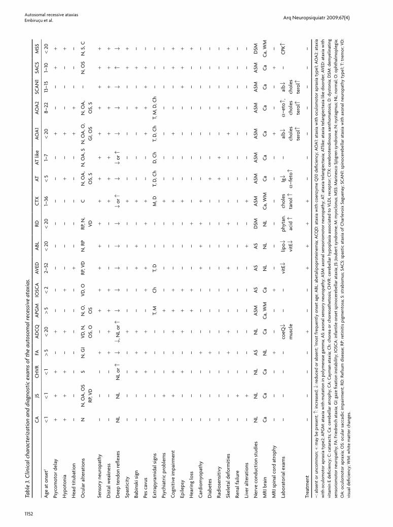
Family history Excluded AD or X-linked
JS CA, CHVR
AVED ABL
RD
FA and variants
AS ASM DSM Normal
APGMSACS*SCAN1
ATAT like†
AOA 1AOA 2CTX*
FAAVEDABL
IOSCA
ATAOA 1AOA 2
SCAN1 CQDAMSSRD
Measure serum cholesterol Measure CoQ 10 muscle
Increased Normal Low Normal
CTX Excluded CTX CQDA Excluded CQDA
Hereditary cerebellar ataxia, onset < 25 years
Brain MR images
ARA
Cerebellar atrophy
No progressive cerebellar
Present Absent
Molar tooth sign
No cerebellar atrophy
Measure total and fractions cholesterol, vit E serum
Only vit E ↓ Normal All ↓
Measure serum phytanic acid
Normal Increased
Molecular testing FRDA
PositiveNegativeMeasure serum albumin, a-fetoprotein, cholesterol
and immnunoglobulins
Progressive ataxia
Abnormal
SA, OA and/or N
Present Absent
Normal
Nerve conduction studies
*Spastic legs. ABL: abetalipoproteinemia; ACQD: ataxia with co-enzyme Q10 deficiency; AD: autossomal dominant; ADVE: atax-ia with vitamin E deficiency; AOA1: Ataxia with oculomotor apraxia type 1; AOA2: Ataxia with oculomotor apraxia type 2; APGM: atax-ia with mutation in polymerase gamma; ARA: autosomal recessive ataxia; AS: axonal sensory neuropathy; ASM: axonal sensoriomo-tor neuropathy; AT: ataxia-telangiectasia; ATlike: ataxia telangiecta-sia like disorder; CA: Cayman ataxia; CHVR: Cerebellar hypoplasia associated to VLDL receptor; CTX: cerebrotendinous xanthomato-sis; DSM: demyelinating sensoriomotor neuropathy; FA: Friedreich ataxia; IOSCA: Infantile onset spinocerebellar ataxia; JS: Joubert syndrome; MSS: Marinesco Sjögren syndrome; N: nystagmus; OA: oculomotor apraxia; RD: Refsum disease; SA: saccades abnormi-ties; SACS: Spastic ataxia of Charlevoix Saguenay; SCAN1: Spi-nocerebellar ataxia with axonal neuropathy type 1
Fig 4. Algorithm for diagnostic of the mains autosomal recessive ataxias.
Arq Neuropsiquiatr 2009;67(4)
1154
Autosomal recessive ataxiasEmbiruçu et al.
ported worldwide, but the largest series remains from Canada1,54-56.
Clinically, SACS is characterized by delay in acquiring independent walk, frequent falls and gait instability55. Dis-ease progression is slow and ataxic gait; dysarthria and spastic paraplegia are the major manifestations in the first two decades. Later, lower limb peripheral neuropathy can also be detected. As disease evolves, pyramidal signs can be masked by progression of peripheral neuropathy, with the exception of the Babinski sign, which is usually pres-ent even in later stages of disease. Distal atrophy, pes ca-vus, and hammer toes are commonly seen as disease ad-vances54-56. In some patients, fundoscopy discloses hyper-myelination of fibers radiating from optic disk and em-bedding the retinal vessels, a very peculiar finding. Hor-izontal nystagmus, saccadic alteration of smooth ocular pursuuit and miccional urgency might be present54,55. Mild mental retardation and cognitive decline were occasional-ly reported54. Patients usually become wheelchair bound after the 3rd or 4th decades of life and life expectancy is reduced as they become bedridden. During pregnancy, disease progression is apparently accelerated in affect-ed women55.
Nerve conduction velocity studies usually disclose an axonal neuropathy with mild demyelination, sensory fi-bers are more severely affected that motor fibers. The most consistent neuroimaging finding is cerebellar vermis atrophy, mostly from its superior portion54-56. Cervical and thoracic spinal cord thinning are occasionally reported55.
At an early stage, SACS can be misdiagnosed as cere-bral palsy55. Diagnosis is based on clinical manifestations and confirmed by mutation analysis of SACS gene located on 13q1154,56. Putative role of its product, sacsin, is to help protein folding, acting as a chaperone56. How sacsin de-ficiency causes neurodegeneration it is not known, but it has been reported to interact with Ataxin-1, the cause of Autosomal Dominant Spinocerebellar Ataxia type 156.
Marinesco-Sjögren syndrome Marinesco-Sjögren syndrome (MSS) is a rare, multisys-
tem disorder, characterized by congenital or early-onset cataracts, developmental delay, cerebellar ataxia and mild to severe mental retardation. Microcephaly, nystagmus, short stature, scoliosis, hypergonadotrophic hypogonad-ism and myopathy are common additional features57,58. Pe-ripheral neuropathy, deafness, optic atrophy, strabismus, spasticity and seizures might be present57. Disease pro-gression is slow and long survival can be expected2.
Brain MRI usually discloses cerebellar atrophy or hy-poplasia. Additional uncommon findings are cortical at-rophy and leucoencephalopathy. Serum creatine kinase is usually elevated and muscle biopsy show chronic myopa-thy with rimmed subsarcolemmal vacuoles57,58.
MSS is caused by mutations in SIL1 gene (encoding a nucleotide exchange factor for heat-shock protein 70 fam-ily member HSPA5). Heat-shock protein 70 family mem-bers are the highly conserved molecular chaperones that assist in stabilization and folding of newly synthesized polypeptides. Decrease of SIL1 gene product leads to a re-duction of protein synthesis in endoplasmic reticulum58,59.
conclusionDifferential diagnosis of ARA is a difficult task, as there
is a clear overlap of clinical manifestations among sever-al previously discussed conditions. Table 3 presents the main characteristics of each these disorders and Fig 4 is a proposed algorithm to help investigation of this group of diseases. Nevertheless, we should be aware that ataxia might be a symptom in many other progressive disorders, affecting primarily white matter (e.g., metachromatic leu-kodystrophy, leukoencephalopathy with vanishing white matter, Paelizeus-Merzbacher disease, X-linked adrenoleu-kodystrophy), neurons (e.g. neuronal lipofuscinosis ceroid, juvenile Tay-Sachs disease), or leading to a more wide-spread brain malformation or systemic manifestations, as it happens in pontocerebellar hypoplasia and congeni-tal disorders of glycosilation (CDG). Non progressive cer-ebellar symptoms are also prominent in ataxic cerebral palsy, an important differential diagnosis for early-onset ARA. It is also important to remind that all spinocerebel-lar ataxias (SCAs) inherited as a dominant trait are out of the scope of this review.
Currently, neuroimaging studies, especially brain MRI, are particularly important in ARA work-up and to help detection of cerebellar malformations and atrophy. Nor-mal MRI is expected in some disorders, as FA and AVED. Nerve conduction velocity studies and, in some cases, electromyography are useful for evaluation of some pa-tients, even in the absence of clinical signs of peripher-al neuropathy or myopathy. Elevation of serum alpha-fe-toprotein is characteristic of AT and AOA2. More specific biochemical tests, as determination of vitamin E and phy-tanic acid should not be neglected, once they can help the diagnosis of some treatable forms of ARA. Determi-nation of serum cholestanol and muscle CoQ10 are per-formed in a few specialized centers around the world but both CTX and ataxia with CoQ deficiency are potential-ly treatable.
Molecular analysis access is limited, but it is feasible for diseases as FA and, in lesser degree, AOA1 and AOA2. Many patients with putative ARA remain undiagnosed, and is expected that new forms of ARA will be recog-nized in the near future. The number of recessive ataxias is already high, and we will probably keep counting new arrivals for the forthcoming years.
Arq Neuropsiquiatr 2009;67(4)
1155
Autosomal recessive ataxiasEmbiruçu et al.
references 1. Fogel BL, Perlman S. Clinical features and molecular genetics of auto-
somal recessive cerebellar ataxias. Lancet Neurol 2007;6:245-257. 2. Palau F, Espinós C. Autosomal recessive cerebellar ataxias. Orphanet J
Rare Diseases 2006;1:47. 3. Bomar JM, Benke PJ, Slattery EL, et al. Mutations in a novel gene en-
coding a CRAL-TRIO domain cause human Cayman ataxia and atax-ia/dystonia in the jittery mouse. Nature Genet 2003;35:264-269.
4. Hayakawa Y, Itoh M, Yamada A, Mitsuda T, Nakagawa T. Expression and localization of Cayman ataxia-related protein, Caytaxin, is regu-lated in a developmental- and spatial-dependent manner. Brain Res 2007;1129:100-109.
5. Parisi MA, Doherty D, Chance PF, Glass IA. Joubert syndrome (and re-lated disorders) (OMIM213300). Eur J Hum Genet 2007;15:511-521.
6. Valente EM, Brancati F, Dallapiccola B. Genotypes and phenotypes of Joubert syndrome and related disorders. Eur J Med Genet 2008;51:1-23.
7. Zaki MS, Abdel-Aleem A, Abdel-Salam G, et al. The molar tooth sign. A new Joubert syndrome and related cerebellar disorders classification system tested in Egyptian families. Neurology 2008;70:556-565.
8. Glass HC, Boycott KM, Adams C, et al. Autosomal recessive cerebellar hypoplasia in the Hutterite population. Dev Med Child Neurol 2005; 47:691-695.
9. Türkmen S, Hoffmann K, Demirhan O, Aruoba D, Humphrey N, Mund-los S. Cerebellar hypoplasia, with quadrupedal locomotion, caused by mutations in the very low-density lipoprotein receptor gene. Eur J Hum Genet 2008;16:1070-1074.
10. Pandolfo M. Friedreich ataxia. Arch Neurol 2008;65:1296-1303.11. Hebert MD, Whittom AA. Gene-based approaches toward Friedreich
ataxia therapeutics. Cell Mol Life Sci 2007;64:3034-3043.12. Gonçalves S, Paupe V, Dassa EP, Rustin P. Deferiprone targets aconitase:
Implication for Friedreich’s ataxia. BMC Neurology 2008;8:1-4.13. Cooper JM, Korlipara LVP, Korlipara PE, Hart PE, Bradley JL, Schapi-
ra AHV. Coenzyme Q10 and vitamin E in Friedreich’s ataxia: predictor of efficacy of vitamin E and coenzyme Q10 therapy. Eur J Neurol 2008; 15:1371-1379.
14. Montero R, Pineda M, Aracil A, et al. Clinical, biochemical and molec-ular aspects of cerebellar ataxia and coenzyme Q10 deficiency. Cerebel-lum 2007;6:118-122.
15. Rötig A, Mollet J, Rio M, Munnich A. Infantile and pediatric quinine deficiency diseases. Mitochondrion 2007;7(Suppl):S112-S121.
16. Quinzii CM, López LC, Naini A, DiMauro S, Hirano M. Human CoQ10 deficiencies. BioFactors 2008;32:113-118.
17. Musumeci O, Naini A, Slonim AE, et al. Familial cerebellar ataxia with muscle coenzyme Q10 deficiency. Neurology 2001;56:849-855.
18. Milone M, Brunetti-Pierri N, Tang L-Y, et al. Sensory ataxic neuropa-thy with ophthalmoparesis caused by POLG mutations. Neuromuscul Disord 2008;18:626-632.
19. Van Goethem G, Luoma P, Rantamäki M, et al. POLG mutations in neu-rodegenerative disorders with ataxia but no muscle involvement. Neu-rology 2004;63:1251-1257.
20. Winterhun S, Ferrari G, He L, Taylor RW, et al. Autosomal recessive mi-tochondrial ataxic syndrome due to mitochondrial polymerase g muta-tions. Neurology 2005;64:1204-1208.
21. Lönnqvist T, Paetau A, Nikali K, von Boguslawski K, Pihko H. Infantile onset spinocerebellar ataxia with sensory neuropathy (IOSCA): neuro-pathological features. J Neurol Sci 1998;161:57-65.
22. Nikali K, Suomalainen A, Saharien J, et al. Infantile onset spinocerebel-lar ataxia is caused by recessive mutations in mitochondrial proteins Twinkle and Twinky. Hum Mol Genet 2005;14:2981-2990.
23. Koskinen T, Valanne L, Ketonen LM, Pihko H. Infantile-onset spinocere-bellar ataxia: MR and CT findings. Am J Neuroradiol 1995;16:1427-1433.
24. Cavalier L, Ouahchi K, Kayden HJ, et al. Ataxia with isolated vitamin E deficiency: heterogeneity of mutations and phenotypic variability in a large number of families. Am J Hum Genet 1998;62:301-310.
25. Marzouki N, Benomar A, Yahyaoui M, et al. Vitamin E deficiency atax-ia with (744del A) mutation on a-TTP gene: genetic and clinical pecu-liarities in Moroccan patients. Eur J Med Genet 2005;48:21-28.
26. Mariotti C, Gellera C, Rimoldi M, et al. Ataxia with isolated vitamin E
deficiency: neurological phenotype, clinical follow-up and novel mu-tations in TTPA gene in Italian families. Neurol Sci 2004;25:130-137.
27. Arita M, Sato Y, Miyata A, et al. Human a-tocoferol transfer protein: cDNA cloning, expression and chromosomal localization. Biochem J 1995;306:437-443.
28. Berriot-Varoqueaux N, Aggerbeck LP, Samson-Bouma ME, Wetterau JR. The role of the microsomal triglyceride transfer protein in abetali-poproteinemia. Annu Rev Nutr 2000;20:663-697.
29. Di Leo E, Magnolo L, Bertolotti M, et al. Variable phenotype expression of homozygous familial hypobetalipoproteinemia due to novel APOB gene mutations. Clin Genet 2008;74:267-273.
30. Zamel R, Khan R, Pollex RL, Hegele RA. Abetalipoproteinemia: two case reports and literature review. Orphanet J Rare Dis 2008;3:19.
31. Wanders RJA, Jansen GA, Skjeldal OH. Refsum disease, Peroxisomes and phytanic acid oxidation: a review. J Neuropathol Exp Neurol 2001; 60:1021-1031.
32. Jansen GA, Waterham HR, Wanders RJA. Molecular basis of refsum dis-ease: sequence variations in phytanoyl-CoA hydroxylase (PHYH) and the PTS2 receptor (PEX7). Hum Mutat 2004;23:209-218.
33. Wierzbicki AS, Lloyd MD, Schofield CJ, Feher MD, Gibberd FB. Ref-sum’s disease: a peroxisomal disorder affecting phytanic acid a-oxida-tion. J Neurochem 2002;80:727-735.
34. Federico A, Dotti MT. Cerebrotendinous xanthomatosis: clinical mani-festations, diagnostic criteria, pathogesnesis, and therapy. J Child Neurol 2003;18:633-638.
35. Barkhof F, Verrips A, Wesseling P, et al. Cerebrotendinous xanthoma-tosis: the spectrum of imaging findings and the correlation with neu-ropathogic findings. Neuroradiology 2000;217:869-876.
36. Gallus GN, Dotti MT, Federico A. Clinical and molecular diagnosis of cerebrotendinous xanthomatosis with a review of the mutations in the CYP27A1 gene. Neurol Sci 2006;27:143-149.
37. Berginer VM, Gross B, Morad K, et al. Chronic diarrhea and juvenile cataracts: think cerebrotendinous xanthomatosis and treat. Pediatrics 2009;123:143-147.
38. Chun HH, Gatti RA. Ataxia-telangiectasia, an evolving phenotype. DNA Repair 2004;3:1187-1196.
39. Taylor AMR, Byrd PJ. Molecular pathology of ataxia telangiectasia. J Clin Pathol 2005;58:1009-1015.
40. Perlman S, Becker-Catania S, Gatti RA. Ataxia-telangiectasia: diagno-sis and treatment. Semin Pediatr Neurol 2003;10:173-182.
41. Farr AK, Shalev B, Crawford TO, Lederman HM, Winkelstein JA, Rep-ka MX. Ocular manifestations of ataxia-telangiectasia. Am J Ophthal-mol 2002;134:891-896.
42. Fernet M, Gribaa M, Salih MAM, Seidahmed MZ, Hall J, Koenig M. Identification and functional consequences of a novel MRE11 muta-tion affecting 10 Saudi Arabian patients with the ataxia telangiectasia-like disorder. Hum Mol Genet 2005;14:307-318.
43. Delia D, Piane M, Buscemi G, et al. MRE11 mutations and impaired ATM-dependent responses in na Italian family with ataxia-telangiecta-sia-like disorder. Hum Mol Genet 2004;13:2155 -2163.
44. Taylor AMR, Groom A, Byrd PJ. Ataxia-telangiectasia-like disorder (ATLD)-its clinical presentation and molecular basis. DNA Repair 2004; 3:1219-1225.
45. Barbot C, Coutinho P, Chorão R, et al. Recessive ataxia with ocular apraxia: review of 22 portuguese patients. Arch Neurl 2001;58:201-205.
46. Le Ber I, Moreira M-C, Rivaud-Péchoux S, et al. Cerebellar ataxia with oculomotor apraxia type 1: clinical and genetic studies. Brain 2003;126: 2761-2772.
47. Ferrarini M, Squintani G, Cavallaro T, Ferrari S, Rizzuto N, Fabrizi GM. A novel mutation of aprataxin associated with ataxia ocular aprax-ia type 1: Phenotypical ang genotypical characterization. J Neurol Sci 2007;260:219-224.
48. Le Ber I, Bouslam N, Rivaud-Péchoux S, et al. Frequency and pheno-typic spectrum of ataxia with oculomotor apraxia 2: a clinical and ge-netic stydy in 18 patients. Brain 2004;127:759-767.
49. Tazir M, Ali-Pacha L, M’Zahem A, et al. Ataxia with ocolumotor aprax-ia type 2: a clinical and genetic study of 19 patients. J Neurol Sci 2009; 278:77-81.
Arq Neuropsiquiatr 2009;67(4)
1156
Autosomal recessive ataxiasEmbiruçu et al.
50. Duquette A, Roddier K, McNabb-Baltar J, et al. Muattions in senataxin responsible for Quebec cluster of ataxia with neuropathy. Ann Neurol 2005;57:408-414.
51. Gueven N, Chen P, Nakamura J, et al. A subgroup of spinocerebellar ataxias defective in DNA damage responses. Neuroscience 2007;145: 1418-1425.
52. Takashima H, Boerkoel CF, John J, et al. Mutation of TDP1, encoding a topoisomerase I-dependent DNA damage repair enzyme, in spinocer-ebellar ataxia with axonal neuropathy. Nat Genet 2002;32:267-272.
53. El-Khamisy SF, Caldecott KW. TDP1-dependent DNA single-strand break repair and neurodegeneration. Mutagenesis 2006;21:219-224.
54. Gücüyener K, Özgul K, Paternotte C, et al. Autosomal recessive spas-tic of Charlevoix-Saguenay in two unrelated Turkish families. Neuro-pediatrics 2001;32:142-146.
55. Bouchard J-P, Richter A, Mathieu J, et al. Autosomal recessive spas-tic ataxia of Charlevoix-Saguenay. Neuromuscul Disord 1998;8: 474-479.
56. Ouyang Y, Segers K, Bouquiaux O, et al. Novel SACS mutation in a Bel-gian family with sacsin-related ataxia. J Neurol Sci 20008;264:73-76.
57. Lagier-Tourenne C, Tranebjaerg L, Chaigne D, et al. Homozygosity mapping of Marinesco-Sjögren syndrome to 5q31. Eur J Hum Genet 2003;11:770-778.
58. Senderek J, Krieger M, Stendel C, et al. Mutations in SIL1 cause Mari-nesco-Sjögren syndrome, a cerebellar ataxia with cataract and myopa-thy. Nat Genet 2005;37 (12):1312-1314.
59. Anttonen A-K, Mahjneh I, Hämäläinen RH, et al. The gene disrupted in Marinesco-Sjögren syndrome encodes SIL1, an HSPA5 cochaperone. Nat Genet 2005;37:1309-1311.


![Autosomal recessive ichthyosis with limb reduction defect ... · including autosomal dominant, autosomal recessive and X-linked inheritance [1,2]. Associated cutaneous and extracutaneous](https://img.pdfslide.net/doc/110x75/5ec8c9b91adfdf12ab3e663c/autosomal-recessive-ichthyosis-with-limb-reduction-defect-including-autosomal.jpg)


