Embed Size (px)
Citation preview
Am. J. Hum. Genet. 67:1320–1326, 2000
1320
Report
Autosomal Recessive Cerebellar Ataxia with Oculomotor Apraxia(Ataxia-Telangiectasia–Like Syndrome) Is Linked to Chromosome 9q34Andrea H. Nemeth,1,2 Elena Bochukova,1 Eimear Dunne,1 Susan M. Huson,2 John Elston,3Mohammed A. Hannan,5 Matthew Jackson,4 Cyril J. Chapman,2 and A. Malcolm R. Taylor6
1Wellcome Trust Centre for Human Genetics, 2Department of Clinical Genetics, The Churchill Hospital, 3Department of Ophthalmology, TheOxford Eye Hospital, and 4Department of Neurology, The Radcliffe Infirmary, Oxford; 5Department of Biomedical Physics, King FaisalSpecialist Hospital and Research Centre, Riyadh, Saudi Arabia; and 6CRC Institute for Cancer Studies, University of Birmingham, Edgbaston,Birmingham, United Kingdom
Ataxia with oculomotor apraxia (ataxia-telangiectasia–like syndrome [AOA]; MIM 208920) is an autosomal re-cessive disorder characterized by ataxia, oculomotor apraxia, and choreoathetosis. These neurological featuresresemble those of ataxia-telangiectasia (AT), but in AOA there are none of the extraneurological features of AT,such as immunodeficiency, neoplasia, chromosomal instability, or sensitivity to ionizing radiation. It is unclearwhether these patients have a true disorder of chromosomal instability or a primary neurodegenerative syndrome,and it has not been possible to identify the defective gene in AOA, since the families have been too small for linkageanalysis. We have identified a new family with AOA, and we show that the patients have no evidence of chromosomalinstability or sensitivity to ionizing radiation, suggesting that AOA in this family is a true primary cerebellar ataxia.We have localized the disease gene, by linkage analysis and homozygosity mapping, to a 15.9-cM interval onchromosome 9q34. This work will ultimately allow the disease gene to be identified and its relevance to othertypes of autosomal recessive cerebellar ataxias to be determined.
Ataxia-telangiectasia (AT) is an autosomal recessive dis-order characterized by immunodeficiency and neoplasia,with laboratory evidence of chromosomal instability andsensitivity to ionizing radiation. The gene that causes AT,known as “ATM,” has been localized to chromosome11q22.3 and is a large gene with homology to cell-cyclecheckpoint genes in other organisms (Savitsky et al.1995). The neurological features of AT are characteristicand include early-onset cerebellar ataxia and oculomotorapraxia (slow or absent voluntary eye movements). Later,patients develop conjunctival telangiectases, a progressiveneurodegenerative syndrome, and sinopulmonary infec-tions and malignancies.
Some patients have been described who have “vari-ant” AT, with few or no clinical features other thanprogressive ataxia. These patients may be divided into
Received August 7, 2000; accepted for publication September 8,2000; electronically published October 5, 2000.
Address for correspondence and reprints: Dr. Andrea H. Nemeth,Wellcome Trust Centre for Human Genetics, Roosevelt Drive, OxfordOX3 7BN, UK. E-mail: [email protected]
� 2000 by The American Society of Human Genetics. All rights reserved.0002-9297/2000/6705-0030$02.00
three groups. The first have laboratory evidence of ATand have been shown to have mutations in the ATMgene on 11q22.3 (McConville et al. 1996; Gilad et al.1998). A second group of variant AT patients have aprogressive cerebellar degeneration (but no telangiec-tasia) and increased cellular and chromosomal instabil-ity, but they do not demonstrate linkage to chromosome11q22.3 (Hernandez et al. 1993). Recently, some of thislatter group of patients have been shown to have mu-tations in hMRE11, a double-strand break-repair genelocated on chromosome 11q21 (Stewart et al. 1999). Athird group of patients have a neurological disorder withan AT-like syndrome, without laboratory evidence of AT,and have normal or moderately impaired cellular andchromosomal stability. This third condition, which isreferred to as “ataxia with oculomotor apraxia” (AOA)or “AT-like syndrome” (ATL) (MIM 208920), was firstdescribed in 14 patients, from 10 families, 6 of whomwere noted to be consanguineous, suggesting autosomalrecessive inheritance (Aicardi et al. 1988). The age atonset of ataxia tended to be in childhood, and, in ad-dition to ataxia of gait, there were also some extrapyr-amidal movements such as chorea, athetosis, and dys-
Reports 1321
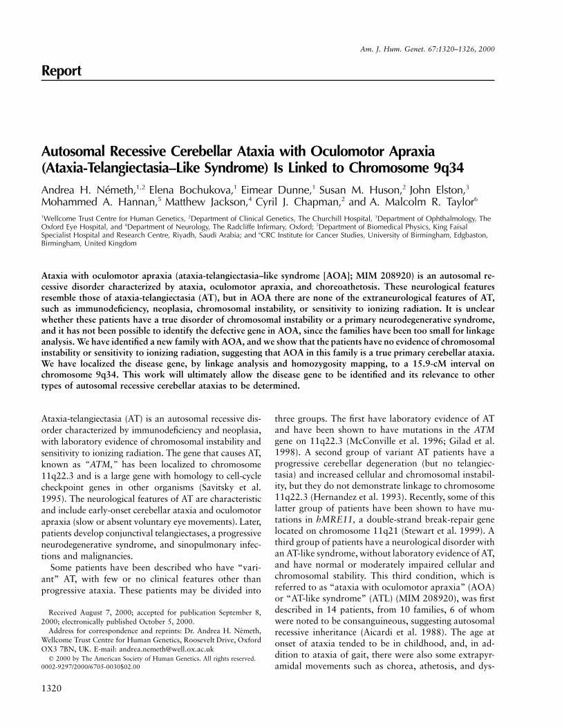
Figure 1 Pedigree with AOA, showing haplotypes along 9q34. Boxes around haplotypes indicate regions of homozygosity by descent inaffected individuals. Arrows indicate meiotic recombination events.
tonia. The patients all had severe oculomotor apraxia.Intellectual function was preserved in half the patientsand was mildly subnormal in the remainder. There wasminimal progression of symptoms after follow-up, al-though some patients were quite disabled. Results ofcomputed tomography (CT) were normal in six patientsand revealed mild cerebellar vermis atrophy in three.Three additional patients, from two consanguineousfamilies, were reported with AOA that presented in earlychildhood (Hannan et al. 1994; Gascon et al. 1995).Again, detailed investigations of sensitivity to acute andchronic ionizing radiation did not reveal abnormalitiestypical of AT. On the basis of lack of laboratory evidencefor chromosomal instability, it was suggested that AOAwas a neurological and genetic entity separate from AT(Aicardi et al. 1988; Hannan et al. 1994; Gascon et al.1995). However, the families reported were too small topermit linkage analysis, and there has been little progressin determining the genetic basis of AOA/ATL. Until nowit has been unclear whether these individuals have mu-
tations in ATM or in a related gene or are, in fact, ge-netically quite distinct.
We have recently identified a new family with AOAand here describe our results of a genome screen thathas identified linkage and homozygosity by descent. Ourresults clearly show that AOA in this family is distinctfrom other AT-like syndromes and will allow the iden-tification of the gene that is mutated in this form ofautosomal recessive cerebellar ataxia.
The family consists of five affected brothers and fourunaffected siblings (two males and two females) (fig. 1).Their parents originated from a small village in the Mirpurdistrict of Azad Kashmir, Pakistan, which comprised ap-proximately eight families in total, and the parents knewthat their great grandfathers were brothers. All the pa-tients developed ataxia during their late childhood orearly teens. The disease progressed into adulthood but,by the time the affected family members were in theirearly twenties, became relatively stable, and further pro-gression had been very slow. Because of their disability,
1322 Am. J. Hum. Genet. 67:1320–1326, 2000
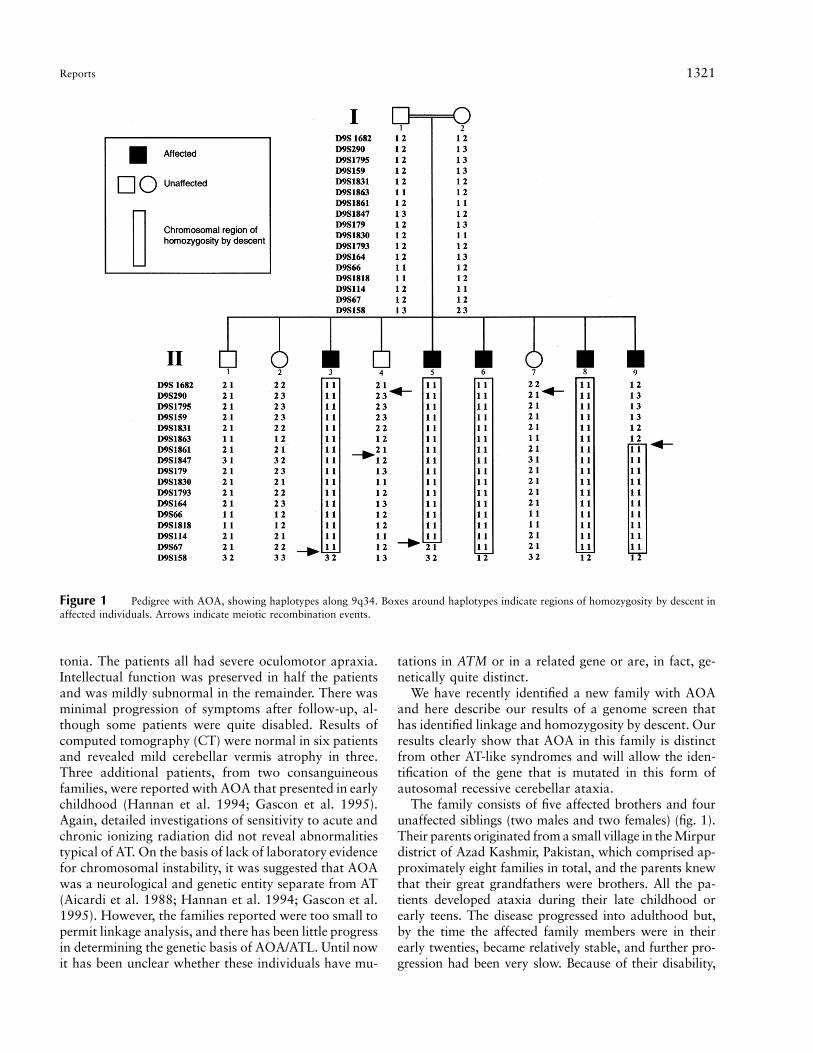
Figure 2 Expression of ATM, hRad50, Nbs1, and hMRE11 inpatient II:5.
Table 1
X-Ray–Induced Chromosome Damage in Lymphocytes from a Family with AOA afterExposure to 1.0-Gray X-Rays at G2
Individual
No. in Individual
CellsAnalyzed
ChromatidGaps
ChromatidBreaks
TriradialChromosomes
QuadriradialChromosomes
II:3 50 8 4 0 0II:5 50 10 1 0 0II:6 50 10 0 0 0Control 1 50 8 4 0 0Control 2 50 12 1 0 0Control 3 50 13 3 0 0AT patient 1 34 80 14 0 0AT patient 2 36 65 15 1 0
none of the patients were able to work. On examination,the patients had severe ataxia of gait with mild ataxia ofthe limbs and trunk. There was mild choreoathetosis withdystonic posturing during walking. The lower-limb deeptendon reflexes were absent and the plantars extensor. Themost severely affected brother had a flattened affect anda masklike face. Ophthalmological examination revealeda severe oculomotor apraxia with abnormal smooth pur-suit movements, absent optokinetic nystagmus, and aglobal saccade palsy, affecting the vertical more than thehorizontal movements. Brain CT of individual II:5 re-vealed prominence of several cerebellar sulci, suggestiveof cerebellar atrophy, and a large cysterna magna. Brainmagnetic resonance imaging of individual II:8 revealed noobvious abnormality. Nerve conduction studies revealedabsent sensory action potentials. None of the patients hadany dysmorphic features, developmental delay, or obviouslearning difficulties. There were no telangiectases and nostandard laboratory abnormalities of immune function.The patients were diagnosed initially, on the basis of theataxia and ophthalmological signs, as having AT. A di-agnosis of variant Friedreich ataxia was also considered.The patients’ cases were reviewed, the discrepancies be-tween their clinical phenotype and both AT and Friedreichataxia were noted, and further investigations wereperformed.
Chromosomes from lymphoblastoid cell lines (LCLs)were prepared according to standard protocols, and lym-phocytes were irradiated as described in Taylor et al.(1987). Whole-cell extracts were made from LCLs ofpatients and controls and were fractionated by sodiumdodecyl sulfate polyacrylamide gel electrophoresis.Western blot analysis was performed using antisera di-rected against the ATM, hMRE11, hRAD50, and Nbs1proteins (Stewart et al. 1999). The western blot was alsoprobed for actin, to standardize for protein loading.
After informed consent was obtained from all familymembers, blood samples were collected and DNA ex-tracted by use of the Nucleon Biosciences DNA-extrac-tion kit. Fluorescently labeled markers from the ABI
PRISM Linkage Mapping Set, version 2, were used forthe initial genome screen. Fine mapping was performedusing markers from Dib et al. (1996) and from the reporton the Sixth International Workshop on Chromosome9 (Chadwick et al. 1998) .
Before performing the genome screen, we performedpower calculations, using SLINK. This indicated powerto detect linkage with a maximum LOD score of 2.91on the assumption that AOA is a fully penetrant, au-tosomal recessive disorder with a disease frequency of1/100,000; allele frequencies for each marker were as-sumed to be equal. Homozygosity by descent was iden-tified by visual inspection. The fine-mapping genotypingdata were analyzed using SIMWALK2 (Sobel and Lange1996). This program allows extended pedigrees to beanalyzed, even in the presence of distant inbreedingloops and in the absence of genotyping information on
Reports 1323
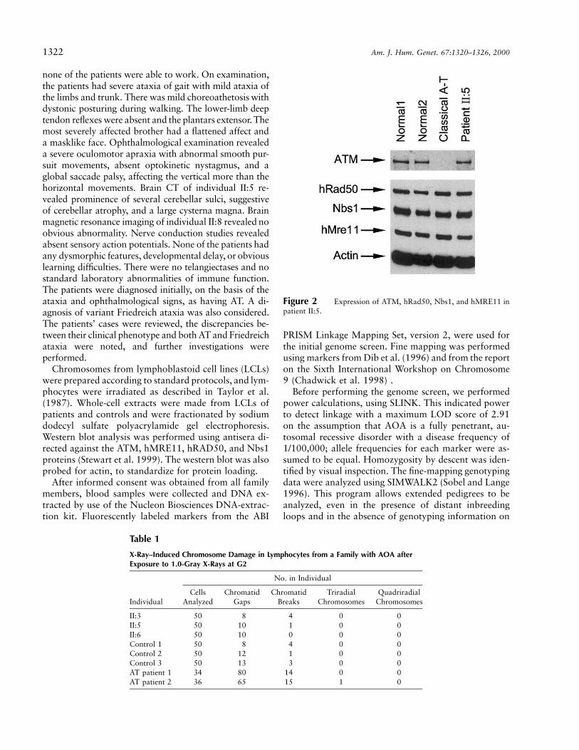
Figure 3 Multipoint location score in family with AOA
the distant ancestors. In the family we studied, the ped-igree must include the great-great-great grandparents ofthe patients (a total of 25 individuals and 17 meioses)for the multipoint computation, although informationis available only on the patients, their siblings, and theirparents. Such pedigrees are difficult to analyze usingother linkage programs, because there are an enormousnumber of underlying configurations that are consistentwith the available data and these greatly increase thecomputation time and memory required. SIMWALK2uses the Markov chain–Monte Carlo (MCMC) algo-rithm to analyze such large pedigrees, because it takesinto account the underlying configurations in proportionto their likelihood. Thus, a configuration that is theo-retically possible but highly unlikely (probably becauseof the large number of recombinations that the config-uration would require) will often not be considered. Be-cause SIMWALK2 uses the MCMC algorithm, its resultsare estimates and not exact. However, SIMWALK2’s es-timates have been found to be in excellent agreementwith the exact results when they are known—for ex-ample, in the analysis of the AT gene (Savitsky et al.1995; Sobel and Lange 1996). SIMWALK2 uses locationscores, which indicate the likelihood of several putativepositions for the trait locus, among the different markerloci. These location scores are directly comparable tomultipoint LOD scores and are presented in log10 units.
The results of the chromosome analysis revealed anabsence of translocation chromosomes in unirradiated
lymphocytes and no evidence of sensitivity to ionizingradiation (table 1). Western blotting revealed normal ex-pression of ATM, as well as hRad50, Nbs1, andhMRE11 (Petrini 1999) (fig. 2). Analysis with poly-morphic markers close to the ATM gene was not con-sistent with linkage to this locus. These investigationsdemonstrated that the patients did not have AT or arelated disorder of chromosomal instability. A diagnosisof Friedreich ataxia was considered; however, the pa-tients did not have the GAA expansion in the FRDAgene, which is found in the majority of patients withFriedreich ataxia (Campuzano et al. 1996). Linkageanalysis to the FRDA locus was performed in case thepatients had a rare homozygous mutation in the FRDAgene, but linkage to 9q13 was also excluded. Therefore,a genome screen was initiated.
The results of two-point linkage analysis revealed themaximum possible LOD score of 2.91, with markerD9S164, at zero recombination. Inspection of haplotypesrevealed homozygosity by descent in 10/10 chromosomesin all affected individuals at marker D9S164 and in 9/10chromosomes in all affected individuals at markerD9S290 (fig. 1). A total of 17 markers were informativeand used for fine mapping in distal 9q34. Multipoint link-age analysis of these markers using SIMWALK2 revealeda maximum location score of 5.32 at markers D9S1861,D9S179, D9S1793, and D9S164 (fig. 3). Examination ofhaplotypes in affected and unaffected siblings revealedcritical recombinations between D9S1863 and D9S1861,
1324 Am. J. Hum. Genet. 67:1320–1326, 2000
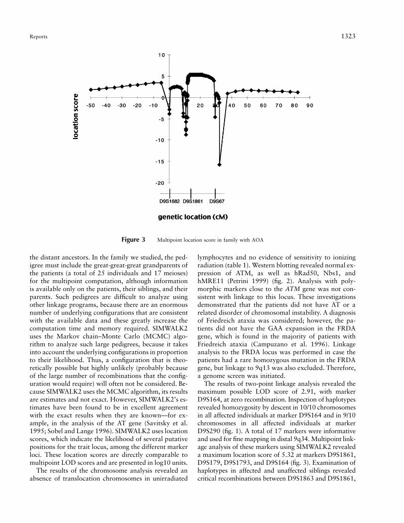
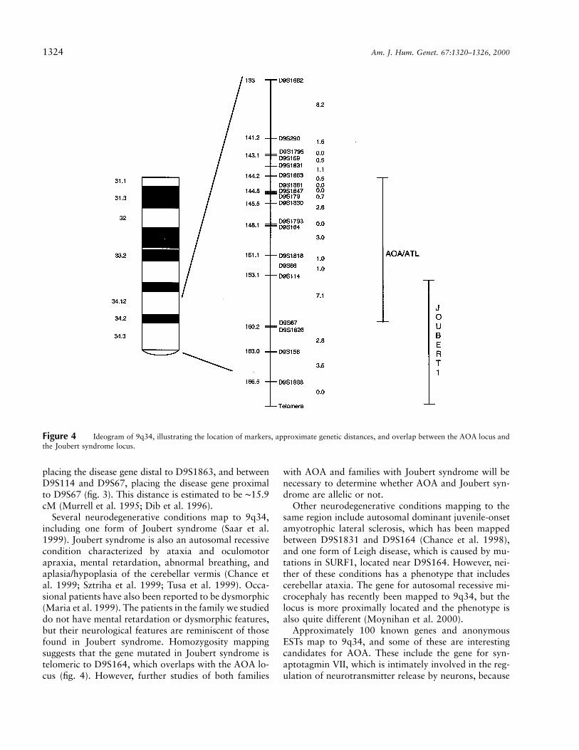
Figure 4 Ideogram of 9q34, illustrating the location of markers, approximate genetic distances, and overlap between the AOA locus andthe Joubert syndrome locus.
placing the disease gene distal to D9S1863, and betweenD9S114 and D9S67, placing the disease gene proximalto D9S67 (fig. 3). This distance is estimated to be ∼15.9cM (Murrell et al. 1995; Dib et al. 1996).
Several neurodegenerative conditions map to 9q34,including one form of Joubert syndrome (Saar et al.1999). Joubert syndrome is also an autosomal recessivecondition characterized by ataxia and oculomotorapraxia, mental retardation, abnormal breathing, andaplasia/hypoplasia of the cerebellar vermis (Chance etal. 1999; Sztriha et al. 1999; Tusa et al. 1999). Occa-sional patients have also been reported to be dysmorphic(Maria et al. 1999). The patients in the family we studieddo not have mental retardation or dysmorphic features,but their neurological features are reminiscent of thosefound in Joubert syndrome. Homozygosity mappingsuggests that the gene mutated in Joubert syndrome istelomeric to D9S164, which overlaps with the AOA lo-cus (fig. 4). However, further studies of both families
with AOA and families with Joubert syndrome will benecessary to determine whether AOA and Joubert syn-drome are allelic or not.
Other neurodegenerative conditions mapping to thesame region include autosomal dominant juvenile-onsetamyotrophic lateral sclerosis, which has been mappedbetween D9S1831 and D9S164 (Chance et al. 1998),and one form of Leigh disease, which is caused by mu-tations in SURF1, located near D9S164. However, nei-ther of these conditions has a phenotype that includescerebellar ataxia. The gene for autosomal recessive mi-crocephaly has recently been mapped to 9q34, but thelocus is more proximally located and the phenotype isalso quite different (Moynihan et al. 2000).
Approximately 100 known genes and anonymousESTs map to 9q34, and some of these are interestingcandidates for AOA. These include the gene for syn-aptotagmin VII, which is intimately involved in the reg-ulation of neurotransmitter release by neurons, because
Reports 1325
it is the major Ca2� sensor for Ca2�-regulated exocytosisat the synapse (reviewed by Schiavo and Stenbeck 1998).Another interesting candidate gene is Barhl1, a novelhomeobox gene that maps to 9q34 and has been sug-gested to be a candidate for Joubert syndrome, as it isexpressed in the developing nervous system (Bulfone etal. 2000). The expression pattern of this gene also makesit an interesting candidate for AOA.
In summary, we have identified a novel locus for aprimary autosomal recessive cerebellar ataxia whose fea-tures resemble those of AT. The clinical features in thefamily we studied are very similar to those of other pa-tients with AOA described in the literature (Aicardi etal. 1988; Hannan et al. 1994; Gascon et al. 1995). How-ever, it is not yet clear whether AOA is genetically ho-mogeneous; this will require the analysis of additionalfamilies. The identification of the disease gene will de-termine whether Joubert syndrome and AOA are allelicand whether other forms of autosomal recessive cere-bellar ataxia are caused by mutations at this locus. Itwill also allow further understanding of the geneticmechanisms underlying autosomal recessive cerebellarataxias.
Acknowledgments
We thank Family “A” for their continuing support of thisproject. We also thank several members of the Monaco lab-oratory, particularly Pat Scudder, Simon Fisher, Elena Maes-trini, and Alessandra Bolino, for valuable help with genotypingand linkage analysis. We thank Mrs. Anne Roberts for helpwith blood sample collection. We also thank Martin Farrallfor help with SIMWALK2, Dr. Alison Shaw for help with ge-ographical and pedigree details, and Dr. Samantha Knight forvaluable comments on the manuscript. This work was sup-ported by a Medical Research Council of Great Britain Cli-nician Scientist Award (to A.H.N.), by an International Fed-eration of Clinical Chemistry grant (to E.B.), and by the CancerResearch Campaign (to A.M.R.T.).
Electronic-Database Information
Accession number and URL for data in this article are asfollows:
Online Mendelian Inheritance in Man (OMIM), http://www.ncbi.nlm.nih.gov/Omim/ (for AOA [MIM 208920])
References
Aicardi J, Barbosa C, Andermann E, Andermann F, MorcosR, Ghanem Q, Fukuyama Y, Awaya Y, Moe P (1988) Ataxia-ocular motor apraxia: a syndrome mimicking ataxia-tel-angiectasia. Ann Neurol 24:497–502
Bulfone A, Menguzzato E, Broccoli V, Marchitiello A, GattusoC, Mariani M, Consalez GG, Martinez S, Ballabio A, Banfi
S (2000) Barhl1, a gene belonging to a new subfamily ofmammalian homeobox genes, is expressed in migrating neu-rons of the CNS. Hum Mol Genet 9:1443–1452
Campuzano V, Montermini L, Molto MD, Pianese L, CosseeM, Cavalcanti F, Monros E, et al (1996) Friedreich’s ataxia:autosomal recessive disease caused by an intronic GAA trip-let repeat expansion. Science 271:1423–1427
Chadwick B, Campbell L, Jackson C, Ozelius L, SlaugenhauptS, Stephenson D, Edwards J, Wiest J, Povey S. Report onthe Sixth International Workshop on Chromosome 9 (Den-ver; October 1998). Available at http://www.gene.ucl.ac.uk/chr9/report98.htm (accessed September 29, 2000)
Chance PF, Cavalier L, Satran D, Pellegrino JE, Koenig M,Dobyns WB (1999) Clinical nosologic and genetic aspectsof Joubert and related syndromes. J Child Neurol 14:660–666
Chance PF, Rabin BA, Ryan SG, Ding Y, Scavina M, Crain B,Griffin JW, Cornblath DR (1998) Linkage of the gene foran autosomal dominant form of juvenile amyotrophic lateralsclerosis to chromosome 9q34. Am J Hum Genet 62:633–640
Dib C, Faure S, Fizames C, Samson D, Drouot N, Vignal A,Millasseau P, Marc S, Hazan J, Seboun E, Lathrop M, Gya-pay G, Morissette J, Weissenbach J (1996) A comprehensivegenetic map of the human genome based on 5,264 micro-satellites. Nature 380:152–154
Gascon GG, Abdo N, Sigut D, Hemidan A, Hannan MA(1995) Ataxia-oculomotor apraxia syndrome. J Child Neu-rol 10:118–122
Gilad S, Chessa L, Khosravi R, Russell P, Galanty Y, Piane M,Gatti RA, Jorgensen TJ, Shiloh Y, Bar-Shira A (1998) Ge-notype-phenotype relationships in ataxia-telangiectasia andvariants. Am J Hum Genet 62:551–561
Hannan MA, Sigut D, Waghray M, Gascon GG (1994) Ataxia-ocular motor apraxia syndrome: an investigation of cellularradiosensitivity of patients and their families. J Med Genet31:953–956
Hassin-Baer S, Bar-Shira A, Gilad S, Galanty Y, Khosravi R,Lossos A, Giladi N, Weitz R, Ben-Zeev B, Goldhammer Y,Shiloh Y (1999) Absence of mutations in ATM, the generesponsible for ataxia telangiectasia in patients with cere-bellar ataxia. J Neurol 246:716–719
Hernandez D, McConville CM, Stacey M, Woods CG, BrownMM, Shutt P, Rysiecki G, Taylor AM (1993) A family show-ing no evidence of linkage between the ataxia telangiectasiagene and chromosome 11q22-23. J Med Genet 30:135–140
Maria BL, Boltshauser E, Palmer SC, Tran TX (1999) Clinicalfeatures and revised diagnostic criteria in Joubert syndrome.J Child Neurol 14:583–590
McConville CM, Stankovic T, Byrd PJ, McGuire GM, YaoQY, Lennox GG, Taylor MR (1996) Mutations associatedwith variant phenotypes in ataxia-telangiectasia. Am J HumGenet 59:320–330
Moynihan L, Jackson AP, Roberts E, Karbani G, Lewis I, CorryP, Turner G, Mueller RF, Lench NJ, Woods CG (2000) Athird novel locus for primary autosomal recessive micro-cephaly maps to chromosome 9q34. Am J Hum Genet 66:724–727
Murrell J, Trofatter J, Rutter M, Cutone S, Stotler C, Rutter
1326 Am. J. Hum. Genet. 67:1320–1326, 2000
J, Long K, Turner A, Deaven L, Buckler A, McCormick MK(1995) A 500-kilobase region containing the tuberous scle-rosis locus (TSC1) in a 1.7-megabase YAC and cosmid con-tig. Genomics 25:59–65
Petrini JHJ (1999) The mammalian Mre11-Rad50-Nbs1 pro-tein complex: integration of functions in the cellular DNA-damage response. Am J Hum Genet 64:1264–1269
Saar K, Al-Gazali L, Sztriha L, Rueschendorf F, Nur-E-KamalM, Reis A, Bayoumi R (1999) Homozygosity mapping infamilies with Joubert syndrome identifies a locus on chro-mosome 9q34.3 and evidence for genetic heterogeneity. AmJ Hum Genet 65:1666–1671
Savitsky K, Bar-Shira A, Gilad S, Rotman G, Ziv Y, VanagaiteL, Tagle DA, et al (1995) A single ataxia telangiectasia genewith a product similar to PI-3 kinase. Science 268:1749–1753
Schiavo G, Stenbeck G (1998) Molecular analysis of neuro-
transmitter release. Essays Biochem 33:29–41Sobel E, Lange K (1996) Descent graphs in pedigree analysis:
applications to haplotyping, location scores, and marker-sharing statistics. Am J Hum Genet 58:1323–1337
Stewart GS, Maser RS, Stankovic T, Bressan DA, Kaplan MI,Jaspers NG, Raams A, Byrd PJ, Petrini JH, Taylor AM(1999) The DNA double-strand break repair gene hMRE11is mutated in individuals with an ataxia-telangiectasia-likedisorder. Cell 99:577-87
Sztriha L, Al-Gazali LI, Aithala GR, Nork M (1999) Joubert’ssyndrome: new cases and review of clinicopathologic cor-relation. Pediatr Neurol 20:274–281
Taylor AMR, Flude E, Laher B, Stacey M, McKay E, Watt J,Green SH, Harding AE (1987) Variant forms of ataxia-tel-angiectasia. J Med Genet 24:669–677
Tusa RJ, Hove MT (1999) Ocular and oculomotor signs inJoubert syndrome. J Child Neurol 14:621–627
![Ataxia telangiectasia: a reviewataxia, oculocutaneous telangiectasia and frequent pul-monary infection [1]. Definition A-T is an autosomal recessive cerebellar ataxia [2]. It has also](https://img.pdfslide.net/doc/110x75/60c0274fdc425b48211dfd10/ataxia-telangiectasia-a-review-ataxia-oculocutaneous-telangiectasia-and-frequent.jpg)