Embed Size (px)
Citation preview
J Clin Pathol 1988;41:188-194
Autosomal recessive distal myopathyH ISAACS, MARGARET E BADENHORST, T WHISTLER
From the Neuromuscular Research Laboratory, Department of Physiology, Witwatersrand University MedicalSchool, Johannesburg, South Africa
SUMMARY Five patients with an autosomally recessively transmitted distal myopathy wereinvestigated. Of these, three belonged to a single sibship. Studies included electromyography,histological examination of muscle tissue, histochemical, electron microscopical, and biochemicalanalyses. One of the cases resembled the Nonaka form while the others were regarded as expressionsof the commoner variety of recessive distal dystrophy.
Distal myopathies, though common in Scandinaviancountries, are rare in other parts of the world. TheScandinavian distal myopathy described by Welanderin 1951 is an inherited disorder with autosomaldominance.' Rare examples of distal myopathy of thedominantly inherited non-Scandinavian variety havebeen reported from remarkably few other areas in theworld, and in these the disease seems to be lessbenign.23 Rare infantile forms of this disorder havealso been recorded."
Sporadic and autosomal recessive inherited distalmyopathies are extremely rare, but, unlike theautosomal dominant, have been reported from manyparts of the world.37"tO The similarity between thereported sporadic and recessive inherited cases makesit likely that the sporadic cases belong to the reces-sively inherited group where only one member of asibship has inherited the disease.
Because of the relative rarity of this disorder,detailed muscle and nerve studies were carried out on asporadic case of distal muscular myopathy, on sib-ships of three, and two patients with the disorder.
Case reports
CASE 1A 25 year old white man of Scottish extractioncomplained of thinning and weakness in both legswhich affected all the muscles below the level of theknees. There was no history of neuromuscular disor-der in the family, the patient was an only child, andhad been aware of the progressive nature of hisdisorder for two years.
Examination ofhim in the standing position showedmild lumbar lordosis; his feet were flat and the smallmuscles undetectable. The anterior, lateral, and pos-
Accepted for publication 18 August 1987
terior compartment muscles in both legs were abnor-mally thin and weak so that the patient was neitherable to stand on his toes or dorsiflex his toes or feet.Inversion and eversion of the ankles was absent. Fromthe knee level upwards he was well built, though theproximal muscles were slightly weaker than expected.Slight weakness was noted in the small muscles of thehands and in the dorsiflexors of the wrists, fingers, andthumb. The tendon reflexes with the exception of theankles were present and equal. All modalities ofsensation were normal and no other abnormalitieswere found. Both parents were examined and found tobe normal.
CASE 2A 35 year old woman gave a 10 year history ofprogressive weakness of the legs and feet. Her parentsand other members of the family, apart from onebrother and a sister, were healthy. She was one of sixsiblings, four of whom were male. Her elder sister,aged 46, had a similar complaint as did her brotheraged 40 years. Examination ofboth parents confirmedthat they were unsymptomatic and this was supportedby electromyography.
She showed severe weakness in the muscles affectingthe lower legs, particularly those below the level of theknees, while mild weakness was present in the moreproximal muscles. The small muscles ofthe hands werealso mildly affected. Reflexes were absent at the levelofthe anklejoint but were present and equal above thislevel.
Sensation and coordination was intact and thefacial musculature was not affected. Intellectualdevelopment was normal.
CASE 3The physical findings were similar to those of her
188
copyright. on 11 N
ovember 2018 by guest. P
rotected byhttp://jcp.bm
j.com/
J Clin P
athol: first published as 10.1136/jcp.41.2.188 on 1 February 1988. D
ownloaded from
Autosomal recessive distal myopathy
.9 ~~~ ~ ~ ~ ~ ~ ~ ~ ~~-0
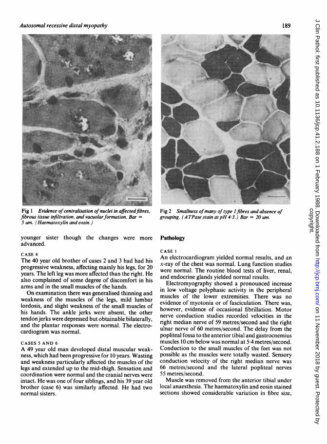
Fig 1 Evidence of centralisation ofnuclei in affectedfibres,fibrous tissue infiltration, and vacuolarformation. BarS urn. (Haematoxylin and eosin.)
younger sister though the changes were moreadvanced.
CASE 4The 40 year old brother of cases 2 and 3 had had hisprogressive weakness, affecting mainly his legs, for 20years. The left leg was more affected than the right. Healso complained of some degree of discomfort in hisarms and in the small muscles of the hands.On examination there was generalised thinning and
weakness of the muscles of the legs, mild lumbarlordosis, and slight weakness of the small muscles ofhis hands. The ankle jerks were absent, the othertendonjerks were depressed but obtainable bilaterally,and the plantar responses were normal. The electro-cardiogram was normal.
CASES 5 AND 6A 49 year old man developed distal muscular weak-ness, which had been progressive for 10 years. Wastingand weakness particularly affected the muscles of thelegs and extended up to the mid-thigh. Sensation andcoordination were normal and the cranial nerves wereintact. He was one of four siblings, and his 39 year oldbrother (case 6) was similarly affected. He had twonormal sisters.
I... _.
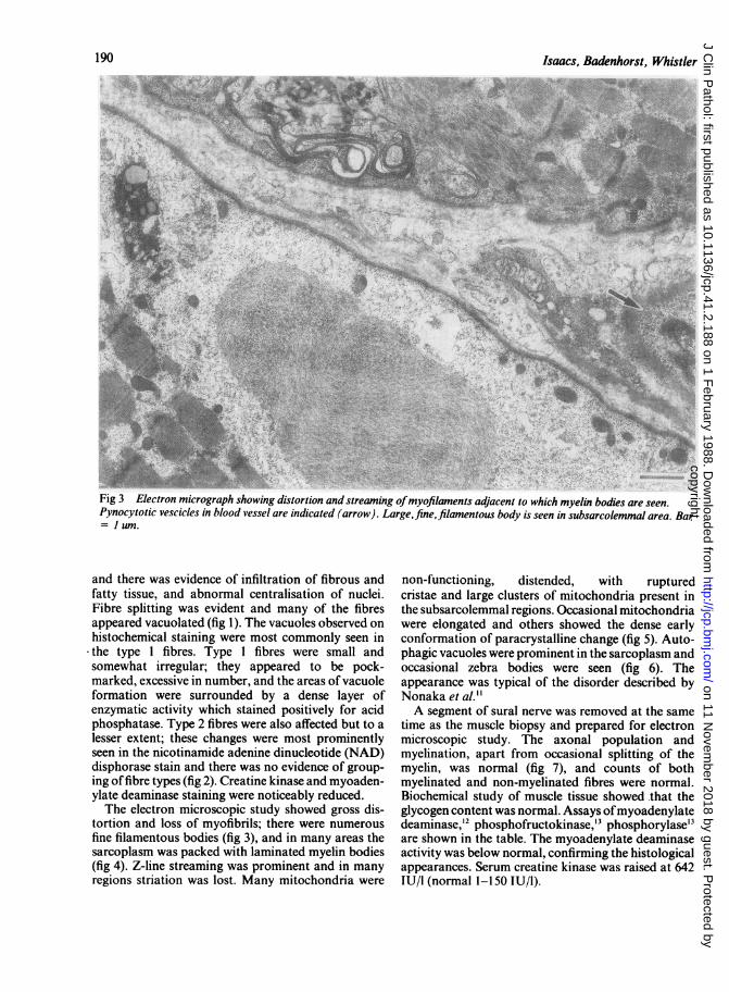
Fig 2 Smallness ofmany of type 1 fibres and absence ofgrouping. (A TPase stain at pH 4 3.) Bar = 20 um.
Pathology
CASE IAn electrocardiogram yielded normal results, and anx-ray of the chest was normal. Lung function studieswere normal. The routine blood tests of liver, renal,and endocrine glands yielded normal results.
Electromyography showed a pronounced increasein low voltage polyphasic activity in the peripheralmuscles of the lower extremities. There was noevidence of myotonia or of fasciculation. There was,however, evidence of occasional fibrillation. Motornerve conduction studies recorded velocities in theright median nerve of 59 metres/second and the rightulnar nerve of 60 metres/second. The delay from thepopliteal fossa to the anterior tibial and gastrocnemiusmuscles 10 cm below was normal at 5 4 metres/second.Conduction to the small muscles of the feet was notpossible as the muscles were totally wasted. Sensoryconduction velocity of the right median nerve was66 metres/second and the lateral popliteal nerves55 metres/second.Muscle was removed from the anterior tibial under
local anaesthesia. The haematoxylin and eosin stainedsections showed considerable variation in fibre size,
189
copyright. on 11 N
ovember 2018 by guest. P
rotected byhttp://jcp.bm
j.com/
J Clin P
athol: first published as 10.1136/jcp.41.2.188 on 1 February 1988. D
ownloaded from
Isaacs, Badenhorst, Whistler
Fig 3 Electron micrograph showing distortion and streaming ofmyofilaments adjacent to which myelin bodies are seen.Pynocytotic vescicles in blood vessel are indicated (arrow). Large,fine, filamentous body is seen in subsarcolemmal area. Bar= 1 um.
and there was evidence of infiltration of fibrous andfatty tissue, and abnormal centralisation of nuclei.Fibre splitting was evident and many of the fibresappeared vacuolated (fig 1). The vacuoles observed onhistochemical staining were most commonly seen in
-the type I fibres. Type 1 fibres were small andsomewhat irregular; they appeared to be pock-marked, excessive in number, and the areas of vacuoleformation were surrounded by a dense layer ofenzymatic activity which stained positively for acidphosphatase. Type 2 fibres were also affected but to alesser extent; these changes were most prominentlyseen in the nicotinamide adenine dinucleotide (NAD)disphorase stain and there was no evidence of group-ing offibre types (fig 2). Creatine kinase and myoaden-ylate deaminase staining were noticeably reduced.The electron microscopic study showed gross dis-
tortion and loss of myofibrils; there were numerousfine filamentous bodies (fig 3), and in many areas thesarcoplasm was packed with laminated myelin bodies(fig 4). Z-line streaming was prominent and in manyregions striation was lost. Many mitochondria were
non-functioning, distended, with rupturedcristae and large clusters of mitochondria present inthe subsarcolemmal regions. Occasional mitochondriawere elongated and others showed the dense earlyconformation of paracrystalline change (fig 5). Auto-phagic vacuoles were prominent in the sarcoplasm andoccasional zebra bodies were seen (fig 6). Theappearance was typical of the disorder described byNonaka et al."A segment of sural nerve was removed at the same
time as the muscle biopsy and prepared for electronmicroscopic study. The axonal population andmyelination, apart from occasional splitting of themyelin, was normal (fig 7), and counts of bothmyelinated and non-myelinated fibres were normal.Biochemical study of muscle tissue showed -that theglycogen content was normal. Assays ofmyoadenylatedeaminase,'2 phosphofructokinase,'3 phosphorylase'3are shown in the table. The myoadenylate deaminaseactivity was below normal, confirming the histologicalappearances. Serum creatine kinase was raised at 642IU/I (normal 1-150 IU/l).
190
,1:,.$:.f:.
copyright. on 11 N
ovember 2018 by guest. P
rotected byhttp://jcp.bm
j.com/
J Clin P
athol: first published as 10.1136/jcp.41.2.188 on 1 February 1988. D
ownloaded from
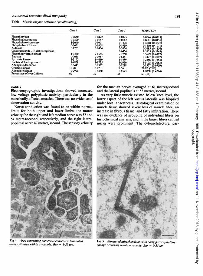
Table Muscle enzyme activities (glmol/min/mg)
Case I Case 2 Case 5 Mean (SD)
Phosphorylase 0 0650 0-0422 0 0325 0-0546 (0 0218)Phosphoglucomutase 0-0390 0-0187 0 0142 0-0601 (0.0155)Phosphohexoisomerase 1 3040 0990 07308 1-4608 (0-3321)Phosphofructokinase 0-0621 0-0308 0-1939 01810 (0-1071)Aldolase 0 1765 0-1454 0-2074 0-3685 (0-1156)Glyceraldehyde-3-P-dehydrogenase 06454 1-5555 (0 5243)Phosphoglycerate kinase 1-3430 2 1555 2-7760 3-5699 (06727)Enolase 07085 03957 08451 07477 (02087)Pyruvate kinase 3 3192 1-4619 1-1489 3-2356 (07853)Lactate dehydrogenase 14859 1-1725 15950 3-0105 (1-2063)Adenylate deaminae 0-0445 0-0332 0-1218 0-1577 (0-0759)Creatine kinase 1078 13 72 18 56 27-07 (786)Adenylate kinase 0 2990 0-8480 06573 1-2068 (04224)Percentage of type 2 fibres 60 32 55 60 (60)
CASE 2Electromyographic investigations showed increasedlow voltage polyphasic activity, particularly in themore badly affected muscles. There was no evidence ofdenervation activity.Nerve conduction was found to be within normal
limits for both upper and lower limbs; the motorvelocity for the right and left median nerve was 52 and54 metres/second, respectively, and the right lateralpopliteal nerve 47 metres/second. The sensory velocity
A~~~~~~~~~~~~
Fig 4 Area containing nwnerous concentric laminatedbodies situated within a vacuole. Bar =1 25 um.
for the median nerves averaged at 61 metres/secondand the lateral popliteals at 53 metres/second.As very little muscle existed below knee level, the
lower aspect of the left vastus lateralis was biopsiedunder local anaesthesia. Histological examination ofmuscle tissue showed severe loss of muscle fibre, anincrease in fibrous tissue, and fatty infiltration. Therewas no evidence of grouping of individual fibres onhistochemical analysis, and in the larger fibres centralnuclei were prominent. The cytoarchitecture, par-
Fig 5 Elongated mitochondrion with early paracrystallinechange occurring within a vacuole. Bar = 0 33 un.
Autosomal recessive distal myopathy 191
t
II
copyright. on 11 N
ovember 2018 by guest. P
rotected byhttp://jcp.bm
j.com/
J Clin P
athol: first published as 10.1136/jcp.41.2.188 on 1 February 1988. D
ownloaded from
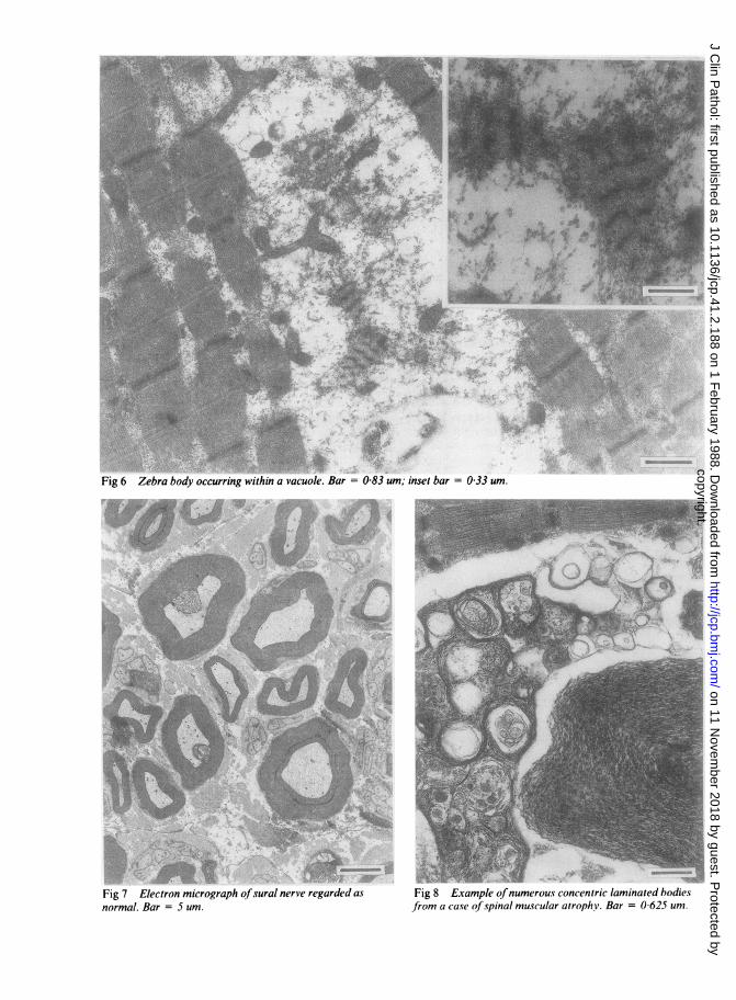
Fig 6 Zebra body occurring within a vacuole. Bar = 0-83 um; inset bar = 0-33 um.
Fig 7 Electron micrograph ofsural nerve regarded as
normal. Bar = 5 um.Fig 8 Example ofnumerous concentric laminated bodiesfrom a case of spinal muscular atrophy. Bar = 0 625 um.
copyright. on 11 N
ovember 2018 by guest. P
rotected byhttp://jcp.bm
j.com/
J Clin P
athol: first published as 10.1136/jcp.41.2.188 on 1 February 1988. D
ownloaded from
Autosomal recessive distal myopathy
ticularly of the smaller fibres, most of which were ofthe type I variety, was grossly disturbed, and subsar-colemmal collections of oxidative material wereobvious on electron microscopical examination.
Muscle enzyme activities were found to be generallynormal apart from a slight decrease in the content ofmyoadenylate deaminase (table). Creatine kinaseactivity was raised and fluctuated between 2800 IUand 3300 IU/l.
CASE 3The histochemical findings were similar, and it wasagain noted that most of the fibres were type 1 andthese displayed a severe disturbance of cytoarchitec-ture. On electron microscopy the changes were moresevere than those seen in case 2, and occasionalmitochondria showed evidence of condensation ofglycogen encased within lysosomes, while in otherareas paracrystalline inclusions were found. The suralnerve study was considered to be within normal limitson both physical appearance and on axonal counts.
Muscle enzyme activities were normal. Creatinekinase activity was 3800 IU/l.
CASE 4Electromyographic findings were similar to those ofcases 2 and 3: there was increased low voltagepolyphasic activity in the more severely affectedmuscles, and no evidence of denervation or spon-taneous activity was noted. The nerve conductionvelocity in the right lateral popliteal was 49-3 metres/second, left lateral popliteal 50 metres/second, and theright median 60 9 metres/second. Sensory conductionof the right median nerve was 62 metres/second, and64 metres/second in the right lateral popliteal.Muscle biopsy studies of the vastus lateralis and the
left sural nerve were similar to those of cases 2 and 3.Selective muscle enzyme activities were normal.Creatine kinase activity was 3150 IU/l. Mitochondrialkinetics were examined and gave normal results at allstages.
CASES 5 AND 6Electromyography showed evidence of low voltagepolyphasic activity, particularly in the more severelyaffected muscles. The right median nerve motor con-duction velocity was 57 metres/second, the rightlateral popliteal 44-4 metres/second, the left lateralpopliteal 50 metres/second. The sensory velocity forthe right median nerve was 66 metres/second and 50metres/second for the right lateral popliteal.A muscle biopsy specimen of the left vastus lateralis
showed fatty and fibrous tissue infiltration. There wasconsiderable variation in fibre size, and many of thefibres had centralised nuclei. Several fibres were ofhyaline appearance and showed evidence of splitting.
193
Histochemical analysis showed that both fibres wereaffected, type 1 fibres more severely so. No evidence offibre type grouping was noted. The muscle electronmicroscopical findings were similar to those of cases 2,3 and 4. The sural nerve study, which included electronmicroscopy, single fibre study, and nerve counts ofboth myelinated and non-myelinated fibres, yieldednormal results. The brother (case 6) was not biopsiedas his clinical picture was identical.
Discussion
In 1975 Miyoshi et al showed that a recessive form ofdistal dystrophy existed in Japan and documented 30such cases.'0 In 1981 Kuhn and Schroder publishedfindings on "a new type of distal myopathy in twobrothers".' These- patients with recessively inheriteddystrophy had distal leg muscle weakness beginning inearly adult life, associated with a pronounced increasein creatine kinase activity, and closely resembledsporadic cases described by other authors.3 8 9 14Edstrom et al described the presence of intermediateskeletin filaments in cases of distal myopathy,'5 and asimilar finding was recorded by Matsubara and Tan-abe.'6Nonaka et al described three cases from two families
of Japanese origin which appear to fit into theautosomal recessive variety and which were character-ised by the presence of rimmed vacuoles seen in themuscle biopsy sections, which possessed an acidphosphatase positive autophagic activity." Thesefindings are similar to those seen in case I and in othercases.'6 17 Sarcoplasmic filamentous inclusions as wellas concentric lamella bodies have also been noted, andit is thought that the continued destruction of themyofibrils is achieved by the activation of lysosomalproteolytic enzymes. Rimmed vacuoles, however,should not be regarded as a specific finding as they maybe seen in other myopathic disorders. Concentriclaminated bodies of various forms are common indistal myopathy, are a non-specific finding, and maybe seen on occasions in other diseases such as limbgirdle dystrophy, and particularly in spinal muscularatrophy (fig 8).
Scopetta et al suggested that the recessiveinheritance for distal dystrophy as opposed to thedominant inheritance exhibited the followingfeatures'8: (i) onset in early adult life; (ii) distal legmuscles affected first; (iii) moderate to severe increasein creatine kinase activity; (iv) electromyographicevidence of myopathic damage despite the presence offibrillation; and (v) a histological picture of a dys-trophic myopathy.Case 1 fits into the category of the sporadic distal
muscular dystrophy of the Nonaka variety; cases 2-6are regarded as examples of the commoner form of
copyright. on 11 N
ovember 2018 by guest. P
rotected byhttp://jcp.bm
j.com/
J Clin P
athol: first published as 10.1136/jcp.41.2.188 on 1 February 1988. D
ownloaded from
194 Isaacs, Badenhorst, Whistler
recessive distal dystrophy in South Africans ofEuropean extraction.We feel that these forms ofdistal myopathy, though
not seen often, have distinctive features which requiretheir inclusion into all modern classifications ofmuscular dystrophy.
We gratefully acknowledge the financial support oftheMedical Research Council of South Africa, theUniversity of the Witwatersrand, and the MuscularDystrophy Research Foundation of South Africa.
References
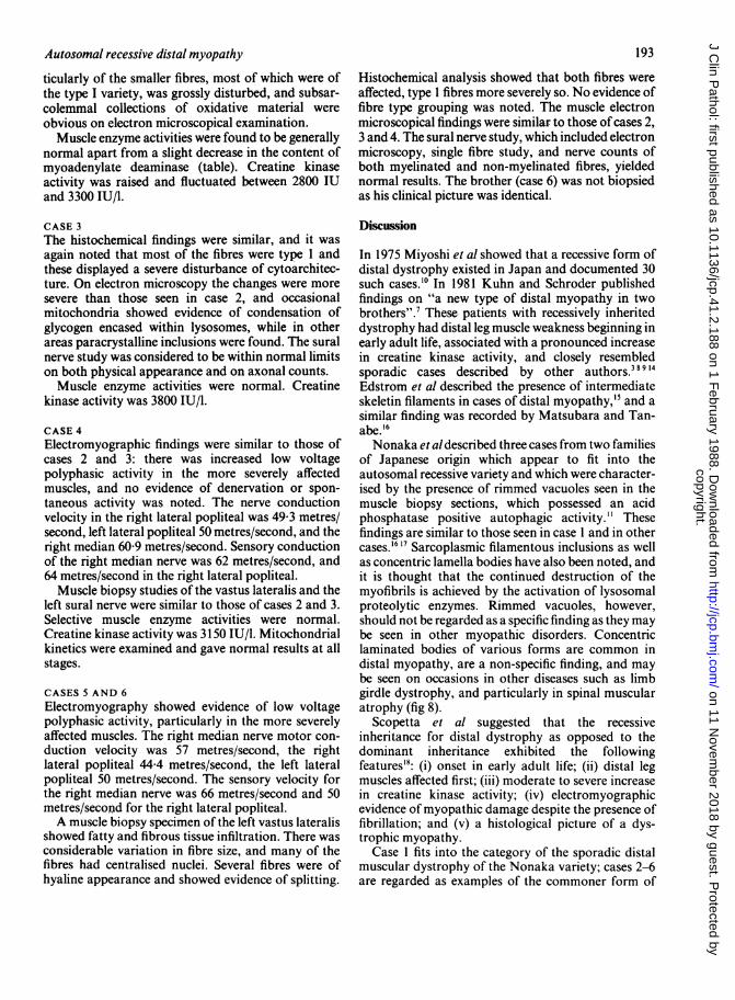
I Welander L. Myopathia distalis tarda hereditaria. Acta MedScand 1951;141(Suppl 265):1-124.
2 Markesbery WR, Griggs RC, Leach RP, Lapham LW. Late onsethereditary distal myopathy. Neurology 1974;24:127-34.
3 Markesbery WR, Griggs RC, Herr B. Distal myopathy: electronmicroscopic and histochemical studies. Neurology 1977;27:727-35.
4 Magee KR, De Jong RN, Arlor A. Hereditary distal myopathywith onset in infancy. Arch Neurol 1965;13:387-90.
5 Van der Doeas de Willebois AEM, Bethlem J, Meyer FH,Simons AJR. Distal myopathy with onset in early infancy.Neurology 1968;18:383-90.
6 Bautista JE, Rafel E, Castilla JM, Alberca R. Hereditary distalmyopathy with onset in early infancy - observation of a family.J Neurol Sci 1978;37:149-58.
7 Kuhn E, Schroder JM. A new type of distal myopathy in twobrothers. J Neurol 1981;226:181-5.
8 Kratz R, Brooke MH. Distal myopathies. In: Vinken PJ,Bruyn GW, eds. Handbook of clinical neurology. Vol 40.Amsterdam: North Holland, 1980:471-83.
9 Miller RG, Blank NK, Layzer RB. Sporadic distal myopathy withearly adult onset. Ann Neurol 1979;5:220-7.
10 Miyoshi K, Tada Y, Iwamasa M, et al. Autosomal recessive distalmyopathy observed characteristically in Japan. JapaneseJournal ofHuman Genetics 1975;20:62-3.
11 Nonaka I, Sunohara N, Ishiura S, Satoyoshi E. Familial distalmyopathy with rimmed vacuole and lamellar (myeloid) bodyformation. J Neurol Sci 1981 ;51:141-55.
12 DiMauro S, Miranda AF, Hays AP, et al. Myoadenylateeaminase deficiency. Muscle biopsy and muscle culture in apatient with gout. J Neurol Sci 1980;47:191-202.
13 Layzer RB, Rowland LP, Ranney HM. Muscle phospho-fructokinase deficiency. Arch Neurol 1967;17:512-23.
14 Vaccario ML, Scoppetta C, Bracaglia R, Uncini A. Sporadic distalmyopathy. J Neurol 1981;224:291-5.
15 Edstrom L, Thornell LE, Eriksson A. A new type of hereditarydistal myopathy with characteristic sarcoplasmic bodies andintermediate (skeletin) filaments. J Neurol Sci 1980;47:171-90.
16 Matsubara S, Tanabe H. Hereditary distal myopathy withfilamentous inclusions. Acia Neurol Scand 1982;65:363-8.
17 Kumamoto T, Fukuhara N, Nagashima M, Kanda T,Wakabayashi M. Distal myopathy: histochemical and ultra-structural studies. Arch Neurol 1982;39:367-71.
18 Scoppetta C, Vaccario ML, Casali C, Di Trapani G, Mennuni G.Distal muscular dystrophy with autosomal recessiveinheritance. Muscle and Nerve 1984;7:478-81.
Requests for reprints to: Dr H Isaacs, NMRL, Departmentof Physiology, University of the Witwatersrand MedicalSchool, Johannesburg, South Africa 2001. copyright.
on 11 Novem
ber 2018 by guest. Protected by
http://jcp.bmj.com
/J C
lin Pathol: first published as 10.1136/jcp.41.2.188 on 1 F
ebruary 1988. Dow
nloaded from