Embed Size (px)
Citation preview
8/6/2019 Avoiding Pitfalls in Trans Vaginal
http://slidepdf.com/reader/full/avoiding-pitfalls-in-trans-vaginal 1/17
A vo id in g Pitfallsin TransvaginalS o n o g r a ph y o f t h e
F e m a l e P e l v i sOksana H. Baltarowich, MD
a,*, Leslie M. Scoutt, MDb
At present, transvaginal sonography (TVS) is the
preferred method for sonographic evaluation of
the female pelvis. Although the transvaginal tech-
nique has numerous advantages in comparison
with transabdominal imaging of the pelvis and
allows for more accurate diagnosis as a result of
increased spatial and soft tissue resolution, one
must be aware of potential pitfalls in technique,
protocols, and interpretation of images that may
lead to errors in diagnosis and inappropriate treat-
ment of the patient. This article examines the pla-ces where pitfalls may occur while analyzing the
components of a TVS examination of the pelvis
in a stepwise method.
KNOW THE OB-GYN HISTORY
Several pitfalls can be avoided even before the
probe is placed into the vagina. Knowledge of the
patient’s clinical history is more important in
imaging the female pelvis than in most other ultra-
sonogaphic examinations because the appear-
ance of the normal anatomy and the type of
potential pathologic conditions vary depending on
the hormonal status of the patient. Before perform-
ing any pelvic sonogram the sonographer should,
at a minimum, know the patient’s age, parity, last
menstrual period, and hormonal status, particularly
infertility treatment, hormone replacement therapy,
and use of SERMs—selective estrogen receptor
modulators (tamoxifen used in the past, raloxifene
used most commonly at present). For example,
the list of differential diagnoses for a small round
cystic mass within a thickened endometrium is
very different for a woman of reproductive age
than for a postmenopausal woman on raloxifene
therapy ( Fig. 1 ). Similarly, a personal history of
certain types of malignancies makes a pelvic
mass more suspicious for recurrence or metas-tasis. Use of assisted reproductive techniques
alerts one to the increased risk of heterotopic preg-
nancies, which have become increasingly popular
over the years. At present, 1% to 3% of pregnan-
cies achieved following assisted reproduction
technology are heterotopic pregnancies.1
One should also be aware of special clinical
circumstances that might influence the sono-
graphic technique or protocol. Transvaginal evalu-
ation of cervical length is routinely conducted in
patients with preterm labor. However, in the case
of preterm rupture of the membranes, transperi-neal sonography may be the preferred method
for cervical evaluation2 because this approach
carries a lower potential risk of infection. If trans-
perineal sonography proves to be inadequate,
then TVS of the cervix may be required.
a Division of Diagnostic Ultrasound, Department of Radiology, Thomas Jefferson University, 132 South 10thStreet, Suite 796B Main Building, Philadelphia, PA 19107-5244, USAb
Ultrasound Service, Department of Radiology, Yale University School of Medicine, 333 Cedar Street, PO Box208042, New Haven, CT 06520-8042, USA* Corresponding author.E-mail address: [email protected]
KEYWORDS
Transvaginal sonography Transvaginal pelvic scanning Pitfalls Pelvic ultrasonography Ultrasonographic scanning techniques
Ultrasound Clin 5 (2010) 177–193doi:10.1016/j.cult.2010.03.0091556-858X/10/$ – see front matter ª 2010 Elsevier Inc. All rights reserved. u
l t r a s o u n d . t h
e c l i n i c s . c
o m
8/6/2019 Avoiding Pitfalls in Trans Vaginal
http://slidepdf.com/reader/full/avoiding-pitfalls-in-trans-vaginal 2/17
Listening to the patient and scanning over the
area where the patient experiences pain help to
avoid missing a diagnosis or performing an inap-
propriate or incomplete study. For example,
a patient who presents for pelvic sonography
with pain may actually be experiencing abdominal
wall pain from causes such as abdominal wall
endometriosis ( Fig. 2 ), malignant lymph node
metastasis, hernia, rectus sheath hematoma, or
abscess. Examination of the anterior abdominal
wall requires a high-frequency linear array trans-
ducer. The curved array probe typically used fortransabdominal scanning does not have adequate
near-field resolution to evaluate the anterior
abdominal wall, and the transvaginal probe does
not have adequate depth or a large enough field
of view to image the more distant anterior subcu-
taneous tissues. In another example, a patient pre-
senting with right-sided pelvic pain, on closer
questioning, may explain that her pain started in
the flank and settled in the right pelvis. Using this
information, transabdominal sonography (TAS) of
the kidneys and TVS with attention to the right ure-
terovesical junction (UVJ) becomes important to
assess for hydronephrosis and a right UVJ
calculus ( Fig. 3 ).3
One should always attempt to answer the clini-
cian’s question. If a good clinician feels a mass
on pelvic examination and it is not identified on
the initial sonogram, then one should be persis-
tent and continue searching the sonogram. Der-
moid cysts of the ovary and pedunculated
subserosal myomas, especially when they are
solitary, are notorious for having high miss rates
when evaluating the female pelvis withultrasonography.
PRESCAN TRANSABDOMINALLY
TAS offers a general pelvic overview, whereas
transvaginal scanning provides a focused or tar-
geted higher-resolution evaluation of pelvic struc-
tures. To avoid tunnel vision, one should first look
at the big picture. Although TVS has replaced TAS
as the primary method for evaluation of the female
pelvis because of the improved resolution affordedby the use of higher frequency transvaginal trans-
ducers, TAS is still considered as a complementary
imaging technique and is particularly important for
evaluation of the upper pelvis. However, a painful
and uncomfortably full bladder is no longer consid-
ered necessary for transabdominal evaluation of
the female pelvis, and although a partially full
bladder provides a more limited view of the pelvis,
in most cases it will be adequate to avoid diag-
nostic errors.4,5
At the authors’ institutions, investigation of the
female pelvis begins by a quick transabdominalprescan, which is performed irrespective of how
full the bladder is at the time of presentation.
Most women have a partially full bladder when
this limited transabdominal scan is performed.
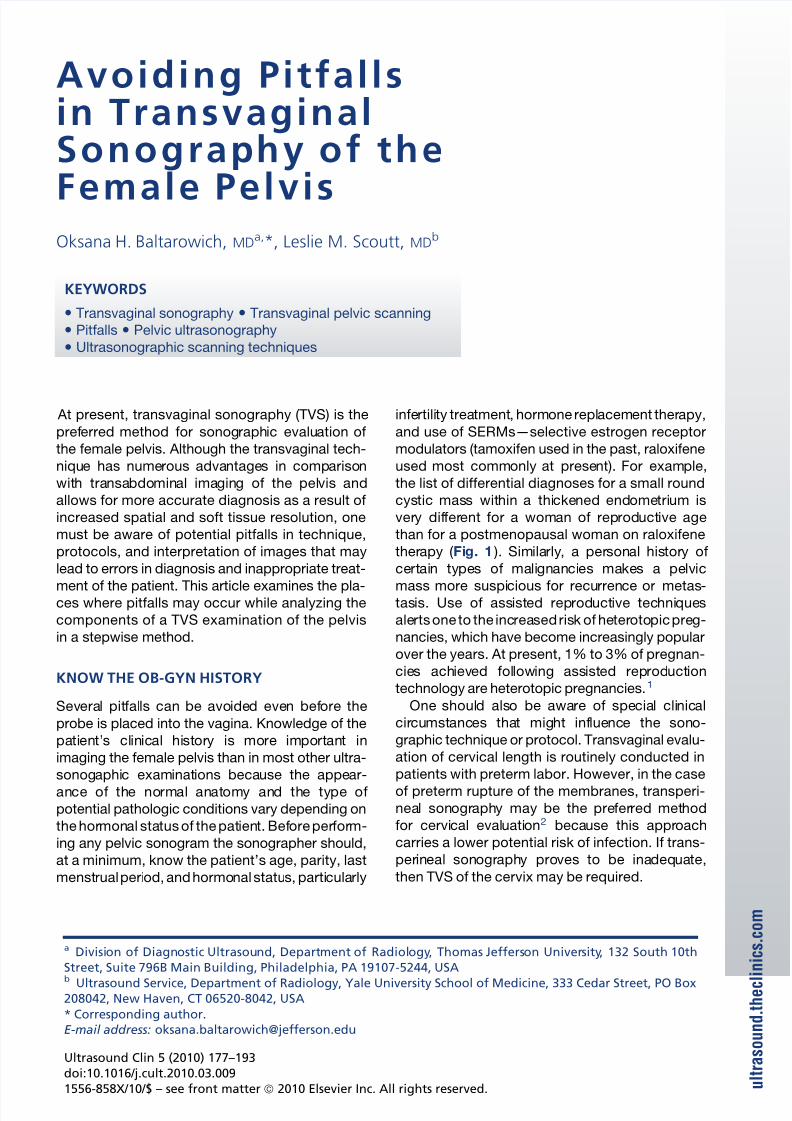
Fig. 1. Transvaginal sagittal sonogram demonstratesa thick endometrium measured as A, containinga small cystic area (arrow ). Without knowledge ofthe patient’s age and gynecologic history, the list ofdifferential diagnoses is long and varied. If the
patient is in child-bearing years, such an appearanceof endometrium could represent an early intrauterinepregnancy. But in this 65-year-old patient on SERM, itrepresented cystic change in a surgically proven endo-metrial polyp.
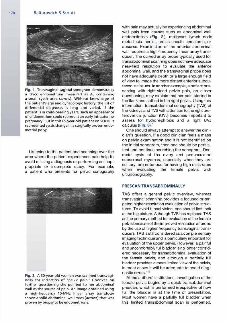
Fig. 2. A 30-year-old woman was scanned transvagi-nally for indication of ‘‘pelvic pain.’’ However, onfurther questioning she pointed to her abdominalwall as the source of pain. An image obtained usinga high-frequency 10-MHz linear array transducershows a solid abdominal wall mass (arrows) that wasproven by biopsy to be endometriosis.
Baltarowich & Scoutt178
8/6/2019 Avoiding Pitfalls in Trans Vaginal
http://slidepdf.com/reader/full/avoiding-pitfalls-in-trans-vaginal 3/17
The patient is then asked to void completely and
the pelvis is subsequently scanned transvaginally.
In a study of 206 patients reported by Benacerraf
and coworkers,5 TVS alone was thought to be
sufficient to visualize all the findings in 83.5% of
patients. An additional 15% of patients required
TAS without a full bladder with only 1.5% of
patients requiring TAS examination with
a completely full bladder to identify all the clinically
important findings.
The transabdominal prescan with an empty or
partially full bladder does not offer an optimal
evaluation of pelvic structures, but is important
because it actually directs the transvaginal ultra-
sound (which way the uterus is pointing),
provides an estimation of the uterine size (best
way to measure the uterus), and is the best
way to search for masses above the fundus of
the uterus such as a pedunculated myoma
( Fig. 4 ) or a large ovarian mass, and to evaluate
for intra-abdominal fluid.6–11 Some ectopic preg-
nancies are seen only on TAS examination. In
addition, the transverse transabdominal scan of
an anteverted uterus may show a true coronal
section of the uterus providing the best visuali-
zation of the fundal contour, which is important
when there is a question of a congenital
anomaly.
If patients are scanned only transvaginally, then
one is likely to miss, incompletely visualize, or
misinterpret a large pelvic mass, incompletely
scan a large uterus, miss a pedunculated subser-
osal myoma, miss an ectopic or intrauterine preg-
nancy in a high pelvic location, misinterpret some
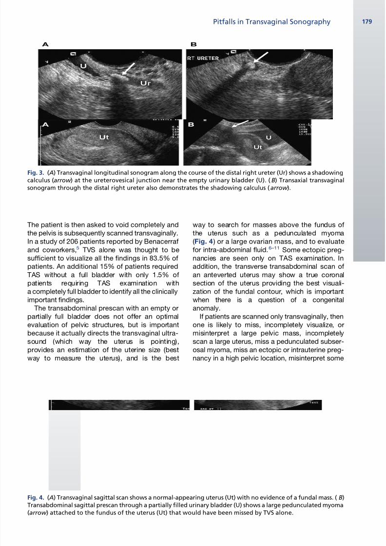
Fig. 3. ( A) Transvaginal longitudinal sonogram along the course of the distal right ureter (Ur) shows a shadowingcalculus (arrow ) at the ureterovesical junction near the empty urinary bladder (U). ( B) Transaxial transvaginalsonogram through the distal right ureter also demonstrates the shadowing calculus (arrow ).
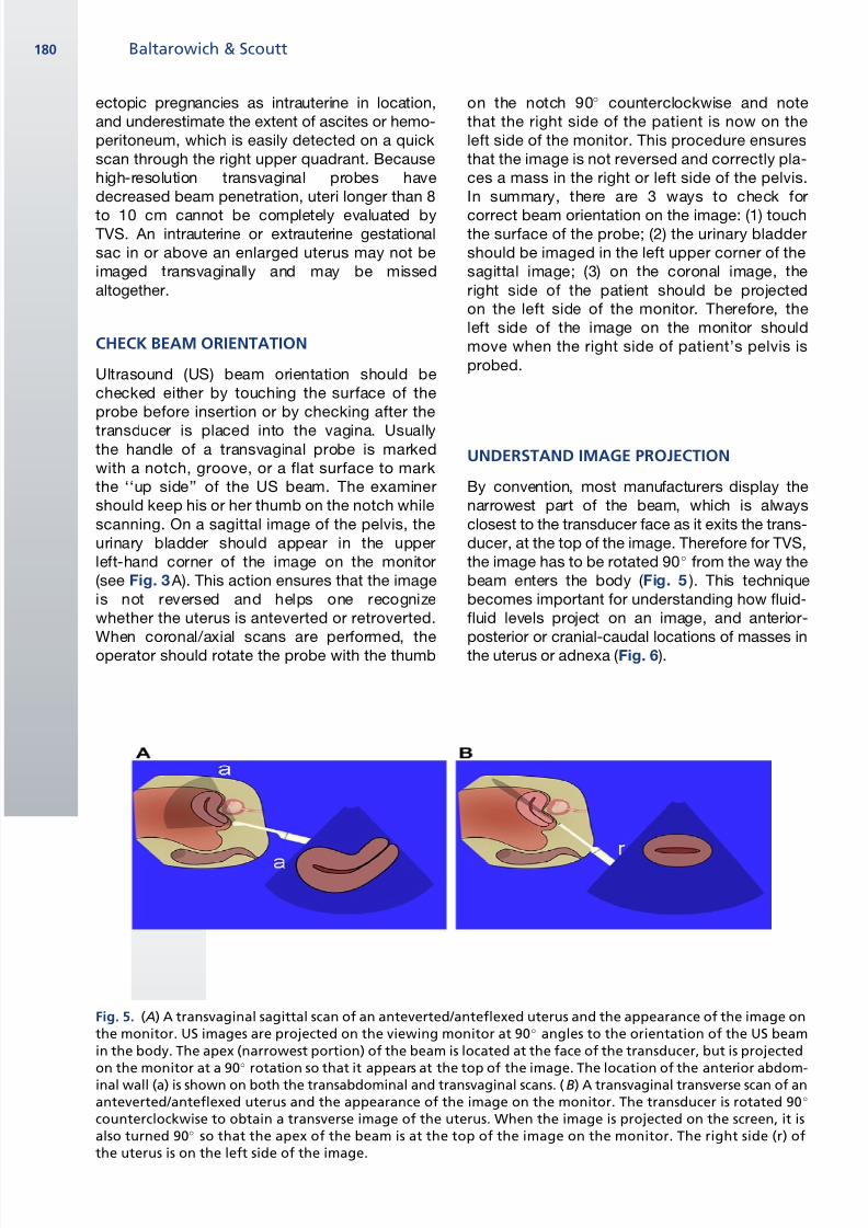
Fig. 4. ( A) Transvaginal sagittal scan shows a normal-appearing uterus (Ut) with no evidence of a fundal mass. ( B)Transabdominal sagittal prescan through a partially filled urinary bladder (U) shows a large pedunculated myoma(arrow ) attached to the fundus of the uterus (Ut) that would have been missed by TVS alone.
Pitfalls in Transvaginal Sonography 179
8/6/2019 Avoiding Pitfalls in Trans Vaginal
http://slidepdf.com/reader/full/avoiding-pitfalls-in-trans-vaginal 4/17
ectopic pregnancies as intrauterine in location,
and underestimate the extent of ascites or hemo-
peritoneum, which is easily detected on a quick
scan through the right upper quadrant. Because
high-resolution transvaginal probes have
decreased beam penetration, uteri longer than 8
to 10 cm cannot be completely evaluated byTVS. An intrauterine or extrauterine gestational
sac in or above an enlarged uterus may not be
imaged transvaginally and may be missed
altogether.
CHECK BEAM ORIENTATION
Ultrasound (US) beam orientation should be
checked either by touching the surface of the
probe before insertion or by checking after the
transducer is placed into the vagina. Usuallythe handle of a transvaginal probe is marked
with a notch, groove, or a flat surface to mark
the ‘‘up side’’ of the US beam. The examiner
should keep his or her thumb on the notch while
scanning. On a sagittal image of the pelvis, the
urinary bladder should appear in the upper
left-hand corner of the image on the monitor
(see Fig. 3 A). This action ensures that the image
is not reversed and helps one recognize
whether the uterus is anteverted or retroverted.
When coronal/axial scans are performed, the
operator should rotate the probe with the thumb
on the notch 90 counterclockwise and note
that the right side of the patient is now on the
left side of the monitor. This procedure ensures
that the image is not reversed and correctly pla-
ces a mass in the right or left side of the pelvis.
In summary, there are 3 ways to check for
correct beam orientation on the image: (1) touchthe surface of the probe; (2) the urinary bladder
should be imaged in the left upper corner of the
sagittal image; (3) on the coronal image, the
right side of the patient should be projected
on the left side of the monitor. Therefore, the
left side of the image on the monitor should
move when the right side of patient’s pelvis is
probed.
UNDERSTAND IMAGE PROJECTION
By convention, most manufacturers display the
narrowest part of the beam, which is always
closest to the transducer face as it exits the trans-
ducer, at the top of the image. Therefore for TVS,
the image has to be rotated 90 from the way the
beam enters the body ( Fig. 5 ). This technique
becomes important for understanding how fluid-
fluid levels project on an image, and anterior-
posterior or cranial-caudal locations of masses in
the uterus or adnexa ( Fig. 6 ).
Fig. 5. ( A) A transvaginal sagittal scan of an anteverted/anteflexed uterus and the appearance of the image onthe monitor. US images are projected on the viewing monitor at 90 angles to the orientation of the US beamin the body. The apex (narrowest portion) of the beam is located at the face of the transducer, but is projectedon the monitor at a 90 rotation so that it appears at the top of the image. The location of the anterior abdom-inal wall (a) is shown on both the transabdominal and transvaginal scans. (B) A transvaginal transverse scan of ananteverted/anteflexed uterus and the appearance of the image on the monitor. The transducer is rotated 90
counterclockwise to obtain a transverse image of the uterus. When the image is projected on the screen, it isalso turned 90 so that the apex of the beam is at the top of the image on the monitor. The right side (r) ofthe uterus is on the left side of the image.
Baltarowich & Scoutt180
8/6/2019 Avoiding Pitfalls in Trans Vaginal
http://slidepdf.com/reader/full/avoiding-pitfalls-in-trans-vaginal 5/17
DOCUMENT THE URINARY BLADDER
Another pitfall is forgetting to look at the urinarybladder before scanning the reproductive organs.
Because the patient is asked to void before the
transvaginal scan, the bladder is typically
collapsed and small, and therefore may be hard
to see. To find the empty bladder, one must delib-
erately angle the probe anteriorly without exerting
pressure. Too much pressure on the bladder and
urethra is uncomfortable for the patient and may
also completely appose the bladder walls, thusmaking the bladder unrecognizable.
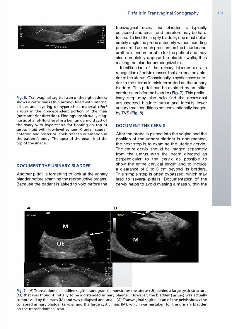
Identification of the urinary bladder aids in
recognition of pelvic masses that are located ante-
rior to the uterus. Occasionally a cystic mass ante-
rior to the uterus is misinterpreted as the urinary
bladder. This pitfall can be avoided by an initial
careful search for the bladder ( Fig. 7 ). This prelim-
inary step may also help find the occasional
unsuspected bladder tumor and identify lower
urinary tract conditions not conventionally imaged
by TVS ( Fig. 8 ).
DOCUMENT THE CERVIX
After the probe is placed into the vagina and the
position of the urinary bladder is documented,
the next step is to examine the uterine cervix.
The entire cervix should be imaged separately
from the uterus with the beam directed as
perpendicular to the cervix as possible to
show the entire cervical length and to include
a clearance of 2 to 3 cm beyond its borders.
This simple step is often bypassed, which maylead to several pitfalls. Documentation of the
cervix helps to avoid missing a mass within the
Fig. 6. Transvaginal sagittal scan of the right adnexashows a cystic mass (thin arrows) filled with internalechoes and layering of hyperechoic material (thick arrow ) in the nondependent portion of the mass
(note anterior direction). Findings are virtually diag-nostic of a fat-fluid level in a benign dermoid cyst ofthe ovary with hyperechoic fat floating on top ofserous fluid with low-level echoes. Cranial, caudal,anterior, and posterior labels refer to orientation inthe patient’s body. The apex of the beam is at thetop of the image.
Fig. 7. ( A) Transabdominal midline sagittal sonogram demonstrates the uterus (Ut) behind a large cystic structure(M) that was thought initially to be a distended urinary bladder. However, the bladder ( arrow ) was actuallycompressed by the mass (M) and was collapsed and small. (B) Transvaginal sagittal scan of the pelvis shows thecollapsed urinary bladder (arrow ) and the large cystic mass (M), which was mistaken for the urinary bladderon the transabdominal scan.
Pitfalls in Transvaginal Sonography 181
8/6/2019 Avoiding Pitfalls in Trans Vaginal
http://slidepdf.com/reader/full/avoiding-pitfalls-in-trans-vaginal 6/17
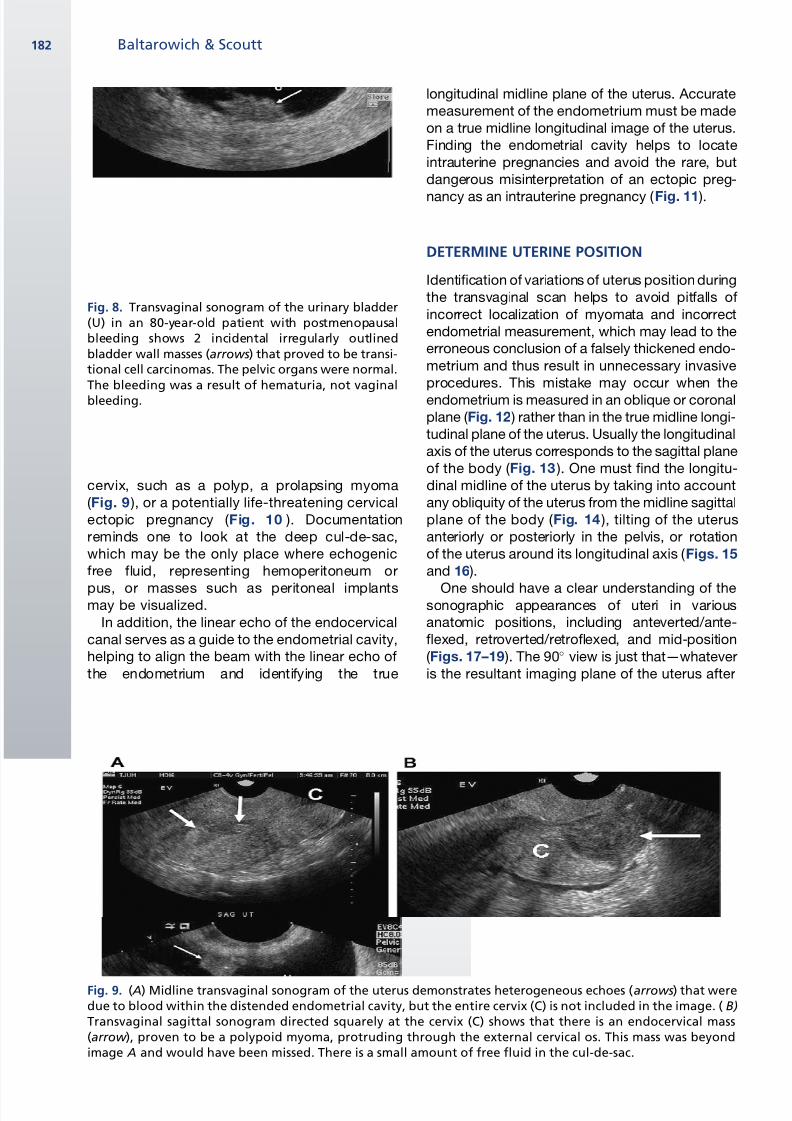
cervix, such as a polyp, a prolapsing myoma
( Fig. 9 ), or a potentially life-threatening cervical
ectopic pregnancy ( Fig. 10 ). Documentation
reminds one to look at the deep cul-de-sac,
which may be the only place where echogenic
free fluid, representing hemoperitoneum or
pus, or masses such as peritoneal implantsmay be visualized.
In addition, the linear echo of the endocervical
canal serves as a guide to the endometrial cavity,
helping to align the beam with the linear echo of
the endometrium and identifying the true
longitudinal midline plane of the uterus. Accurate
measurement of the endometrium must be made
on a true midline longitudinal image of the uterus.
Finding the endometrial cavity helps to locate
intrauterine pregnancies and avoid the rare, but
dangerous misinterpretation of an ectopic preg-
nancy as an intrauterine pregnancy ( Fig. 11 ).
DETERMINE UTERINE POSITION
Identification of variations of uterus position during
the transvaginal scan helps to avoid pitfalls of
incorrect localization of myomata and incorrect
endometrial measurement, which may lead to the
erroneous conclusion of a falsely thickened endo-
metrium and thus result in unnecessary invasive
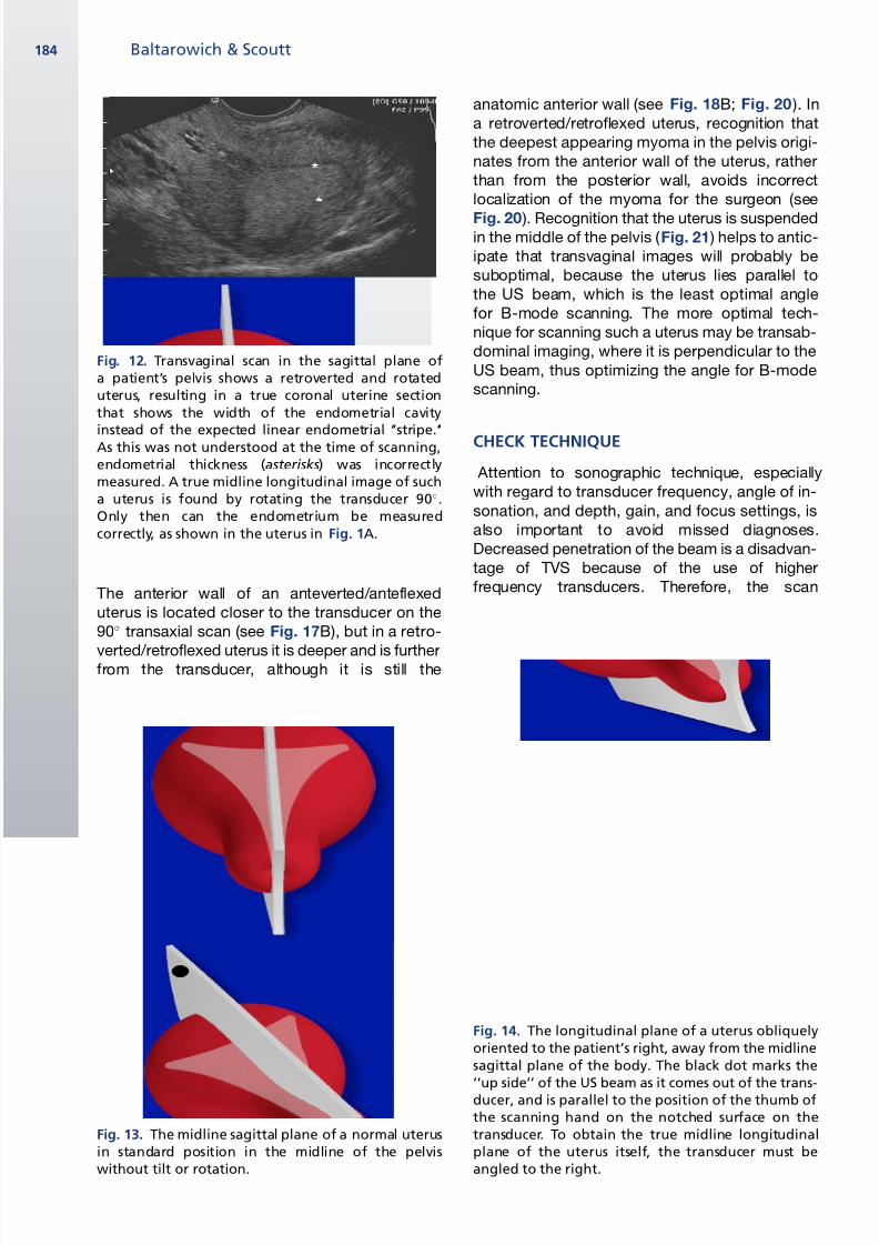
procedures. This mistake may occur when the
endometrium is measured in an oblique or coronal
plane ( Fig. 12 ) rather than in the true midline longi-
tudinal plane of the uterus. Usually the longitudinal
axis of the uterus corresponds to the sagittal plane
of the body ( Fig. 13 ). One must find the longitu-
dinal midline of the uterus by taking into account
any obliquity of the uterus from the midline sagittal
plane of the body ( Fig. 14 ), tilting of the uterus
anteriorly or posteriorly in the pelvis, or rotation
of the uterus around its longitudinal axis ( Figs. 15
and 16 ).
One should have a clear understanding of thesonographic appearances of uteri in various
anatomic positions, including anteverted/ante-
flexed, retroverted/retroflexed, and mid-position
( Figs. 17–19 ). The 90 view is just that—whatever
is the resultant imaging plane of the uterus after
Fig. 8. Transvaginal sonogram of the urinary bladder(U) in an 80-year-old patient with postmenopausalbleeding shows 2 incidental irregularly outlinedbladder wall masses (arrows) that proved to be transi-tional cell carcinomas. The pelvic organs were normal.The bleeding was a result of hematuria, not vaginalbleeding.
Fig. 9. ( A) Midline transvaginal sonogram of the uterus demonstrates heterogeneous echoes (arrows) that weredue to blood within the distended endometrial cavity, but the entire cervix (C) is not included in the image. ( B)
Transvaginal sagittal sonogram directed squarely at the cervix (C) shows that there is an endocervical mass(arrow ), proven to be a polypoid myoma, protruding through the external cervical os. This mass was beyondimage A and would have been missed. There is a small amount of free fluid in the cul-de-sac.
Baltarowich & Scoutt182
8/6/2019 Avoiding Pitfalls in Trans Vaginal
http://slidepdf.com/reader/full/avoiding-pitfalls-in-trans-vaginal 7/17
one rotates the transducer 90 counterclockwise
from the midline longitudinal plane of the given
uterus. Depending on the anatomic position of
the uterus in the pelvis, the 90 view may be
a true transverse/transaxial, oval-shaped section(see Figs. 17 and 18 ), a coronal/frontal cut (see
Fig. 19 ), or a nonstandard oblique section that
includes parts of the uterine corpus and cervix.
The true transaxial sections or 90 views of ante-
verted/anteflexed and retroverted/retroflexed
uteri have an oval shape, but the location of theanterior wall is different (see Figs. 17 and 18 ).
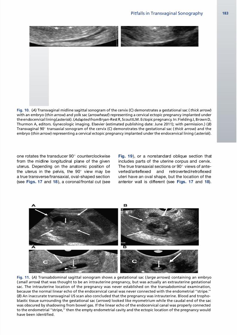
Fig. 10. ( A) Transvaginal midline sagittal sonogram of the cervix (C) demonstrates a gestational sac ( thick arrow )with an embryo (thin arrow ) and yolk sac (arrowhead ) representing a cervical ectopic pregnancy implanted underthe endocervical lining(asterisk ). ( AdaptedfromBryan-Rest R, ScouttLM. Ectopic pregnancy. In: Fielding J, Brown D,Thurmon A, editors. Gynecologic imaging. Elsevier (estimated publishing date: June 2011); with permission.) (B)Transvaginal 90 transaxial sonogram of the cervix (C) demonstrates the gestational sac (thick arrow ) and theembryo (thin arrow ) representing a cervical ectopic pregnancy implanted under the endocervical lining ( asterisk ).
Fig. 11. ( A) Transabdominal sagittal sonogram shows a gestational sac (large arrows) containing an embryo( small arrow ) that was thought to be an intrauterine pregnancy, but was actually an extrauterine gestationalsac. The intrauterine location of the pregnancy was never established on the transabdominal examination,because the normal linear echo of the endocervical canal was never connected with the endometrial ‘‘stripe.’’(B) An inaccurate transvaginal US scan also concluded that the pregnancy was intrauterine. Blood and tropho-blastic tissue surrounding the gestational sac (arrows) looked like myometrium while the caudal end of the sacwas obscured by shadowing from bowel gas. If the linear echo of the endocervical canal was properly connectedto the endometrial ‘‘stripe,’’ then the empty endometrial cavity and the ectopic location of the pregnancy wouldhave been identified.
Pitfalls in Transvaginal Sonography 183
8/6/2019 Avoiding Pitfalls in Trans Vaginal
http://slidepdf.com/reader/full/avoiding-pitfalls-in-trans-vaginal 8/17
The anterior wall of an anteverted/anteflexed
uterus is located closer to the transducer on the
90 transaxial scan (see Fig. 17B), but in a retro-
verted/retroflexed uterus it is deeper and is furtherfrom the transducer, although it is still the
anatomic anterior wall (see Fig. 18B; Fig. 20 ). In
a retroverted/retroflexed uterus, recognition that
the deepest appearing myoma in the pelvis origi-
nates from the anterior wall of the uterus, rather
than from the posterior wall, avoids incorrect
localization of the myoma for the surgeon (see
Fig. 20 ). Recognition that the uterus is suspendedin the middle of the pelvis ( Fig. 21 ) helps to antic-
ipate that transvaginal images will probably be
suboptimal, because the uterus lies parallel to
the US beam, which is the least optimal angle
for B-mode scanning. The more optimal tech-
nique for scanning such a uterus may be transab-
dominal imaging, where it is perpendicular to the
US beam, thus optimizing the angle for B-mode
scanning.
CHECK TECHNIQUE
Attention to sonographic technique, especially
with regard to transducer frequency, angle of in-
sonation, and depth, gain, and focus settings, is
also important to avoid missed diagnoses.
Decreased penetration of the beam is a disadvan-
tage of TVS because of the use of higher
frequency transducers. Therefore, the scan
Fig. 12. Transvaginal scan in the sagittal plane ofa patient’s pelvis shows a retroverted and rotateduterus, resulting in a true coronal uterine sectionthat shows the width of the endometrial cavity
instead of the expected linear endometrial ‘‘stripe.’’As this was not understood at the time of scanning,endometrial thickness (asterisks) was incorrectlymeasured. A true midline longitudinal image of sucha uterus is found by rotating the transducer 90.Only then can the endometrium be measuredcorrectly, as shown in the uterus in Fig. 1A.
Fig. 13. The midline sagittal plane of a normal uterusin standard position in the midline of the pelviswithout tilt or rotation.
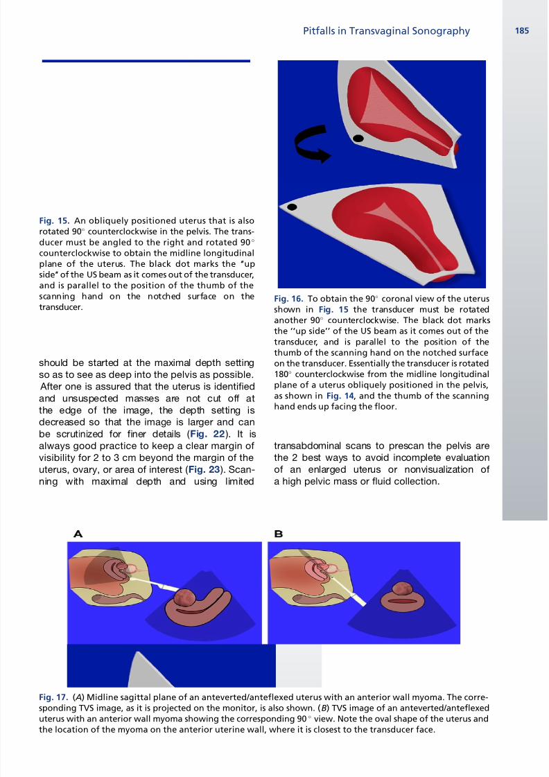
Fig. 14. The longitudinal plane of a uterus obliquelyoriented to the patient’s right, away from the midlinesagittal plane of the body. The black dot marks the‘‘up side’’ of the US beam as it comes out of the trans-ducer, and is parallel to the position of the thumb ofthe scanning hand on the notched surface on thetransducer. To obtain the true midline longitudinalplane of the uterus itself, the transducer must beangled to the right.
Baltarowich & Scoutt184
8/6/2019 Avoiding Pitfalls in Trans Vaginal
http://slidepdf.com/reader/full/avoiding-pitfalls-in-trans-vaginal 9/17
should be started at the maximal depth setting
so as to see as deep into the pelvis as possible.
After one is assured that the uterus is identified
and unsuspected masses are not cut off at
the edge of the image, the depth setting isdecreased so that the image is larger and can
be scrutinized for finer details ( Fig. 22 ). It is
always good practice to keep a clear margin of
visibility for 2 to 3 cm beyond the margin of the
uterus, ovary, or area of interest ( Fig. 23 ). Scan-
ning with maximal depth and using limited
transabdominal scans to prescan the pelvis are
the 2 best ways to avoid incomplete evaluation
of an enlarged uterus or nonvisualization of
a high pelvic mass or fluid collection.
Fig. 15. An obliquely positioned uterus that is alsorotated 90 counterclockwise in the pelvis. The trans-ducer must be angled to the right and rotated 90
counterclockwise to obtain the midline longitudinalplane of the uterus. The black dot marks the ‘‘up
side’’ of the US beam as it comes out of the transducer,and is parallel to the position of the thumb of thescanning hand on the notched surface on thetransducer.
Fig. 17. ( A) Midline sagittal plane of an anteverted/anteflexed uterus with an anterior wall myoma. The corre-sponding TVS image, as it is projected on the monitor, is also shown. (B) TVS image of an anteverted/anteflexeduterus with an anterior wall myoma showing the corresponding 90 view. Note the oval shape of the uterus andthe location of the myoma on the anterior uterine wall, where it is closest to the transducer face.
Fig. 16. To obtain the 90 coronal view of the uterusshown in Fig. 15 the transducer must be rotatedanother 90 counterclockwise. The black dot marksthe ‘‘up side’’ of the US beam as it comes out of thetransducer, and is parallel to the position of thethumb of the scanning hand on the notched surfaceon the transducer. Essentially the transducer is rotated180 counterclockwise from the midline longitudinalplane of a uterus obliquely positioned in the pelvis,as shown in Fig. 14, and the thumb of the scanning
hand ends up facing the floor.
Pitfalls in Transvaginal Sonography 185
8/6/2019 Avoiding Pitfalls in Trans Vaginal
http://slidepdf.com/reader/full/avoiding-pitfalls-in-trans-vaginal 10/17
Gain settings, including the time-gain-compen-
sation (TGC) curve and overall gain, should be
adjusted so that false echoes are not created in
fluid. The best way to decide if the echoes in
a mass or fluid collection are real or artifactual is
to compare the echogenicity with a known fluid
structure, ideally the urinary bladder or ovarian
follicle, at a similar depth. If the urinary bladder is
anechoic and the pelvic mass or fluid contains
echoes, then the echoes are real ( Fig. 24 ). To
test this hypothesis, start by lowering the gain until
the bladder and the mass or pelvic fluid are free of
echoes. Now slowly increase the gain. If the mass
fills in with echoes before the urinary bladder, then
the echoes are real. If they fill in at the same time,
then the echoes are artifactual. This test becomes
very important in analysis of pelvic fluid that may
represent hemoperitoneum or in analysis of
complex ovarian cystic masses.
BLIND SPOTS IN THE PELVIS
One must be aware of the blind spots in the pelvis
on transvaginal scanning. These blind spots
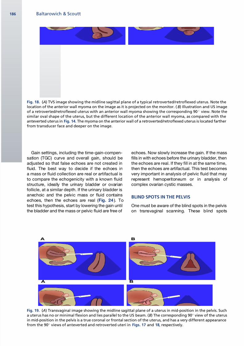
Fig. 18. ( A) TVS image showing the midline sagittal plane of a typical retroverted/retroflexed uterus. Note thelocation of the anterior wall myoma on the image as it is projected on the monitor. ( B) Illustration and US imageof a retroverted/retroflexed uterus with an anterior wall myoma showing the corresponding 90 view. Note thesimilar oval shape of the uterus, but the different location of the anterior wall myoma, as compared with the
anteverted uterus in Fig. 14. The myoma on the anterior wall of a retroverted/retroflexed uterus is located fartherfrom transducer face and deeper on the image.
Fig. 19. ( A) Transvaginal image showing the midline sagittal plane of a uterus in mid-position in the pelvis. Sucha uterus has no or minimal flexion and lies parallel to the US beam. (B) The corresponding 90 view of the uterusin mid-position in the pelvis is a true coronal or frontal section of the uterus, and has a very different appearancefrom the 90 views of anteverted and retroverted uteri in Figs. 17 and 18, respectively.
Baltarowich & Scoutt186
8/6/2019 Avoiding Pitfalls in Trans Vaginal
http://slidepdf.com/reader/full/avoiding-pitfalls-in-trans-vaginal 11/17
include areas in the upper pelvis above the fundus
of the uterus, laterally along the pelvic side walls,
deep in the cul-de-sac, and markedly anterior to
the uterus. Evaluation of these areas is especially
important when searching for ovaries or ectopic
pregnancies. Transabdominal imaging is helpful
in such cases.
MANEUVER THE PROBE
Maneuvers with the transvaginal probe may help
to solve diagnostic dilemmas in the pelvis. Exert-
ing pressure on masses with the probe helps to
check for mobility, separate masses in close
proximity, separate a mass from the ovary, or
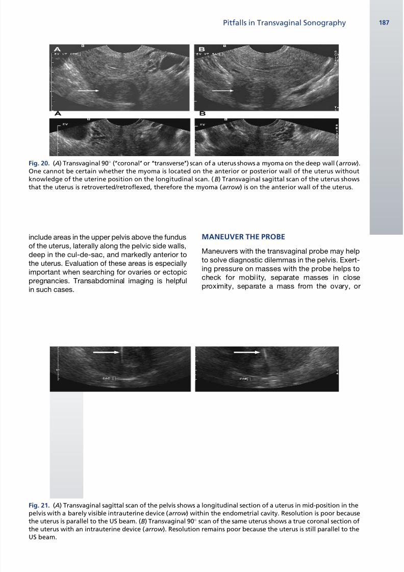
Fig. 20. ( A) Transvaginal 90 (‘‘coronal’’ or ‘‘transverse’’) scan of a uterus shows a myoma on the deep wall (arrow ).One cannot be certain whether the myoma is located on the anterior or posterior wall of the uterus withoutknowledge of the uterine position on the longitudinal scan. ( B) Transvaginal sagittal scan of the uterus showsthat the uterus is retroverted/retroflexed, therefore the myoma (arrow ) is on the anterior wall of the uterus.
Fig. 21. ( A) Transvaginal sagittal scan of the pelvis shows a longitudinal section of a uterus in mid-position in thepelvis with a barely visible intrauterine device (arrow ) within the endometrial cavity. Resolution is poor becausethe uterus is parallel to the US beam. (B) Transvaginal 90 scan of the same uterus shows a true coronal section ofthe uterus with an intrauterine device (arrow ). Resolution remains poor because the uterus is still parallel to theUS beam.
Pitfalls in Transvaginal Sonography 187
8/6/2019 Avoiding Pitfalls in Trans Vaginal
http://slidepdf.com/reader/full/avoiding-pitfalls-in-trans-vaginal 12/17
elicit pain that helps to localize and identify
pathology. Using the nonscanning hand for
pressing and simultaneously pulling down on
the anterior abdominal wall with the intention of
displacing an ovary in a high location into the
pelvis is a helpful technique, and one should be
persistent about identifying both ovaries toensure a complete examination. Either the scan-
ning hand with the probe or the palpating hand
on the abdominal wall may be used to cause
movement of the internal contents of a complex
cystic mass or to prove that echoes are real by
causing particles to move. Using the energy of
color or spectral Doppler to move particles in
fluid, called acoustic streaming, may also be
helpful.12,13 Whenever there appears to be a sep-
tated cystic mass, especially if it is elongated, the
probe should be maneuvered by twisting and
turning movements of the hand to try to unravel
a potentially folded dilated tubular mass( Fig. 25 ) because a hydrosalpinx has much
different clinical consequences for the patient
than a septated ovarian mass. Another useful
maneuver is to change location of the transducer
tip from the anterior to the posterior fornix while
applying varying degrees of pressure on the
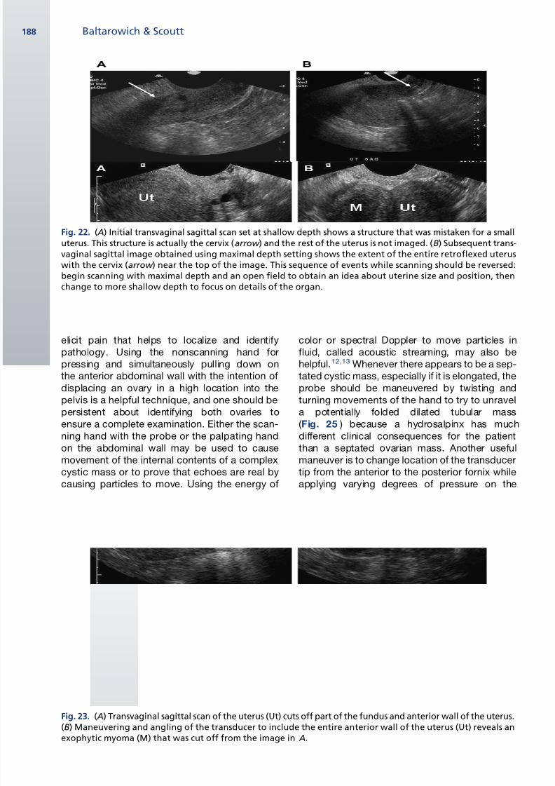
Fig. 22. ( A) Initial transvaginal sagittal scan set at shallow depth shows a structure that was mistaken for a smalluterus. This structure is actually the cervix (arrow ) and the rest of the uterus is not imaged. (B) Subsequent trans-vaginal sagittal image obtained using maximal depth setting shows the extent of the entire retroflexed uteruswith the cervix (arrow ) near the top of the image. This sequence of events while scanning should be reversed:
begin scanning with maximal depth and an open field to obtain an idea about uterine size and position, thenchange to more shallow depth to focus on details of the organ.
Fig. 23. ( A) Transvaginal sagittal scan of the uterus (Ut) cuts off part of the fundus and anterior wall of the uterus.(B) Maneuvering and angling of the transducer to include the entire anterior wall of the uterus (Ut) reveals anexophytic myoma (M) that was cut off from the image in A.
Baltarowich & Scoutt188
8/6/2019 Avoiding Pitfalls in Trans Vaginal
http://slidepdf.com/reader/full/avoiding-pitfalls-in-trans-vaginal 13/17
cervix to change the position of the uterus in the
pelvis to a more favorable scan plane ( Figs. 26
and 27 ).
DOPPLER EVALUATION
Doppler investigation is helpful in evaluating many
types of pelvic pathology. Color or spectralDoppler US helps to differentiate a pelvic vessel
from a hydrosalpinx ( Fig. 28 ). One can avoid misin-
terpretation of axially imaged uterine vessels for
ovaries, so-called pseudo-ovaries, simply by using
color Doppler US or scanning at 90 to elongate
the vessels.
Using Doppler to help evaluate unusual cystic
pelvic masses may help to avoid misdiagnosis of
a vascular mass, such as an iliac artery aneurysm
for an ovarian cyst, which could lead to unneces-sary surgery in a postmenopausal woman.
Doppler evaluation should also be considered for
any unusual area of myometrial echogenicity that
includes myometrial inhomogeneity, irregular
cystic changes, or an area of tortuous tubular
cystic structures. Such an area may be caused
by a uterine arterial venous malformation, which
is a congenital or acquired vascular plexus of
arteries and veins within the uterine myometrium
or endometrium. Proper diagnosis would help to
avoid dilation and curettage or surgery, which
may cause massive hemorrhage or on occasion
might lead to hysterectomy.
Although Doppler US investigation is often help-
ful in the evaluation of pelvic masses, one should
exercise caution concerning the role of Doppler
US in the diagnosis of ovarian torsion. The US
diagnosis of ovarian torsion is not as certain as
testicular torsion. The spectrum of Doppler US
findings in ovarian torsion ranges from complete
lack of arterial and venous flow through variations
in arterial and venous flow to completely normal
blood flow in the ovary.14,15 Because it is possibleto demonstrate normal blood flow in an ovary
undergoing torsion ( Fig. 29 ), one must first look
for signs of torsion on the gray-scale image. These
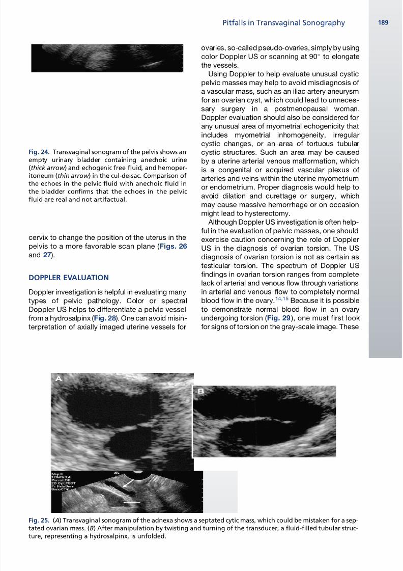
Fig. 24. Transvaginal sonogram of the pelvis shows anempty urinary bladder containing anechoic urine(thick arrow ) and echogenic free fluid, and hemoper-itoneum (thin arrow ) in the cul-de-sac. Comparison ofthe echoes in the pelvic fluid with anechoic fluid inthe bladder confirms that the echoes in the pelvic
fluid are real and not artifactual.
Fig. 25. ( A) Transvaginal sonogram of the adnexa shows a septated cytic mass, which could be mistaken for a sep-tated ovarian mass. (B) After manipulation by twisting and turning of the transducer, a fluid-filled tubular struc-ture, representing a hydrosalpinx, is unfolded.
Pitfalls in Transvaginal Sonography 189
8/6/2019 Avoiding Pitfalls in Trans Vaginal
http://slidepdf.com/reader/full/avoiding-pitfalls-in-trans-vaginal 14/17
signs include an enlarged edematous ovary with
peripherally displaced follicles or an ovarian
mass that serves as the lead for the twist around
a fulcrum. The twisted vascular pedicle sign, which
is a round mass composed of several concentric
stripes of alternating echogenicity creating a target
appearance adjacent to the twisted ovary, is
another reliable sign of ovarian or adnexal
torsion.16 Any one of these findings in the pres-ence of significant pelvic pain should lead to the
proper diagnosis, despite the presence of blood
flow in an adnexal mass. Waiting for complete
absence of arterial and venous blood flow to
make a diagnosis of ovarian torsion usually leads
to a diagnosis of ovarian necrosis, which is not
reversible. It is far more important to make the
diagnosis of ovarian torsion in a salvageable ovary
before infarction has occurred. A potential pitfall in
the demonstration of blood flow by color Doppler
US is flash artifact that can mimic blood flow.
Any evidence of blood flow seen on color DopplerUS scan must be confirmed with a spectral
tracing, which will confirm real flow by demon-
strating a venous or arterial waveform.
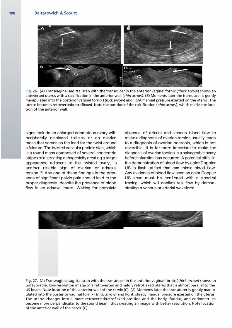
Fig. 26. ( A) Transvaginal sagittal scan with the transducer in the anterior vaginal fornix (thick arrow ) shows ananteverted uterus with a calcification in the anterior wall (thin arrow ). (B) Moments later the transducer is gentlymanipulated into the posterior vaginal fornix (thick arrow ) and light manual pressure exerted on the uterus. Theuterus becomes retroverted/retroflexed. Note the position of the calcification ( thin arrow ), which marks the loca-tion of the anterior wall.
Fig. 27. ( A) Transvaginal sagittal scan with the transducer in the anterior vaginal fornix (thick arrow ) shows anunfavorable, low-resolution image of a retroverted and mildly retroflexed uterus that is almost parallel to theUS beam. Note location of the anterior wall of the cervix (C). (B) Moments later the transducer is gently manip-ulated into the posterior vaginal fornix (thick arrow ) and light, steady manual pressure exerted on the uterus.The uterus changes into a more retroverted/retroflexed position and the body, fundus, and endometriumbecome more perpendicular to the sound beam, thus creating an image with better resolution. Note locationof the anterior wall of the cervix (C).
Baltarowich & Scoutt190
8/6/2019 Avoiding Pitfalls in Trans Vaginal
http://slidepdf.com/reader/full/avoiding-pitfalls-in-trans-vaginal 15/17
Another word of caution concerns the ‘‘ring of
fire,’’ which refers to concentric enhancement of
vessels on color or power Doppler interrogation
around the perimeter of a mass, originallydescribed in ectopic pregnancy.17 Unfortunately,
the ‘‘ring of fire’’ is not a reliable sign of ectopic
pregnancy. The ‘‘ring of fire’’ is also caused by
circumferential increased vascularity in the wall
of the corpus luteum, which is a much more
common event than ectopic pregnancy. A cysticmass with a thick echogenic rim and a ‘‘ring of
fire’’ within the ovary is much more likely to be
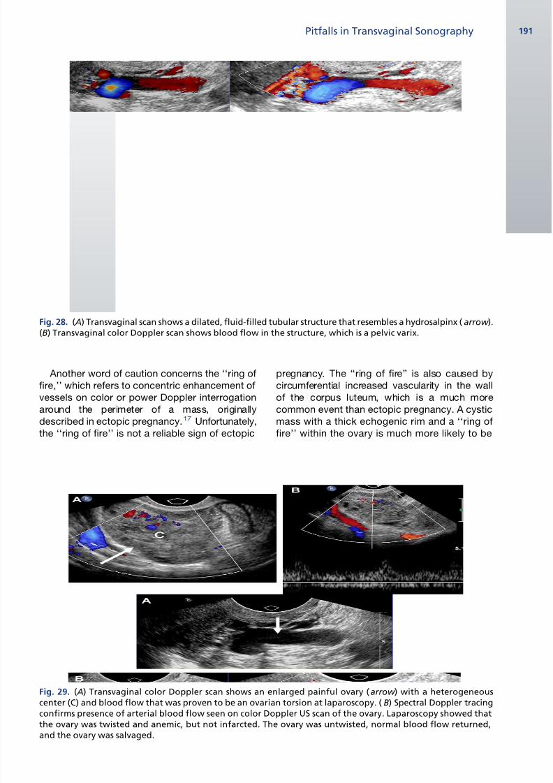
Fig. 28. ( A) Transvaginal scan shows a dilated, fluid-filled tubular structure that resembles a hydrosalpinx ( arrow ).(B) Transvaginal color Doppler scan shows blood flow in the structure, which is a pelvic varix.
Fig. 29. ( A) Transvaginal color Doppler scan shows an enlarged painful ovary ( arrow ) with a heterogeneouscenter (C) and blood flow that was proven to be an ovarian torsion at laparoscopy. ( B) Spectral Doppler tracingconfirms presence of arterial blood flow seen on color Doppler US scan of the ovary. Laparoscopy showed thatthe ovary was twisted and anemic, but not infarcted. The ovary was untwisted, normal blood flow returned,and the ovary was salvaged.
Pitfalls in Transvaginal Sonography 191
8/6/2019 Avoiding Pitfalls in Trans Vaginal
http://slidepdf.com/reader/full/avoiding-pitfalls-in-trans-vaginal 16/17
a corpus luteum ( Fig. 30 ) than the rare (<0.5% of
all ectopic pregnancies) ovarian ectopic
pregnancy.
THINK BEYOND GYNECOLOGIC CONDITIONSFOR PELVIC PAIN
Considering nongynecologic sources of pain in the
pelvis expands the differential diagnosis and may
lead to correct diagnosis, for which a good clinical
history is also critical. Lower urinary tract abnor-
malities often present as pelvic pain. Distal ureteral
calculi may be diagnosed by TVS, when the
calculus is at the ureterovesical junction, thus
sparing a young female patient the significant radi-
ation dose from a computer tomographic scan
(see Fig. 3 ). Ureteric jets in the bladder may be
easier to detect with transvaginal color Doppler
US. Unsuspected bladder masses have been
observed in young women, even during evaluation
of early pregnancy. Bladder masses have alsobeen found to be the source of bleeding in elderly
women complaining of ‘‘postmenopausal
bleeding,’’ which was actually unrecognized
hematuria (see Fig. 8 ). Conditions related to the
gastrointestinal tract may also present as pelvic
pain. Abnormally thickened large or small bowel
loops may be observed in inflammatory bowel
diseases. Pericolonic abscesses, such as those
associated with diverticulitis, may be well visual-
ized. Occasionally pelvic pain from a low appendix
with acute inflammation mimics ovarian pain. This
situation may occur when the inflamed tip of
a pelvic appendix touches the ovary or
surrounding tissues and incites inflammation
( Fig. 31 ).
TVS has become the preferred method for sono-
graphic evaluation of the pelvis because of its
higher resolution, better detail, and more confi-
dence in diagnosis of pelvic pathology. Unfortu-
nately, there are numerous opportunities for
missed diagnosis or incorrect interpretation with
TVS. Knowledge of the pitfalls and a systematic,
orderly approach for examination of every patienthelps to minimize the potential for errors.
ACKNOWLEDGMENTS
Artwork was created by John D’Agostino of Tin
Can Productions.
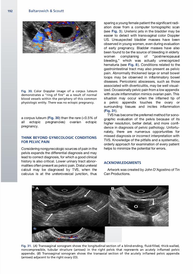
Fig. 30. Color Doppler image of a corpus luteum
demonstrates a ‘‘ring of fire’’ as a result of normalblood vessels within the periphery of this commonphysiologic entity. There was no ectopic pregnancy.
Fig. 31. ( A) Transvaginal sonogram shows the longitudinal section of a blind-ending, fluid-filled, thick-walled,noncompressible, tubular structure (arrows) in the right pelvis that represents an acutely inflamed pelvicappendix. (B) Transvaginal sonogram shows the transaxial section of the acutely inflamed pelvic appendix(arrows) adjacent to the right ovary (O).
Baltarowich & Scoutt192
8/6/2019 Avoiding Pitfalls in Trans Vaginal
http://slidepdf.com/reader/full/avoiding-pitfalls-in-trans-vaginal 17/17
REFERENCES
1. Fernandez H, Gervaise A. Ectopic pregnancies
after infertility treatment: modern diagnosis and
therapeutic strategy. Hum Reprod Update 2004;
10:503–13.
2. Hertzberg BS, Livingston E, DeLong DM, et al. Ultra-sonographic evaluation of the cervix: transperineal
versus endovaginal imaging. J Ultrasound Med
2001;20:1071–8.
3. Laing FC, Benson CB, DiSalvo DN, et al. Distal
ureteral calculi: detection with vaginal US. Radiology
1994;192:545–8.
4. Benacerraf BR. Filling of the bladder for pelvic sono-
grams: an ancient form of torture. J Ultrasound Med
2003;22:239–41.
5. Benacerraf BR, Shipp TD, Bromley B. Is the full
bladder still necessary for pelvic sonography? J
Ultrasound Med 2000;19:237–41.
6. Tessler FN, Schiller VL, Perrella RR, et al. Transabdo-
minal versus endovaginal pelvic sonography:
prospective study. Radiology 1989;170:553–6.
7. Mendelson EB, Bohm-Velez M, Joseph N, et al.
Gynecologic imaging: comparison of transabdomi-
nal and transvaginal sonography. Radiology 1988;
166:321–4.
8. Leibman AJ, Kruse B, McSweeney MB. Transvaginal
sonography: comparison with transabdominal
sonography in the diagnosis of pelvic masses. AJR
Am J Roentgenol 1988;151(1):89–92.
9. Coleman BG, Arger PH, Grumbach K, et al. Transva-
ginal and transabdominal ultrasound: prospective
comparison. Radiology 1988;168:639–43.
10. Andolf E, Jorgensen C. A prospective comparison of
transabdominal and transvaginal ultrasound with
surgical findings in gynecologic disease. J Ultra-
sound Med 1990;9:71–5.11. Hill LM, Breckle R. Value of a postvoid scan during
adnexal sonography. Am J Obstet Gynecol 1985;
152:23–5.
12. Edwards A, Clarke L, Piessens S, et al. Acoustic
streaming: a new technique for assessing
adnexal cysts. Ultrasound Obstet Gynecol 2003;
22:74–8.
13. Clarke L, Edwards A, Pollard K. Acoustic streaming in
ovarian cysts. J Ultrasound Med 2005;24:617–21.
14. Albayram F, Hamper UM. Ovarian and adnexal
torsion: spectrum of sonographic findings with
pathologic correlation. J Ultrasound Med 2001;20:
1083–9.
15. Pena JE, Ufberg D, Cooney N, et al. Usefulness of
Doppler sonography in the diagnosis of ovarian
torsion. Fertil Steril 2000;73:1047–50.
16. Lee EJ, Kwon HC, Joo HJ, et al. Diagnosis of ovarian
torsion with color Doppler sonography: depiction of
twisted vascular pedicle. J Ultrasound Med 1998;
17:83–9.
17. Pellerito JS, Taylor KJ, Quedens-Case C, et al.
Ectopic pregnancy: evaluation with endovaginal
color flow imaging. Radiology 1992;183:407–11.
Pitfalls in Transvaginal Sonography 193


![[Slideshare]avoiding linguistic pitfalls](https://img.pdfslide.net/doc/110x75/5588e512d8b42a28148b4642/slideshareavoiding-linguistic-pitfalls.jpg)