Embed Size (px)
Citation preview
Bacteria cause biodeterioration in medieval fresco paint in Siena (Italy)
Claudio Milanesi^*, Franco Baldiº, Sara Borinˉ, Lorenzo Brusettiˉ, Fabrizio Ciampolin^, Fabrizio Iacopini˜, Mauro Cresti^
^) Department of Environmental Science ‘G. Sarfatti’, University of Siena, Via Mattioli 4, 53100 Siena, Italy, *E-mail: [email protected] º) Department of Environmental Science, Cà Foscari University of Venezia, Calle Larga S. Marta, 30121 Venezia, Italy
ˉ) Department of Food Science and Technology, University of Milano, Via Celoria 2, 20133 Milano, Italy˜) A.T.I., Via Ciurini 6, 50051 Castelfiorentino, Firenze, Italy
Materials and methods Sampling and culture of fresco fragments
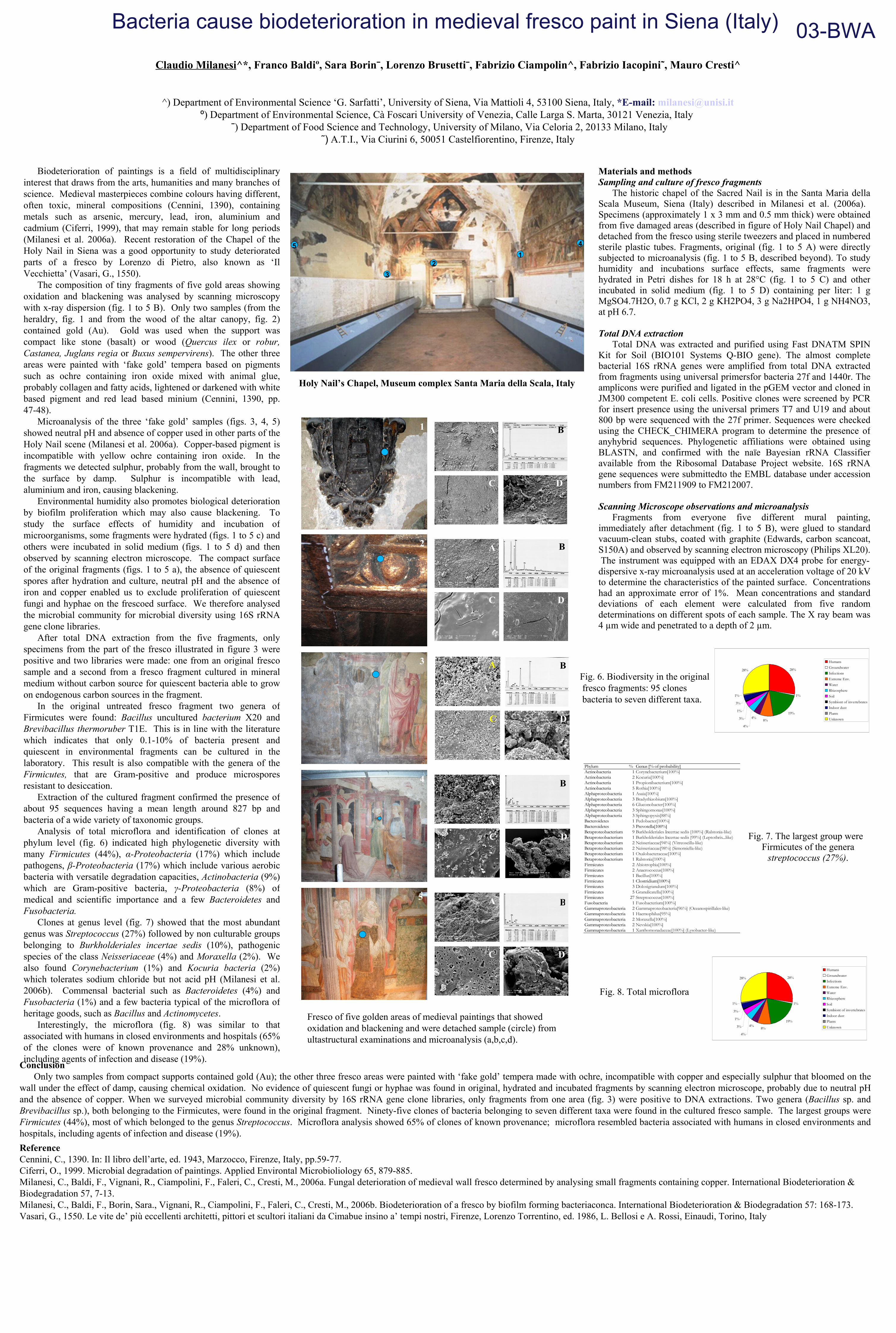
The historic chapel of the Sacred Nail is in the Santa Maria della Scala Museum, Siena (Italy) described in Milanesi et al. (2006a). Specimens (approximately 1 x 3 mm and 0.5 mm thick) were obtained from five damaged areas (described in figure of Holy Nail Chapel) and detached from the fresco using sterile tweezers and placed in numbered sterile plastic tubes. Fragments, original (fig. 1 to 5 A) were directly subjected to microanalysis (fig. 1 to 5 B, described beyond). To study humidity and incubations surface effects, same fragments were hydrated in Petri dishes for 18 h at 28°C (fig. 1 to 5 C) and other incubated in solid medium (fig. 1 to 5 D) containing per liter: 1 g MgSO4.7H2O, 0.7 g KCl, 2 g KH2PO4, 3 g Na2HPO4, 1 g NH4NO3, at pH 6.7.
Total DNA extraction
Total DNA was extracted and purified using Fast DNATM SPIN Kit for Soil (BIO101 Systems Q-BIO gene). The almost complete bacterial 16S rRNA genes were amplified from total DNA extracted from fragments using universal primersfor bacteria 27f and 1440r. The amplicons were purified and ligated in the pGEM vector and cloned in JM300 competent E. coli cells. Positive clones were screened by PCR for insert presence using the universal primers T7 and U19 and about 800 bp were sequenced with the 27f primer. Sequences were checked using the CHECK_CHIMERA program to determine the presence of anyhybrid sequences. Phylogenetic affiliations were obtained using BLASTN, and confirmed with the naïe Bayesian rRNA Classifier available from the Ribosomal Database Project website. 16S rRNA gene sequences were submittedto the EMBL database under accession numbers from FM211909 to FM212007.
Scanning Microscope observations and microanalysis Fragments from everyone five different mural painting,
immediately after detachment (fig. 1 to 5 B), were glued to standard vacuum-clean stubs, coated with graphite (Edwards, carbon scancoat, S150A) and observed by scanning electron microscopy (Philips XL20). The instrument was equipped with an EDAX DX4 probe for energy-dispersive x-ray microanalysis used at an acceleration voltage of 20 kV to determine the characteristics of the painted surface. Concentrations had an approximate error of 1%. Mean concentrations and standard deviations of each element were calculated from five random determinations on different spots of each sample. The X ray beam was 4 µm wide and penetrated to a depth of 2 µm.
Biodeterioration of paintings is a field of multidisciplinary interest that draws from the arts, humanities and many branches of science. Medieval masterpieces combine colours having different, often toxic, mineral compositions (Cennini, 1390), containing metals such as arsenic, mercury, lead, iron, aluminium and cadmium (Ciferri, 1999), that may remain stable for long periods (Milanesi et al. 2006a). Recent restoration of the Chapel of the Holy Nail in Siena was a good opportunity to study deteriorated parts of a fresco by Lorenzo di Pietro, also known as ‘Il Vecchietta’ (Vasari, G., 1550).
The composition of tiny fragments of five gold areas showing oxidation and blackening was analysed by scanning microscopy with x-ray dispersion (fig. 1 to 5 B). Only two samples (from the heraldry, fig. 1 and from the wood of the altar canopy, fig. 2) contained gold (Au). Gold was used when the support was compact like stone (basalt) or wood (Quercus ilex or robur, Castanea, Juglans regia or Buxus sempervirens). The other three areas were painted with ‘fake gold’ tempera based on pigments such as ochre containing iron oxide mixed with animal glue, probably collagen and fatty acids, lightened or darkened with white based pigment and red lead based minium (Cennini, 1390, pp. 47-48).
Microanalysis of the three ‘fake gold’ samples (figs. 3, 4, 5) showed neutral pH and absence of copper used in other parts of the Holy Nail scene (Milanesi et al. 2006a). Copper-based pigment is incompatible with yellow ochre containing iron oxide. In the fragments we detected sulphur, probably from the wall, brought to the surface by damp. Sulphur is incompatible with lead, aluminium and iron, causing blackening.
Environmental humidity also promotes biological deterioration by biofilm proliferation which may also cause blackening. To study the surface effects of humidity and incubation of microorganisms, some fragments were hydrated (figs. 1 to 5 c) and others were incubated in solid medium (figs. 1 to 5 d) and then observed by scanning electron microscope. The compact surface of the original fragments (figs. 1 to 5 a), the absence of quiescent spores after hydration and culture, neutral pH and the absence of iron and copper enabled us to exclude proliferation of quiescent fungi and hyphae on the frescoed surface. We therefore analysed the microbial community for microbial diversity using 16S rRNA gene clone libraries.
After total DNA extraction from the five fragments, only specimens from the part of the fresco illustrated in figure 3 were positive and two libraries were made: one from an original fresco sample and a second from a fresco fragment cultured in mineral medium without carbon source for quiescent bacteria able to grow on endogenous carbon sources in the fragment.
In the original untreated fresco fragment two genera of Firmicutes were found: Bacillus uncultured bacterium X20 and Brevibacillus thermoruber T1E. This is in line with the literature which indicates that only 0.1-10% of bacteria present and quiescent in environmental fragments can be cultured in the laboratory. This result is also compatible with the genera of the Firmicutes, that are Gram-positive and produce microspores resistant to desiccation.
Extraction of the cultured fragment confirmed the presence of about 95 sequences having a mean length around 827 bp and bacteria of a wide variety of taxonomic groups.
Analysis of total microflora and identification of clones at phylum level (fig. 6) indicated high phylogenetic diversity with many Firmicutes (44%), α-Proteobacteria (17%) which include pathogens, β-Proteobacteria (17%) which include various aerobic bacteria with versatile degradation capacities, Actinobacteria (9%) which are Gram-positive bacteria, γ-Proteobacteria (8%) of medical and scientific importance and a few Bacteroidetes and Fusobacteria.
Clones at genus level (fig. 7) showed that the most abundant genus was Streptococcus (27%) followed by non culturable groups belonging to Burkholderiales incertae sedis (10%), pathogenic species of the class Neisseriaceae (4%) and Moraxella (2%). We also found Corynebacterium (1%) and Kocuria bacteria (2%) which tolerates sodium chloride but not acid pH (Milanesi et al. 2006b). Commensal bacterial such as Bacteroidetes (4%) and Fusobacteria (1%) and a few bacteria typical of the microflora of heritage goods, such as Bacillus and Actinomycetes.
Interestingly, the microflora (fig. 8) was similar to that associated with humans in closed environments and hospitals (65% of the clones were of known provenance and 28% unknown), including agents of infection and disease (19%).
Holy Nail’s Chapel, Museum complex Santa Maria della Scala, Italy
Fig. 6. Biodiversity in the original fresco fragments: 95 clones bacteria to seven different taxa.
Fig. 7. The largest group were Firmicutes of the genera
streptococcus (27%).
Conclusion Only two samples from compact supports contained gold (Au); the other three fresco areas were painted with ‘fake gold’ tempera made with ochre, incompatible with copper and especially sulphur that bloomed on the wall under the effect of damp, causing chemical oxidation. No evidence of quiescent fungi or hyphae was found in original, hydrated and incubated fragments by scanning electron microscope, probably due to neutral pH and the absence of copper. When we surveyed microbial community diversity by 16S rRNA gene clone libraries, only fragments from one area (fig. 3) were positive to DNA extractions. Two genera (Bacillus sp. and Brevibacillus sp.), both belonging to the Firmicutes, were found in the original fragment. Ninety-five clones of bacteria belonging to seven different taxa were found in the cultured fresco sample. The largest groups were Firmicutes (44%), most of which belonged to the genus Streptococcus. Microflora analysis showed 65% of clones of known provenance; microflora resembled bacteria associated with humans in closed environments and hospitals, including agents of infection and disease (19%).
ReferenceCennini, C., 1390. In: Il libro dell’arte, ed. 1943, Marzocco, Firenze, Italy, pp.59-77.Ciferri, O., 1999. Microbial degradation of paintings. Applied Environtal Microbioliology 65, 879-885. Milanesi, C., Baldi, F., Vignani, R., Ciampolini, F., Faleri, C., Cresti, M., 2006a. Fungal deterioration of medieval wall fresco determined by analysing small fragments containing copper. International Biodeterioration & Biodegradation 57, 7-13. Milanesi, C., Baldi, F., Borin, Sara., Vignani, R., Ciampolini, F., Faleri, C., Cresti, M., 2006b. Biodeterioration of a fresco by biofilm forming bacteriaconca. International Biodeterioration & Biodegradation 57: 168-173.Vasari, G., 1550. Le vite de’ più eccellenti architetti, pittori et scultori italiani da Cimabue insino a’ tempi nostri, Firenze, Lorenzo Torrentino, ed. 1986, L. Bellosi e A. Rossi, Einaudi, Torino, Italy
03-BWA
1
2
3
4
5
A
A
A
A
B
B
B
B
C
C
C
C
C
A
D
D
D
D
D
Fig. 8. Total microflora
28%
1%
19%
8%4%
4%
3%
1%
3%
1%
28%
HumansGroundwaterInfectionsExtreme Env.WaterRhizosphereSoilSymbiont of invertebratesIndoor dustPlantsUnknown
28%
1%
19%
8%4%
4%
3%
1%
3%
1%
28%
HumansGroundwaterInfectionsExtreme Env.WaterRhizosphereSoilSymbiont of invertebratesIndoor dustPlantsUnknown
Phylum % Genus [% of probability]Actinobacteria 1 Corynebacterium[100%]Actinobacteria 2 Kocuria[100%]Actinobacteria 1 Propionibacterium[100%]Actinobacteria 5 Rothia[100%]Alphaproteobacteria 1 Asaia[100%]Alphaproteobacteria 3 Bradyrhizobium[100%]Alphaproteobacteria 6 Gluconobacter[100%]Alphaproteobacteria 3 Sphingomonas[100%]Alphaproteobacteria 3 Sphingopyxis[88%]Bacteroidetes 1 Pedobacter[100%]Bacteroidetes 3 Prevotella[100%]Betaproteobacterium 9 Burkholderiales Incertae sedis [100%] (Ralstonia-like)Betaproteobacterium 1 Burkholderiales Incertae sedis [99%] (Leptothrix_like)Betaproteobacterium 2 Neisseriaceae[94%] (Vitreoscilla-like)Betaproteobacterium 2 Neisseriaceae[98%] (Simonsiella-like)Betaproteobacterium 1 Oxalobacteraceae[100%]Betaproteobacterium 1 Ralstonia[100%]Firmicutes 2 Abiotrophia[100%]Firmicutes 2 Anaerococcus[100%]Firmicutes 1 Bacillus[100%]Firmicutes 1 Clostridium[100%]Firmicutes 3 Dolosigranulum[100%]Firmicutes 5 Granulicatella[100%]Firmicutes 27 Streptococcus[100%]Fusobacteria 1 Fusobacterium[100%]Gammaproteobacteria 2 Gammaproteobacteria[96%] (Oceanospirillales-like)Gammaproteobacteria 1 Haemophilus[95%]Gammaproteobacteria 2 Moraxella[100%]Gammaproteobacteria 2 Nevskia[100%]Gammaproteobacteria 1 Xanthomonadaceae[100%] (Lysobacter-like)
12
3
45
B
Fresco of five golden areas of medieval paintings that showed oxidation and blackening and were detached sample (circle) fromultastructural examinations and microanalysis (a,b,c,d).