Embed Size (px)
Citation preview
Bacterial chromosome segregation: structure andDNA binding of the Soj dimer — a conservedbiological switch
Thomas A Leonard*, P Jonathan Butlerand Jan Lowe
MRC Laboratory of Molecular Biology, Cambridge, UK
Soj and Spo0J of the Gram-negative hyperthermophile
Thermus thermophilus belong to the conserved ParAB
family of bacterial proteins implicated in plasmid and
chromosome partitioning. Spo0J binds to DNA near the
replication origin and localises at the poles following
initiation of replication. Soj oscillates in the nucleoid
region in an ATP- and Spo0J-dependent fashion. Here,
we show that Soj undergoes ATP-dependent dimerisation
in solution and forms nucleoprotein filaments with DNA.
Crystal structures of Soj in three nucleotide states demon-
strate that the empty and ADP-bound states are mono-
meric, while a hydrolysis-deficient mutant, D44A, is
capable of forming a nucleotide ‘sandwich’ dimer. Soj
ATPase activity is stimulated by Spo0J or the N-terminal
20 amino-acid peptide of Spo0J. Our analysis shows that
dimerisation and activation involving a peptide containing
a Lys/Arg is conserved for Soj, ParA and MinD and their
modulators Spo0J, ParB and MinE, respectively. By homol-
ogy to the nitrogenase iron protein and the GTPases Ffh/
FtsY, we suggest that Soj dimerisation and regulation
represent a conserved biological switch.
The EMBO Journal (2005) 24, 270–282. doi:10.1038/
sj.emboj.7600530; Published online 6 January 2005
Subject Categories: structural biology; microbiology
& pathogens
Keywords: chromosome segregation; MinCD; ParAB; Soj;
Spo0J
Introduction
The equipartitioning of newly replicated chromosomes into
the daughter cells is a crucial but poorly understood step in
bacterial cell division (Gordon and Wright, 2000). The plas-
mid-partitioning proteins SopAB of F factor and ParAB of
Escherischia coli plasmid P1 are required for faithful DNA
segregation (Nordstrom and Austin, 1989; Hiraga, 1992) and
their chromosomally encoded homologues, Soj and Spo0J,
have been shown to be implicated in the partitioning of
chromosomal DNA (Draper and Gober, 2002). Soj and
Spo0J may function to orient the oriC regions toward the
poles (Sharpe and Errington, 1996; Glaser et al, 1997; Lewis
and Errington, 1997; Lin et al, 1997; Mohl and Gober, 1997).
Spo0J (ParB) is a classical helix–turn–helix DNA-binding
protein (Khare et al, 2004; Leonard et al, 2004), which binds
directly to cis-acting centromere-like elements, parS, located
in the origin-proximal region of the chromosome (Lin and
Grossman, 1998). Complexes of ParB proteins bound to
newly replicated nucleoids have been detected as discrete
bipolar foci coincident with oriC in living cells; their duplica-
tion and abrupt separation strongly advocate the existence of
an active mitotic-like mechanism of DNA segregation in
bacteria (Glaser et al, 1997; Gordon et al, 1997; Lin et al,
1997; Mohl and Gober, 1997; Niki and Hiraga, 1997). Hence,
the prokaryotic origin-proximal region appears to be the
counterpart of the eukaryotic centromere (Wheeler and
Shapiro, 1997).
Soj and other ParA proteins are members of a large family
of ATPases that include the bacterial cell division regulator
MinD, nitrogenase iron protein involved in biological nitro-
gen fixation and the anion pump ATPase ArsA (Koonin,
1993). Soj has been shown to associate with the promoter
regions and inhibit the transcription of several early sporula-
tion genes in vivo, an effect which is antagonised by Spo0J
(Ireton et al, 1994; Quisel et al, 1999; Quisel and Grossman,
2000). Soj is believed to repress transcription by binding to
single-stranded DNA in the open transcription complex
(Cervin et al, 1998; Quisel et al, 1999; Quisel and
Grossman, 2000). It is also known to play a role in the
formation of condensed Spo0J foci on oriC (Marston and
Errington, 1999), but deletion of Soj alone does not seem to
have a significant effect on chromosome segregation (Ireton
et al, 1994). However, recent work on the roles of the DNA-
binding protein RacA and DivIVA of Bacillus subtilis in
prespore chromosome segregation has indicated a level of
redundancy in the system: specifically, in the absence of Soj
and RacA, a DdivIVA-like defect in prespore chromosome
segregation is observed and deletion of RacA, Soj and
Spo0J results in the elimination of the oriC specificity of
orientation of the prespore chromosome altogether (Wu and
Errington, 2002). Moreover, Soj is required together with
Spo0J for stable maintenance of a plasmid bearing a parS
site, indicating that it does function in parS-Spo0J-mediated
partitioning (Lin and Grossman, 1998). Localisation studies
of Soj have shown dynamic oscillation in spo0Jþ cells
compared with static nucleoid association in a Dspo0J back-
ground. It has been deduced that ParB of Caulobacter cres-
centus acts as a nucleotide exchange factor for ParA,
stimulating the rapid exchange of ADP for ATP (Quisel et al,
1999; Easter and Gober, 2002; Figge et al, 2003). The
N-terminal regions of ParB of C. crescentus, ParB of plasmid
P1 and SopB of F plasmid have all been shown to be the
determinants for interaction with ParA/SopA (Radnedge et al,
1998; Figge et al, 2003; Ravin et al, 2003).Received: 14 September 2004; accepted: 29 November 2004;published online: 6 January 2005
*Corresponding author. MRC Laboratory of Molecular Biology,Hills Road, Cambridge CB2 2QH, UK.Tel.: þ 44 1223 252 696; Fax: þ 44 1223 213 556;E-mail: [email protected] or [email protected]
The EMBO Journal (2005) 24, 270–282 | & 2005 European Molecular Biology Organization | All Rights Reserved 0261-4189/05
www.embojournal.org
The EMBO Journal VOL 24 | NO 2 | 2005 &2005 European Molecular Biology Organization
EMBO
THE
EMBOJOURNAL
THE
EMBOJOURNAL
270
In the case of the nitrogenase iron protein, the ATPase acts
as a molecular switch during biological nitrogen fixation,
coupling nucleotide hydrolysis to electron transfer in a multi-
protein complex (Schindelin et al, 1997). Nucleotide hydro-
lysis is achieved by the formation of a nucleotide ‘sandwich’
dimer. It has been postulated that MinD forms a similar
dimer dependent on ATP (Hu et al, 2003; Lutkenhaus and
Sundaramoorthy, 2003). However, published structures of
MinD proteins have shown them to be exclusively mono-
meric (Cordell and Lowe, 2001; Hayashi et al, 2001; Sakai
et al, 2001), even when co-crystallised with the nonhydroly-
sable ATP analogue, AMPPCP (Hayashi et al, 2001).
The MinD protein of E. coli regulates division site selection
in conjunction with the division inhibitor MinC and the
topological specificity factor MinE, and exhibits an oscillatory
pattern similar to that of Soj (Raskin and de Boer, 1999).
MinCD oscillation is dependent on MinE, which stimulates
MinD ATPase activity in vitro (Hu and Lutkenhaus, 2001), in
a seemingly analogous fashion to Spo0J-dependent oscilla-
tion of Soj (Quisel et al, 1999). MinD undergoes ATP- and
membrane-dependent self-association by virtue of a con-
served C-terminal amphipathic helix which associates with
the lipid bilayer (Lutkenhaus and Sundaramoorthy, 2003).
Members of the ParA family, including Soj, do not contain
this helix and do not associate with the membrane, but have
been shown to be dynamically associated with the nucleoid
in vivo (Marston and Errington, 1999; Quisel et al, 1999;
Ebersbach and Gerdes, 2001).
Here, we present structural and biochemical evidence that
Thermus thermophilus Soj forms an ATP-dependent ‘sand-
wich’ dimer, shows dimerisation-dependent DNA-binding
activity, forming nucleoprotein filaments, and that ATP hy-
drolysis is activated by a short N-terminal Spo0J peptide. We
present data suggesting that a conserved mechanism of
activation most likely exists for Spo0J activation of Soj,
ParB activation of ParA and MinE activation of MinD. We
hypothesise that the nucleotide-dependent dimerisation of
the ATPase acts as a molecular switch, which regulates
binding to the nucleoid, the membrane or another surface
within the bacterial cell. The emerging parallels between the
MinCDE and Soj–Spo0J systems suggest a conserved me-
chanism of spatiotemporal regulation of protein localisation
in bacteria.
Results and discussion
Soj undergoes ATP-dependent dimerisation in solution
We investigated the oligomeric state of Soj in solution by size
exclusion chromatography (Figure 1) and sedimentation ve-
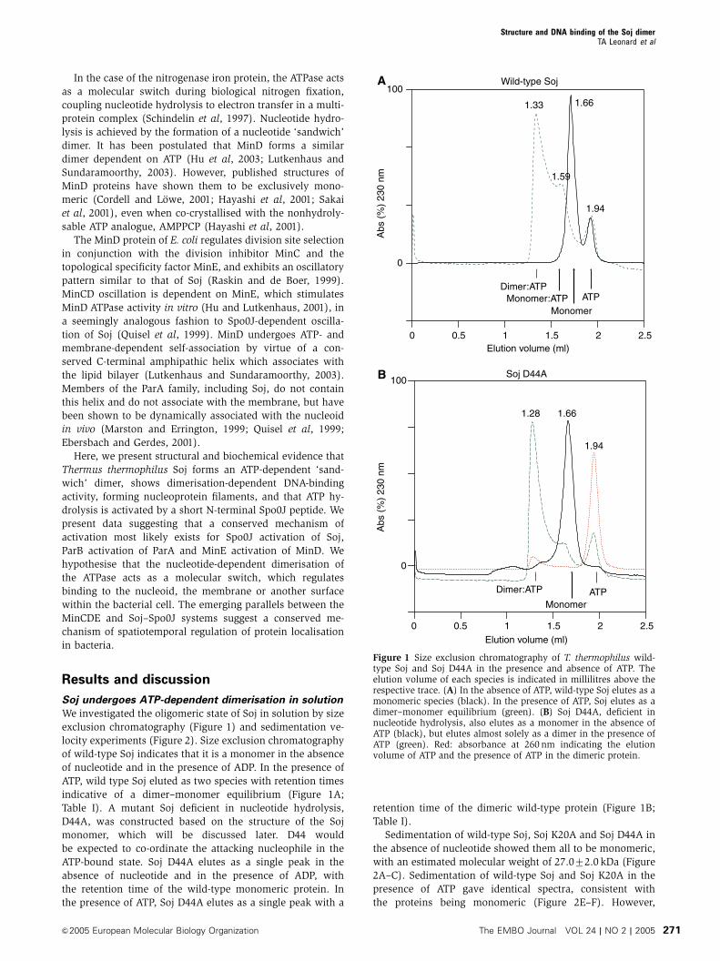
locity experiments (Figure 2). Size exclusion chromatography
of wild-type Soj indicates that it is a monomer in the absence
of nucleotide and in the presence of ADP. In the presence of
ATP, wild type Soj eluted as two species with retention times
indicative of a dimer–monomer equilibrium (Figure 1A;
Table I). A mutant Soj deficient in nucleotide hydrolysis,
D44A, was constructed based on the structure of the Soj
monomer, which will be discussed later. D44 would
be expected to co-ordinate the attacking nucleophile in the
ATP-bound state. Soj D44A elutes as a single peak in the
absence of nucleotide and in the presence of ADP, with
the retention time of the wild-type monomeric protein. In
the presence of ATP, Soj D44A elutes as a single peak with a
retention time of the dimeric wild-type protein (Figure 1B;
Table I).
Sedimentation of wild-type Soj, Soj K20A and Soj D44A in
the absence of nucleotide showed them all to be monomeric,
with an estimated molecular weight of 27.072.0 kDa (Figure
2A–C). Sedimentation of wild-type Soj and Soj K20A in the
presence of ATP gave identical spectra, consistent with
the proteins being monomeric (Figure 2E–F). However,
Abs
(%
) 23
0 nm
0
100
Elution volume (ml)
Abs
(%
) 23
0 nm
0
100
0 0.5 1 1.5 2 2.5
0 0.5 1 1.5 2 2.5
Elution volume (ml)
1.28
1.661.33
1.94
1.66
1.59
Dimer:ATP
MonomerMonomer:ATP ATP
Dimer:ATP
MonomerATP
1.94
Wild-type Soj
Soj D44A
A
B
Figure 1 Size exclusion chromatography of T. thermophilus wild-type Soj and Soj D44A in the presence and absence of ATP. Theelution volume of each species is indicated in millilitres above therespective trace. (A) In the absence of ATP, wild-type Soj elutes as amonomeric species (black). In the presence of ATP, Soj elutes as adimer–monomer equilibrium (green). (B) Soj D44A, deficient innucleotide hydrolysis, also elutes as a monomer in the absence ofATP (black), but elutes almost solely as a dimer in the presence ofATP (green). Red: absorbance at 260 nm indicating the elutionvolume of ATP and the presence of ATP in the dimeric protein.
Structure and DNA binding of the Soj dimerTA Leonard et al
&2005 European Molecular Biology Organization The EMBO Journal VOL 24 | NO 2 | 2005 271
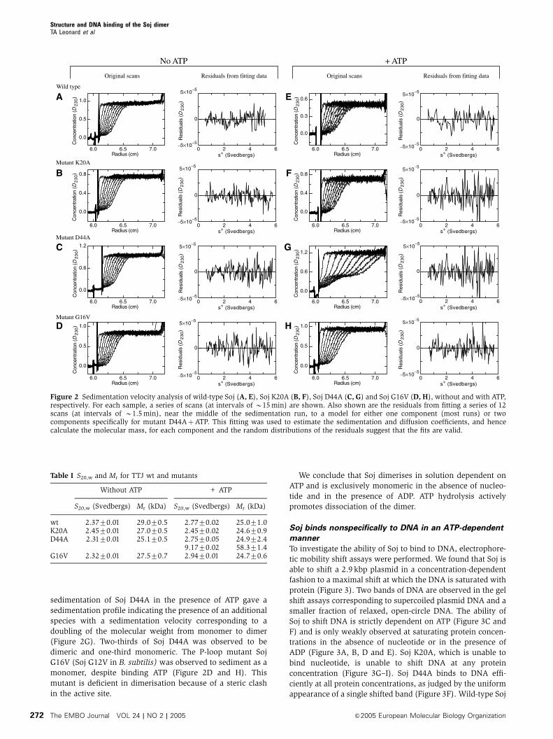
sedimentation of Soj D44A in the presence of ATP gave a
sedimentation profile indicating the presence of an additional
species with a sedimentation velocity corresponding to a
doubling of the molecular weight from monomer to dimer
(Figure 2G). Two-thirds of Soj D44A was observed to be
dimeric and one-third monomeric. The P-loop mutant Soj
G16V (Soj G12V in B. subtilis) was observed to sediment as a
monomer, despite binding ATP (Figure 2D and H). This
mutant is deficient in dimerisation because of a steric clash
in the active site.
We conclude that Soj dimerises in solution dependent on
ATP and is exclusively monomeric in the absence of nucleo-
tide and in the presence of ADP. ATP hydrolysis actively
promotes dissociation of the dimer.
Soj binds nonspecifically to DNA in an ATP-dependent
manner
To investigate the ability of Soj to bind to DNA, electrophore-
tic mobility shift assays were performed. We found that Soj is
able to shift a 2.9 kbp plasmid in a concentration-dependent
fashion to a maximal shift at which the DNA is saturated with
protein (Figure 3). Two bands of DNA are observed in the gel
shift assays corresponding to supercoiled plasmid DNA and a
smaller fraction of relaxed, open-circle DNA. The ability of
Soj to shift DNA is strictly dependent on ATP (Figure 3C and
F) and is only weakly observed at saturating protein concen-
trations in the absence of nucleotide or in the presence of
ADP (Figure 3A, B, D and E). Soj K20A, which is unable to
bind nucleotide, is unable to shift DNA at any protein
concentration (Figure 3G–I). Soj D44A binds to DNA effi-
ciently at all protein concentrations, as judged by the uniform
appearance of a single shifted band (Figure 3F). Wild-type Soj
6.0 6.5 7.0
0.0
0.5
1.0
Radius (cm)
Con
cent
ratio
n (D
23
0)
6.0 6.5 7.0
0.0
0.4
0.8
Radius (cm)
Con
cent
ratio
n (D
23
0)
6.0 6.5 7.0
0.0
0.6
1.2
Radius (cm)
Con
cent
ratio
n (D
23
0)
6.0 6.5 7.0
0.0
0.5
1.0
Radius (cm)
Con
cent
ratio
n (D
23
0)
0 2 4 6
0
s∗ (Svedbergs)
Res
idua
ls (D
23
0)
0 2 4 6
0
s∗ (Svedbergs)
Res
idua
ls (D
23
0)
0 2 4 6
0
s∗ (Svedbergs)
Res
idua
ls (D
23
0)
0 2 4 6
0
s∗ (Svedbergs)
Res
idua
ls (D
23
0)
6.0 6.5 7.0
0.0
0.3
0.6
Radius (cm)
Con
cent
ratio
n (D
23
0)
6.0 6.5 7.0
0.0
0.4
0.8
Radius (cm)
Con
cent
ratio
n (D
23
0)
6.0 6.5 7.0
0.0
0.6
1.2
Con
cent
ratio
n (D
23
0)
Radius (cm)
6.0 6.5 7.0
0.0
0.5
1.0
Radius (cm)
Con
cent
ratio
n (D
23
0)
0 2 4 6
0
s∗ (Svedbergs)
Res
idua
ls (D
23
0)
0 2 4 6
0
s∗ (Svedbergs)
Res
idua
ls (D
23
0)
0 2 4 6
0
s∗ (Svedbergs)
Res
idua
ls (D
23
0)
No ATP + ATP
Original scans Residuals from fitting data Original scans Residuals from fitting data
Wild type
Mutant K20A
Mutant D44A
Mutant G16V
0 2 4 6
0
s∗ (Svedbergs)
Res
idua
ls (D
23
0)
A
B
C
D
E
F
G
H
−5×10⁻⁵
5×10⁻⁵
−5×10⁻⁵
5×10⁻⁵
−5×10⁻⁵
5×10⁻⁵
−5×10⁻⁵
5×10⁻⁵
−5×10⁻⁵
5×10⁻⁵
−5×10⁻⁵
5×10⁻⁵
−5×10⁻⁵
5×10⁻⁵
−5×10⁻⁵
5×10⁻⁵
Figure 2 Sedimentation velocity analysis of wild-type Soj (A, E), Soj K20A (B, F), Soj D44A (C, G) and Soj G16V (D, H), without and with ATP,respectively. For each sample, a series of scans (at intervals of B15 min) are shown. Also shown are the residuals from fitting a series of 12scans (at intervals of B1.5 min), near the middle of the sedimentation run, to a model for either one component (most runs) or twocomponents specifically for mutant D44AþATP. This fitting was used to estimate the sedimentation and diffusion coefficients, and hencecalculate the molecular mass, for each component and the random distributions of the residuals suggest that the fits are valid.
Table I S20,w and Mr for TTJ wt and mutants
Without ATP + ATP
S20,w (Svedbergs) Mr (kDa) S20,w (Svedbergs) Mr (kDa)
wt 2.3770.01 29.070.5 2.7770.02 25.071.0K20A 2.4570.01 27.070.5 2.4570.02 24.670.9D44A 2.3170.01 25.170.5 2.7570.05 24.972.4
9.1770.02 58.371.4G16V 2.3270.01 27.570.7 2.9470.01 24.770.6
Structure and DNA binding of the Soj dimerTA Leonard et al
The EMBO Journal VOL 24 | NO 2 | 2005 &2005 European Molecular Biology Organization272
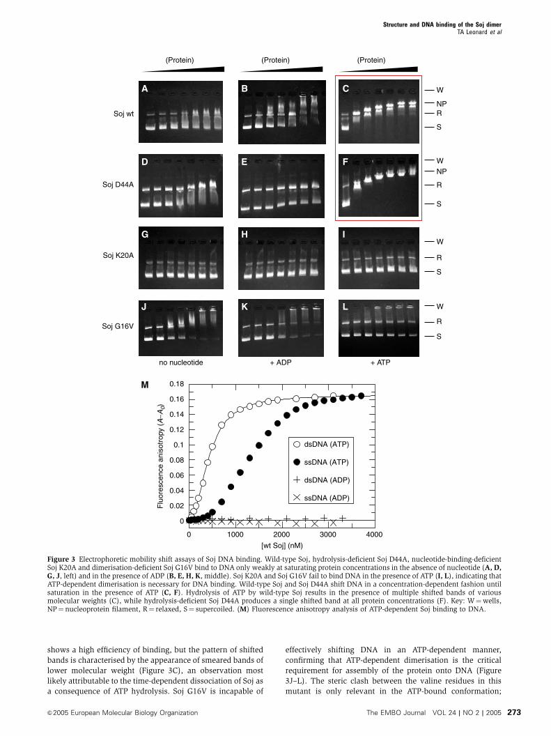
shows a high efficiency of binding, but the pattern of shifted
bands is characterised by the appearance of smeared bands of
lower molecular weight (Figure 3C), an observation most
likely attributable to the time-dependent dissociation of Soj as
a consequence of ATP hydrolysis. Soj G16V is incapable of
effectively shifting DNA in an ATP-dependent manner,
confirming that ATP-dependent dimerisation is the critical
requirement for assembly of the protein onto DNA (Figure
3J–L). The steric clash between the valine residues in this
mutant is only relevant in the ATP-bound conformation;
Soj wt
Soj D44A
Soj K20A
no nucleotide + ADP + ATP
Soj G16V
W
W
W
W
R
NP
NP
R
S
S
R
R
S
S
(Protein)(Protein) (Protein)
A B C
D E F
G H I
J K L
40003000200010000
0.18
0.16
0.14
0.12
0.1
0.08
0.06
0.04
0.02
0
dsDNA (ATP)
ssDNA (ATP)
dsDNA (ADP)
ssDNA (ADP)
Flu
ores
cenc
e an
isot
ropy
(A
−A0)
[wt Soj] (nM)
M
Figure 3 Electrophoretic mobility shift assays of Soj DNA binding. Wild-type Soj, hydrolysis-deficient Soj D44A, nucleotide-binding-deficientSoj K20A and dimerisation-deficient Soj G16V bind to DNA only weakly at saturating protein concentrations in the absence of nucleotide (A, D,G, J, left) and in the presence of ADP (B, E, H, K, middle). Soj K20A and Soj G16V fail to bind DNA in the presence of ATP (I, L), indicating thatATP-dependent dimerisation is necessary for DNA binding. Wild-type Soj and Soj D44A shift DNA in a concentration-dependent fashion untilsaturation in the presence of ATP (C, F). Hydrolysis of ATP by wild-type Soj results in the presence of multiple shifted bands of variousmolecular weights (C), while hydrolysis-deficient Soj D44A produces a single shifted band at all protein concentrations (F). Key: W¼wells,NP¼nucleoprotein filament, R¼ relaxed, S¼ supercoiled. (M) Fluorescence anisotropy analysis of ATP-dependent Soj binding to DNA.
Structure and DNA binding of the Soj dimerTA Leonard et al
&2005 European Molecular Biology Organization The EMBO Journal VOL 24 | NO 2 | 2005 273
hence, at high protein concentrations in the absence of
nucleotide and in the presence of ADP, the protein shows a
notable propensity to shift the DNA (Figure 3J–K). This
effect, also observed to an extent with the wild-type protein
(Figure 3A–B), is most likely a concentration-dependent
effect that ‘dimerises’ the protein in the absence of nucleo-
tide. We conclude that ATP-dependent dimerisation of Soj is
critical for DNA binding.
While the ATP-dependent polymerisation of Soj on DNA
could be a simple charge effect, it is consistent with in vivo
localisation studies which show that wild-type Soj dynami-
cally associates with the nucleoid in an Spo0J-dependent
fashion and remains statically associated with the nucleoid
in a Dspo0J background (Marston and Errington, 1999; Quisel
et al, 1999). In addition, Quisel et al showed that the P-loop
mutant G12V of B. subtilis does not associate with the
nucleoid in a Dspo0J background and its dynamic behaviour
is abolished. Furthermore, the MinD protein of E. coli has
been shown to undergo surface-dependent polymerisation.
MinD assembles into protein filaments in an ATP- and
phospolipid-dependent manner (Lackner et al, 2003), sup-
porting in vivo localisation studies which show that MinD
localises to the polar membrane (Raskin and de Boer, 1999)
and oscillates in membrane-associated coiled structures that
extend between the cell poles (Shih et al, 2003). It is worth-
while mentioning at this point that the similarity between the
Soj and MinD proteins is so great that, it appears to us,
mistakes have been made in the assignment of these proteins,
such that there are in fact two published ‘MinD’ structures
which are more than likely members of the Soj family, lacking
the MinD-characteristic C-terminal amphipathic helix which
mediates membrane association (Szeto et al, 2002; Hu and
Lutkenhaus, 2003).
Soj binds preferentially to double-stranded DNA
To investigate whether Soj binds preferentially to double-
stranded or single-stranded DNA, fluorescence anisotropy
measurements (Figure 3M) were made using a 50-fluores-
cein-labelled oligonucleotide (see Materials and methods).
There is no change in the anisotropy of the DNA for Soj in the
presence of ADP, indicating that Soj:ADP does not bind to
either double-stranded or single-stranded DNA. In the pre-
sence of ATP, however, Soj binds to both double-stranded and
single-stranded DNA. Binding of Soj to double-stranded DNA
can be fitted by the Hill equation for cooperative binding,
yielding a Hill coefficient of 2.11. Binding of Soj to single-
stranded DNA is approximately 3.5-fold less efficient and
cannot be fitted by the Hill equation, although a sigmoidal
curve is apparent. We conclude that Soj only binds DNA in
the ATP-bound form and that Soj binds preferentially to
double-stranded DNA in a cooperative fashion.
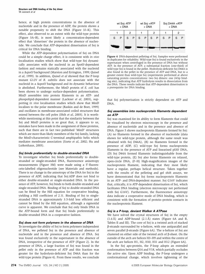
Soj does not form polymers in the absence of DNA
To investigate the ability of Soj to form polymers independent
of DNA, we pelleted Soj in the presence and absence of
nucleotide and in the presence and absence of DNA. Soj
was found exclusively in the supernatant in the absence of
DNA, irrespective of the presence of ATP (Figure 2). In the
presence of DNA, a large fraction of Soj was found in the
pellet only in the presence of ATP and this fraction was
greater for the hydrolysis-deficient Soj D44A than for the
wild-type protein (Figure 4). From these results, we conclude
that Soj polymerisation is strictly dependent on ATP and
DNA.
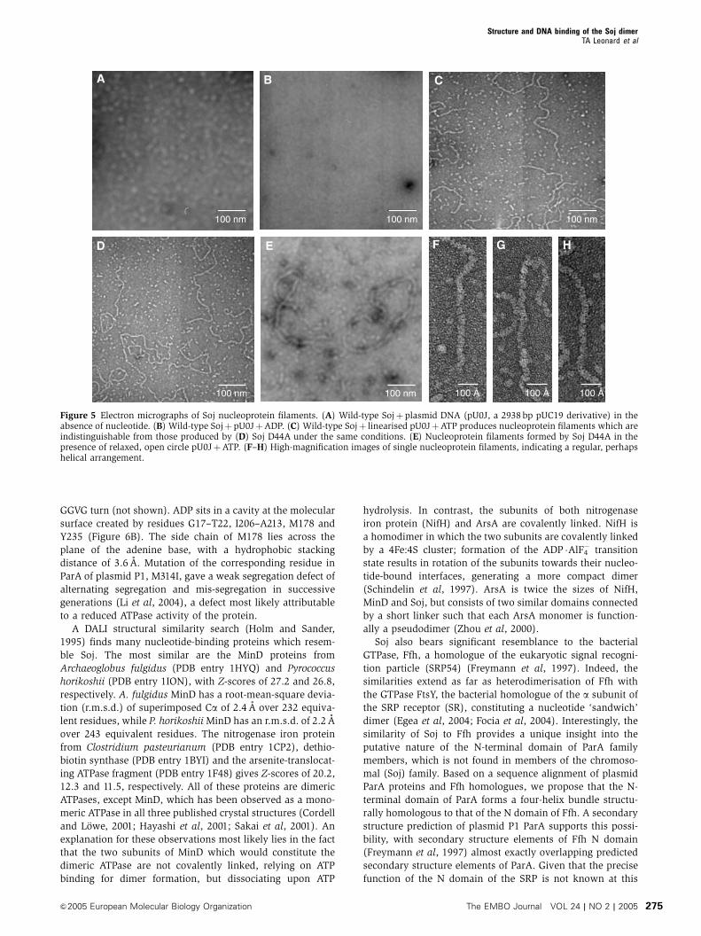
Soj assembles into nucleoprotein filaments dependent
on ATP
Soj was examined for its ability to form filaments that could
be visualised by electron miscroscopy in the presence and
absence of nucleotide and in the presence and absence of
DNA. Figure 5 shows nucleoprotein filaments formed by Soj:
(A) no filaments formed in the absence of nucleotide (data
shown for wild-type protein, although the same result was
obtained with Soj D44A), (B) no filaments formed in the
presence of ADP, (C) wild-type Soj forms nucleoprotein
filaments in the presence of ATP and linearised pU0J DNA,
(D) Soj D44A formed filaments indistinguishable from the
wild-type protein, (E) Soj also forms filaments on relaxed,
open-circle DNA, (F–H) High-magnification images of the
nucleoprotein filament, indicating that they most likely
have a regular, perhaps helical structure. Taken together
with the results of the pelleting and gel shift assays, we
have demonstrated that Soj forms nucleoprotein filaments
in an ATP- and DNA-dependent manner. Soj G16V indicates
that, critically, it is ATP-dependent dimerisation of Soj, which
facilitates DNA binding (electron microscopy not performed
with Soj G16V). Furthermore, the fluorescence anisotropy
data indicate a cooperative mode of DNA binding, which is
consistent with the formation of protein–protein contacts in
the nucleoprotein filament.
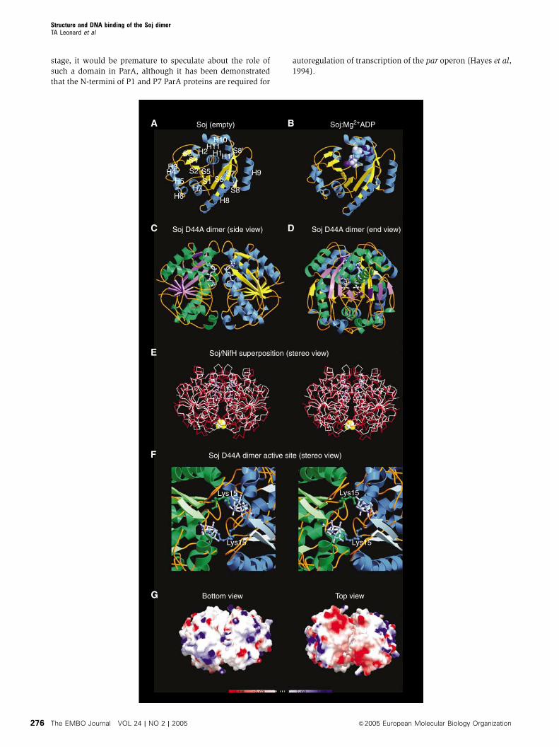
Soj is a P-loop, deviant Walker A ATPase
We have solved the crystal structures of Soj in the empty
(1.6 A) and ADP-bound (2.1 A) states (Figure 6A and B,
Tables II and III). The core of Soj is a twisted arch of stacked
b-strands surrounded by a-helices, with one antiparallel and
seven parallel b-strands (Figure 6A). The a-helices of Soj are
clustered on either side of the twisted arch of b-sheet. On the
outside of the arch are helices H3–H9 and within the centre of
the arch are helices H1, H2, H10, H11 and H12 (Figure 6A).
In the Soj apo-protein, the P-loop adopts an extended
conformation between Q14 and V18, which partially occludes
the active site. Upon ADP binding, the P-loop undergoes a
conformational change, which involves tightening of the
S P S S S SSP P P P P
wt Soj−ATP+ DNA
wt Soj +ATP+ DNA
Soj D44A +ATP+ DNA
1 2 1 1 22
Figure 4 DNA-dependent pelleting of Soj. Samples were performedin duplicates for reliability. Wild-type Soj is found exclusively in thesupernatant when centrifuged in the presence of DNA but withoutATP. In the presence of ATP, a substantial fraction (one-third) ofwild-type Soj is found in the pellet. Hydrolysis-deficient Soj D44A isalso found in the pellet in the presence of ATP and DNA, but to agreater extent than wild-type Soj (experiments performed at abovesaturating protein concentrations: two Soj dimers: one 24 bp bind-ing site), indicating that ATP hydrolysis results in dissociation fromthe DNA. These results indicate that ATP-dependent dimerisation isa prerequisite for DNA binding.
Structure and DNA binding of the Soj dimerTA Leonard et al
The EMBO Journal VOL 24 | NO 2 | 2005 &2005 European Molecular Biology Organization274
GGVG turn (not shown). ADP sits in a cavity at the molecular
surface created by residues G17–T22, I206–A213, M178 and
Y235 (Figure 6B). The side chain of M178 lies across the
plane of the adenine base, with a hydrophobic stacking
distance of 3.6 A. Mutation of the corresponding residue in
ParA of plasmid P1, M314I, gave a weak segregation defect of
alternating segregation and mis-segregation in successive
generations (Li et al, 2004), a defect most likely attributable
to a reduced ATPase activity of the protein.
A DALI structural similarity search (Holm and Sander,
1995) finds many nucleotide-binding proteins which resem-
ble Soj. The most similar are the MinD proteins from
Archaeoglobus fulgidus (PDB entry 1HYQ) and Pyrococcus
horikoshii (PDB entry 1ION), with Z-scores of 27.2 and 26.8,
respectively. A. fulgidus MinD has a root-mean-square devia-
tion (r.m.s.d.) of superimposed Ca of 2.4 A over 232 equiva-
lent residues, while P. horikoshii MinD has an r.m.s.d. of 2.2 A
over 243 equivalent residues. The nitrogenase iron protein
from Clostridium pasteurianum (PDB entry 1CP2), dethio-
biotin synthase (PDB entry 1BYI) and the arsenite-translocat-
ing ATPase fragment (PDB entry 1F48) gives Z-scores of 20.2,
12.3 and 11.5, respectively. All of these proteins are dimeric
ATPases, except MinD, which has been observed as a mono-
meric ATPase in all three published crystal structures (Cordell
and Lowe, 2001; Hayashi et al, 2001; Sakai et al, 2001). An
explanation for these observations most likely lies in the fact
that the two subunits of MinD which would constitute the
dimeric ATPase are not covalently linked, relying on ATP
binding for dimer formation, but dissociating upon ATP
hydrolysis. In contrast, the subunits of both nitrogenase
iron protein (NifH) and ArsA are covalently linked. NifH is
a homodimer in which the two subunits are covalently linked
by a 4Fe:4S cluster; formation of the ADP �AlF4� transition
state results in rotation of the subunits towards their nucleo-
tide-bound interfaces, generating a more compact dimer
(Schindelin et al, 1997). ArsA is twice the sizes of NifH,
MinD and Soj, but consists of two similar domains connected
by a short linker such that each ArsA monomer is function-
ally a pseudodimer (Zhou et al, 2000).
Soj also bears significant resemblance to the bacterial
GTPase, Ffh, a homologue of the eukaryotic signal recogni-
tion particle (SRP54) (Freymann et al, 1997). Indeed, the
similarities extend as far as heterodimerisation of Ffh with
the GTPase FtsY, the bacterial homologue of the a subunit of
the SRP receptor (SR), constituting a nucleotide ‘sandwich’
dimer (Egea et al, 2004; Focia et al, 2004). Interestingly, the
similarity of Soj to Ffh provides a unique insight into the
putative nature of the N-terminal domain of ParA family
members, which is not found in members of the chromoso-
mal (Soj) family. Based on a sequence alignment of plasmid
ParA proteins and Ffh homologues, we propose that the N-
terminal domain of ParA forms a four-helix bundle structu-
rally homologous to that of the N domain of Ffh. A secondary
structure prediction of plasmid P1 ParA supports this possi-
bility, with secondary structure elements of Ffh N domain
(Freymann et al, 1997) almost exactly overlapping predicted
secondary structure elements of ParA. Given that the precise
function of the N domain of the SRP is not known at this
A B C
D E F G H
100 nm100 nm100 nm
100 Å 100 Å 100 Å100 nm100 nm
Figure 5 Electron micrographs of Soj nucleoprotein filaments. (A) Wild-type Sojþplasmid DNA (pU0J, a 2938 bp pUC19 derivative) in theabsence of nucleotide. (B) Wild-type SojþpU0JþADP. (C) Wild-type Sojþ linearised pU0JþATP produces nucleoprotein filaments which areindistinguishable from those produced by (D) Soj D44A under the same conditions. (E) Nucleoprotein filaments formed by Soj D44A in thepresence of relaxed, open circle pU0JþATP. (F–H) High-magnification images of single nucleoprotein filaments, indicating a regular, perhapshelical arrangement.
Structure and DNA binding of the Soj dimerTA Leonard et al
&2005 European Molecular Biology Organization The EMBO Journal VOL 24 | NO 2 | 2005 275
stage, it would be premature to speculate about the role of
such a domain in ParA, although it has been demonstrated
that the N-termini of P1 and P7 ParA proteins are required for
autoregulation of transcription of the par operon (Hayes et al,
1994).
A B
C
E
D
F
S8'
S8
H9S7S6S1
S5S2S4
S3 H1H2
H5
H6H7
H8
H10H11
H12H3H4
G Bottom view Top view
Soj (empty) Soj:Mg2+ADP
Soj D44A dimer (side view) Soj D44A dimer (end view)
Soj/NifH superposition (stereo view)
Soj D44A dimer active site (stereo view)
Lys15
Lys15 Lys15
Lys15
Structure and DNA binding of the Soj dimerTA Leonard et al
The EMBO Journal VOL 24 | NO 2 | 2005 &2005 European Molecular Biology Organization276
Hydrolysis-deficient Soj forms a nucleotide ‘sandwich’
dimer
We have crystallised the hydrolysis-deficient mutant of Soj,
Soj D44A, in the ATP-bound state (Tables II and III), showing
conclusively for the first time the ability of this family of
proteins to form a nucleotide ‘sandwich’ dimer (Figure 6C–D)
which is structurally homologous to the nitrogenase iron
protein dimer (Schindelin et al, 1997) (Figure 6E). The
dimer interface is made between the nucleotide-binding
surfaces of each monomer and is stabilised by networks of
H-bonds between residues on adjacent chains and water-
mediated hydrogen bonds. The ‘signature’ lysine, K15, of
each chain stabilises the a and g phosphates of the ATP
moiety bound by the adjacent chain; the amino group is in
2.96 A co-ordination distance to the a-phosphate and 2.76 A
from the g-phosphate. The P-loops and switch II motifs of
each chain are found very close to each other in the dimer
structure (Figure 6F). In the region of the P-loops, the amide
nitrogens of G16 and G17 from adjacent chains stabilise the
g-phosphate of one ATP moiety.
The dimeric structure of Soj complexed with ATP contains
a cleft on both sides of the dimer, which extends part of the
way down the dimer interface and in which the two surfaces
are complementary in terms of both shape and charge
distribution. The cleft itself is almost exclusively lined by
hydrophobic residues contributed by each monomer
(Figure 6G). A comparison of the active sites of Soj and
NifH indicates that the nucleotides adopt different conforma-
tions. The two ATP molecules in the Soj active site chamber
adopt a ‘kinked’ conformation in which the g-phosphate is
Table II Crystallographic data
Crystal l (A) Resol. (A) I/sIa Rmb (%) Multipl.c Compl.d (%)
1. Soj (apoprotein), space group P43212, a¼ b¼ 61.35 A, c¼ 124.53 AKAu(CN)2 0.9202 1.8 14.3(3.8) 0.094 7.0 99.8Na2WO4 0.9793 1.8 10.8(3.1) 0.072 3.7 99.9NATI 0.9393 1.6 14.0(4.3) 0.096 7.0 99.8
2. Soj:Mg2+ADP, space group P43212, a¼ b¼ 61.38 A, c¼ 126.66 ANATI 1.4880 2.1 14.3(4.0) 0.055 3.6 99.2
3. Soj D44A:Mg2+ATP, space group P212121, a¼ 87.43 A, b¼ 95.91 A, c¼ 123.94 ANATI 0.934 1.8 14.8(6.2) 0.067 4.0 99.9
aSignal-to-noise ratio for the highest resolution of intensities.bRm:
Ph
PI|I(h,I)�I(h)|/
Ph
PII(h,I) where I(h,I) are symmetry-related intensities and I(h) is the mean intensity of the reflection with unique
index h.cMultiplicity for unique reflections.dCompleteness for unique reflections.
Table III Refinement statistics
Soj (apoprotein) Soj:Mg2+ADP Soj D44A:Mg2+ATP
Model 5-247, 254 H2O 5-247, ADP Chains A-D, 5-247,4 ATP, 1279 H2O
Diffraction data NATI, 1.6 A NATI, 2.1 A NATI, 1.8 AR-factor, R-freea 0.229, 0.252 0.218, 0.266 0.1815, 0.2156B average/bondedb 29.66 A2/2.70 A2 50.01/0.75 A2 20.29 A2/2.46 A2
Geometry bonds/anglesc 0.006 A/1.371 0.009 A/1.151 0.005 A/1.291Ramachandrand 92.1%/0.0% 92.7%/0.0% 92.0%/0.0%PDB IDe 1wcv 2bej 2bek
aIn all, 5% of reflections were randomly selected for determination of the free R-factor, prior to any refinement.bTemperature factors averaged for all atoms and r.m.s.d. of temperature factors between bonded atoms.cR.m.s.d. from ideal geometry for bond lengths and restraint angles (Engh, 1991).dPercentage of residues in the ‘most favoured’ region of the Ramachandran plot and percentage of outliers (PROCHECK; Laskowski et al, 1993).eProtein Data Bank identifiers for co-ordinates.
Figure 6 Crystal structures of Soj. (A) Crystal structure of Soj in the empty state at 1.6 A. The arrangement of the sheet and helices follows thatof the MinD family of ATPases (Cordell and Lowe, 2001; Hayashi et al, 2001; Sakai et al, 2001). (B) Structure of Soj:Mg2þADP at 2.1 A.Nucleotide binding is coupled to rearrangement of the P-loop. (C) Structure of hydrolysis-deficient Soj D44A in the dimeric state (side view),indicating the symmetrical assembly of the two monomers and the close proximity of the two nucleotides. (D) End view of Soj D44A dimer. (E)Structure of Soj D44A superimposed on the structure of nitrogenase iron protein from A. vinelandii (PDB ID: 1n2c) (Schindelin et al, 1997). Thehigh structural homology between the two proteins indicates that the dimeric structure of Soj is correct. (F) Stereo view of the Soj D44A dimeractive site. The nucleotide-binding surface of each monomer contributes to the formation of the active site chamber, which accommodates twomolecules of ATP. Each monomer also contributes a universally conserved lysine (Lys15), which stabilises the negative charges on theopposing ATP. (G) Bottom and top views of the electrostatic surface potential maps of the Soj D44A dimer. The top view clearly indicates twopatches of negative charge (one on each monomer) and, importantly, the existence of a cleft between the two monomers in which the surfacesare entirely complementary.
Structure and DNA binding of the Soj dimerTA Leonard et al
&2005 European Molecular Biology Organization The EMBO Journal VOL 24 | NO 2 | 2005 277
accommodated without structural rearrangement of the
switch II region and K15 of the opposing subunit stabilises
the a- and b-phosphates. In the NifH active site, ADP adopts
an extended conformation in which the conserved lysine
(K10) of the opposing subunit stabilises the b-phosphate
and the switch II region undergoes a conformational change,
placing the P-loop G16 of each subunit B4 A apart compared
to B10 A in the nucleotide-free protein. This results in a
rotation of B131 of each monomer towards the subunit
interface, closing the dimer into a more compact structure
in the complex. AlF4� in the NifH dimer structure mimics the
transition state of the hydrolysis reaction by adopting a
planar conformation in which octahedral co-ordination is
completed by the terminal oxygen of the b-phosphate of
ADP and the attacking nucleophilic water, which is co-
ordinated by the side-chain carboxyl oxygen of D39 (D44 in
Soj). Based on the structure of NifH with molybdenum iron
protein, we suggest that formation of the transition state
closes the dimer interface such that Soj is competent for
interaction with Spo0J.
The effects of several mutations of the Walker A box have
been reported for B. subtilis Soj. Wild-type Soj oscillates
within the nucleoid region of the cell over a time course of
several minutes, but in a Dspo0J background, Soj remains
statically associated with the nucleoid (Quisel et al, 1999).
The mutation K16Q in B. subtilis Soj has been observed to
abolish the in vivo oscillatory behaviour of Soj, its association
with the nucleoid and its response to Spo0J, consistent with a
lack of nucleotide binding (Quisel et al, 1999). This observa-
tion is supported by the equivalent mutation, K20A, in T.
thermophilus Soj, which abolishes nucleotide binding. A
second mutant, D125A, in B. subtilis Soj is also predicted
not to bind nucleotide. A third mutation, G12V, however
exhibits a different localisation pattern. Like wild-type Soj,
Soj G12V localises to the cell poles in Spo0Jþ cells, but,
unlike wild-type Soj, it also localises to the poles in a spo0J
null mutant (Quisel et al, 1999). Mutation of the equivalent
residue, G16V, in the structure of dimeric T. thermophilus Soj
indicates a steric clash between V16 of each monomer and
P121 of the switch II region, V16 of the adjacent monomer
and the g-phosphate of ATP, which would prevent dimerisa-
tion of Soj. Soj G16V sediments as a monomer in the presence
of ATP (Figure 2H), confirming that it is deficient in dimer-
isation, and from this we conclude that nucleoprotein fila-
ment formation is dependent on ATP-mediated dimerisation.
Soj ATPase is activated by the N-terminal 20 amino
acids of Spo0J
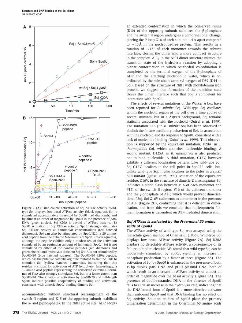
The ATPase activity of wild-type Soj was assayed using the
malachite green method of Chan et al (1986). Wild-type Soj
displays low basal ATPase activity (Figure 7A). Soj K20A
displays no detectable ATPase activity, a consequence of its
failure to bind nucleotide. We found that wild-type Soj can be
moderately stimulated by Spo0J, yielding an increase in
phosphate production by a factor of three (Figure 7A). The
activation of Soj by Spo0J is enhanced in the presence of both
19 bp duplex parS DNA and pU0J plasmid DNA, both of
which result in an increase in ATPase activity of almost an
order of magnitude over the basal activity (Figure 7A). The
presence of double-stranded DNA in the absence of Spo0J
fails to elicit an increase in the hydrolysis rate, indicating that
the DNA-bound form of Spo0J is a more effective activator
than unbound Spo0J and that DNA binding has no effect on
Soj activity. Solution studies of Spo0J place the primary
dimerisation determinant in the C-terminal 60 amino acids
0
2
4
6
8
0
5
10
15
0E+00 2E − 08 4E − 08 6E − 08 8E − 08
mol
Pi p
rodu
ced/
mol
Soj
0 50 100 150 200
Time/min
mol
Pi p
rodu
ced/
mol
Soj
mol Spo0J/peptide
wt Soj / Soj + parS
Soj + Spo0J
Soj + Spo0J:parS
+ Spo0J
+ Spo0JN20peptide
+ FtsA − Cpeptide
+ controlpeptide 2
+ controlpeptide 1
Soj D44A +Spo0JN20
+ Spo0JN20R10A
Soj K20A
A
B
Figure 7 (A) Time course activation of Soj ATPase activity. Wild-type Soj displays low basal ATPase activity (black squares). Soj isstimulated approximately three-fold by Spo0J (red diamonds) andby almost an order of magnitude by Spo0J in the presence of parSDNA (green circles). Soj K20A is devoid of ATPase activity. (B)Spo0J activation of Soj ATPase activity. Spo0J strongly stimulatesSoj ATPase activity at nanomolar concentrations (red hatcheddiamonds). Soj can also be stimulated by Spo0JN20, a 20 amino-acid peptide from the extreme N-terminus of Spo0J (black squares),although the peptide exhibits only a modest 8% of the activationstimulated by an equimolar amount of full-length Spo0J. Soj is notstimulated by either of the control peptides (red diamonds andgreen circles) and hydrolysis-deficient Soj D44A is not stimulated bySpo0JN20 (blue hatched squares). The Spo0JN20 R10A peptide,which has the putative catalytic arginine mutated to alanine, fails tostimulate Soj (yellow hatched diamonds), indicating that thisresidue is critical for activation of ATP hydrolysis. Interestingly, a19 amino-acid peptide representing the conserved extreme C-termi-nus of FtsA also strongly stimulates Soj, but to a lesser extent thanSpo0JN20. The kinetics of activation by Spo0JN20 and full-lengthSpo0J indicate possible cooperativity of binding and activation,consistent with dimeric Spo0J binding dimeric Soj.
Structure and DNA binding of the Soj dimerTA Leonard et al
The EMBO Journal VOL 24 | NO 2 | 2005 &2005 European Molecular Biology Organization278
of the protein, but biochemical and structural studies have
also shown that the N-terminal and central DNA-binding
domains dimerise, a requirement for HTH-mediated DNA
binding (Leonard et al, 2004). We hypothesise that Spo0J
undergoes DNA-dependent dimerisation of its N-termini and
that this is coupled to activation of ATP hydrolysis by Soj.
Biochemical studies of P1 ParB, C. crescentus ParB and F
plasmid SopB have mapped the ParA/SopA interaction de-
terminant to the extreme N-terminus of the protein (Surtees
and Funnell, 1999; Figge et al, 2003; Ravin et al, 2003).
Alignments of putative Spo0J proteins indicated to us that
the region responsible for Soj activation lay in the first 20
amino acids, given the high conservation observed. This
region of seemingly flexible nature is not visible in the
structure of Spo0J (Leonard et al, 2004). We found that the
hydrolysis rate of Soj could be strongly and specifically
stimulated by this N-terminal 20 amino-acid peptide,
Spo0JN20 (Figure 7B). The peptide did not activate Soj to
the same extent as Spo0J, exhibiting 8% activation compared
with full-length Spo0J at equimolar concentrations
(Figure 7B). This is in agreement with our observation that
the context of the N-termini is important for activation.
Additional protein–protein contacts may be made in the
case of full-length Spo0J, thereby increasing affinity for the
binding site, and there is likely an entropic effect of both
ligands being supplied by a dimeric Spo0J molecule rather
than a monomeric ligand binding to two sites. Soj was not
activated by two control peptides (Figure 7B). Interestingly,
the peptide CASVGSWIKRLNSWLRKEF exhibited moderate
stimulation of Soj (75% when compared with TTJN20)
(Figure 7B). This 19 amino-acid peptide represents the ex-
treme C-terminus of E. coli FtsA, conservation of which was
first recognised by Lowe and van den Ent (Lowe and van den
Ent, 2001), and which is also disordered in the crystal
structure (van den Ent and Lowe, 2000). Alignments of the
extreme C-terminus of FtsA and the extreme N-termini of
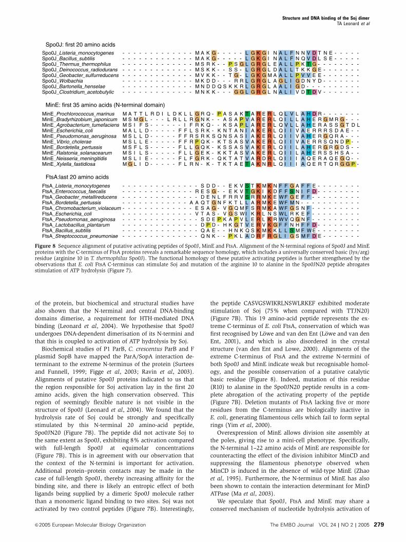
both Spo0J and MinE indicate weak but recognisable homol-
ogy, and the possible conservation of a putative catalytic
basic residue (Figure 8). Indeed, mutation of this residue
(R10) to alanine in the Spo0JN20 peptide results in a com-
plete abrogation of the activating property of the peptide
(Figure 7B). Deletion mutants of FtsA lacking five or more
residues from the C-terminus are biologically inactive in
E. coli, generating filamentous cells which fail to form septal
rings (Yim et al, 2000).
Overexpression of MinE allows division site assembly at
the poles, giving rise to a mini-cell phenotype. Specifically,
the N-terminal 1–22 amino acids of MinE are responsible for
counteracting the effect of the division inhibitor MinCD and
suppressing the filamentous phenotype observed when
MinCD is induced in the absence of wild-type MinE (Zhao
et al, 1995). Furthermore, the N-terminus of MinE has also
been shown to contain the interaction determinant for MinD
ATPase (Ma et al, 2003).
We speculate that Spo0J, FtsA and MinE may share a
conserved mechanism of nucleotide hydrolysis activation of
Spo0J_Listeria_monocytogenesSpo0J_Bacillus_subtilisSpo0J_Thermus_thermophilusSpo0J_Deinococcus_radioduransSpo0J_Geobacter_sulfurreducensSpo0J_WolbachiaSpo0J_Bartonella_henselaeSpo0J_Clostridium_acetobutylic
MinE_Prochlorococcus_marinusMinE_Bradyrhizobium_japonicumMinE_Agrobacterium_tumefaciensMinE_Escherichia_coliMinE_Pseudomonas_aeruginosaMinE_Vibrio_choleraeMinE_Bordetella_pertussisMinE_Ralstonia_solanacearumMinE_Neisseria_meningitidisMinE_Xylella_fastidiosa
FtsA_Listeria_monocytogenesFtsA_Enterococcus_faecalisFtsA_Geobacter_metallireducensFtsA_Bordetella_pertussisFtsA_Chromobacterium_violaceumFtsA_Escherichia_coliFtsA_Pseudomonas_aeruginosaFtsA_Lactobacillus_plantarumFtsA_Bacillus_subtilisFtsA_Streptococcus_pneumoniae
--------
MMMMMMMMMM
----------
--------
ASSASSSSSG
----------
--------
TMILLLFILL
----------
--------
TGFLLLLLII
----------
--------
LLSDDESSED
----------
--------
R---------
----------
--------
D---------
----------
--------
I---------
----------
--------
LL--------
----------
--------
DR--------
----------
--------
KL-FFFFFFF
----------
--------
LLIFFFLLLL
----------
--------
LRFLRRLLFR
---A------
MMMMMMMM
GGRSSPGGGN
-R-AEV----
AASSVKNN
RNKRRQQER-
SEDQSTSDQQ
KKRKKDDK
QKQKKKKKKK
DSETAADPAN
GGKKKDDK
----S-----
DGNGGSEDEK
------Q-
P--KQKKKQT
--LN--P---
------S-
AAKNNTSKKK
--FFVVKH--
--PST-K-
SSSTSSSTTT
EERKGGAKHP
--SSGRKG
AAAAAAAAAA
KKRTQSPGNK
--G--RRG
KPPNSSSSTE
VVVLMWVTKL
LLLLLLLL
TVLIIVVVVT
STSLFILVQA
GGGGGGGG
AAAAAAAAAA
TGRASKEESD
KKRRKRRR
RRRKKKKKRK
KKRRRRRRKR
GGGGGGGG
EEEEEEEEDN
MIMMMLLVMF
IILLMLLL
RRRRRRRRRR
KKKKKNKKKR
NNEDAAAN
LLLLLLLLLL
NDEEASRGKG
AAAAAGAA
QQQQQQQQQQ
FFWWWWWFLL
LLLLLLLL
LIVIIIIIII
FFFFFLVFLI
FFLLLIII
VLLIIIIIII
GSGMGRQNSG
NNPTPGGV
LLLVVVLLII
ANENSKGHMS
NQKKVDDD
AAAAAAAAAA
FIF-NENFFM
VVTKVN-T
HHHEHEHHQQ
FFF-FFFFWF
DDGGEY-D
DEEREREEEE
ED-----DED
TL-EED-V
RRRRRRRRRR
---------E
NS------
-GARGSGSAT
----------
EE------
-MSSQQRSQQ
----------
--------
-RSDRNGHER
----------
--------
-GGAADDSGG
----------
--------
--TE-PSAQG
----------
--------
--D------P
----------
--------
--L-------
----------
Spo0J: first 20 amino acids
MinE: first 35 amino acids (N-terminal domain)
FtsA:last 20 amino acids
Figure 8 Sequence alignment of putative activating peptides of Spo0J, MinE and FtsA. Alignment of the N-terminal regions of Spo0J and MinEproteins with the C-terminus of FtsA proteins reveals a remarkable sequence homology, which includes a universally conserved basic (lys/arg)residue (arginine 10 in T. thermophilus Spo0J). The functional homology of these putative activating peptides is further strengthened by theobservations that E. coli FtsA C-terminus can stimulate Soj and mutation of the arginine 10 to alanine in the Spo0JN20 peptide abrogatesstimulation of ATP hydrolysis (Figure 7).
Structure and DNA binding of the Soj dimerTA Leonard et al
&2005 European Molecular Biology Organization The EMBO Journal VOL 24 | NO 2 | 2005 279
their respective interaction partners. There are two possibi-
lities for the mechanism of activation: the first involves
activation of ATP hydrolysis, the result of which is destabi-
lisation of the dimer and a shift of the equilibrium towards
the monomeric form. The second mechanism involves nu-
cleotide exchange of the monomer following ATP hydrolysis,
the result of which is a shift in the equilibrium to the ATP-
bound, dimeric state. We find the mechanism of activation
rather than nucleotide exchange more attractive because it
involves interaction of a dimeric activator (Spo0J, MinE)
containing two ligands with dimeric ATPase (Soj, MinD
respectively) containing two binding sites, and couples di-
merisation of the ATPase to protein:protein complex assem-
bly, as is observed for the interaction of NifH with
molybdenum iron protein (Schindelin et al, 1997).
The structure of the nitrogenase iron protein in complex
with molybdenum iron protein shows that the ATP-bound
state, and hence ‘closed’ conformation of the dimer, is
necessary for productive interaction with its binding partner
(Schindelin et al, 1997). We hypothesise that the ATP-depen-
dent dimerisation of Soj acts as an identical molecular switch
which regulates its putative interaction with Spo0J, and, by
extension, that dimerisation of MinD regulates its interaction
with MinC/MinE. We predict that the identification of a
hydrolysis-deficient mutant which is constitutively dimeric
in the presence of ATP will be a useful tool in the character-
isation of physiologically relevant protein:protein complexes
and their intracellular functions. ATPases of this deviant
Walker A family play fundamental roles in a diverse range
of cellular processes, from nitrogen fixation and anion extru-
sion to bacterial division site selection and plasmid and
chromosome segregation.
Finally, the similarities between MinDE and Soj/Spo0J are
wide-ranging. MinD and Soj oscillate (jump) between places
in the cell: MinD oscillates by binding to the membrane,
while Soj oscillates by binding to the nucleoid. MinD and Soj
have the same three-dimensional structure apart from the
amphiphatic helix on MinD that is involved in membrane
binding, and most likely form the same ATP-dependent
dimer. MinD and Soj ATPase activity is activated by a short,
disordered peptide located on a dimeric binding partner
(MinE N-terminus, Spo0J N-terminus, respectively) and
both MinD and Soj bind to extended surfaces (MinD: mem-
brane, Soj: DNA) and binding is regulated by dimerisation.
Both the MinDE and the Soj/Spo0J system are involved in
accurate positioning of molecules (MinC and region of the
nucleoid, respectively). The same oscillatory mechanisms
involving surface-assisted polymerisation may be used to
position the septum and the origins of replication.
Materials and methods
Protein expression and purificationSoj from T. thermophilus HB27 (ATCC BAA-163D) was cloned intopHis17 to generate a C-terminally hexa-histidine-tagged protein.Vectors expressing the mutant proteins Soj G16V, K20A and D44Awere constructed using the QuikChange protocol (Stratagene).Single colonies of C41 (DE3) were used to inoculate 2� 65 mlcultures of 2�TY, 0.4% glucose and 100 mg/ml ampicillin, andgrown overnight at 371C. The cultures were used to inoculate 12 lof 2�TYþ 0.4% glucose, which was induced with 1 mM IPTGwhen OD600¼ 0.6 and the temperature reduced to 251C. The cellswere grown overnight. Soj proteins were purifed by NiNTA follo-wed by heparin affinity chromatography and gel filtration on a
Sephacryl S200 column (Amersham Biosciences), equilibrated inTENþ 100 mM NaCl, pH 8.5. The wild-type and mutant proteinseluted as a single peak at a position, indicating them to bemonomeric.
Crystallisation and data collection1152 crystallisation conditions were screened using in-housenanolitre crystallisation robotics (Stock et al, 2004). Crystals ofthe native protein were grown using the sitting drop vapourdiffusion technique using 6% PEG 3350, 0.15 M NaCl and 0.4 M KIas the crystallisation solution. Drops composed of 1ml protein at2 mg/ml and 1 ml crystallisation solution were incubated overnightat 191C. Crystals grew in space group P43212 with cell dimensionsa¼ b¼ 61.35 A and c¼ 124.53 A, and were frozen in mother liquorplus 25% glycerol. Heavy metal derivatives were made by addingKAu(CN)2 or Na2WO4 solutions to the drop to a final concentrationof 4 mM. The drops were incubated overnight at 191C and thecrystals flash frozen as for the natives. The native and derivativedata sets were collected at ID29 ESRF, Grenoble, France.
Co-crystals of Soj:ADP were obtained by adding 1 mM ATPgS and2 mM MgSO4 to the protein solution prior to screening of crystal-lisation conditions. Crystals containing ADP (ATPgS apparentlypartly hydrolysed) grew in 8% PEG 550-MMEþ 8% PEG 20 K, 0.1 Msodium acetate, pH 5.5 and 0.2 M KSCN. The crystals were frozenunder the same cryoprotectant as the natives and also belong tospace group P43212 with unit cell dimensions a¼ b¼ 61.38 A andc¼ 126.66 A. A native data set to 2.1 A resolution was collected onbeam line 14-2 at the SRS facility, Daresbury Laboratory, UK.
Crystals of the Soj D44A dimer were grown by adding 250 mMCHES, pH 10.0, 1 mM ATP and 5 mM MgSO4 to the protein solutionand concentrating it from 2 to 8 mg/ml prior to screening ofcrystallisation conditions. Crystals were obtained in 200 mMimidazole, pH 7.6, and 10–20% isopropanol. Crystals grew in spacegroup P212121 with unit cell dimensions a¼ 87.43 A, b¼ 95.91 A andc¼ 123.94 A. A native data set to 1.8 A resolution was collected onbeam line ID14-1 at the ESRF, Grenoble, France.
All crystals were indexed and integrated using the MOSFLMpackage and further processed using the CCP4 package. Thestructure of Soj apoprotein was solved by mulitple isomorphousreplacement (MIR), while Soj:ADP and the Soj D44A dimer weresolved by molecular replacement. REFMAC (Murshudov et al, 1999)was used for TLS refinement of Soj:ADP.
Analytical ultracentrifugationSedimentation velocity experiments were performed in a BeckmanOptima XL-A analytical ultracentrifuge with an An60-Ti rotor, withthe samples in various buffers as described in the text.
Sedimentation velocity was at 60 000 or 50 000 rev min�1, 5.01C,with scans of the single cell taken at 0-min intervals (to obtain scansas closely spaced as possible; in practice about 1.5 min apart).Adjacent sets of data were analysed by the method of Stafford(1994, 1997) using the program DCDTþ (Philo, 2000).
Size exclusion chromatographySize exclusion chromatography was performed on a calibratedSuperdex 200 3.2. Precision Column (Amersham Biosciences).Samples were applied in a volume of 10ml at 2 mg/ml. The columnwas equilibrated in 50 mM CHES, 100 mM NaCl, 5 mMMgSO470.5 mM ATP, pH 10.0 (the high pH was required to preventprecipitation of the protein upon the addition of ATP; we checkedthat the protein eluted as a single peak consistent with a monomerin the absence of ATP at the same elution volume as in 50 mM Tris–HCl, pH 8.5).
DNA-binding assaysThe ability of wild-type Soj, Soj D44A, Soj K20A and Soj G16V tobind to double-stranded, plasmid DNA was assayed in the absenceof nucleotide and in the presence of Mg2þADP or Mg2þATP.Binding reactions were performed in a volume of 10ml in 50 mMTris–HCl, pH 8.5, 5 mM MgSO4. Each reaction contained 200 fmol ofpUC19 and 0–100 pmol Soj70.75 mM ADP or ATP. Reactions wereincubated for 10 min at 251C, mixed with gel loading buffer, run ona 1% agarose gel in 0.5�TBþ 1 mM MgSO4 buffer and stained withethidium bromide.
Structure and DNA binding of the Soj dimerTA Leonard et al
The EMBO Journal VOL 24 | NO 2 | 2005 &2005 European Molecular Biology Organization280
Fluorescence anisotropyFluorescence anisotropy measurements were collected using aPerkin-Elmer LS50B luminescence spectrometer. A 50-fluorescei-nated oligonucleotide (50-AAAACAAACCCAAAACAAACCC-30) wasused as a fluorescein-labelled single-stranded DNA and annealed toits complementary, unlabelled oligonucleotide to create fluorescein-labelled double-stranded DNA. The binding buffer was 20 mM Tris(pH 8.5), 100 mM NaCl, 5 mM MgSO4, 1 mM ADP or ATP. Wild-typeSoj was serially titrated into the cuvette, which contained 10 nM 50-fluoresceinated DNA. The measurements were performed at 298 K.The data were plotted and the curves fitted using the programGraFit.
Pelleting assaysWild-type Soj and Soj D44A were pelleted in the presence andabsence of ATP and in the presence and absence of pU0J DNA.Reactions were performed in a volume of 30ml. Each reactioncontained 750 pmol wild-type Soj/Soj D44A, 50 mM Tris–HCl, pH8.5, 5 mM MgSO471 mM ATP. For pelleting in the presence of pU0JDNA, reactions contained 1.5 pmol of pU0J. Samples werecentrifuged at 100 000 r.p.m. for 1 h at ambient temperature. Thesupernatants were carefully removed and mixed with an equalvolume of SDS gel loading buffer. The pellets were washed with30ml buffer and then solubilised in 30ml SDS gel loading buffer. Anequal volume of buffer was then added to normalise theconcentrations of components in the supernatant and pellet. Avolume of 30ml of each sample was then run on a 12.5% denaturingpolyacrylamide gel.
Electron microscopySupercoiled or XmnI digested pU0J DNA (40–400 ng) was incubatedwith Soj, Soj K20A or Soj D44A (0–5 mg) in 50 mM Tris–HCl, pH 8.5,5 mM MgSO470.75 mM ATP. The complexes were incubated at251C for 10 min, after which they were applied to glow dischargedcarbon-coated grids for 30 s. The grids were washed with one dropof distilled water, and stained with three drops of 2% uranyl acetatebefore being blotted to dryness. Images were taken on a PhilipEM208 electron microscope at � 50 000 magnification.
ATPase activity assaysThe ATPase activity of Soj was assayed by the spectrophotometricdetection of inorganic phosphate following termination ofthe reaction with an acidic solution containing malachite greenreagent (1:1:2:2 ratio of 5.72%. ammonium molybdate, (w/v) in6 N HCl, 2.32% (w/v) polyvinyl alcohol (Sigma), 0.08712%(w/v) malachite green (Sigma) and distilled water, respectively).Reactions were performed in a volume of 50ml containing 50 mMHEPES, pH 8.0 at 371C for 3.5 h. Each reaction contained 1.82 nmolSoj and 50 nmol ATP. Spo0J activation was assayed by the additionof 0–2 nmol T. thermophilus Spo0J to the reaction. Activationby Spo0J was assayed in the presence of an equimolar amountof parS duplex and in the presence of the 2.9 kbp plasmidpUOJ (Leonard et al, 2004), such that there was an equimolaramount of 24 bp binding sites. The ability of the N-terminal 20amino acids of Spo0J to activate Soj was assayed by incubation ofthe ATPase with increasing concentrations of the 20 amino-acid peptide MSRKPSGLGRGLEALLPKTG (Spo0JN20) and themutant peptide MSRKPSGLGAGLEALLPKTG (Spo0JN20R10A).Control reactions contained the peptides PEGDIPAIYR and ILFPEG-DIPAIYRYGL. The sequence of the E. coli FtsA C-terminalpeptide (FtsA-C) was CASVGSWIKRLNSWLRKEF. Reactions wereterminated by the addition of 200ml malachite green reagent.The colour was allowed to stabilise for 5 min before the absorbancewas measured at 630 nm. A calibration curve was constructed using0–13 nmol inorganic phosphate standards and samples werenormalised for acid hydrolysis of ATP by the malachite greenreagent.
Acknowledgements
We thank the staff at ID14-1 and ID29 of ESRF (Grenoble, France)and the staff of ID14-1 of Daresbury Synchrotron Radiation Source(UK) for assistance with data collection. We thank HenriqueFerreira and Jeff Errington (Sir William Dunn School of Pathology,Oxford, UK) for providing us with plasmid pU0J.
References
Cervin MA, Spiegelman GB, Raether B, Ohlsen K, Perego M, HochJA (1998) A negative regulator linking chromosome segregationto developmental transcription in Bacillus subtilis. Mol Microbiol29: 85–95
Chan KM, Delfert D, Junger KD (1986) A direct colorimetric assayfor Ca2+-stimulated ATPase activity. Anal Biochem 157: 375–380
Cordell SC, Lowe J (2001) Crystal structure of the bacterial celldivision regulator MinD. FEBS Lett 492: 160–165
Draper GC, Gober JW (2002) Bacterial chromosome segregation.Annu Rev Microbiol 56: 567–597
Easter Jr J, Gober JW (2002) ParB-stimulated nucleotide exchangeregulates a switch in functionally distinct ParA activities. Mol Cell10: 427–434
Ebersbach G, Gerdes K (2001) The double par locus of virulencefactor pB171: DNA segregation is correlated with oscillation ofParA. Proc Natl Acad Sci USA 98: 15078–15083
Egea PF, Shan SO, Napetschnig J, Savage DF, Walter P, Stroud RM(2004) Substrate twinning activates the signal recognition particleand its receptor. Nature 427: 215–221
Engh RA (1991) Accurate bond and angle parameters for X-rayprotein structure refinement. Acta Crystallogr Sect A 47: 392–400
Figge RM, Easter J, Gober JW (2003) Productive interaction be-tween the chromosome partitioning proteins, ParA and ParB, isrequired for the progression of the cell cycle in Caulobactercrescentus. Mol Microbiol 47: 1225–1237
Focia PJ, Shepotinovskaya IV, Seidler JA, Freymann DM (2004)Heterodimeric GTPase core of the SRP targeting complex. Science303: 373–377
Freymann DM, Keenan RJ, Stroud RM, Walter P (1997) Structure ofthe conserved GTPase domain of the signal recognition particle.Nature 385: 361–364
Glaser P, Sharpe ME, Raether B, Perego M, Ohlsen K, Errington J(1997) Dynamic, mitotic-like behavior of a bacterial protein
required for accurate chromosome partitioning. Genes Dev 11:1160–1168
Gordon GS, Sitnikov D, Webb CD, Teleman A, Straight A, Losick R,Murray AW, Wright A (1997) Chromosome and low copy plasmidsegregation in E. coli: visual evidence for distinct mechanisms.Cell 90: 1113–1121
Gordon GS, Wright A (2000) DNA segregation in bacteria. Annu RevMicrobiol 54: 681–708
Hayashi I, Oyama T, Morikawa K (2001) Structural and functionalstudies of MinD ATPase: implications for the molecular recogni-tion of the bacterial cell division apparatus. EMBO J 20:1819–1828
Hayes F, Radnedge L, Davis MA, Austin SJ (1994) The homologousoperons for P1 and P7 plasmid partition are autoregulated fromdissimilar operator sites. Mol Microbiol 11: 249–260
Hiraga S (1992) Chromosome and plasmid partition in Escherichiacoli. Annu Rev Biochem 61: 283–306
Holm L, Sander C (1995) Dali: a network tool for protein structurecomparison. Trends Biochem Sci 20: 478–480
Hu Z, Lutkenhaus J (2001) Topological regulation of cell division inE. coli. Spatiotemporal oscillation of MinD requires stimulation ofits ATPase by MinE and phospholipid. Mol Cell 7: 1337–1343
Hu Z, Lutkenhaus J (2003) A conserved sequence at the C-terminusof MinD is required for binding to the membrane and targetingMinC to the septum. Mol Microbiol 47: 345–355
Hu Z, Saez C, Lutkenhaus J (2003) Recruitment of MinC, aninhibitor of Z-ring formation, to the membrane in Escherichiacoli: role of MinD and MinE. J Bacteriol 185: 196–203
Ireton K, Gunther NWt, Grossman AD (1994) spo0J is required fornormal chromosome segregation as well as the initiation ofsporulation in Bacillus subtilis. J Bacteriol 176: 5320–5329
Khare D, Ziegelin G, Lanka E, Heinemann U (2004) Sequence-specific DNA binding determined by contacts outside the helix-
Structure and DNA binding of the Soj dimerTA Leonard et al
&2005 European Molecular Biology Organization The EMBO Journal VOL 24 | NO 2 | 2005 281
turn-helix motif of the ParB homolog KorB. Nat Struct Mol Biol 11:656–663
Koonin EV (1993) A superfamily of ATPases with diverse functionscontaining either classical or deviant ATP-binding motif. J MolBiol 229: 1165–1174
Lackner LL, Raskin DM, de Boer PA (2003) ATP-dependent interac-tions between Escherichia coli Min proteins and the phospholipidmembrane in vitro. J Bacteriol 185: 735–749
Laskowski RA, Moss DS, Thornton JM (1993) Main-chain bondlengths and bond angles in protein structures. J Mol Biol 231:1049–1067
Leonard TA, Butler PJ, Lowe J (2004) Structural analysis of thechromosome segregation protein Spo0J from Thermus thermo-philus. Mol Microbiol 53: 419–432
Lewis PJ, Errington J (1997) Direct evidence for active segregationof oriC regions of the Bacillus subtilis chromosome and co-localization with the SpoOJ partitioning protein. Mol Microbiol25: 945–954
Lin DC, Grossman AD (1998) Identification and characterization ofa bacterial chromosome partitioning site. Cell 92: 675–685
Lin DC, Levin PA, Grossman AD (1997) Bipolar localization of achromosome partition protein in Bacillus subtilis. Proc Natl AcadSci USA 94: 4721–4726
Li Y, Dabrazhynetskaya A, Youngren B, Austin S (2004) The role ofPar proteins in the active segregation of the P1 plasmid. MolMicrobiol 53: 93–102
Lowe J, van den Ent F (2001) Conserved sequence motif at the C-terminus of the bacterial cell-division protein FtsA. Biochimie 83:117–120
Lutkenhaus J, Sundaramoorthy M (2003) MinD and role of thedeviant Walker A motif, dimerization and membrane binding inoscillation. Mol Microbiol 48: 295–303
Ma LY, King G, Rothfield L (2003) Mapping the MinE site involved ininteraction with the MinD division site selection protein ofEscherichia coli. J Bacteriol 185: 4948–4955
Marston AL, Errington J (1999) Dynamic movement of the ParA-likeSoj protein of B. subtilis and its dual role in nucleoid organizationand developmental regulation. Mol Cell 4: 673–682
Mohl DA, Gober JW (1997) Cell cycle-dependent polar localizationof chromosome partitioning proteins in Caulobacter crescentus.Cell 88: 675–684
Murshudov GN, Vagin AA, Lebedev A, Wilson KS, Dodson EJ(1999) Efficient anisotropic refinement of macromolecular struc-tures using FFT. Acta Crystallogr D Biol Crystallogr 55 (Part 1):247–255
Niki H, Hiraga S (1997) Subcellular distribution of actively parti-tioning F plasmid during the cell division cycle in E. coli. Cell 90:951–957
Nordstrom K, Austin SJ (1989) Mechanisms that contributeto the stable segregation of plasmids. Annu Rev Genet 23:37–69
Philo JS (2000) A method for directly fitting the time derivative ofsedimentation velocity data and an alternative algorithm forcalculating sedimentation coefficient distribution functions.Anal Biochem 279: 151–163
Quisel JD, Grossman AD (2000) Control of sporulation geneexpression in Bacillus subtilis by the chromosome partitioningproteins Soj (ParA) and Spo0J (ParB). J Bacteriol 182:3446–3451
Quisel JD, Lin DC, Grossman AD (1999) Control of development byaltered localization of a transcription factor in B. subtilis. Mol Cell4: 665–672
Radnedge L, Youngren B, Davis M, Austin S (1998) Probing thestructure of complex macromolecular interactions by homologspecificity scanning: the P1 and P7 plasmid partition systems.EMBO J 17: 6076–6085
Raskin DM, de Boer PA (1999) Rapid pole-to-pole oscillation of aprotein required for directing division to the middle of Escherichiacoli. Proc Natl Acad Sci USA 96: 4971–4976
Ravin NV, Rech J, Lane D (2003) Mapping of functional domains inF plasmid partition proteins reveals a bipartite SopB-recognitiondomain in SopA. J Mol Biol 329: 875–889
Sakai N, Yao M, Itou H, Watanabe N, Yumoto F, Tanokura M,Tanaka I (2001) The three-dimensional structure of septum site-determining protein MinD from Pyrococcus horikoshii OT3 incomplex with Mg-ADP. Structure (Camb) 9: 817–826
Schindelin H, Kisker C, Schlessman JL, Howard JB, Rees DC (1997)Structure of ADP�AIF4(�)-stabilized nitrogenase complex andits implications for signal transduction. Nature 387: 370–376
Sharpe ME, Errington J (1996) The Bacillus subtilis soj-spo0J locusis required for a centromere-like function involved in presporechromosome partitioning. Mol Microbiol 21: 501–509
Shih YL, Le T, Rothfield L (2003) Division site selection inEscherichia coli involves dynamic redistribution of Min proteinswithin coiled structures that extend between the two cell poles.Proc Natl Acad Sci USA 100: 7865–7870
Stafford WF (1997) Sedimentation velocity spins a new weave foran old fabric. Curr Opin Biotechnol 8: 14–24
Stafford III WF (1994) Boundary analysis in sedimentation velocityexperiments. Methods Enzymol 240: 478–501
Stock D, Perisic O, Lowe L (2004) Robotic nanolitre protein crystal-lisation at the MRC Laboratory of Molecular Biology. Prog BiophysMol Biol (In press). doi:10.1016/j.pbiomolbio.2004.07.009
Surtees JA, Funnell BE (1999) P1 ParB domain structure includestwo independent multimerization domains. J Bacteriol 181:5898–5908
Szeto TH, Rowland SL, Rothfield LI, King GF (2002) Membranelocalization of MinD is mediated by a C-terminal motif that isconserved across eubacteria, archaea, and chloroplasts. Proc NatlAcad Sci USA 99: 15693–15698
van den Ent F, Lowe J (2000) Crystal structure of the cell divisionprotein FtsA from Thermotoga maritima. EMBO J 19: 5300–5307
Wheeler RT, Shapiro L (1997) Bacterial chromosome segregation: isthere a mitotic apparatus? Cell 88: 577–579
Wu LJ, Errington J (2002) A large dispersed chromosomal regionrequired for chromosome segregation in sporulating cells ofBacillus subtilis. EMBO J 21: 4001–4011
Yim L, Vandenbussche G, Mingorance J, Rueda S, Casanova M,Ruysschaert JM, Vicente M (2000) Role of the carboxy terminusof Escherichia coli FtsA in self-interaction and cell division.J Bacteriol 182: 6366–6373
Zhao CR, de Boer PA, Rothfield LI (1995) Proper placement of theEscherichia coli division site requires two functions that areassociated with different domains of the MinE protein. ProcNatl Acad Sci USA 92: 4313–4317
Zhou T, Radaev S, Rosen BP, Gatti DL (2000) Structure of the ArsAATPase: the catalytic subunit of a heavy metal resistance pump.EMBO J 19: 4838–4845
Structure and DNA binding of the Soj dimerTA Leonard et al
The EMBO Journal VOL 24 | NO 2 | 2005 &2005 European Molecular Biology Organization282


![SOJ Genetic Science - Symbiosis Online Publishing · SOJ Genetic Science. Editorial Perspective. Open Access. SOJ Genetic Science (SOJGS) [1] is an open Access Publication which aims](https://img.pdfslide.net/doc/110x75/5c8b437d09d3f2fa728c03a0/soj-genetic-science-symbiosis-online-publishing-soj-genetic-science-editorial.jpg)