Embed Size (px)
Citation preview
RESEARCH Open Access
BAP1 cancer syndrome: malignant mesothelioma,uveal and cutaneous melanoma, and MBAITsMichele Carbone1,2*, Laura Korb Ferris6, Francine Baumann1, Andrea Napolitano1,3, Christopher A Lum1,4,Erin G Flores1, Giovanni Gaudino1, Amy Powers1,2,5, Peter Bryant-Greenwood1,2,5, Thomas Krausz7, Elizabeth Hyjek8,Rachael Tate9, Joseph Friedberg10, Tracey Weigel11, Harvey I Pass12 and Haining Yang1,2
Abstract
Background: BRCA1–associated protein 1 (BAP1) is a tumor suppressor gene located on chromosome 3p21.Germline BAP1 mutations have been recently associated with an increased risk of malignant mesothelioma, atypicalmelanocytic tumors and other neoplasms. To answer the question if different germline BAP1 mutations maypredispose to a single syndrome with a wide phenotypic range or to distinct syndromes, we investigated thepresence of melanocytic tumors in two unrelated families (L and W) with germline BAP1 mutations and increasedrisk of malignant mesothelioma.
Methods: Suspicious cutaneous lesions were clinically and pathologically characterized and compared to thosepresent in other families carrying BAP1 mutations. We then conducted a meta-analysis of all the studies reportingBAP1-mutated families to survey cancer risk related to the germline BAP1 mutation (means were compared usingt-test and proportions were compared with Pearson χ2 test or two-tailed Fisher’s exact test).
Results: Melanocytic tumors: of the five members of the L family studied, four (80%) carried a germline BAP1mutation (p.Gln684*) and also presented one or more atypical melanocytic tumors; of the seven members of Wfamily studied, all carried a germline BAP1 mutation (p.Pro147fs*48) and four of them (57%) presented one or moreatypical melanocytic tumors, that we propose to call “melanocytic BAP1-mutated atypical intradermal tumors”(MBAITs). Meta-analysis: 118 individuals from seven unrelated families were selected and divided into aBAP1-mutated cohort and a BAP1-non-mutated cohort. Malignant mesothelioma, uveal melanoma, cutaneousmelanoma, and MBAITs prevalence was significantly higher in the BAP1-mutated cohort (p≤ 0.001).
Conclusions: Germline BAP1 mutations are associated with a novel cancer syndrome characterized by malignantmesothelioma, uveal melanoma, cutaneous melanoma and MBAITs, and possibly by other cancers. MBAITs providephysicians with a marker to identify individuals who may carry germline BAP1 mutations and thus are at high risk ofdeveloping associated cancers.
Keywords: BAP1, Mesothelioma, Melanoma, Cancer syndrome, MBAITs
BackgroundHereditary cancer syndromes are caused by mutationsin genes conferring high relative risks of cancer amongcarriers [1]. The majority of the hereditary cancer syn-dromes are inherited in an autosomal dominant man-ner and the cancers occur early in life. Remarkably,
benign mucocutaneous lesions (e.g. café-au-lait spots inneurofibromatosis type I, hamartomas in Cowden syn-drome, and hyperpigmented macules in Peutz–Jegherssyndrome) are often the first clues that allow physiciansto diagnose patients with these syndromes [2]. BAP1(BRCA1–associated protein 1) is a member of the ubi-quitin C-terminal hydrolase subfamily of deubiquitinat-ing enzymes that catalyze the removal of ubiquitin fromprotein substrates [3], e.g. monoubiquitinated histoneH2A [4]. Also, a multi-protein complex containingBAP1 is involved in regulation of cell transcription [5].
* Correspondence: [email protected] of Hawai‘i Cancer Center, 677 Ala Moana Boulevard, Suite 901,Honolulu 96813, HI, USA2Pathology Department, University of Hawai‘i at Mānoa John. A. Burns Schoolof Medicine, 651 Ilalo Street, MEB 401, Honolulu 96813, HI, USAFull list of author information is available at the end of the article
© 2012 Carbone et al.; licensee BioMed Central Ltd. This is an Open Access article distributed under the terms of the CreativeCommons Attribution License (http://creativecommons.org/licenses/by/2.0), which permits unrestricted use, distribution, andreproduction in any medium, provided the original work is properly cited.
Carbone et al. Journal of Translational Medicine 2012, 10:179http://www.translational-medicine.com/content/10/1/179
We recently identified two unrelated families (L andW) with germline BAP1 mutations and increased risk ofmalignant mesothelioma (MM) and possibly other can-cers [6]. In parallel with our study, Wiesner and collea-gues reported germline BAP1 mutations in twounrelated families with “atypical melanocytic tumors”and other cancers [7]. In a subsequent paper, Wiesnerand colleagues called the same melanocytic lesions“atypical Spitz tumors” (ASTs), but noted that in con-trast to ASTs these lesions had a different histology andwere characterized by the presence of both BAP1 andBRAF mutations [8]. BRAF mutations are indeed com-mon in melanocytic nevi (80%) and in cutaneous mela-nomas (CMs) (65%) but are rare in ASTs [8]. Otherinvestigators independently confirmed the association ofgermline BAP1 mutations and other cancers in add-itional families [9,10]. In two of these families, skinlesions morphologically, histologically, and molecularlysimilar to the previously reported “atypical melanocytictumors” or “ASTs” were investigated and identified butinstead called “nevoid melanoma-like melanocytic prolif-erations” (NEMMPs) [10].To verify if germline BAP1 mutations are associated
with distinct syndromes or with a single syndrome exhi-biting a wide phenotypic range, we first investigated thepresence of melanocytic tumors in our two families andcompared them to those published in the literature, andthen conducted a pooled analysis of individuals fromstudies reporting BAP1-mutated families to comparecancer risk in the 63 mutated vs the 55 non-mutatedindividuals from those families.We demonstrate that germline BAP1 mutations are
associated with a novel cancer syndrome characterizedby MM, uveal melanoma (UVM), CM, MBAITs and pos-sibly by other tumors.
MethodsPatients and family historiesWe studied members of the L and W families. Writtenand informed consent was obtained from all participantsin the study according to the guidelines set forth by theInstitutional Review Board of the University of Hawai‘i.Family histories were collected through interviews, writtenquestionnaires, and medical and/or pathological reports.
Germline BAP1 sequencingGenomic DNA was extracted from whole blood andanalyzed using bidirectional sequencing of the BAP1gene in our Hawaii Cancer Consortium CLIA/CAP cer-tified laboratory.
Clinical and pathological studiesBAP1-mutated family members were evaluated by aboard certified dermatologist specialized in melanocytic
lesions (L.K.F.). Biopsies from five members of L family(L-III-18, L-IV-4, L-IV-5, L-IV-13, and L-IV-15) andseven members of W family (W-III-4, W-IV-8, W-IV-9,W-IV-12, W-IV-13, W-IV-21, and W-IV-25) were col-lected. In total, 13 lesions were collected from theL family members and 11 from the W family members.A dermatoscopic analysis was conducted of all suspi-cious lesions after their excision.Immunohistochemistry (IHC) was performed on par-
affin tissue sections with a monoclonal antibody againstBAP1 (1:200, C-4, Santa Cruz Biotechnology) usingLeica-BOND III automated IHC and ISH system accord-ing to a modified manufacturer protocol using BondPolymer Refine Detection kit (Leica Microsystems) fol-lowing antigen retrieval Solution 1 (Leica Microsystems).The pathological evaluation of the tissue sections andthe immune-staining were independently conductedat the University of Hawai‘i and at the University ofChicago with 100% diagnostic concordance.Tumor tissue sections were microdissected with a laser
capture microscope (Molecular Machine Industries) by aboard certified dermatopathologist (C.A.L.). DNA wasextracted and purified with a QIAamp DNA FFPE TissueKit (Qiagen) according to the manufacturer’s instruc-tions. BRAF (exon15) mutations were detected by multi-plex PCR amplification of tumor DNA, followed byautomated fluorescent labeling, hybridization to a Bio-FilmChip Microarray, and signal detection using theAutoGenomics INFINITY Analyzer (AutoGenomics).
Meta-analysisTo identify all reports of germline BAP1-mutated fam-ilies, we searched the PubMed database using the searchterms “germline” and “BAP1”. This search yielded 11results. The inclusion criteria were: at least two genera-tions and five members tested for germline BAP1 muta-tions, one positive germline BAP1 mutation found in atleast one member of the family, and cancer statusassessed for each of the tested members. Among the 11results, four studies published between August 2011 andApril 2012 satisfied the inclusion criteria, with a total ofseven selected families [6,7,9,10]. Moreover, the findingsof this current paper were included in the analysis. Weasked the authors of the four selected papers for add-itional data: we collected gender, age of death or age oflast follow up, germline BAP1 mutation result, all diag-nosed cancers, and age of each cancer diagnosis. Theseavailable data allowed us to avoid most of the problemsarising with formal meta-analyses and to conduct apooled analysis of the individual data. To assess ifgermline BAP1 mutations were associated with anincreased risk of cancer or MBAITs, we built twocohorts from these families: one cohort constituting63 germline BAP1-mutated patients and the second
Carbone et al. Journal of Translational Medicine 2012, 10:179 Page 2 of 7http://www.translational-medicine.com/content/10/1/179
cohort constituting 55 non-mutated patients. Both co-hort numbers were higher than 30 cases, thus theywere considered as following a normal distribution.Means were compared using the t-test if the Barlett’stest for equal variances was non-significant; otherwisemeans were compared by the Welch’s test. Proportionswere compared with Pearson χ2 test, or two-tailedFisher’s exact test when an expected number wasfewer than five. Because of the very small numbers foreach site of cancer, the odds ratio (OR) of only theoverall cancer risk was calculated with its 95% confi-dence interval (CI). Statistical tests were performedwith STATA (version 12.0) software.
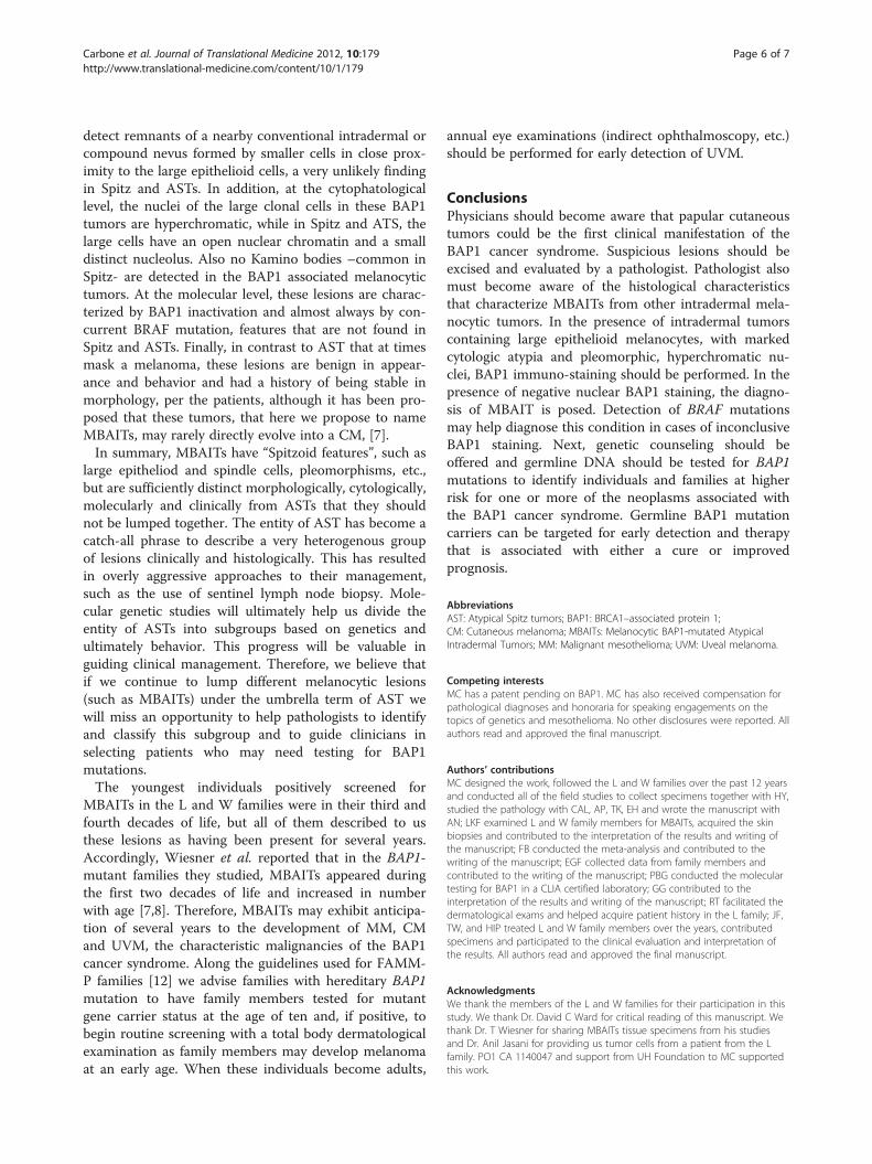
ResultsWe reviewed the published morphology and histologyand some of the original tissue sections from the pub-lished melanocytic skin lesions [7-9]. In addition, weinvestigated for the presence of these melanocytictumors in members of the BAP1-mutated families previ-ously found to have developed MM and UVM [6]. Thesetumors were indistinguishable. Thus, pathologists haveused different names to identify these pink to tan skintumors of about 0.2-1.0 cm in diameter, which macro-scopically resemble dermal nevi, but that histologicallyand at the molecular level do not fit any previous diag-nostic nomenclature. In this paper we will use the termMBAITs (Melanocytic BAP1-mutated Atypical Intrader-mal Tumors) for all the described BAP1-mutated atyp-ical melanocytic proliferations with similar histology andmolecular characterization that were previously diag-nosed using various terminologies (Figure 1).Specifically, we analyzed suspicious cutaneous tumors
from five members of L family and seven members ofW family. Of the five members of L family, four (80%)presented one or more MBAITs. The age range of posi-tively screened individuals was 36–64 years. One ofthem positive for MBAITs (L-III-18) had already beendiagnosed with both MM and UVM.Of the seven members of W family, four (57%) pre-
sented one or more MBAITs. The age range of thescreened individuals was 26–61 years. One of them, posi-tive for MBAITs (W-III-04), had already been diagnosedwith MM. The individual W-IV-21, already diagnosedwith MM, resulted negative for MBAITs screening, butreported a recently diagnosed breast carcinoma.All patients communicated that all of the MBAITs
looked and behaved as benign (present for a long time,not growing or changing).Histologically, MBAITs presented as intradermal
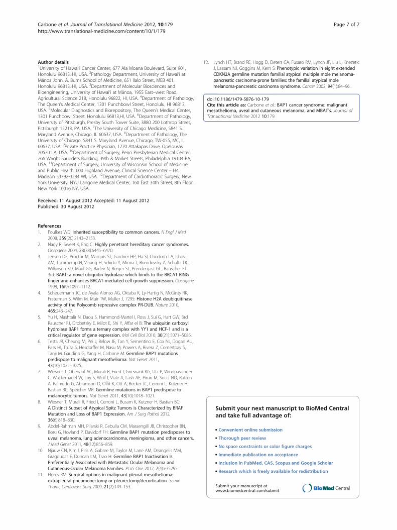
lesions with large epithelioid and spindled melanocytes,with marked cytologic atypia and pleomorphic, hyper-chromatic nuclei, and they were often associated with anearby compound or intradermal nevus (Figure 2). The
large epithelioid and spindle cells (i.e., the MBAITs cells)showed no mitotic activity, a finding confirmed by thenegative Ki67 immunostaining (not shown). Kaminobodies were not identified. In the MBAITs cells –but notin the nearby smaller nevus cells- , IHC showed negativeBAP1 nuclear staining with variable cytoplasmic stain-ing, suggesting loss of heterozygosity (LOH) of the wild-type BAP1 allele (Figure 2).Of the five members of L family, four (80%) carried
the germline BAP1 mutation (p.Gln684*). The familymember L-IV-5 did not have MBAITs and was notmutated in BAP1. All of the seven members of W familycarried the germline BAP1 mutations (p.Pro147fs*48).BRAF genotyping was performed on all the nine diag-
nosed MBAITs and BRAFV600E mutations were identi-fied in all of them, confirming the extremely highprevalence of this mutation as originally reported byWiesner and colleagues [7], and confirmed by Njauwand colleagues [10].The characteristics of the pooled families are described
in Table 1. Among the seven families, 118 patients wereselected. The results of the meta-analysis are presentedin Table 2. The BAP1-mutated cohort included 63patients and the BAP1-non-mutated cohort 55 patients.The mean ages of follow up were comparable in bothcohorts (53.2 years, 95%CI: 49.0-57.4 in the BAP1mutated cohort, 51.0 years, 95%CI: 46.3-55.8 in the non-mutated cohort). The proportion of women was higherin the BAP1-mutated cohort compared to the non-mutated cohort (63.3% and 43.6% respectively, p = 0.034).
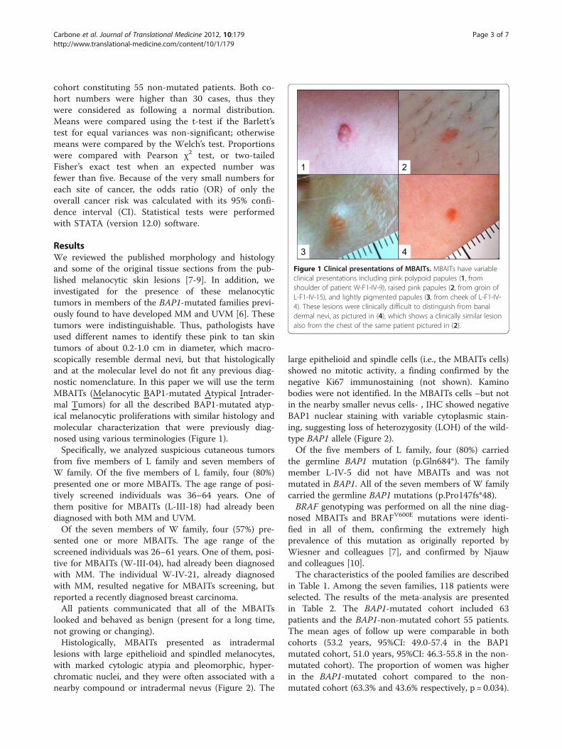
Figure 1 Clinical presentations of MBAITs. MBAITs have variableclinical presentations including pink polypoid papules (1, fromshoulder of patient W-F1-IV-9), raised pink papules (2, from groin ofL-F1-IV-15), and lightly pigmented papules (3, from cheek of L-F1-IV-4). These lesions were clinically difficult to distinguish from banaldermal nevi, as pictured in (4), which shows a clinically similar lesionalso from the chest of the same patient pictured in (2).
Carbone et al. Journal of Translational Medicine 2012, 10:179 Page 3 of 7http://www.translational-medicine.com/content/10/1/179
At this time we do not have an explanation for thisfinding, although it appears possible that BAP1 muta-tion may carry a higher risk of lethality in utero formales.The overall prevalence of cancer was significantly
higher in the BAP1-mutated cohort compared with thenon-mutated cohort (63.5% and 9.1% respectively,p < 0.001). Five tumors were observed in the non-mutated cohort: two prostate cancers, one breast cancer,one Hodgkin’s lymphoma and one Non-Hodgkin’slymphoma. The odds ratio (OR) of cancer risk in thegermline BAP1-mutated cohort versus the non-mutatedcohort was 17.39 (95% CI: 6.07-49.83). MM, UVM, CMand MBAITs prevalence was significantly higher in theBAP1-mutated cohort compared with the non-mutatedcohort (Table 2). No significant difference was found be-tween the two cohorts concerning the rates of othercancers.
DiscussionWe performed a meta-analysis of all the published stud-ies with BAP1-mutated families. To avoid the overesti-mation of the cancer risks estimates usually due topublication bias in meta-analyses, we chose to compareBAP1 mutated vs non-mutated patients from the samefamilies. We demonstrate that germline BAP1 mutationsare associated with a significant increased overall risk of
cancer and particularly of MM, UVM and CM (Table 2).In addition, two thirds of patients from the germlineBAP1-mutated cohort presented MBAITs, while thesetumors were not detected in the non-mutated indivi-duals. Our results reveal that germline BAP1 mutationscause a novel autosomal dominant hereditary cancersyndrome, the BAP1 cancer syndrome, characterizedpredominantly by MM, UVM, CM and by MBAITs andpossibly by other cancers. In fact, because of the rela-tively high incidence of carcinomas in the general popu-lation, a larger number of BAP1-mutant family membershave to be studied before ruling out the possibility thatadditional cancers are linked to the BAP1 cancersyndrome.Early diagnosis is crucial for curative resection of CM
and UVM. For its anatomical localization, MM is atumor in which early diagnosis is particularly difficult; itis indeed often diagnosed in advanced stages whenpatients have median survivals of 6–12 months. How-ever, when MM patients are diagnosed at Stage 1a, sur-vivals of five or more years are not uncommon [11].Indeed, a high degree of suspicion allowed us to detectfour MM in the L and W families at an early stage andthese patients experienced survivals of 5–10 and, hope-fully, many more years.MBAITs provide physicians with a marker to identify
individuals who may carry germline BAP1 mutations
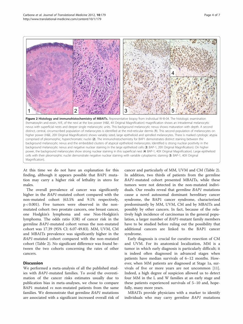
Figure 2 Histology and immunohistochemistry of MBAITs. Representative biopsy from individual W-III-04. The histologic examination(hematoxylin and eosin, H/E, of the nevi at the low power (H&E, 4X Original Magnification) magnification shows an intradermal melanocyticnevus with superficial nests and deeper single melanocytic units. This background melanocytic nevus shows maturation with depth. A seconddistinct, central, circumscribed population of melanocytes is identified at the mid-reticular dermis (1). This second population of melanocytes onhigher power (H&E, 20X Original Magnification) shows variably sized, large epithelioid and spindled melanocytes. There is marked cytologic atypiacomprised of pleomorphic, hyperchromatic nuclei (2). The immunohistochemistry for BAP1 demonstrates distinct staining between thebackground melanocytic nevus and the embedded clusters of atypical epithelioid melanocytes. Identified is strong nuclear positivity in thebackground melanocytic nevus and negative nuclear staining in the large epithelioid cells (3: BAP-1, 20X Original Magnification). On higherpower, the background melanocytes show strong nuclear staining in this superficial nest (4: BAP-1, 40X Original Magnification). Large epithelioidcells with their pleomorphic nuclei demonstrate negative nuclear staining with variable cytoplasmic staining (5: BAP-1, 40X OriginalMagnification).
Carbone et al. Journal of Translational Medicine 2012, 10:179 Page 4 of 7http://www.translational-medicine.com/content/10/1/179
and thus are at high risk of developing CM, UVM andMM. We identified MBAITs among the majority ofgermline BAP1 mutation carriers in the L and W fam-ilies. Our meta-analysis demonstrates that the prevalenceof MBAITs is significantly higher in germline BAP1 mu-tation carriers compared to controls. MBAITs have vari-able papular macroscopic appearance similar to dermalnevi; nevertheless they present histological (Figure 2),immunohistochemical (Figure 2) and molecular (BAP1and BRAF mutations) features that allow theircharacterization in the broad spectrum of melanocyticlesions. We debated how to call these tumors, and weconcluded that it was best to give them a new name tomake a clear distinction between these tumors, andother melanocytic lesions, such as Spitz nevus and ASTs.Briefly, Spitz nevus consists of proliferation of largeepithelioid or spindle shaped melanocytes, or a mixtureof the two. At all ages, spindle cells are the most com-mon cell type. Spitz nevi composed wholly of epithelioidcells occur mainly in early childhood. Spitz nevi gothrough the same junctional, compound and intradermalphases as common acquired nevi, but most are removedwhen they are compound lesions (when they have anepidermal and dermal component). Spitz nevi areroughly symmetrical and at any given level (epidermis,junction, upper dermis, lower dermis) the lesion showssimilar architecture and cell type from side to side. Thecellularity of the lesion and the size of the cells and theirnuclei decrease toward the base of the Spitz nevus (socalled maturation) and this is associated with loss ofproliferative activity that is instead often present in theupper parts of the nevus. Also the architecture at thebase (deep aspect) is infiltrative rather than expansile/pushing, the nevus cells lying dispersed between dermalcollagen. In addition there is no nevus associate withthem (unless it is part of a combined nevus). ATS areSpitzoid lesions that have some features that overlapwith melanoma making the differential diagnosis chal-lenging: for example, there is no maturation towards thedeeper part of the dermis, and instead the AST cellsshow mitotic activity.
Instead, the tumors found in these BAP1 mutatedpatients show large epithelioid clonal cells (that resemblethose found in Spitz nevus and in ASTs) but these cellsare present only in the dermis (there is no epidermalcomponent). In contrast to Spitz nevi there is no matur-ation towards the deeper part of the dermis, and in con-trast to ASTs, Ki67 stain (a marker of cell proliferation)consistently showed absence of mitotic figures. In almostall of these BAP1 associated tumors, it was possible to
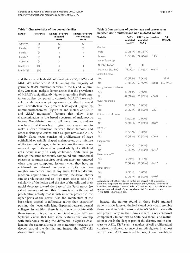
Table 1 Characteristics of the pooled families
Family Reference Number of BAP1-mutatedN=63
Number of BAP1-non-mutated
N=55
Family W [6] 12 12
Family L [6] 16 32
Family 1 [7] 4 3
Family 2 [7] 9 2
FUM036 [9] 8 1
Family 562 [10] 4 3
Family 729 [10] 10 2
Table 2 Comparisons of gender, age and cancer ratesbetween BAP1-mutated and non-mutated cohorts
Variable BAP1-mutatedN=63*
BAP1-non-mutatedN=55
p-value OR(95%CI)
Gender
Male 22 (36.7%) 31 (56.4%)
Female 38 (63.3%) 24 (43.6%) 0.034 -
Age of follow-up
Number known 46 42
Mean age (Std. Err.) 53.2 (2.1) 51.0 (2.3) 0.4851 -
At least 1 cancer
Yes 40 (63.5%) 5 (9.1%) 17.39
No 23 (36.5%) 50 (90.9%) <0.001 (6.07-49.83)
Malignant mesothelioma
Yes 13 (21.0%) 0 (0.0%)
No 49 (79.0%) 55 (100%) <0.001 -
Uveal melanoma
Yes 11 (17.7%) 0 (0.0%)
No 51 (82.3%) 55 (100%) 0.001 -
Cutaneous melanoma
Yes 8 (12.9%) 0 (0.0%)
No 54 (87.1%) 55 (100%) 0.007} -
MBAITs**
Yes 24 (66.7%) 0 (0.0%)
No 12 (33.3%) 12 (100%) <0.001 -
Lung cancer
Yes 3 (4.8%) 0 (0.0%)
No 59 (95.2%) 55 (100%) 0.246} -
Breast cancer***
Yes 3 (7.9%) 1 (4.1%)
No 35 (91.9%) 23 (95.9%) 0.621} -
Renal cancer
Yes 2 (3.3%) 0 (0.0%)
No 60 (96.7%) 55 (100%) 0.497} -
Abbreviations: OR: Odds Ratio; CI: confidence interval; *: 62 informative, 1BAP1-mutated patient had cancer of unknown origin, **: calculated only inindividuals belonging to present study, ref. 7 and ref. 10, ***: calculated only inwomen, -: not calculated; NS: non significant; Std. Err.: standard error;}: two-tailed Fisher’s exact test.
Carbone et al. Journal of Translational Medicine 2012, 10:179 Page 5 of 7http://www.translational-medicine.com/content/10/1/179
detect remnants of a nearby conventional intradermal orcompound nevus formed by smaller cells in close prox-imity to the large epithelioid cells, a very unlikely findingin Spitz and ASTs. In addition, at the cytophatologicallevel, the nuclei of the large clonal cells in these BAP1tumors are hyperchromatic, while in Spitz and ATS, thelarge cells have an open nuclear chromatin and a smalldistinct nucleolus. Also no Kamino bodies –common inSpitz- are detected in the BAP1 associated melanocytictumors. At the molecular level, these lesions are charac-terized by BAP1 inactivation and almost always by con-current BRAF mutation, features that are not found inSpitz and ASTs. Finally, in contrast to AST that at timesmask a melanoma, these lesions are benign in appear-ance and behavior and had a history of being stable inmorphology, per the patients, although it has been pro-posed that these tumors, that here we propose to nameMBAITs, may rarely directly evolve into a CM, [7].In summary, MBAITs have “Spitzoid features”, such as
large epitheliod and spindle cells, pleomorphisms, etc.,but are sufficiently distinct morphologically, cytologically,molecularly and clinically from ASTs that they shouldnot be lumped together. The entity of AST has become acatch-all phrase to describe a very heterogenous groupof lesions clinically and histologically. This has resultedin overly aggressive approaches to their management,such as the use of sentinel lymph node biopsy. Mole-cular genetic studies will ultimately help us divide theentity of ASTs into subgroups based on genetics andultimately behavior. This progress will be valuable inguiding clinical management. Therefore, we believe thatif we continue to lump different melanocytic lesions(such as MBAITs) under the umbrella term of AST wewill miss an opportunity to help pathologists to identifyand classify this subgroup and to guide clinicians inselecting patients who may need testing for BAP1mutations.The youngest individuals positively screened for
MBAITs in the L and W families were in their third andfourth decades of life, but all of them described to usthese lesions as having been present for several years.Accordingly, Wiesner et al. reported that in the BAP1-mutant families they studied, MBAITs appeared duringthe first two decades of life and increased in numberwith age [7,8]. Therefore, MBAITs may exhibit anticipa-tion of several years to the development of MM, CMand UVM, the characteristic malignancies of the BAP1cancer syndrome. Along the guidelines used for FAMM-P families [12] we advise families with hereditary BAP1mutation to have family members tested for mutantgene carrier status at the age of ten and, if positive, tobegin routine screening with a total body dermatologicalexamination as family members may develop melanomaat an early age. When these individuals become adults,
annual eye examinations (indirect ophthalmoscopy, etc.)should be performed for early detection of UVM.
ConclusionsPhysicians should become aware that papular cutaneoustumors could be the first clinical manifestation of theBAP1 cancer syndrome. Suspicious lesions should beexcised and evaluated by a pathologist. Pathologist alsomust become aware of the histological characteristicsthat characterize MBAITs from other intradermal mela-nocytic tumors. In the presence of intradermal tumorscontaining large epithelioid melanocytes, with markedcytologic atypia and pleomorphic, hyperchromatic nu-clei, BAP1 immuno-staining should be performed. In thepresence of negative nuclear BAP1 staining, the diagno-sis of MBAIT is posed. Detection of BRAF mutationsmay help diagnose this condition in cases of inconclusiveBAP1 staining. Next, genetic counseling should beoffered and germline DNA should be tested for BAP1mutations to identify individuals and families at higherrisk for one or more of the neoplasms associated withthe BAP1 cancer syndrome. Germline BAP1 mutationcarriers can be targeted for early detection and therapythat is associated with either a cure or improvedprognosis.
AbbreviationsAST: Atypical Spitz tumors; BAP1: BRCA1–associated protein 1;CM: Cutaneous melanoma; MBAITs: Melanocytic BAP1-mutated AtypicalIntradermal Tumors; MM: Malignant mesothelioma; UVM: Uveal melanoma.
Competing interestsMC has a patent pending on BAP1. MC has also received compensation forpathological diagnoses and honoraria for speaking engagements on thetopics of genetics and mesothelioma. No other disclosures were reported. Allauthors read and approved the final manuscript.
Authors’ contributionsMC designed the work, followed the L and W families over the past 12 yearsand conducted all of the field studies to collect specimens together with HY,studied the pathology with CAL, AP, TK, EH and wrote the manuscript withAN; LKF examined L and W family members for MBAITs, acquired the skinbiopsies and contributed to the interpretation of the results and writing ofthe manuscript; FB conducted the meta-analysis and contributed to thewriting of the manuscript; EGF collected data from family members andcontributed to the writing of the manuscript; PBG conducted the moleculartesting for BAP1 in a CLIA certified laboratory; GG contributed to theinterpretation of the results and writing of the manuscript; RT facilitated thedermatological exams and helped acquire patient history in the L family; JF,TW, and HIP treated L and W family members over the years, contributedspecimens and participated to the clinical evaluation and interpretation ofthe results. All authors read and approved the final manuscript.
AcknowledgmentsWe thank the members of the L and W families for their participation in thisstudy. We thank Dr. David C Ward for critical reading of this manuscript. Wethank Dr. T Wiesner for sharing MBAITs tissue specimens from his studiesand Dr. Anil Jasani for providing us tumor cells from a patient from the Lfamily. PO1 CA 1140047 and support from UH Foundation to MC supportedthis work.
Carbone et al. Journal of Translational Medicine 2012, 10:179 Page 6 of 7http://www.translational-medicine.com/content/10/1/179
Author details1University of Hawai‘i Cancer Center, 677 Ala Moana Boulevard, Suite 901,Honolulu 96813, HI, USA. 2Pathology Department, University of Hawai‘i atMānoa John. A. Burns School of Medicine, 651 Ilalo Street, MEB 401,Honolulu 96813, HI, USA. 3Department of Molecular Biosciences andBioengineering, University of Hawai‘i at Mānoa, 1955 East–west Road,Agricultural Science 218, Honolulu 96822, HI, USA. 4Department of Pathology,The Queen’s Medical Center, 1301 Punchbowl Street, Honolulu, HI 96813,USA. 5Molecular Diagnostics and Biorepository, The Queen’s Medical Center,1301 Punchbowl Street, Honolulu 96813,HI, USA. 6Department of Pathology,University of Pittsburgh, Presby South Tower Suite, 3880 200 Lothrop Street,Pittsburgh 15213, PA, USA. 7The University of Chicago Medicine, 5841 S.Maryland Avenue, Chicago, IL 60637, USA. 8Department of Pathology, TheUniversity of Chicago, 5841 S. Maryland Avenue, Chicago, TW-055, MC, IL60637, USA. 9Private Practice Physician, 1270 Attakapas Drive, Opelousas70570 LA, USA. 10Department of Surgery, Penn Presbyterian Medical Center,266 Wright Saunders Building, 39th & Market Streets, Philadelphia 19104 PA,USA. 11Department of Surgery, University of Wisconsin School of Medicineand Public Health, 600 Highland Avenue, Clinical Science Center – H4,Madison 53792-3284 WI, USA. 12Department of Cardiothoracic Surgery, NewYork University, NYU Langone Medical Center, 160 East 34th Street, 8th Floor,New York 10016 NY, USA.
Received: 11 August 2012 Accepted: 11 August 2012Published: 30 August 2012
References1. Foulkes WD: Inherited susceptibility to common cancers. N Engl J Med
2008, 359(20):2143–2153.2. Nagy R, Sweet K, Eng C: Highly penetrant hereditary cancer syndromes.
Oncogene 2004, 23(38):6445–6470.3. Jensen DE, Proctor M, Marquis ST, Gardner HP, Ha SI, Chodosh LA, Ishov
AM, Tommerup N, Vissing H, Sekido Y, Minna J, Borodovsky A, Schultz DC,Wilkinson KD, Maul GG, Barlev N, Berger SL, Prendergast GC, Rauscher FJ3rd: BAP1: a novel ubiquitin hydrolase which binds to the BRCA1 RINGfinger and enhances BRCA1-mediated cell growth suppression. Oncogene1998, 16(9):1097–1112.
4. Scheuermann JC, de Ayala Alonso AG, Oktaba K, Ly-Hartig N, McGinty RK,Fraterman S, Wilm M, Muir TW, Muller J, 7295: Histone H2A deubiquitinaseactivity of the Polycomb repressive complex PR-DUB. Nature 2010,465:243–247.
5. Yu H, Mashtalir N, Daou S, Hammond-Martel I, Ross J, Sui G, Hart GW, 3rdRauscher FJ, Drobetsky E, Milot E, Shi Y, Affar el B: The ubiquitin carboxylhydrolase BAP1 forms a ternary complex with YY1 and HCF-1 and is acritical regulator of gene expression. Mol Cell Biol 2010, 30(21):5071–5085.
6. Testa JR, Cheung M, Pei J, Below JE, Tan Y, Sementino E, Cox NJ, Dogan AU,Pass HI, Trusa S, Hesdorffer M, Nasu M, Powers A, Rivera Z, Comertpay S,Tanji M, Gaudino G, Yang H, Carbone M: Germline BAP1 mutationspredispose to malignant mesothelioma. Nat Genet 2011,43(10):1022–1025.
7. Wiesner T, Obenauf AC, Murali R, Fried I, Griewank KG, Ulz P, WindpassingerC, Wackernagel W, Loy S, Wolf I, Viale A, Lash AE, Pirun M, Socci ND, RuttenA, Palmedo G, Abramson D, Offit K, Ott A, Becker JC, Cerroni L, Kutzner H,Bastian BC, Speicher MR: Germline mutations in BAP1 predispose tomelanocytic tumors. Nat Genet 2011, 43(10):1018–1021.
8. Wiesner T, Murali R, Fried I, Cerroni L, Busam K, Kutzner H, Bastian BC:A Distinct Subset of Atypical Spitz Tumors is Characterized by BRAFMutation and Loss of BAP1 Expression. Am J Surg Pathol 2012,36(6):818–830.
9. Abdel-Rahman MH, Pilarski R, Cebulla CM, Massengill JB, Christopher BN,Boru G, Hovland P, Davidorf FH: Germline BAP1 mutation predisposes touveal melanoma, lung adenocarcinoma, meningioma, and other cancers.J Med Genet 2011, 48(12):856–859.
10. Njauw CN, Kim I, Piris A, Gabree M, Taylor M, Lane AM, Deangelis MM,Gragoudas E, Duncan LM, Tsao H: Germline BAP1 Inactivation IsPreferentially Associated with Metastatic Ocular Melanoma andCutaneous-Ocular Melanoma Families. PLoS One 2012, 7(4):e35295.
11. Flores RM: Surgical options in malignant pleural mesothelioma:extrapleural pneumonectomy or pleurectomy/decortication. SeminThorac Cardiovasc Surg 2009, 21(2):149–153.
12. Lynch HT, Brand RE, Hogg D, Deters CA, Fusaro RM, Lynch JF, Liu L, KnezeticJ, Lassam NJ, Goggins M, Kern S: Phenotypic variation in eight extendedCDKN2A germline mutation familial atypical multiple mole melanoma-pancreatic carcinoma-prone families: the familial atypical molemelanoma-pancreatic carcinoma syndrome. Cancer 2002, 94(1):84–96.
doi:10.1186/1479-5876-10-179Cite this article as: Carbone et al.: BAP1 cancer syndrome: malignantmesothelioma, uveal and cutaneous melanoma, and MBAITs. Journal ofTranslational Medicine 2012 10:179.
Submit your next manuscript to BioMed Centraland take full advantage of:
• Convenient online submission
• Thorough peer review
• No space constraints or color figure charges
• Immediate publication on acceptance
• Inclusion in PubMed, CAS, Scopus and Google Scholar
• Research which is freely available for redistribution
Submit your manuscript at www.biomedcentral.com/submit
Carbone et al. Journal of Translational Medicine 2012, 10:179 Page 7 of 7http://www.translational-medicine.com/content/10/1/179