Embed Size (px)
Citation preview
7Basaloid Tumors: Basal Cell Adenoma and Basal Cell Adenocarcinoma
Background
Basaloid tumors of the salivary gland are among the most diagnostically challenging areas of salivary gland FNA cytopathology. The primary tumors included in this group are basal cell adenoma, basal cell adenocarcinoma, and the solid variant of adenoid cystic carcinoma. In addition, various other salivary gland tumors, such as cellular pleomorphic adenoma, can also exhibit basaloid features and will be considered as differential diagnostic entities within this section.
Basal cell adenomas are rare salivary gland tumors comprised of basaloid cells and lacking the chondromyxoid matrix material characteristic of pleomorphic adenomas. In the past, they have been classified as “monomorphic adenomas,” but this nonspe-cific terminology is to be avoided in favor of the more specific designation recommended by the WHO - “basal cell adenoma.” Basal cell adenomas represent 1%–3% of all salivary gland neoplasms, and they arise primarily in older adults, usually in the sixth to seventh decade. A somewhat histologically similar basaloid tumor that occurs in infants is known as sialoblastoma. Over 75% of basal cell adenomas occur in the parotid gland; they are rarely seen in minor salivary glands. There are 3 subtypes of basal cell adenoma: solid, tubulotrabecular, and membranous. Most patients present with a solitary firm nodule between 1 and 3 cm that is slowly enlarging. Occasionally, basal cell adenomas can be cystic.
115
116 7. Basaloid Tumors
CELLULARNORMAL TISSUE?
BASAL CELL ADENOMABASAL CELLADENOCARCINOMASOLID ADENOID CYSTICCARCINOMAMETASTASISSIALOBLASTOMA
ADENOID CYSTICCARCINOMA
MATRIX?
FIBRILLAR?
BASALOID?
ABSENT
ABSENTABSENT
PRESENTPRESENT
MATRIXSPHERESAND
TUBULES?
PRESENTPRESENT ABSENT
CYSTIC
PRESENT
PRESENT
PRESENT
INFLAMMATORY ANDLYMPHOMA
ASSESS GENERALCOMPONENTS
SATISFACTORY UNSATISFACTORY
ASSESS SAMPLE ADEQUACY
SALIVARYGLANDFNA
THYROIDFNA
ASSESS GENERALCOMPONENTS
SATISFACTORY UNSATISFACTORY
ASSESS ADEQUACY
ASSESS GENERALCOMPONENTS
SATISFACTORY UNSATISFACTORY
ASSESS SAMPLE ADEQUACY
SALIVARYGLANDFNA
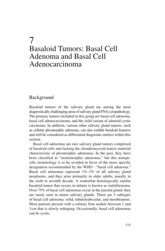
Fig. 7.1. Algorithm for basaloid tumors.
The membranous subtype of basal cell adenoma is the most cytologically and histologically distinctive of the 3 subtypes. In contrast to the tubulotrabecular and solid subtypes, the membra-nous subtype is often multinodular and sometimes multifocal. It is also unusual because of its occasional association with mul-tiple synchronous dermal cylindromas, trichoepitheliomas, and spiradenomas, to which basal cell adenoma can bear a remarkable microscopic resemblance. For this reason, the membranous sub-type of basal cell adenoma has also been known as “dermal ana-logue tumor.” The condition of multiple cutaneous adnexal tumors and synchronous salivary gland basal cell adenomas, which can be disfiguring, is called Brooke-Spiegler syndrome. It is an autosomal dominant disease caused by mutations in the tumor suppressor gene that encodes the CYLD protein (an inhibitor of NF-kB).
Basal cell adenocarcinoma is a rare salivary gland neoplasm that is the malignant counterpart of basal cell adenoma. It is a low-grade malignancy with a very good clinical prognosis; although it has a tendency for local recurrence (approximately 35% of cases), meta-static disease is uncommon. Basal cell adenocarcinoma accounts for less than 2% of malignant epithelial salivary gland tumors. The majority occur in the superficial lobe of the parotid gland, although occasional cases have been reported in the submandibular gland and the minor salivary glands. The average age at diagnosis is 60 years, with a broad age range from third to tenth decade, with no gen-der predilection. Salivary gland enlargement is the main presenting symptom, and uncommonly, mild pain or tenderness may also be present. Like its benign counterpart, basal cell adenocarcinomas can be solid, tubulotrabecular, or the membranous subtype. The solid subtype is the most common. Most basal cell adenocarcinomas are believed to arise de novo, although a small subset may develop from a pre-existing basal cell adenoma.
FNA is highly sensitive at detecting basaloid neoplasms such as basal cell adenoma and adenocarcinoma, but distinction between several of the basaloid entities in the differential diagnosis is often not possible. As will be discussed, some cases of basal cell tumor can be recognized by FNA, but many will receive a descriptive signout and differential diagnosis. Most basal cell adenocarcinomas are microscopically identical to basal cell adenomas except for the pres-ence of an invasive histologic growth pattern. Because FNA does not
Background 117
118 7. Basaloid Tumors
detect parenchymal invasion, basal cell adenomas and adenocarci-nomas are, for the most part, indistinguishable by FNA.
General Diagnostic Approach
Using the algorithm (Fig. 7.1), aspirates comprised of epithelial cells that lack the characteristic matrix of pleomorphic adenoma, and that exhibit basaloid cytologic features (scant cytoplasm and dark nuclei), lead to a differential diagnosis that includes adenoid cystic carcinoma, basal cell adenoma, basal cell adenocarcinoma, and other lesions with basaloid features. The classic cribriform type of adenoid cystic carcinoma is basaloid but can be distinguished by its acellular matrix spheres and branching matrix tubules; however, the solid form of adenoid cystic carcinoma must be considered in the differential diagnosis with the solid form of basal cell adenoma and adenocarcinoma.
Diagnostic Criteria
Basal cell adenoma and adenocarcinoma are classic basaloid tumors that in most cases exhibit identical cytomorphologic features – basal cell adenocarcinoma is distinguished from basal cell adenoma by an infiltrative growth pattern in histologic specimens. There is a small subset of basal cell adenocarcinomas that exhibits nuclear atypia and may show mitotic activity and/or necrosis. Such features are rare in basal cell adenocarcinomas, but when present, would exclude basal cell adenoma. For the majority of FNA cases, a general rule of thumb is that basal cell adenoma and basal cell adenocarcinoma cannot be reliably distinguished on the basis of cytologic features.
Basal cell adenoma and basal cell adenocarcinoma cannot be reliably distinguished on the basis of cytologic features.
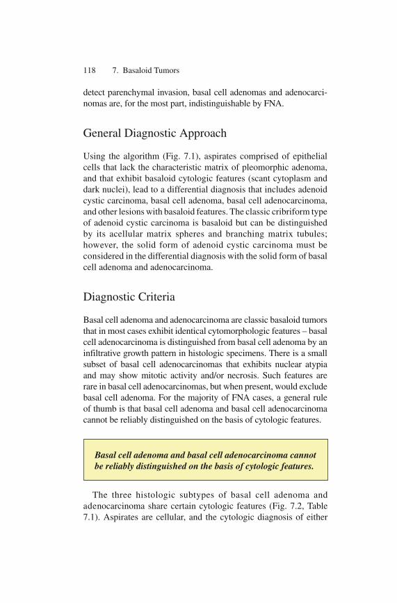
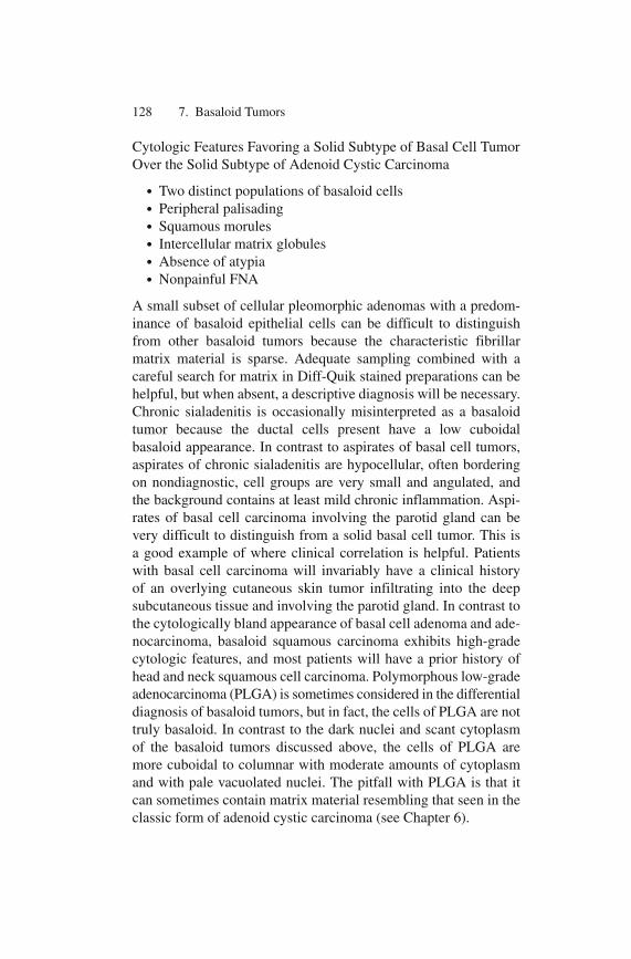
The three histologic subtypes of basal cell adenoma and adenocarcinoma share certain cytologic features (Fig. 7.2, Table 7.1). Aspirates are cellular, and the cytologic diagnosis of either
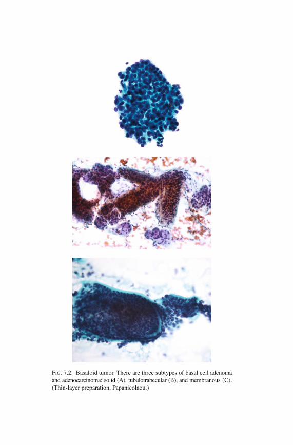
Fig. 7.2. Basaloid tumor. There are three subtypes of basal cell adenoma and adenocarcinoma: solid (A), tubulotrabecular (B), and membranous (C). (Thin-layer preparation, Papanicolaou.)
120 7. Basaloid Tumors
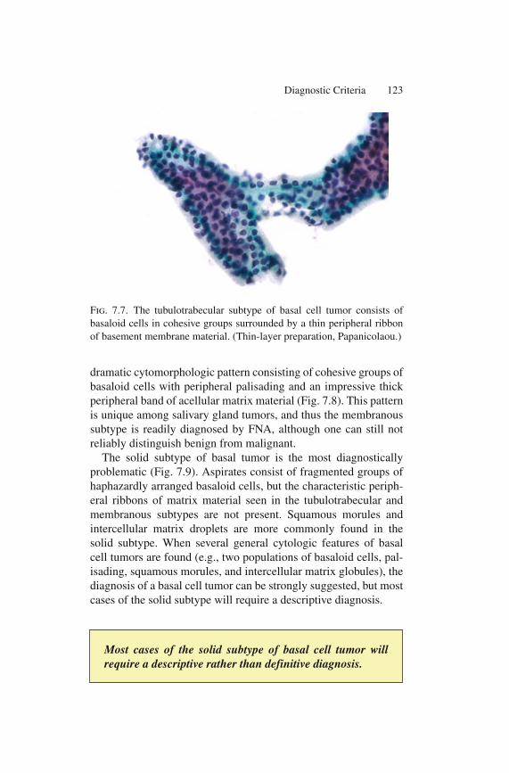
basal cell adenoma or adenocarcinoma rests on identifying two populations of basaloid cells: a group of small oval cells with bland hyperchromatic nuclei, scant cytoplasm, and indistinct nucleoli, and a group of larger oval to polygonal cells with moder-ate amounts of delicate pale cytoplasm (Fig. 7.3). The basaloid cells are uniform and haphazardly arranged in variably-sized clus-ters or trabeculae, often with peripheral palisading of the smaller population of cells (Fig. 7.4). Squamous morules, a characteristic feature of basal cell tumors, are sometimes present in well-sam-pled cases (Fig. 7.5). All three subtypes can have small dense, nonfibrillary intercellular globules of acellular matrix material that is blue-green using Papanicolaou stains and metachromatic using Diff-Quik stains (Fig. 7.6).
Shared Cytologic Features of Basal Cell Adenoma and Adenocarcinoma
● Cellular aspirate● Two populations of basaloid cells● Haphazardly arranged cells● Peripheral palisading● Squamous morules● Intercellular matrix globules
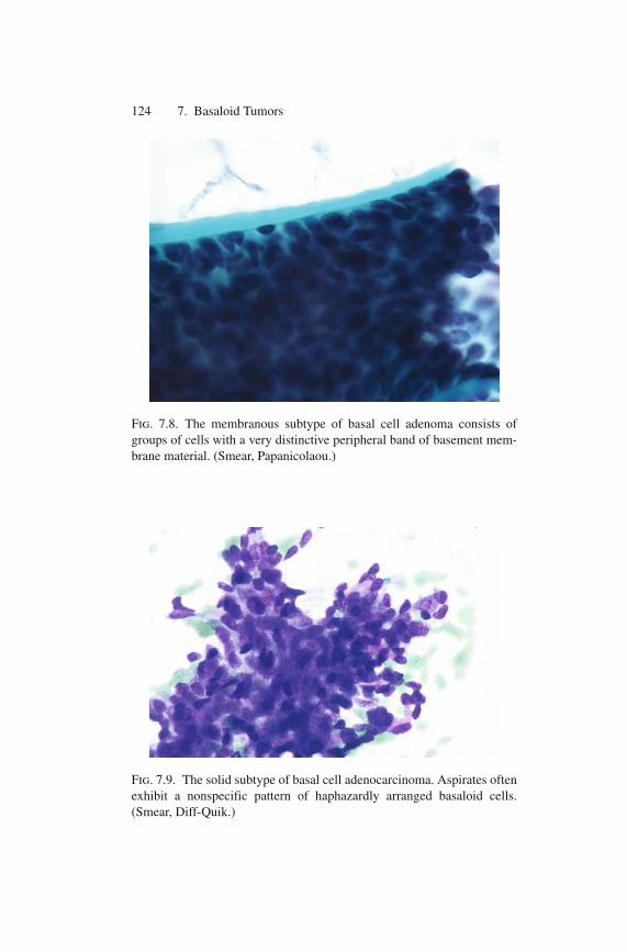
The tubulotrabecular subtype of basal cell adenoma and adeno-carcinoma is characterized by branching tubules and trabeculae of basaloid cells with a thin peripheral ribbon of acellular matrix
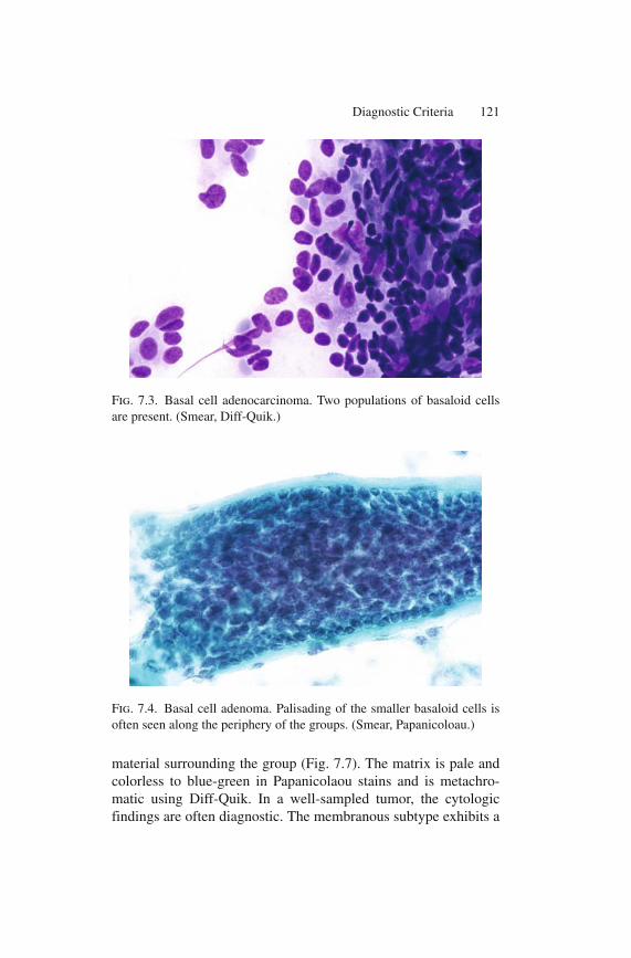
Table 7.1. Cytologic features of the three subtypes of basal cell adenoma and adenocarcinoma. CharacteristicSubtype Cytoarchitecture Cytology FNA Diagnosis
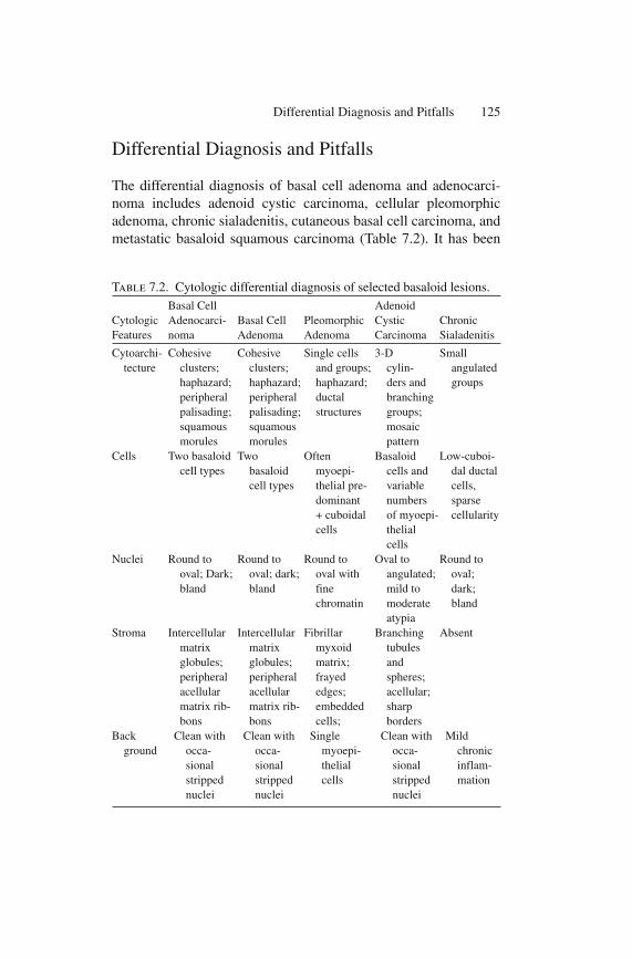
Solid Fragmented groups Squamous morules Usually of haphazard and intercellular descriptive basaloid cells matrix dropletsTubulotrabecular Branching Thin peripheral Sometimes tubules matrix ribbon diagnosticMembranous Cohesive Thick peripheral Usually trabecular and matrix ribbon diagnostic insular groups
material surrounding the group (Fig. 7.7). The matrix is pale and colorless to blue-green in Papanicolaou stains and is metachro-matic using Diff-Quik. In a well-sampled tumor, the cytologic findings are often diagnostic. The membranous subtype exhibits a
Fig. 7.3. Basal cell adenocarcinoma. Two populations of basaloid cells are present. (Smear, Diff-Quik.)
Fig. 7.4. Basal cell adenoma. Palisading of the smaller basaloid cells is often seen along the periphery of the groups. (Smear, Papanicoloau.)
Diagnostic Criteria 121
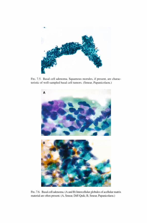
Fig. 7.5. Basal cell adenoma. Squamous morules, if present, are charac-teristic of well-sampled basal cell tumors. (Smear, Papanicolaou.)
Fig. 7.6. Basal cell adenoma. (A and B) Intercellular globules of acellular matrix material are often present. (A, Smear, Diff-Quik; B, Smear, Papanicolaou.)
dramatic cytomorphologic pattern consisting of cohesive groups of basaloid cells with peripheral palisading and an impressive thick peripheral band of acellular matrix material (Fig. 7.8). This pattern is unique among salivary gland tumors, and thus the membranous subtype is readily diagnosed by FNA, although one can still not reliably distinguish benign from malignant.
The solid subtype of basal tumor is the most diagnostically problematic (Fig. 7.9). Aspirates consist of fragmented groups of haphazardly arranged basaloid cells, but the characteristic periph-eral ribbons of matrix material seen in the tubulotrabecular and membranous subtypes are not present. Squamous morules and intercellular matrix droplets are more commonly found in the solid subtype. When several general cytologic features of basal cell tumors are found (e.g., two populations of basaloid cells, pal-isading, squamous morules, and intercellular matrix globules), the diagnosis of a basal cell tumor can be strongly suggested, but most cases of the solid subtype will require a descriptive diagnosis.
Fig. 7.7. The tubulotrabecular subtype of basal cell tumor consists of basaloid cells in cohesive groups surrounded by a thin peripheral ribbon of basement membrane material. (Thin-layer preparation, Papanicolaou.)
Diagnostic Criteria 123
Most cases of the solid subtype of basal cell tumor will require a descriptive rather than definitive diagnosis.
124 7. Basaloid Tumors
Fig. 7.8. The membranous subtype of basal cell adenoma consists of groups of cells with a very distinctive peripheral band of basement mem-brane material. (Smear, Papanicolaou.)
Fig. 7.9. The solid subtype of basal cell adenocarcinoma. Aspirates often exhibit a nonspecific pattern of haphazardly arranged basaloid cells. (Smear, Diff-Quik.)
Differential Diagnosis and Pitfalls
The differential diagnosis of basal cell adenoma and adenocarci-noma includes adenoid cystic carcinoma, cellular pleomorphic adenoma, chronic sialadenitis, cutaneous basal cell carcinoma, and metastatic basaloid squamous carcinoma (Table 7.2). It has been
Differential Diagnosis and Pitfalls 125
Table 7.2. Cytologic differential diagnosis of selected basaloid lesions.
Cytologic Features
Basal Cell Adenocarci-noma
Basal Cell Adenoma
Pleomorphic Adenoma
Adenoid Cystic Carcinoma
Chronic Sialadenitis
Cytoarchi- tecture
Cohesive clusters; haphazard; peripheral palisading; squamous morules
Cohesive clusters; haphazard; peripheral palisading; squamous morules
Single cells and groups; haphazard; ductal structures
3-D cylin-ders and branching groups; mosaic pattern
Small angulated groups
Cells Two basaloid cell types
Two basaloid cell types
Often myoepi-thelial pre-dominant + cuboidal cells
Basaloid cells and variable numbers of myoepi-thelial cells
Low-cuboi-dal ductal cells, sparse cellularity
Nuclei Round to oval; Dark; bland
Round to oval; dark; bland
Round to oval with fine chromatin
Oval to angulated; mild to moderate atypia
Round to oval; dark; bland
Stroma Intercellular matrix globules; peripheral acellular matrix rib-bons
Intercellular matrix globules; peripheral acellular matrix rib-bons
Fibrillar myxoid matrix; frayed edges; embedded cells;
Branching tubules and spheres; acellular; sharp borders
Absent
Back ground
Clean with occa-sional stripped nuclei
Clean with occa-sional stripped nuclei
Single myoepi-thelial cells
Clean with occa-sional stripped nuclei
Mild chronic inflam-mation
126 7. Basaloid Tumors
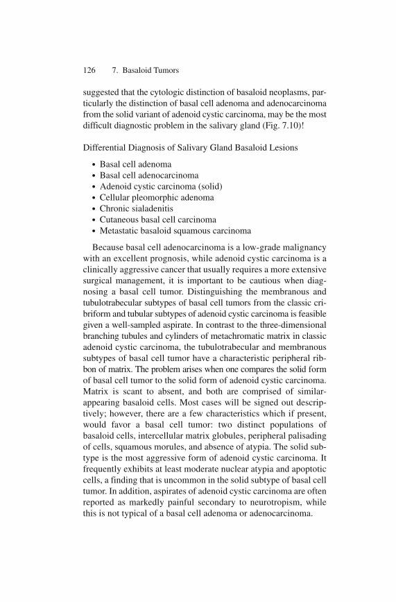
suggested that the cytologic distinction of basaloid neoplasms, par-ticularly the distinction of basal cell adenoma and adenocarcinoma from the solid variant of adenoid cystic carcinoma, may be the most difficult diagnostic problem in the salivary gland (Fig. 7.10)!
Differential Diagnosis of Salivary Gland Basaloid Lesions
● Basal cell adenoma● Basal cell adenocarcinoma● Adenoid cystic carcinoma (solid)● Cellular pleomorphic adenoma● Chronic sialadenitis● Cutaneous basal cell carcinoma● Metastatic basaloid squamous carcinoma
Because basal cell adenocarcinoma is a low-grade malignancy with an excellent prognosis, while adenoid cystic carcinoma is a clinically aggressive cancer that usually requires a more extensive surgical management, it is important to be cautious when diag-nosing a basal cell tumor. Distinguishing the membranous and tubulotrabecular subtypes of basal cell tumors from the classic cri-briform and tubular subtypes of adenoid cystic carcinoma is feasible given a well-sampled aspirate. In contrast to the three-dimensional branching tubules and cylinders of metachromatic matrix in classic adenoid cystic carcinoma, the tubulotrabecular and membranous subtypes of basal cell tumor have a characteristic peripheral rib-bon of matrix. The problem arises when one compares the solid form of basal cell tumor to the solid form of adenoid cystic carcinoma. Matrix is scant to absent, and both are comprised of similar-appearing basaloid cells. Most cases will be signed out descrip-tively; however, there are a few characteristics which if present, would favor a basal cell tumor: two distinct populations of basaloid cells, intercellular matrix globules, peripheral palisading of cells, squamous morules, and absence of atypia. The solid sub-type is the most aggressive form of adenoid cystic carcinoma. It frequently exhibits at least moderate nuclear atypia and apoptotic cells, a finding that is uncommon in the solid subtype of basal cell tumor. In addition, aspirates of adenoid cystic carcinoma are often reported as markedly painful secondary to neurotropism, while this is not typical of a basal cell adenoma or adenocarcinoma.
Differential Diagnosis and Pitfalls 127
Fig. 7.10. The differential diagnosis of the solid subtype of basal cell adenoma (A), cellular pleomorphic adenoma (B), and solid adenoid cystic carcinoma (C) is among the most challenging and will often require a descriptive diagnosis. (Smears, Papanicolaou.)
128 7. Basaloid Tumors
Cytologic Features Favoring a Solid Subtype of Basal Cell Tumor Over the Solid Subtype of Adenoid Cystic Carcinoma
● Two distinct populations of basaloid cells● Peripheral palisading● Squamous morules● Intercellular matrix globules● Absence of atypia● Nonpainful FNA
A small subset of cellular pleomorphic adenomas with a predom-inance of basaloid epithelial cells can be difficult to distinguish from other basaloid tumors because the characteristic fibrillar matrix material is sparse. Adequate sampling combined with a careful search for matrix in Diff-Quik stained preparations can be helpful, but when absent, a descriptive diagnosis will be necessary. Chronic sialadenitis is occasionally misinterpreted as a basaloid tumor because the ductal cells present have a low cuboidal basaloid appearance. In contrast to aspirates of basal cell tumors, aspirates of chronic sialadenitis are hypocellular, often bordering on nondiagnostic, cell groups are very small and angulated, and the background contains at least mild chronic inflammation. Aspi-rates of basal cell carcinoma involving the parotid gland can be very difficult to distinguish from a solid basal cell tumor. This is a good example of where clinical correlation is helpful. Patients with basal cell carcinoma will invariably have a clinical history of an overlying cutaneous skin tumor infiltrating into the deep subcutaneous tissue and involving the parotid gland. In contrast to the cytologically bland appearance of basal cell adenoma and ade-nocarcinoma, basaloid squamous carcinoma exhibits high-grade cytologic features, and most patients will have a prior history of head and neck squamous cell carcinoma. Polymorphous low-grade adenocarcinoma (PLGA) is sometimes considered in the differential diagnosis of basaloid tumors, but in fact, the cells of PLGA are not truly basaloid. In contrast to the dark nuclei and scant cytoplasm of the basaloid tumors discussed above, the cells of PLGA are more cuboidal to columnar with moderate amounts of cytoplasm and with pale vacuolated nuclei. The pitfall with PLGA is that it can sometimes contain matrix material resembling that seen in the classic form of adenoid cystic carcinoma (see Chapter 6).
Ancillary Techniques
The immunohistochemical profile of basal cell adenoma and ade-nocarcinoma includes reactivity with markers of both epithelial and myoepithelial differentiation such as cytokeratin, smooth muscle actin, calponin, S-100, and p63. This pattern is nonspecific, being similar to that of many of the other mixed epithelial-myoepithelial tumors of salivary gland origin, including pleomorphic adenoma and adenoid cystic carcinoma. Specific molecular markers of basal cell tumors have not been identified, except for inherited forms of the membranous subtype of basal cell tumor (Brooke-Spiegler syndrome) that contain mutations in the tumor suppressor gene encoding the CYLD protein.
Clinical Management and Prognosis
Basal cell adenomas are treated by complete surgical excision with negative margins, usually involving superficial parotidectomy. Unlike pleomorphic adenomas, which can result in a high degree of morbid-ity due to recurrent disease, most basal cell adenomas are nonrecur-rent. The exception is that approximately one-fourth of membranous basal cell adenomas have been reported to recur; this is probably related to the multinodular nature of this particular subtype. The clini-cal management of basal cell adenocarcinoma is similar to that of its benign counterpart: conservative surgical resection with negative margins. Basal cell adenocarcinomas are low-grade salivary gland cancers. While they exhibit local infiltrative growth, including focal vascular and perineural invasion detectable by histologic examination, they rarely metastasize, or result in mortality; the overall prognosis is excellent. Local recurrence occurs in about one-third of cases, and is the primary complication associated with this cancer.
Suggested Reading
Choi HR, Batsakis JG, Callender DL, Prieto VG, Luna MA, El Naggar AK. Molecular analysis of chromosome 16q regions in dermal analogue tumors of salivary glands: a genetic link to dermal cylindroma? Am J Surg Pathol 2002;26:778–783.
Clinical Management and Prognosis 129
130 7. Basaloid Tumors
Ellis G. Basal cell adenocarcinoma. In: Barnes L, Eveson JW, Reichart P. Sidransky D (eds).. World Health Organization Classification of Tumours: Head and Neck Tumours. Lyon: IARC Press, 2005;229–230.
Kawahara A, Harada H, Akiba J, Yokoyama T, Kage M. Fine-needle aspiration cytology of basal cell adenoma of the parotid gland: char-acteristic cytological features and diagnostic pitfalls. Diagn Cytopathol 2007;35:85–90.
Muller S, Barnes L. Basal cell adenocarcinoma of the salivary glands : report of seven cases and review of the literature. Cancer 1996;78:2471–2477.
Nagao T, Sugano I, Ishida Y, et al. Basal cell adenocarcinoma of the salivary glands: comparison with basal cell adenoma through assess-ment of cell proliferation, apoptosis, and expression of p53 and bcl-2. Cancer 1998;82:439–442.

http://www.springer.com/978-0-387-76622-5