Embed Size (px)
Citation preview
Basolateral amygdala input to the medial prefrontalcortex controls obsessive-compulsive disorder-likechecking behaviorTingting Suna,1, Zihua Songa,1, Yanghua Tianb,1, Wenbo Tiana, Chunyan Zhub, Gongjun Jib, Yudan Luob, Shi Chenc,Likui Wangc, Yu Maoa,d, Wen Xiee, Hui Zhonge, Fei Zhaof, Min-Hua Luof, Wenjuan Taoa, Haitao Wanga, Jie Lia,Juan Lia, Jiangning Zhoua, Kai Wangb,2, and Zhi Zhanga,2
aHefei National Laboratory for Physical Sciences at the Microscale, Department of Biophysics and Neurobiology, University of Science and Technology ofChina, Hefei, Anhui 230027, People’s Republic of China; bDepartment of Neurology, The First Affiliated Hospital of Anhui Medical University, Hefei, Anhui230022, People’s Republic of China; cDepartment of Pain Management, The First Affiliated Hospital of Anhui Medical University, Hefei, Anhui 230022,People’s Republic of China; dDepartment of Anesthesiology, The First Affiliated Hospital of Anhui Medical University, Hefei, Anhui 230022, People’sRepublic of China; eDepartment of Psychology, Anhui Mental Health Center, Hefei, Anhui 230026, People’s Republic of China; and fState Key Laboratory ofVirology, Chinese Academy of Sciences Center for Excellence in Brain Science and Intelligence Technology, Wuhan Institute of Virology, Chinese Academy ofSciences, Wuhan 430071, People’s Republic of China
Edited by Solomon H. Snyder, Johns Hopkins University School of Medicine, Baltimore, MD, and approved January 9, 2019 (received for review August24, 2018)
Obsessive-compulsive disorder (OCD) affects ∼1 to 3% of theworld’s population. However, the neural mechanisms underlyingthe excessive checking symptoms in OCD are not fully understood.Using viral neuronal tracing in mice, we found that glutamatergicneurons from the basolateral amygdala (BLAGlu) project onto bothmedial prefrontal cortex glutamate (mPFCGlu) and GABA (mPFCGABA)neurons that locally innervate mPFCGlu neurons. Next, we developedan OCD checking mouse model with quinpirole-induced repetitivechecking behaviors. This model demonstrated decreased glutamater-gic mPFC microcircuit activity regulated by enhanced BLAGlu inputs.Optical or chemogenetic manipulations of this maladaptive circuitryrestored the behavioral response. These findings were verified in amouse functional magnetic resonance imaging (fMRI) study, in whichthe BLA–mPFC functional connectivity was increased in OCD mice.Together, these findings define a unique BLAGlu→mPFCGABA→Glu cir-cuit that controls the checking symptoms of OCD.
OCD checking symptoms | neural circuit | BLA | mPFC
Obsessive-compulsive disorder (OCD) is a common and de-bilitating neuropsychiatric disorder characterized by per-
sistent intrusive thoughts (obsessions) and repetitive actions(compulsions) (1–3). The etiology and pathology of OCD remainpoorly understood, making therapy challenging for clinicians (4).Dysfunction of the cortico-striato-thalamo-cortical (CSTC)
circuits, including serotonin, dopamine, and glutamate systems,is thought to underlie certain types of OCD symptoms (5–9).However, a single model is insufficient to clarify OCD patho-physiology, because different OCD symptom dimensions (e.g.,symmetry/ordering vs. contamination/washing) may have differ-ent underlying neural substrates (10). For example, the CSTCcircuits have been proposed to mediate fear of contamination inOCD, while discrete circuits in the insular cortex have been elu-cidated as the primary loci of symptom generation (11). Notably,pharmacological options for the treatment of OCD remain fairlylimited. Selective serotonin reuptake inhibitors are the first-linedrugs for the treatment of OCD, but 40 to 60% of patients re-spond poorly to this treatment (4). This suggests that integrationof other brain structures beyond the CSTC circuits may be re-quired to establish causal links in OCD pathophysiology (12).Preliminary findings with glutamatergic neurotransmission-
modulating agents have been promising for the treatment ofOCD (13, 14). However, it has not been clear whether defects inthe glutamatergic system are primary cause and how the preciseglutamate circuits beyond segregated CSTC pathways encode OCDsymptoms such as compulsive checking. Therefore, we targeted the
glutamate projecting system to identify the precise circuit and toexamine its function in compulsive checking behavior.The amygdaloid complex, which is composed of the baso-
lateral amygdala (BLA), lateral amygdala, and central amygdala,is known for its relevance to fear, anxiety, and reward (15, 16).Imaging studies have shown significant alterations in the volumeand activity of the amygdala in OCD patients (17). Even thebrain regions in the CSTC pathways have been reported to in-teract with the amygdala in processing cognition and emotion(18). For example, the reciprocal glutamatergic connectionsbetween the BLA and the medial prefrontal cortex (mPFC) havebeen highly implicated in fear acquisition, expression, and ex-tinction (19, 20). This raises the possibility that the amygdala—inparticular the BLA, which consists of ∼90% glutamatergic neu-rons (21)—could be an important site for processing OCDchecking symptoms. Based on the evidence linking the amygdalaand CSTC brain regions with emotional regulation, here weaddressed the pathological causes of the OCD checking symptoms
Significance
The pathophysiology underlying obsessive-compulsive disor-der (OCD) remains unclear, leading to major challenges in thetreatment of OCD patients. Here, we defined a projection fromthe basolateral amygdala glutamate neurons to the medialprefrontal cortex glutamate and GABA neurons and describedthe putative importance of this circuit in manifesting thechecking symptoms of OCD in mice. In addition, the abovemajor findings were further verified in an fMRI mouse study.These findings raise the possibility of developing optimaltreatments for OCD that involve the use of nondrug ap-proaches, such as transcranial magnetic stimulation, that targetthe converging pathways.
Author contributions: K.W. and Z.Z. designed research; T.S., Z.S., Y.T., and W. Tian per-formed research; C.Z., G.J., S.C., L.W., Y.M., W.X., H.Z., F.Z., M.-H.L., W. Tao, H.W., Jie Li,Juan Li, J.Z., and Z.Z. contributed new reagents/analytic tools; T.S. and Y.L. analyzed data;and T.S. and Z.Z. wrote the paper.
The authors declare no conflict of interest.
This article is a PNAS Direct Submission.
Published under the PNAS license.1T.S., Z.S., and Y.T. contributed equally to this work.2To whom correspondence may be addressed. Email: [email protected] [email protected].
This article contains supporting information online at www.pnas.org/lookup/suppl/doi:10.1073/pnas.1814292116/-/DCSupplemental.
Published online February 11, 2019.
www.pnas.org/cgi/doi/10.1073/pnas.1814292116 PNAS | February 26, 2019 | vol. 116 | no. 9 | 3799–3804
NEU
ROSC
IENCE
Dow
nloa
ded
by g
uest
on
Apr
il 2,
202
0
by defining the precise BLA circuits through which excessivechecking states are orchestrated by glutamate under OCDconditions.
ResultsA Mouse Model of Checking Behavior. To identify the neural cir-cuitry controlling OCD checking behaviors, it is necessary toconsider behavior in animals that resembles the checkingsymptoms of OCD patients. Given that rats treated with thedopamine agonist quinpirole develop checking behavior, wedeveloped a convenient mouse model of OCD-like checkingbehavior in favor of optogenetic manipulation (22–24). To exploremouse behavior after chronic quinpirole treatment, we designedan experimental setup in which mice received 10% sucrose solu-tion and water (Fig. 1 A and B). Compared with saline-treatedmice, the quinpirole-treated mice rapidly exhibited repetitivechecking behaviors, revisiting the home base and container ofsucrose more excessively than any other place after 30 d oftraining. The frequency and the total time of sucrose solutiondrinking were increased (Fig. 1 C and D), while the consumptionof sucrose solution did not change (SI Appendix, Fig. S1A). Fur-thermore, the quinpirole mice displayed anxietylike behaviors inopen-field testing, compared with the control group (SI Appendix,Fig. S1B). Notably, these behavioral characteristics lasted for 21d after quinpirole withdrawal (SI Appendix, Fig. S1 C and D),which is much longer than the duration observed in quinpirole-treated rats (22, 23). Of note, upon chronic quinpirole treatmentwithout the environmental training with sucrose and an opaquehome base in the mouse cage, the mice displayed no repetitivechecking behavior (SI Appendix, Fig. S1 E and F).To rule out the possibility that the compulsive drinking behavior
was due to sucrose preference, we exchanged the positions ofsucrose and water (Fig. 1E). Strikingly, the frequency and time ofdrinking water, but not sucrose, increased in the quinpirole mice(Fig. 1 F and G). These findings suggest that the mice followed arigid route as well as checking behavior accompanied by anxiety,which are prominent features in OCD checkers (10).To search the specific brain regions involved in the develop-
ment of OCD-like checking behavior, we investigated the ex-pression of c-Fos protein in the brain after 30 d of drinking
training (25). Compared with saline-treated mice, drinking train-ing induced massive c-Fos expression in the mPFC, BLA, andprimary somatosensory cortex of quinpirole mice (Fig. 1 H andI and SI Appendix, Fig. S2). In contrast, these differences werenot observed on days 6, 10, and 14 after quinpirole treatment (SIAppendix, Fig. S3), and the mice had no repetitive checking be-havior at these time points (Fig. 1 C and D).Because altered volume and activation have been reported in the
amygdala of OCD patients (17), we set out to target the amygdala-projecting system to verify the changes in OCD mice. Using resting-state fMRI, we found significant perturbations in the brain functionalconnectivity of BLA–mPFC in OCD mice compared with controls(SI Appendix, Fig. S4). To the best of our knowledge, there havebeen no previous reports in mice describing the BLA–mPFC path-way in the process of OCD checking symptoms (26, 27). We nextfocused on the role of this pathway in OCD-like checking symptoms.
The BLAGlu Projects onto mPFC Neurons. A retrograde trans-monosynaptic tracing system was employed to characterize theBLA–mPFC contacts. Cre-dependent adeno-associated helperviruses (AAV-Ef1α-DIO-TVA-GFP and AAV-Ef1α-DIO-RVG)were injected into the mPFC of Ca2+/calmodulin-dependent proteinkinase II (CaMKII, an enzyme in glutamatergic neurons)-Cre miceand glutamic acid decarboxylase 2 (GAD2, a GABA-synthesizingenzyme)-Cremice. After 3 wk, rabies virus (RV) (EnvA-pseudotypedRV-ΔG-DsRed) was injected into the same site (Fig. 2A). The pres-ence of these helper viruses enabled the RV to spread to mono-synapses retrogradely (28). In addition to the well-characterizedmPFC inputs, such as the ventral hippocampus and the thalamus(SI Appendix, Fig. S5), we found intensely DsRed-labeled neu-rons in the BLA (Fig. 2 B and C). Quantification showed thatthe BLA projected preferentially onto GABAergic neurons inthe mPFC (Fig. 2D). Moreover, the DsRed signal was colo-calized with the glutamate antibody based on immunofluo-rescence staining (Fig. 2E). These results indicate that both mPFCglutamatergic (mPFCGlu) and GABAergic (mPFCGABA) neuronsreceive BLA glutamatergic (BLAGlu) neuron projections.To characterize the functional connections within the BLA–
mPFC pathway, Cre-dependent channelrhodopsin-2 (AAV-DIO-ChR2-mCherry) virus was infused into the BLA of CaMKII-Cre
Fig. 1. Mouse model of OCD-like checking behav-ior. (A) Diagram of drinking behavior. (B) Timelinefor training paradigm. (C and D) Frequency and du-ration of sucrose/water drinking of mice treatedwith quinpirole or saline [n = 7 to 9 mice per group,(saline, sucrose) vs. (quinpirole, sucrose), frequency:F1,14 = 18.39, P < 0.001; drink time: F1,14 = 8.96, P =0.0097]. (E–G) After exchanging the positions of su-crose solution and water (E), the frequency (F) andduration of water drinking (G) were increased inquinpirole mice relative to saline mice on day 39 (n =7 to 9 mice per group, frequency: t6 = −3.41, P =0.014; drink time: t6 = −3.11, P = 0.021). (H and I)Distribution of c-Fos–positive neurons in the mPFCand BLA in mice treated with quinpirole or saline(n = 5 to 7 slices from three mice per group, mPFC:t12 = −4.31, P = 0.001; BLA: t9 = −7.08, P < 0.001).Average c-Fos–positive neurons per 0.04 mm2 imag-ing area. (Scale bars: 50 μm.) Data are means ± SEM.*P < 0.05, **P < 0.01, ***P < 0.001. Two-wayrepeated-measures ANOVA with Bonferroni posthoc analysis for C and D; paired t test for F and G;unpaired t test for I.
3800 | www.pnas.org/cgi/doi/10.1073/pnas.1814292116 Sun et al.
Dow
nloa
ded
by g
uest
on
Apr
il 2,
202
0
mice (Fig. 3A). We observed mCherry+ (glutamate) cell bodies inthe BLA and numerous mCherry+ fibers in the mPFC (Fig. 3B).At −70 mV holding potential, optical stimulation of ChR2-containing BLAGlu terminals in the mPFC reliably elicited ex-citatory postsynaptic currents (EPSCs) in mPFCGlu neurons,which were blocked by the AMPA receptor antagonist 6,7-dini-troquinoxaline-2,3-dione in brain slices (Fig. 3 C and D). At 0 mVholding potential, inhibitory postsynaptic currents (IPSCs) wereelicited in the same neurons by photostimulation and were elim-inated by the GABAA receptor antagonist bicuculline (Fig. 3 E–G). Notably, the latency to light-evoked EPSCs was lower thanthat to IPSCs. In addition, EPSCs were also induced in mPFCGABA
neurons by photostimulation (Fig. 3H). These results reveal amicrocircuit organization wherein mPFCGlu neurons are in-nervated by local mPFCGABA interneurons, both of which receive
direct BLAGlu inputs (Fig. 3I). Light stimulation of mPFCGABA
neurons elicited IPSCs in local glutamate neurons in GAD2-Cremice by mPFC injection of AAV-DIO-ChR2-mCherry, confirm-ing this hypothesis (SI Appendix, Fig. S6).
Increased Excitability of mPFC-Projecting BLAGlu Neurons ControlsOCD-Like Checking Behavior. To determine the activity of mPFC-projecting BLAGlu neurons in OCD, whole-cell recordings wereperformed in brain slices from mice with mPFC infusion of theretrograde tracer Fluoro-Gold (FG) (Fig. 4A). We observed thatBLA FG+ neurons were mostly glutamatergic (87.57 ± 0.028%,Fig. 4 B and C), consistent with reports that the BLA consistsof 90% glutamatergic neurons (21). In response to a series ofcurrent injections, we found an increase in the spike number ofBLA FG+ neurons from quinpirole mice compared with those
Fig. 2. BLAGlu neurons preferentially synapse on mPFCGABA. (A) Schematic of the retrograde transmonosynaptic RV tracing strategy. (B and C, Top) Typicalimages of injection sites and viral expression within the mPFC of CaMKII-Cre (B) and GAD2-Cre (C) mice. Starter cells (yellow) coexpressing AAV-DIO-TVA-GFP,AAV-DIO-RVG (green), and RV-EnvA-ΔG-DsRed (red). The blue boxes in the Left images depict the area shown in the Right boxes. (B and C, Bottom) DsRed-labeled neurons within the BLA traced from the mPFC. [Scale bars: 500 μm (Left), 50 μm (Right).] (D) Quantification of DsRed (RV)-labeled cells in the BLA (n =3 to 5 slices from four mice per group, t6 = −3.03, P = 0.023). (E) DsRed signals were colocalized with the glutamate immunofluorescence in the BLA. (Scalebars: 25 μm.) Data are means ± SEM. *P < 0.05. Unpaired t test for D.
Fig. 3. Synaptic connectivity of the BLAGlu→mPFCGABA→Glu
circuit. (A) Schematic of viral injection and whole-cellrecording in brain slices. (B) Representative imagesof the injection sites and viral expression in the BLA(Left) and mPFC (Right). (Scale bars: 50 μm.) (C) EPSCswere recorded at −70 mV from an mPFC neuron af-ter photostimulation of BLAGlu fibers before andafter bath application of 10 μM 6,7-dinitroquinoxaline-2,3-dione (DNQX). (D) Summarized data of the light-evoked EPSC amplitude (n = 10 neurons, t9 = −5.77,P = 0.0003). (E) Typical current traces recorded fromthe same mPFC neuron after photostimulation ofBLAGlu fibers. The box in the Left image depicts thearea shown in the Right image. (F) Summarized datashowing the amplitude of light-evoked IPSCs beforeand after bath application of 10 μM bicuculline (n = 4neurons, t3 = 2.54, P = 0.042). (G) Recording of anmPFC neuron filled with biocytin showing glutamate.(Scale bars: 10 μm.) (H) EPSCs recorded from anmPFCGABA neuron after photostimulation (20 Hz) ofBLAGlu fibers before and after bath application of10 μM DNQX. (I) Schematic illustration of BLAGlu pro-jections onto mPFC neurons. Data are means ± SEM.*P < 0.05, ***P < 0.001. Paired t test for D and F.
Sun et al. PNAS | February 26, 2019 | vol. 116 | no. 9 | 3801
NEU
ROSC
IENCE
Dow
nloa
ded
by g
uest
on
Apr
il 2,
202
0
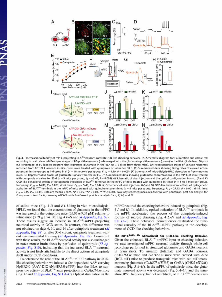
of saline mice (Fig. 4 D and E). Using in vivo microdialysis-HPLC, we found that the concentration of glutamate in the mPFCwas increased in the quinpirole mice (35.07 ± 9.03 μM) relative tosaline mice (3.59 ± 1.54 μM, Fig. 4 F–H and SI Appendix, Fig. S7).These results suggest an increase in BLAGlu
–mPFC-projectingneuronal activity in OCD states. In contrast, this difference wasnot obtained on days 6, 10, and 14 after quinpirole treatment (SIAppendix, Fig. S8) or after 30-d chronic quinpirole treatment with-out environmental training (SI Appendix, Fig. S9). Consistentwith these results, the BLAGlu neuronal activity was also unchangedin naïve mouse brain slices by perfusion of quinpirole (SI Ap-pendix, Fig. S10), indicating that the increased BLAGlu neuronalactivity is not likely attributable to the direct actions of quinpiroleitself under OCD conditions.To determine the role of the BLAGlu→mPFC pathway in OCD-
like checking behavior, we infused a Cre-dependent AAV carryingNpHR3.0 (AAV-DIO-eNpHR3.0-eYFP) into the BLA to sup-press the activity of BLAGlu axon projections in CaMKII-Cre mice(Fig. 4I and SI Appendix, Fig. S11 A–C). Optical stimulation in the
mPFC restored the checking behaviors induced by quinpirole (Fig.4 J and K). In addition, optical activation of BLAGlu terminals inthe mPFC accelerated the process of the quinpirole-inducedroutine of sucrose drinking (Fig. 4 L–N and SI Appendix, Fig.S11 D–F). These behavioral consequences established the func-tional causality of the BLAGlu→mPFC pathway in the develop-ment of OCD-like checking behaviors.
The mPFCGABA→Glu Microcircuit for OCD-Like Checking Behavior.Given the enhanced BLAGlu→mPFC input in checking behavior,we next investigated mPFC neuronal activity through whole-cellrecordings performed in visualized glutamate and GABA neuronsin brain slices. To visualize glutamate and GABA neurons,CaMKII-Cre mice and GAD2-Cre mice were crossed with Ai14(RCL-tdT) mice to produce transgenic mice with red tdTomato-expressing glutamate (CaMKII-tdTOM) or GABA (GAD2-tdTOM)neurons (Fig. 5 A–H). In the mPFC of quinpirole mice, the gluta-mate neuronal activity was decreased (Fig. 5 A–C), and the mini-ature IPSC frequency, but not amplitude, of mPFCGlu neurons was
Fig. 4. Increased excitability of mPFC-projecting BLAGlu neurons controls OCD-like checking behavior. (A) Schematic diagram for FG injection and whole-cellrecording in brain slices. (B) Example images of FG-positive neurons (red) merged with the glutamate-positive neurons (green) in the BLA. (Scale bars: 50 μm.)(C) Percentage of FG-labeled neurons that expressed glutamate in the BLA (n = 5 slices from three mice). (D) Representative traces of voltage responsesrecorded from FG+ BLA neurons in slices from mice treated with quinpirole or saline for 30 d. (E) Summarized data showing firing rates of evoked actionpotentials in the groups as indicated in D (n = 18 neurons per group, F1,34 = 9.19, P = 0.005). (F) Schematic of microdialysis-HPLC detection in freely movingmice. (G) Representative traces of glutamate signals from the mPFC. (H) Summarized data showing glutamate concentrations in the mPFC of mice treatedwith quinpirole or saline for 30 d (n = 5 mice per group, t8 = −3.44, P = 0.009). (I) Schematic of viral injection and the optical configuration in vivo. (J and K)OCD-like behavioral effects of optogenetic inhibition of BLAGlu terminals in the mPFC of mice treated with quinpirole 15 times (n = 5 to 7 mice per group,frequency: F1,10 = 14.88, P = 0.003; drink time: F1,10 = 5.48, P = 0.04). (L) Schematic of viral injection. (M and N) OCD-like behavioral effects of optogeneticactivation of BLAGlu terminals in the mPFC of mice treated with quinpirole seven times (n = 5 mice per group, frequency: F1,8 = 27.13, P < 0.001; drink time:F1,8 = 6.45, P = 0.035). Data are means ± SEM. *P < 0.05, **P < 0.01, ***P < 0.001. Two-way repeated-measures ANOVA with Bonferroni post hoc analysis forE; unpaired t test for H; one-way ANOVA with Bonferroni post hoc analysis for J, K, M, and N.
3802 | www.pnas.org/cgi/doi/10.1073/pnas.1814292116 Sun et al.
Dow
nloa
ded
by g
uest
on
Apr
il 2,
202
0
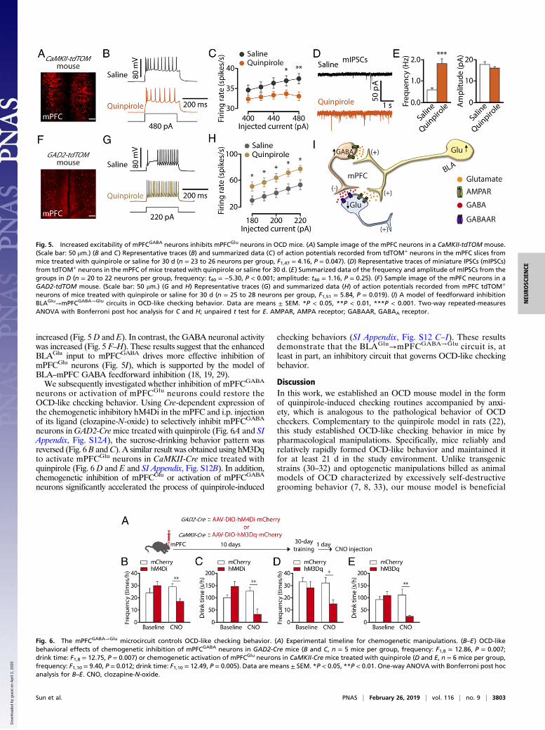
increased (Fig. 5D and E). In contrast, the GABA neuronal activitywas increased (Fig. 5 F–H). These results suggest that the enhancedBLAGlu input to mPFCGABA drives more effective inhibition ofmPFCGlu neurons (Fig. 5I), which is supported by the model ofBLA–mPFC GABA feedforward inhibition (18, 19, 29).We subsequently investigated whether inhibition of mPFCGABA
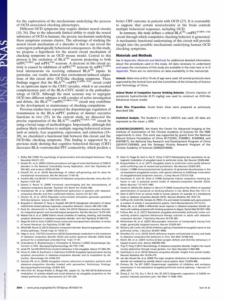
neurons or activation of mPFCGlu neurons could restore theOCD-like checking behavior. Using Cre-dependent expression ofthe chemogenetic inhibitory hM4Di in the mPFC and i.p. injectionof its ligand (clozapine-N-oxide) to selectively inhibit mPFCGABA
neurons inGAD2-Cremice treated with quinpirole (Fig. 6A and SIAppendix, Fig. S12A), the sucrose-drinking behavior pattern wasreversed (Fig. 6B andC). A similar result was obtained using hM3Dqto activate mPFCGlu neurons in CaMKII-Cre mice treated withquinpirole (Fig. 6 D and E and SI Appendix, Fig. S12B). In addition,chemogenetic inhibition of mPFCGlu or activation of mPFCGABA
neurons significantly accelerated the process of quinpirole-induced
checking behaviors (SI Appendix, Fig. S12 C–I). These resultsdemonstrate that the BLAGlu→mPFCGABA→Glu circuit is, atleast in part, an inhibitory circuit that governs OCD-like checkingbehavior.
DiscussionIn this work, we established an OCD mouse model in the formof quinpirole-induced checking routines accompanied by anxi-ety, which is analogous to the pathological behavior of OCDcheckers. Complementary to the quinpirole model in rats (22),this study established OCD-like checking behavior in mice bypharmacological manipulations. Specifically, mice reliably andrelatively rapidly formed OCD-like behavior and maintained itfor at least 21 d in the study environment. Unlike transgenicstrains (30–32) and optogenetic manipulations billed as animalmodels of OCD characterized by excessively self-destructivegrooming behavior (7, 8, 33), our mouse model is beneficial
Fig. 5. Increased excitability of mPFCGABA neurons inhibits mPFCGlu neurons in OCD mice. (A) Sample image of the mPFC neurons in a CaMKII-tdTOM mouse.(Scale bar: 50 μm.) (B and C) Representative traces (B) and summarized data (C) of action potentials recorded from tdTOM+ neurons in the mPFC slices frommice treated with quinpirole or saline for 30 d (n = 23 to 26 neurons per group, F1,47 = 4.16, P = 0.047). (D) Representative traces of miniature IPSCs (mIPSCs)from tdTOM+ neurons in the mPFC of mice treated with quinpirole or saline for 30 d. (E) Summarized data of the frequency and amplitude of mIPSCs from thegroups in D (n = 20 to 22 neurons per group, frequency: t40 = −5.30, P < 0.001; amplitude: t40 = 1.16, P = 0.25). (F) Sample image of the mPFC neurons in aGAD2-tdTOM mouse. (Scale bar: 50 μm.) (G and H) Representative traces (G) and summarized data (H) of action potentials recorded from mPFC tdTOM+
neurons of mice treated with quinpirole or saline for 30 d (n = 25 to 28 neurons per group, F1,51 = 5.84, P = 0.019). (I) A model of feedforward inhibitionBLAGlu→mPFCGABA→Glu circuits in OCD-like checking behavior. Data are means ± SEM. *P < 0.05, **P < 0.01, ***P < 0.001. Two-way repeated-measuresANOVA with Bonferroni post hoc analysis for C and H; unpaired t test for E. AMPAR, AMPA receptor; GABAAR, GABAA receptor.
Fig. 6. The mPFCGABA→Glu microcircuit controls OCD-like checking behavior. (A) Experimental timeline for chemogenetic manipulations. (B–E) OCD-likebehavioral effects of chemogenetic inhibition of mPFCGABA neurons in GAD2-Cre mice (B and C, n = 5 mice per group, frequency: F1,8 = 12.86, P = 0.007;drink time: F1,8 = 12.75, P = 0.007) or chemogenetic activation of mPFCGlu neurons in CaMKII-Cre mice treated with quinpirole (D and E, n = 6 mice per group,frequency: F1,10 = 9.40, P = 0.012; drink time: F1,10 = 12.49, P = 0.005). Data are means ± SEM. *P < 0.05, **P < 0.01. One-way ANOVA with Bonferroni post hocanalysis for B–E. CNO, clozapine-N-oxide.
Sun et al. PNAS | February 26, 2019 | vol. 116 | no. 9 | 3803
NEU
ROSC
IENCE
Dow
nloa
ded
by g
uest
on
Apr
il 2,
202
0
for the exploration of the mechanisms underlying the processof OCD-associated checking phenotypes.Different OCD symptoms may require distinct neural circuits
(10, 34). Due to the inherently limited ability to study the neuralsubstrates of OCD in humans, the precise mechanism underlyingthese symptoms remains elusive. The advantage of studying theneural circuit mechanism of a disorder is that it can reveal theconvergent pathologically behavioral consequences. In this study,we propose a hypothesis for the neural circuit mechanism ofchecking symptoms in an OCD mouse model. Central to thisprocess is the excitation of BLAGlu neurons projecting to bothmPFCGABA and mPFCGlu neurons. A decrease in this circuit ac-tivity is caused by inhibition of mPFCGlu neurons by mPFCGABA
local interneurons via receiving enhanced BLAGlu inputs. Inparticular, our results showed that environment-induced adapta-tions of this circuit drive OCD-like checking symptoms. Thesefindings suggest that the BLAGlu→mPFCGABA→Glu circuit couldbe an upstream input to the CSTC circuitry, which is an essentialcomplementary part of the BLA–CSTC model in the pathophys-iology of OCD. Although the most accurate way to segregateOCD into different subtypes is still a matter of active investigationand debate, the BLAGlu→mPFCGABA→Glu circuit may contributeto the development or maintenance of checking compulsions.Previous studies have reported the dopaminergic regulation of
transmission in the BLA–mPFC pathway of distinct executivefunctions in vivo (35). In the current study, we dissected theprecise organization of the BLAGlu→mPFCGABA→Glu circuit byusing a broad range of methodologies. Importantly, although thispathway likely contributes to multiple ongoing behavioral actionssuch as anxiety, fear acquisition, expression, and extinction (18–20), we elucidated a functional link between this circuit and theOCD-like checking symptoms. This finding is supported by aprevious study showing that cognitive behavioral therapy (CBT)decreases BLA–ventromedial PFC connectivity, which predicts a
better CBT outcome in patients with OCD (27). It is reasonableto suppose that certain neurocircuitry in the brain controlsmultiple behavioral responses, including OCD.In summary, this study defines a critical BLAGlu→mPFCGABA→Glu
circuit through which compulsive checking behavior is generated.A mechanistic functional understating of this circuit will provideinsight into the possible mechanisms underlying human OCDchecking symptoms.
Materials and MethodsSee SI Appendix, Materials and Methods for additional detailed informationabout the procedures used in this study. All data necessary to understandand assess the conclusions of this study are available in the main text or in SIAppendix. There are no restrictions on data availability in the manuscript.
Animals.Male mice at 8 to 10 wk of age were used. All animal protocols wereapproved by the Animal Care and Use Committee of the University of Scienceand Technology of China.
Animal Model of Compulsive Sucrose Drinking Behavior. Chronic injection ofquinpirole hydrochloride (0.75 mg/kg) was used to construct an OCD-likebehavioral mouse model.
Brain Slice Preparation. Acute brain slices were prepared as previouslydescribed (36).
Statistical Analysis. The Student’s t test or ANOVA was used. All data areexpressed as the mean ± SEM.
ACKNOWLEDGMENTS. We thank the Center for Advanced Imaging at theInstitute of Automation of the Chinese Academy of Sciences for the fMRIexperiments in mice. This work was supported by grants from the NationalNatural Science Foundation of China (91732303, 31600851, 81600964, and81870877), the National Key Research and Development Program of China(2016YFC1305900), and the Strategic Priority Research Program of theChinese Academy of Sciences (XDB02010000).
1. Ridley RM (1994) The psychology of perserverative and stereotyped behaviour. ProgNeurobiol 44:221–231.
2. Kessler RC, et al. (2005) Lifetime prevalence and age-of-onset distributions of DSM-IVdisorders in the National Comorbidity Survey Replication. Arch Gen Psychiatry 62:593–602, and erratum (2005) 62:768.
3. Kalueff AV, et al. (2016) Neurobiology of rodent self-grooming and its value fortranslational neuroscience. Nat Rev Neurosci 17:45–59.
4. Franklin ME, Foa EB (2011) Treatment of obsessive compulsive disorder. Annu Rev ClinPsychol 7:229–243.
5. Saxena S, Rauch SL (2000) Functional neuroimaging and the neuroanatomy ofobsessive-compulsive disorder. Psychiatr Clin North Am 23:563–586.
6. Chamberlain SR, et al. (2008) Orbitofrontal dysfunction in patients with obsessive-compulsive disorder and their unaffected relatives. Science 321:421–422.
7. Ahmari SE, et al. (2013) Repeated cortico-striatal stimulation generates persistentOCD-like behavior. Science 340:1234–1239.
8. Burguière E, Monteiro P, Feng G, Graybiel AM (2013) Optogenetic stimulation of lateralorbitofronto-striatal pathway suppresses compulsive behaviors. Science 340:1243–1246.
9. Pauls DL, Abramovitch A, Rauch SL, Geller DA (2014) Obsessive-compulsive disorder:An integrative genetic and neurobiological perspective. Nat Rev Neurosci 15:410–424.
10. Mataix-Cols D, et al. (2004) Distinct neural correlates of washing, checking, and hoardingsymptom dimensions in obsessive-compulsive disorder. Arch Gen Psychiatry 61:564–576.
11. Nagai M, Kishi K, Kato S (2007) Insular cortex and neuropsychiatric disorders: A reviewof recent literature. Eur Psychiatry 22:387–394.
12. Milad MR, Rauch SL (2012) Obsessive-compulsive disorder: Beyond segregated cortico-striatal pathways. Trends Cogn Sci 16:43–51.
13. Häge A, et al.; TACTICS Consortium (2016) Glutamatergic medication in the treatmentof obsessive compulsive disorder (OCD) and autism spectrum disorder (ASD)–Studyprotocol for a randomised controlled trial. Trials 17:141.
14. Chakrabarty K, Bhattacharyya S, Christopher R, Khanna S (2005) Glutamatergic dys-function in OCD. Neuropsychopharmacology 30:1735–1740.
15. Janak PH, Tye KM (2015) From circuits to behaviour in the amygdala. Nature 517:284–292.16. Simon D, Adler N, Kaufmann C, Kathmann N (2014) Amygdala hyperactivation during
symptom provocation in obsessive-compulsive disorder and its modulation by dis-traction. Neuroimage Clin 4:549–557.
17. Szeszko PR, et al. (2004) Amygdala volume reductions in pediatric patients withobsessive-compulsive disorder treated with paroxetine: Preliminary findings.Neuropsychopharmacology 29:826–832.
18. Felix-Ortiz AC, Burgos-Robles A, Bhagat ND, Leppla CA, Tye KM (2016) Bidirectionalmodulation of anxiety-related and social behaviors by amygdala projections to themedial prefrontal cortex. Neuroscience 321:197–209.
19. Klavir O, Prigge M, Sarel A, Paz R, Yizhar O (2017) Manipulating fear associations via op-togenetic modulation of amygdala inputs to prefrontal cortex. Nat Neurosci 20:836–844.
20. Burgos-Robles A, et al. (2017) Amygdala inputs to prefrontal cortex guide behavioramid conflicting cues of reward and punishment. Nat Neurosci 20:824–835.
21. Carlsen J (1988) Immunocytochemical localization of glutamate decarboxylase in therat basolateral amygdaloid nucleus, with special reference to GABAergic innervationof amygdalostriatal projection neurons. J Comp Neurol 273:513–526.
22. Szechtman H, Sulis W, Eilam D (1998) Quinpirole induces compulsive checking be-havior in rats: A potential animal model of obsessive-compulsive disorder (OCD).Behav Neurosci 112:1475–1485.
23. Amato D, Milella MS, Badiani A, Nencini P (2006) Compulsive-like effects of repeatedadministration of quinpirole on drinking behavior in rats. Behav Brain Res 172:1–13.
24. Eilam D (2017) From an animal model to human patients: An example of a translationalstudy on obsessive compulsive disorder (OCD). Neurosci Biobehav Rev 76:67–76.
25. Hoffman GE, SmithMS, Verbalis JG (1993) c-Fos and related immediate early gene productsas markers of activity in neuroendocrine systems. Front Neuroendocrinol 14:173–213.
26. Phillips ML, et al. (2000) A differential neural response in obsessive-compulsive disorder pa-tients withwashing comparedwith checking symptoms to disgust. PsycholMed 30:1037–1050.
27. Fullana MA, et al. (2017) Basolateral amygdala-ventromedial prefrontal cortex con-nectivity predicts cognitive behavioural therapy outcome in adults with obsessive-compulsive disorder. J Psychiatry Neurosci 42:378–385.
28. Wickersham IR, et al. (2007) Monosynaptic restriction of transsynaptic tracing fromsingle, genetically targeted neurons. Neuron 53:639–647.
29. McGarry LM, Carter AG (2016) Inhibitory gating of basolateral amygdala inputs to theprefrontal cortex. J Neurosci 36:9391–9406.
30. Shmelkov SV, et al. (2010) Slitrk5 deficiency impairs corticostriatal circuitry and leadsto obsessive-compulsive-like behaviors in mice. Nat Med 16:598–602.
31. Welch JM, et al. (2007) Cortico-striatal synaptic defects and OCD-like behaviours inSapap3-mutant mice. Nature 448:894–900.
32. Ting JT, Feng G (2011) Neurobiology of obsessive-compulsive disorder: Insights into neuralcircuitry dysfunction through mouse genetics. Curr Opin Neurobiol 21:842–848.
33. Szechtman H, et al. (2016) Obsessive-compulsive disorder: Insights from animal models.Neurosci Biobehav Rev 76:254–279.
34. van den Heuvel OA, et al. (2009) The major symptom dimensions of obsessive-compulsivedisorder are mediated by partially distinct neural systems. Brain 132:853–868.
35. Floresco SB, Tse MT (2007) Dopaminergic regulation of inhibitory and excitatorytransmission in the basolateral amygdala-prefrontal cortical pathway. J Neurosci 27:2045–2057.
36. Zhang Z, Cai YQ, Zou F, Bie B, Pan ZZ (2011) Epigenetic suppression of GAD65 ex-pression mediates persistent pain. Nat Med 17:1448–1455.
3804 | www.pnas.org/cgi/doi/10.1073/pnas.1814292116 Sun et al.
Dow
nloa
ded
by g
uest
on
Apr
il 2,
202
0