Embed Size (px)
Citation preview
Bcr/Abl Interferes with the Fanconi Anemia/BRCAPathway: Implications in the Chromosomal Instability ofChronic Myeloid Leukemia CellsAntonio Valeri1, Maria Eugenia Alonso-Ferrero1, Paula Rıo1, Marıa Roser Pujol2,3, Jose A. Casado1, Laura
Perez1, Ariana Jacome1, Xabier Agirre4, Maria Jose Calasanz4, Helmut Hanenberg5,6, Jordi Surralles2,3,
Felipe Prosper4, Beatriz Albella1, Juan A. Bueren1*
1 Centro de Investigaciones Energeticas, Medioambientales y Tecnologicas (CIEMAT) and Centro de Investigacion Biomedica en Red de Enfermedades Raras (CIBERER),
Madrid, Spain, 2 Departamento de Genetica y Microbiologia, Universitat Autonoma de Barcelona, Bellaterra, Spain, 3 Centro de Investigacion Biomedica en Red de
Enfermedades Raras (CIBERER), Barcelona, Spain, 4 Fundacion para la Investigacion Medica Aplicada (CIMA), Clınica Universidad de Navarra, Pamplona, Spain,
5 Department of Pediatric Oncology, Hematology and Immunology, Children’s Hospital, Duesseldorf, Germany, 6 Department of Pediatrics, Wells Center for Pediatric
Research, Riley Hospital for Children, Indianapolis, Indiana, United States of America
Abstract
Chronic myeloid leukemia (CML) is a malignant clonal disorder of the hematopoietic system caused by the expression of theBCR/ABL fusion oncogene. Although it is well known that CML cells are genetically unstable, the mechanisms accounting forthis genomic instability are still poorly understood. Because the Fanconi anemia (FA) pathway is believed to control severalmechanisms of DNA repair, we investigated whether this pathway was disrupted in CML cells. Our data show that CML cellshave a defective capacity to generate FANCD2 nuclear foci, either in dividing cells or after DNA damage. Similarly, humancord blood CD34+ cells transduced with BCR/ABL retroviral vectors showed impaired FANCD2 foci formation, whereasFANCD2 monoubiquitination in these cells was unaffected. Soon after the transduction of CD34+ cells with BCR/ABLretroviral vectors a high proportion of cells with supernumerary centrosomes was observed. Similarly, BCR/ABL induced ahigh proportion of chromosomal abnormalities, while mediated a cell survival advantage after exposure to DNA cross-linking agents. Significantly, both the impaired formation of FANCD2 nuclear foci, and also the predisposition of BCR/ABLcells to develop centrosomal and chromosomal aberrations were reverted by the ectopic expression of BRCA1. Takentogether, our data show for the first time a disruption of the FA/BRCA pathway in BCR/ABL cells, suggesting that thisdefective pathway should play an important role in the genomic instability of CML by the co-occurrence of centrosomalamplification and DNA repair deficiencies.
Citation: Valeri A, Alonso-Ferrero ME, Rıo P, Pujol MR, Casado JA, et al. (2010) Bcr/Abl Interferes with the Fanconi Anemia/BRCA Pathway: Implications in theChromosomal Instability of Chronic Myeloid Leukemia Cells. PLoS ONE 5(12): e15525. doi:10.1371/journal.pone.0015525
Editor: Sue Cotterill, St Georges University of London, United Kingdom
Received August 7, 2010; Accepted October 8, 2010; Published December 28, 2010
Copyright: � 2010 Valeri et al. This is an open-access article distributed under the terms of the Creative Commons Attribution License, which permitsunrestricted use, distribution, and reproduction in any medium, provided the original author and source are credited.
Funding: This work was supported by grants from the European Program "Life Sciences, Genomics and Biotechnology for Health" (CONSERT; Ref LSHB-CT-2004-5242), Centro de Investigacion en Red de Enfermedades Raras (CIBERER), Comision Interministerial de Ciencia y Tecnologıa (SAF2005-00058, SAF 2009-11936 andSAF 2009-027) and Genoma Espana (FANCOGENE). The funders had no role in study design, data collection and analysis, decision to publish, or preparation of themanuscript.
Competing Interests: H.H. may receive royalties based on a license agreement between Indiana University and Takara Shuzo, Ltd., resulting from the sale of thefibronectin fragment CH296 (RetronectinH). The other authors declare no competing financial interests.
* E-mail: [email protected]
Introduction
Chronic Myeloid Leukemia (CML) is a clonal hematopoietic
disorder generated by a t(9;22)(q34;q11) translocation resulting in a
BCR/ABL oncogene[1,2] that encodes for a tyrosine kinase BCR/
ABL-p210 oncoprotein[3]. Although several genetic defects are
accumulated in CML cells during the progression from the chronic
phase towards the accelerated and blast crisis phases (see review in
[4]), studies in mice transplanted with BCR/ABL transduced cells
demonstrated that this oncogene is the causative agent of CML[5].
In addition to a differentiation arrest, failures in the genomic
surveillance and DNA repair of CML cells account for the natural
malignant progression of the disease (see review in[6]). Although
the mechanisms by which BCR/ABL interferes with the genomic
stability of the cell are still poorly understood, the effects of this
oncoprotein upon DNA damage, apoptosis and DNA repair are
considered critical processes facilitating the accumulation of
mutations during the progress to blast crisis (see review in [7]).
Moreover, increasing evidence has been published showing that
BCR/ABL induces reactive oxygen species (ROSs) causing
oxidative damage to CML cells[8], and therefore a variety of
DNA lesions, including the highly mutagenic double strand breaks
(DSBs)[9,10]. These effects, together with the reported effect of
this oncoprotein on the efficacy and/or the fidelity of different
DNA repair mechanisms[9,11,12] contribute to explain the
mutator phenotype of CML cells.
Concerning the mechanisms by which BCR/ABL affects the
repair of the DSBs, previous studies have shown that this
oncoprotein interferes both with the non-homologous end joining
(NHEJ) pathway and with pathways that utilize homologous
templates. Regarding the effects of BCR/ABL on classic NHEJ,
Deutsch et al observed that the catalytic subunit of DNA-
PLoS ONE | www.plosone.org 1 December 2010 | Volume 5 | Issue 12 | e15525
dependent protein kinase (DNA-PKcs), a key protein in this major
DNA repair system in mammalians, was down-regulated in CML
cells[13]. In addition to NHEJ, BCR/ABL has also been involved
in the aberrant regulation of the two pathways that utilize
homologous templates, the faithful homology directed repair
(HDR) and the mutagenic single strand annealing (SSA).
Interestingly, previous studies have shown that BRCA1, a critical
protein for preserving the genomic integrity by promoting
homologous recombination[14], is nearly undetectable in CML
cells[15]. On the other hand, more recent studies have shown that
BCR/ABL specifically promotes the repair of DSBs through SSA,
a mutagenic pathway that involve sequence repeats[16,17].
Because the Fanconi anemia (FA) pathway is believed to control
several DNA repair pathways, and therefore the genomic stability of
the cell (see review in[18]), we ought to investigate the integrity of
this pathway in CML cells. Thirteen FA proteins have been
identified in the FA pathway, each of them participating in one of
the three FA protein complexes. The upstream complex – the FA
core complex - is integrated by eight FA proteins (FANCA,
FANCB, FANCC, FANCE, FANCF, FANCG, FANCL, FANCM)
and two FA associated proteins (FAAP24 and FAAP100). A second
complex is formed by FANCD2 and FANCI, which work together
in the FA-ID complex. Because of the E3 ligase activity (FANCL) of
the FA core complex, FANCD2 and FANCI can be monoubiqui-
tinated and then loaded onto chromatin, forming large nuclear foci
in response to DNA damage or replication arrest. Finally,
monoubiquitinated FANCD2/FANCI interact with downstream
FA proteins such as FANCJ/BRIP1, FANCN/PALB2 and
FANCD1/BRCA2, which form stable complexes with proteins
participating in HDR, like BRCA1 and RAD51[19,20].
The results presented in this study demonstrate for the first time
that CML cells are characterized by a defective FA/BRCA
pathway, downstream FANCD2 monoubiquitination. In particu-
lar we demonstrate that BCR/ABL interferes with the formation
of nuclear FANCD2 foci, in a process that can be reverted by the
ectopic expression of BRCA1. The consequences of this defect
upon the genetic stability of BCR/ABL cells are shown.
Materials and Methods
Culture of CD34+ cells from CML patients and healthyumbilical cord blood
Studies were approved by the authors’ Institutional Review
Board and conducted under the Declaration of Helsinki rules.
Chronic myeloid leukemia CD34+ cells were obtained from the
peripheral blood (PB) of CML patients, after previous informed
consents approved by the ethical Committee of the Clınica
Universitaria de Navarra. Healthy CD34+ cells were obtained from
umbilical cord bloods (CB) scheduled for discard, after written
informed consents of the mother. Mononuclear cells (MNCs) were
obtained by fractionation in Ficoll-hypaque according to manufac-
turer’s instructions (GE Healthcare, Stockholm, Sweden). Purified
CD34+ cells were obtained using a MACS CD34 Micro-Bead kit
(Miltenyi Biotec, Gladbach, Germany). For the expansion of CML
CD34+ cells, samples were cultured in StemSpan medium (Stem
Cell Technologies, Vancouver, BC, Canada) supplemented with
100 ng/ml human stem cell factor (hSCF; Peprotech, London,
UK), 100 ng/ml Flt3-L (Invitrogen, Carlsbad, CA), 20 ng/ml IL-6
(Peprotech) and 20 ng/ml IL-3 (Biosource).
Cell linesHuman-derived Mo7e (a megakaryoblastic leukaemia cell line
without the BCR/ABL fusion) and Mo7e-p210 cells (Mo7e cells
transfected with p210 isoform of BCR-ABL1) [21,22] were cultured
in RPMI medium (Gibco, NY) supplemented with 10% fetal
bovine serum (FBS; Lonza, Belgium), 2 mM L-glutamine (Gibco),
100 U/mL penicillin/streptomycin (Gibco) at 37uC in a humid-
ified atmosphere with 5% CO2. Mo7e cells were grown in medium
supplemented with 10 ng/ml of hr-IL3 (Biosource). FA-A LCLs
were cultured in RPMI medium (Gibco) supplemented with 15%
FBS, 2 mM L-glutamine (Gibco), 100 U/mL penicillin/strepto-
mycin (Gibco) at 37uC in a humidified atmosphere with 5% CO2.
Retroviral vectors productionThe control retroviral vector (RV) used in these experiments was
the MIG-R1 retroviral vector which consist on a MSCV-IRES-
GFP vector. The MIG-210 is derived from the MIG-R1, and
contains the full-length b3a2 BCR/ABL cDNA under the control of
the MSCV promoter. Both vectors were kindly provided by Bryan
G. Druker (Oregon Health and Science University, Portland, OR).
Where indicated, cells were transduced with similar RVs (MIN-210
and its respective MIN-R1 control, kindly provided by W. S. Pear).
In these vectors, a truncated version of the NGFR (DNGFR) cDNA
was used instead of the EGFP marker gene, to facilitate the
immunoselection of transduced cells. In order to ectopically express
BRCA1 in control and BCR/ABL positive cells, S11Brca1-IRES-
Neo and S11-IRES-Neo RVs were generated. Retroviral vectors
were produced and titrated as previously described[23].
Retroviral transduction of hematopoietic progenitorsfrom human cord blood
Human CB CD34+ cells were pre-stimulated for 48 h with
StemSpan (StemCell technologies) supplemented with 300 ng/ml
hSCF (Peprotech), 100 ng/ml hTPO (R&DSystems, Minneapolis,
MN) and 100 ng/ml Flt3-L (Invitrogen, Carlsbad, CA). Pre-
stimulated cells were re-suspended at a density of 56105 cells/ml
in retroviral supernatant medium supplemented with FBS (20%
final concentration) and growth factors. Cells were then added to
retronectin-coated wells (Retronectin, Takara Shuzo, Otsu, Japan)
preloaded with the correspondent retroviral vector. Supernatants
were replaced every 12 h by new virus containing medium. A total
of four transduction cycles were routinely conducted[23].
When hCB CD34+ cells were transduced with MIN-R1 and
MIN-210 and purified by immunomagnetic cell sorting with anti-
NGFR beads (Miltenyi Biotech) 2 days after transduction
following manufacturer’s instructions. Purified populations con-
tained at least 95% of NGFR+ cells.
DNA damage treatments and drugs exposureTo conduct Western Blotting and Immunofluorescence assays,
cells were treated with Imatinib 1 mM (Novartis Pharma, Basel,
Switzerland) for 24 hours and then treated with 40 nM of
mitomycin C (MMC) for 16 hours. For inhibition studies cells
were treated for 16 hours with MMC and afterwards 1 hour with
40 mM of MG132 (Sigma-Aldrich, ST.Louis, MO) or 20 mM of
LY294002 (Cell Signaling Technology, Inc, Danvers, MA). To test
the cellular resistance to MMC, clonogenic assays were conducted
in semisolid medium (Methocult H4434, StemCell Technologies)
containing increasing concentrations of the drug, plated in
triplicate on 35-mm plastic culture dishes (Nunc, Roskilde,
Denmark) and cultured at 37uC, 5% CO2 and fully humidified
air. Fourteen days after plating, the total number of colonies was
scored under an inverted microscope.
Chromosomal breakage analysesCB CD34+ cells were transduced with MIN-210 or its respective
MIN-R1 control and purified two days afterwards, prior to re-infect
Defective Fanconi Anemia Pathway in CML
PLoS ONE | www.plosone.org 2 December 2010 | Volume 5 | Issue 12 | e15525
these samples with Neor or BRCA1/Neor vectors. Samples were left
untreated or treated with 0.1 mg/ml of diepoxibutane (DEB) for a
72-h period. To obtain metaphases, colcemid (0.1 mg/ml; Gibco)
was added 24 h prior to harvesting. Cells were then treated with
hypotonic solution (0.075 M KCl, Sigma), fixed in methanol:acetic
acid (3:1 vol/vol), dropped onto clean slides and air-dried O.N.
following standard cytogenetic procedures. Slides were stained with
10% Giemsa in phosphate buffer, pH 6.8. Fifty to seventy
metaphases per sample were analyzed for chromosome aberrations
including gaps, chromosome and chromatid breaks, acentric
fragments, and chromosome- and chromatid-type exchanges.
Flow cytometry analysesFor cell cycle analyses, aliquots of 105 cells were washed twice in
PBS and fixed in 4.5 mL ice-cold 1% methanol-free formaldehyde
in PBS for 15 min on ice. After centrifugation, 5 mL of 70%
ethanol (0–4uC) was added, and cells were stored at 220uC at least
for 2 hours. After washing, cells were resuspended in 1 mL
propidium iodide solution (5 mg/mL; Molecular Probes) with
100 mg/mL DNase-free RNase A (Sigma) and incubated for
30 min at 37uC. Finally, cells were analyzed by flow cytometry
(Coulter XL) with linear fluorescence of propidium iodide (DNA
content) from 10,000 events with doublet discrimination.
Western Blot analysesWestern blot analyses were performed using extracts of purified
CD34+ cells collected by centrifugation. After electrophoresis,
proteins were transferred to a nitrocellulose membrane. After
blocking, the membrane was incubated at 4uC O.N. with the
primary antibodies (anti-FANCD2, Abcam ab2187) diluted in
blocking solution, washed extensively and incubated with the
appropriate secondary antibody using the Western Breeze
Immunodetection Kit (Invitrogen), according to methods previ-
ously described[24].
Immunofluorescence studiesIn these studies cells were fixed with 3.7% paraformaldehyde in
PBS for 15 minutes followed by permeabilization with 0.5% Triton
X-100 in PBS for 5 min. After blocking for 30 minutes in Blocking
buffer (10% FBS, 0.1% NP-40 in PBS) cells were incubated with
rabbit polyclonal anti-FANCD2 (Abcam, ab2187-50), mouse
monoclonal anti-cH2AX (Upstate, JBW301), and mouse monoclo-
nal anti-BRCA1 (Oncogene, ab-1 and ab-3). For centrosomal
staining, cells were fixed and permeabilized and incubated with
primary antibody against c-tubulin (Abcam, ab16504). Cells were
subsequently washed three times in TBS (50 mM Tris-HCl
(pH 8.0), 150 mM NaCl) and incubated with anti-mouse or anti-
rabbit Texas Red (Molecular Probes, Leiden, The Netherlands) as
secondary antibodies. After 45 min. cells were washed three times
with TBS and the slides were mounted in Moviol with 4,6-
diamidino-2 phenylindole (DAPI). Slides were analyzed with a
fluorescence microscope Axioplan2 (Carl Zeiss, Gottingen, Ger-
many) using a 100x/1.45 oil working distance 0.17-mm objective.
The proportion of cells with nuclear foci was scored as described
elsewhere[24] after analyzing 200 cells per slide. Immunofluores-
cent images were acquired with an AxioCam MRm (Carl Zeiss) and
were processed with AxioVision version 4.6.3 (Carl Zeiss) and Corel
Photo-Paint 11 (Corel, Ottawa, Canada).
Statistical analysisResults are shown as the mean6s.e. Differences between groups
were assessed using the two tailed Student’s t-test. The data from the
clonogenic assays were calculated as survival percentages respect to
control cultures. The survival data were fitted by least squares only
for experiments with at least three available data points. The IC50
value was obtained algebraically, solving the fitted quadratic
equation for the value of dose where the estimated percentage of
surviving cells would equal 50%. The processing and statistical
analysis of the data was performed by using the Statgraphics Plus
5.0 software package (Manugistics, Inc. Rockville, MD).
Results
BCR/ABL interferes with the formation of FANCD2nuclear foci in hematopoietic progenitors from chronicmyeloid leukemia patients
Because of the relevance of the FA/BRCA pathway in the
control of DNA repair, we first investigated whether CML cells
had a disruption in this pathway. To this aim, and given that the
loading of FANCD2 to chromatin constitutes a central process in
the FA/BRCA pathway[20], we determined the proportion of
cells with nuclear FANCD2 foci, both in a cell line stably
transfected with the BCR/ABL oncogene (Mo7e-p210 cells) and in
the control parental cells (Mo7e cells). As shown in Figure 1a, the
proportion of Mo7e-p210 cells with FANCD2 nuclear foci was
significantly reduced compared to control Mo7e cells. This effect
was evident in samples not exposed to any DNA damaging agent,
and also in cells treated with the DNA cross-linking drug MMC.
To investigate whether data obtained in Mo7e-p210 cells was
reproduced in primary CML cells, similar studies were conducted
with peripheral blood (PB) CD34+ cells from CML patients at
diagnosis, and from healthy CD34+ cells obtained from cord blood
(CB) samples. Similar to Mo7e cells, the proportion of CD34+ cells
with FANCD2 foci was significantly reduced when samples were
obtained from CML patients, compared to healthy CD34+ cells.
As in Mo7e cells, differences between both groups were significant,
both in untreated and in MMC-treated cells (Figure 1b).
The inhibition of FANCD2 foci in Mo7e-p210 cells and in
primary CML cells could result from either a direct effect of the
BCR/ABL oncogene, or through accumulated mutations that may
have occurred along the culture of the cell line or during the
progression of the disease of CML patients. To understand
whether this effect was directly generated by the BCR/ABL
oncogene, CB CD34+ cells were transduced with a control MIG-
R1 vector (only expressing the EGFP marker gene) or with the
oncogenic MIG-210 vector (expressing both the BCR/ABL and the
EGFP genes), and exposed to MMC seven days afterwards.
Additionally, one aliquot of these samples was incubated with
imatinib 24 h prior to MMC treatment, to evaluate whether
potential effects mediated by BCR/ABL were dependent on the
tyrosine kinase activity of the oncoprotein (See schematic
experimental protocol in Figure 2a). To ensure that FANCD2
foci were scored exclusively in cells that had been transduced with
either MIG-R1 or MIG-210 vectors, only green fluorescent cells
were considered for the analysis of nuclear FANCD2 foci.
Consistent with the results in Figure 1, the proportion of MMC-
treated CD34+ cells with FANCD2 foci was significantly inhibited
when samples were transduced with MIG-210, as compared to the
control MIG-R1 vector (Figure 2a). Moreover, while imatinib did
not significantly affect the formation of nuclear FANCD2 foci in
cells transduced with the control vector, this drug significantly
increased the proportion of MIG-210 transduced CD34+ cells with
FANCD2 foci (see Figure 2a and representative pictures in
Figure 2b). The effect of BCR/ABL upon the formation of nuclear
FANCD2 foci was confirmed in MIG-210-transduced CD34+
cells, by the observation that only EGFP-negative (untransduced)
cells contained evident nuclear FANCD2 foci; while EGFP
Defective Fanconi Anemia Pathway in CML
PLoS ONE | www.plosone.org 3 December 2010 | Volume 5 | Issue 12 | e15525
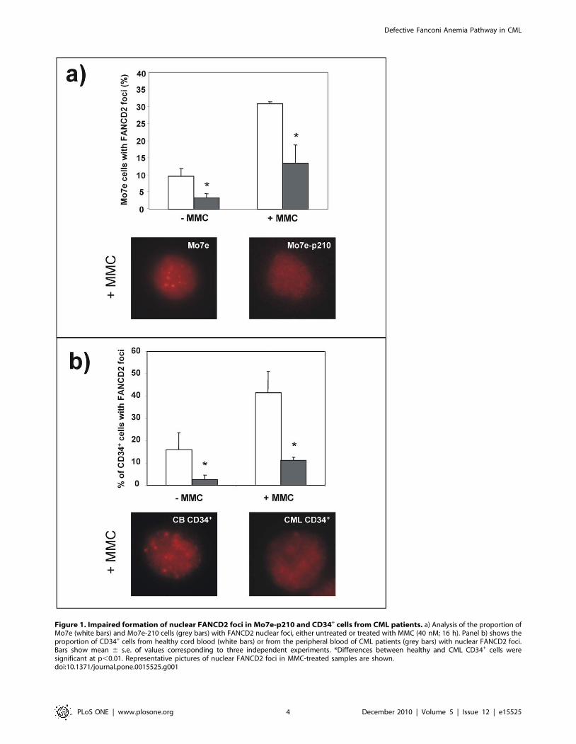
Figure 1. Impaired formation of nuclear FANCD2 foci in Mo7e-p210 and CD34+ cells from CML patients. a) Analysis of the proportion ofMo7e (white bars) and Mo7e-210 cells (grey bars) with FANCD2 nuclear foci, either untreated or treated with MMC (40 nM; 16 h). Panel b) shows theproportion of CD34+ cells from healthy cord blood (white bars) or from the peripheral blood of CML patients (grey bars) with nuclear FANCD2 foci.Bars show mean 6 s.e. of values corresponding to three independent experiments. *Differences between healthy and CML CD34+ cells weresignificant at p,0.01. Representative pictures of nuclear FANCD2 foci in MMC-treated samples are shown.doi:10.1371/journal.pone.0015525.g001
Defective Fanconi Anemia Pathway in CML
PLoS ONE | www.plosone.org 4 December 2010 | Volume 5 | Issue 12 | e15525
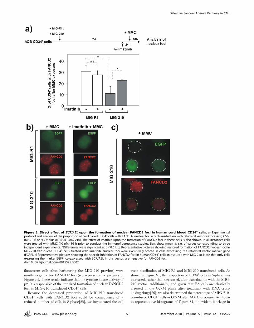
fluorescent cells (thus harbouring the MIG-210 provirus) were
mostly negative for FANCD2 foci (see representative pictures in
Figure 2c). These results indicate that the tyrosine kinase activity of
p210 is responsible of the impaired formation of nuclear FANCD2
foci in MIG-210 transduced CD34+ cells.
Because the decreased proportion of MIG-210 transduced
CD34+ cells with FANCD2 foci could be consequence of a
reduced number of cells in S-phase[25], we investigated the cell
cycle distribution of MIG-R1 and MIG-210 transduced cells. As
shown in Figure S1, the proportion of CD34+ cells in S-phase was
increased, rather than decreased, after transduction with the MIG-
210 vector. Additionally, and given that FA cells are classically
arrested in the G2/M phase after treatment with DNA cross-
linking drugs[26], we also determined the percentage of MIG-210-
transduced CD34+ cells in G2/M after MMC exposure. As shown
in representative histograms of Figure S1, no evident blockage in
Figure 2. Direct effect of BCR/ABL upon the formation of nuclear FANCD2 foci in human cord blood CD34+ cells. a) Experimentalprotocol and analysis of the proportion of cord blood CD34+ cells with FANCD2 nuclear foci after transduction with retroviral vectors expressing EGFP(MIG-R1) or EGFP plus BCR/ABL (MIG-210). The effect of imatinib upon the formation of FANCD2 foci in these cells is also shown. In all instances cellswere treated with MMC (40 nM) 16 h prior to conduct the immunofluorescence studies. Bars show mean 6 s.e. of values corresponding to threeindependent experiments. *Differences were significant at p,0.01. b) Representative pictures showing restored formation of FANCD2 nuclear foci inMIG-210-transduced CD34+ cells treated with imatinib. Nuclear foci were exclusively scored in cells expressing the retroviral vector marker gene(EGFP). c) Representative pictures showing the specific inhibition of FANCD2 foci in human CD34+ cells transduced with MIG-210. Note that only cellsexpressing the marker EGFP, co-expressed with BCR/ABL in this vector, are negative for FANCD2 foci.doi:10.1371/journal.pone.0015525.g002
Defective Fanconi Anemia Pathway in CML
PLoS ONE | www.plosone.org 5 December 2010 | Volume 5 | Issue 12 | e15525
the G2/M phase was observed in MMC-treated MIG-210-
transduced CD34+ cells at doses conventionally used for FA
diagnosis (40 nM).
We also speculated that the inhibitory effects of BCR/ABL upon
the formation of FANCD2 foci in MMC-treated cells could be due
to a potentially lower generation of DSBs in BCR/ABL cells. To rule
out this possibility, we determined the generation of DSBs in MIG-
R1 and MIG-210 transduced CB CD34+ cells by means of the
analysis of c-H2AX nuclear foci. A higher number of DSBs was
observed in MIG-210 cells compared to MIG-R1 cells in the
absence of MMC treatment (Figure S2), consistent with the
reported effects of BCR/ABL on the generation of ROSs[8,9,10].
After MMC, a similar proportion of cells with c-H2AX foci was
observed in control and p210-transduced cells (Figure S2). These
observations indicate that the low proportion of BCR/ABL cells with
FANCD2 foci is not attributable to an impaired generation of DSBs.
The impaired formation of nuclear FANCD2 foci in BCR/ABL cells is not due to a defect in FANCD2monoubiquitination
Since the formation of nuclear FANCD2 foci requires the
monoubiquitination of this protein[27], we then investigated
whether BCR/ABL was able to inhibit FANCD2 monoubiquitina-
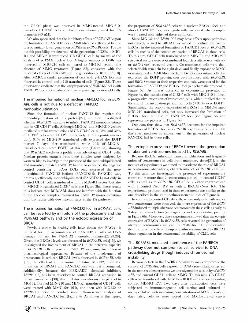
tion in CB CD34+ cells. Although MIG-R1 and MIG-210 vectors
mediated similar transductions of CB CD34+ cells (28% and 32%
of CD34+ cells were EGFP+, respectively, at 48 h post-transduc-
tion), 91% of MIG-210 transduced cells expressed the EGFP
marker 7 days after transduction, while 29% of MIG-R1
transduced cells were EGFP+ at this time (Figure 3a), showing
that BCR/ABL mediates a proliferation advantage in CD34+ cells.
Nuclear protein extracts from these samples were analyzed by
western blot to investigate the presence of the monoubiquitinated
and non-ubiquitinated FANCD2 bands. As expected, the negative
control consisting of FA-A LCLs only expressed the non-
ubiquitinated FANCD2 isoform (FANCD2-S). FANCD2 was,
however, efficiently monoubiquitinated (FANCD2-L) not only in
control CD34+ cells transduced with the MIG-R1 vector, but also
in MIG-210 transduced CD34+ cells (see Figure 3b). These results
thus indicate that BCR/ABL does not interfere with the function
of the FA core complex, required for FANCD2 monoubiquitina-
tion, but rather with downstream steps in the FA pathway.
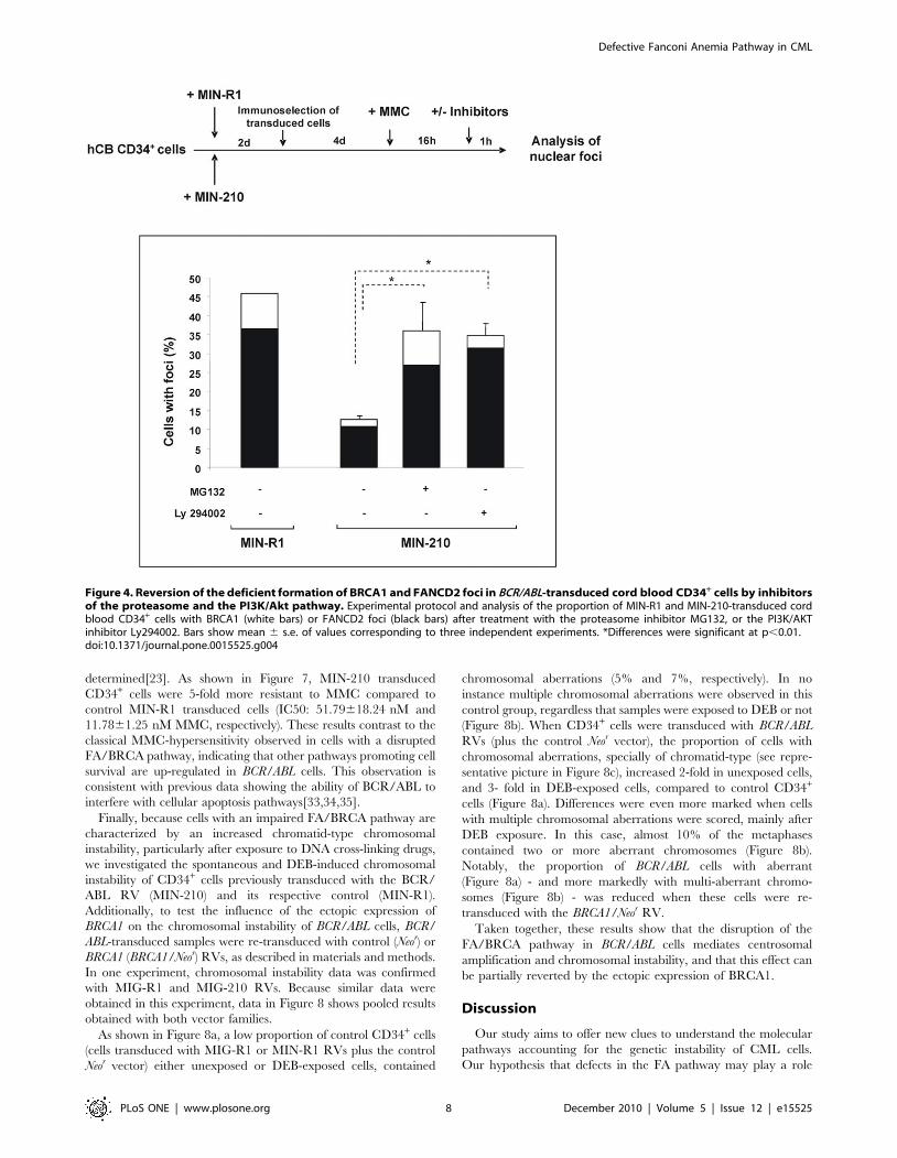
The impaired formation of FANCD2 foci in BCR/ABL cellscan be reverted by inhibitors of the proteasome and thePI3K/Akt pathway and by the ectopic expression ofBRCA1
Previous studies in healthy cells have shown that BRCA1 is
required for the accumulation of FANCD2 at sites of DNA
damage but not for FANCD2 monoubiquitination[27,28,29].
Given that BRCA1 levels are decreased in BCR/ABL cells[15], we
investigated the involvement of BRCA1 in the defective capacity
of BCR/ABL cells to generate FANCD2 foci, using two different
pharmacological approaches. Because of the involvement of
proteasome in reduced BRCA1 levels observed in BCR/ABL cells
[15], the effect of a proteasome inhibitor, MG132, upon the
formation of BRCA1 and FANCD2 foci was first investigated.
Additionally, because the PI3K/AKT chemical inhibitor,
LY294002, has been described to control BRCA1 activation in
breast cancer cells [30], this inhibitor was also used in parallel to
MG132. Purified MIN-210 and MIN-R1 transduced CD34+ cells
were treated with MMC for 16 h, and then with MG132 or
LY294002 prior to conduct immunofluorescence analyses of
BRCA1 and FANCD2 foci (Figure 4). As shown in this figure,
the proportion of BCR/ABL cells with nuclear BRCA1 foci, and
also of FANCD2 foci, was significantly increased when samples
were treated with either of these inhibitors.
Since MG132 and LY294002 may have effects upon pathways
not directly related to BRCA1, we aimed to confirm the role of
BRCA1 in the impaired formation of FANCD2 foci of BCR/ABL
cells by means of the ectopic expression of BRCA1 in these cells.
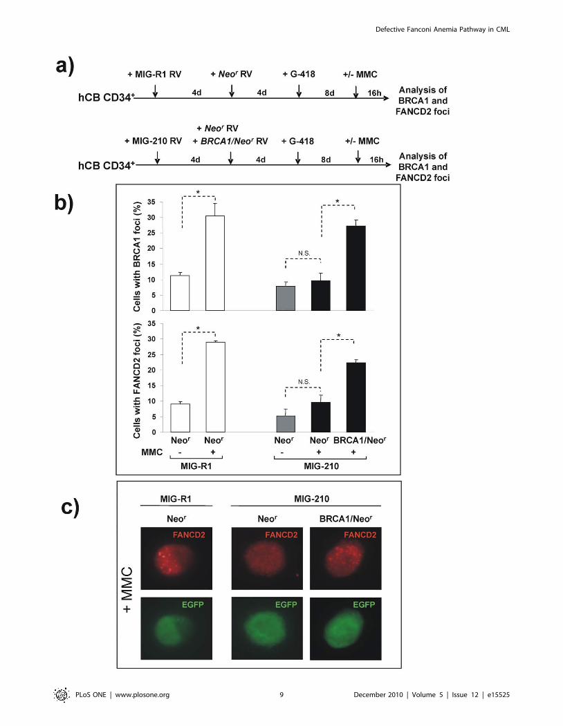
To this aim, CD34+ cells transduced with MIG-R1 and MIG-210
retroviral vectors were re-transduced four days afterwards with neor
or BRCA1/neor retroviral vectors. Co-transduced cells were then
selected with geneticin for eight days, and finally exposed to MMC
or maintained in MMC-free medium. Geneticin-resistant cells that
expressed the EGFP protein, thus co-transduced with BCR/ABL
and BRCA1 vectors or their respective controls, were scored for the
formation of FANCD2 and BRCA1 foci (see schematic protocol in
Figure 5a). As it was observed in experiments presented in
Figure 3a, the transduction of CD34+ cells with MIG-210 induced
a progressive expansion of transduced cells, which implied that at
the end of the incubation period most cells (.90%) were EGFP+.
Significantly, the ectopic expression of BRCA1 in MMC-treated
MIG-210 transduced cells, not only increased the formation of
BRCA1 foci, but also of FANCD2 foci (see Figure 5b and
representative pictures in Figure 5c).
Our data thus show that BCR/ABL accounts for the impaired
formation of BRCA1 foci in BCR/ABL expressing cells, and that
this effect mediates an impairment in the generation of nuclear
FANCD2 foci in these cells.
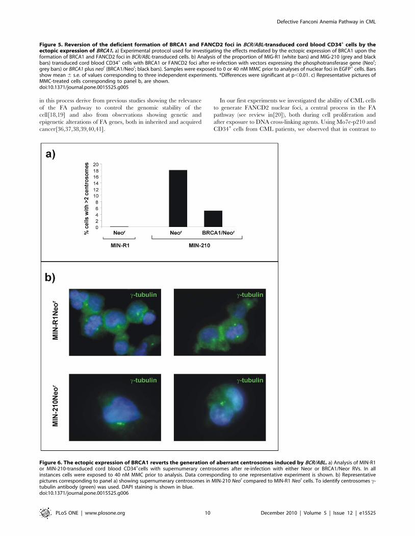
The ectopic expression of BRCA1 reverts the generationof aberrant centrosomes induced by BCR/ABL
Because BRCA1 inhibition caused amplification and fragmen-
tation of centrosomes in cells from mammary tissue[31], in the
next set of experiments we aimed to investigate the role of BRCA1
in centrosome aberrations, characteristic of BCR/ABL cells[32].
To this aim, we investigated the presence of supernumerary
centrosomes (more than 2 centrosomes per cell) in control CD34+
cells, as well as in BCR/ABL CD34+ cells, either re-transduced
with a control Neor RV or with a BRCA1/Neor RV. The
experimental protocol used in these experiments was similar to the
one described in the immunofluorescence studies of Figure 5.
In contrast to control CD34+ cells, where only cells with one or
two centrosomes were observed, the mere expression of the BCR/
ABL induced multiple aberrant centrosomes in these cells as early as
9 days post-transduction (see Figure 6a and representative pictures
in Figure 6b). Moreover, these experiments showed that the ectopic
expression of BRCA1 in BCR/ABL cells reverted the generation of
aberrant centrosomes induced by BCR/ABL. This observation
demonstrates the role of disrupted pathways associated to BRCA1
down-regulation in the centrosomal instability of CML cells.
The BCR/ABL-mediated interference of the FA/BRCApathway does not compromise cell survival to DNAcross-linking drugs though induces chromosomalinstability
Because defects in the FA/BRCA pathway may compromise the
survival of BCR/ABL cells exposed to DNA cross-linking drugs[26]
in the next set of experiments we investigated the sensitivity of BCR/
ABL and control CD34+ cells to MMC. To this aim, CB CD34+
cells were transduced with the MIN-210 RV and the corresponding
control MIN-R1 RV. Two days after transduction, cells were
subjected to immunomagnetic cell sorting and cultured in
methylcellulose with increasing concentrations of MMC. Fourteen
days later, colonies were scored and MMC-survival curves
Defective Fanconi Anemia Pathway in CML
PLoS ONE | www.plosone.org 6 December 2010 | Volume 5 | Issue 12 | e15525
Figure 3. Efficient monoubiquitination of FANCD2 in BCR/ABL- transduced cord blood CD34+ cells. a) Flow cytometry analysis showingthe proportion of cord blood CD34+ cells expressing the retroviral marker EGFP, 7 days after transduction with MIG-R1 or MIG-210 vectors. b) Westernblot analysis of monoubiquitinated (FANCD2-L) and non ubiquitinated FANCD2 (FANCD2-S) in samples shown in panel a. As a negative control ofFANCD2 ubiquitination, LCLs from a FA-A patient was also included. Ratios between FANCD2-L/FANCD2-S are shown.doi:10.1371/journal.pone.0015525.g003
Defective Fanconi Anemia Pathway in CML
PLoS ONE | www.plosone.org 7 December 2010 | Volume 5 | Issue 12 | e15525
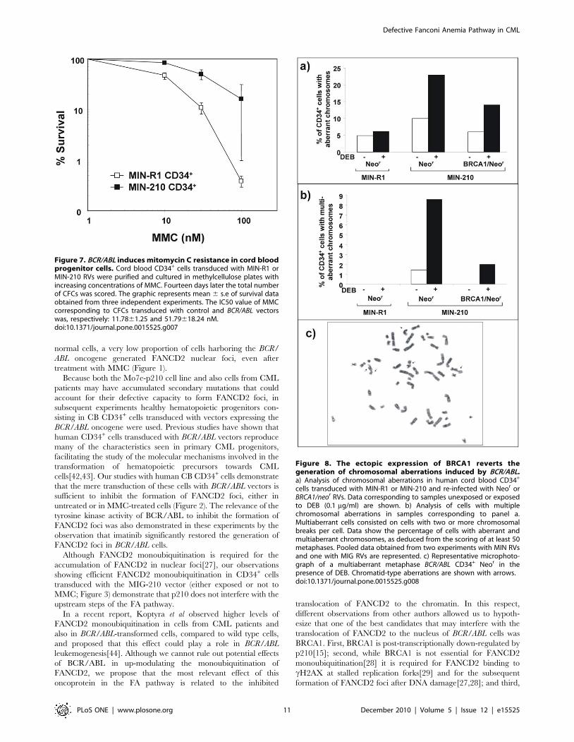
determined[23]. As shown in Figure 7, MIN-210 transduced
CD34+ cells were 5-fold more resistant to MMC compared to
control MIN-R1 transduced cells (IC50: 51.79618.24 nM and
11.7861.25 nM MMC, respectively). These results contrast to the
classical MMC-hypersensitivity observed in cells with a disrupted
FA/BRCA pathway, indicating that other pathways promoting cell
survival are up-regulated in BCR/ABL cells. This observation is
consistent with previous data showing the ability of BCR/ABL to
interfere with cellular apoptosis pathways[33,34,35].
Finally, because cells with an impaired FA/BRCA pathway are
characterized by an increased chromatid-type chromosomal
instability, particularly after exposure to DNA cross-linking drugs,
we investigated the spontaneous and DEB-induced chromosomal
instability of CD34+ cells previously transduced with the BCR/
ABL RV (MIN-210) and its respective control (MIN-R1).
Additionally, to test the influence of the ectopic expression of
BRCA1 on the chromosomal instability of BCR/ABL cells, BCR/
ABL-transduced samples were re-transduced with control (Neor) or
BRCA1 (BRCA1/Neor) RVs, as described in materials and methods.
In one experiment, chromosomal instability data was confirmed
with MIG-R1 and MIG-210 RVs. Because similar data were
obtained in this experiment, data in Figure 8 shows pooled results
obtained with both vector families.
As shown in Figure 8a, a low proportion of control CD34+ cells
(cells transduced with MIG-R1 or MIN-R1 RVs plus the control
Neor vector) either unexposed or DEB-exposed cells, contained
chromosomal aberrations (5% and 7%, respectively). In no
instance multiple chromosomal aberrations were observed in this
control group, regardless that samples were exposed to DEB or not
(Figure 8b). When CD34+ cells were transduced with BCR/ABL
RVs (plus the control Neor vector), the proportion of cells with
chromosomal aberrations, specially of chromatid-type (see repre-
sentative picture in Figure 8c), increased 2-fold in unexposed cells,
and 3- fold in DEB-exposed cells, compared to control CD34+
cells (Figure 8a). Differences were even more marked when cells
with multiple chromosomal aberrations were scored, mainly after
DEB exposure. In this case, almost 10% of the metaphases
contained two or more aberrant chromosomes (Figure 8b).
Notably, the proportion of BCR/ABL cells with aberrant
(Figure 8a) - and more markedly with multi-aberrant chromo-
somes (Figure 8b) - was reduced when these cells were re-
transduced with the BRCA1/Neor RV.
Taken together, these results show that the disruption of the
FA/BRCA pathway in BCR/ABL cells mediates centrosomal
amplification and chromosomal instability, and that this effect can
be partially reverted by the ectopic expression of BRCA1.
Discussion
Our study aims to offer new clues to understand the molecular
pathways accounting for the genetic instability of CML cells.
Our hypothesis that defects in the FA pathway may play a role
Figure 4. Reversion of the deficient formation of BRCA1 and FANCD2 foci in BCR/ABL-transduced cord blood CD34+ cells by inhibitorsof the proteasome and the PI3K/Akt pathway. Experimental protocol and analysis of the proportion of MIN-R1 and MIN-210-transduced cordblood CD34+ cells with BRCA1 (white bars) or FANCD2 foci (black bars) after treatment with the proteasome inhibitor MG132, or the PI3K/AKTinhibitor Ly294002. Bars show mean 6 s.e. of values corresponding to three independent experiments. *Differences were significant at p,0.01.doi:10.1371/journal.pone.0015525.g004
Defective Fanconi Anemia Pathway in CML
PLoS ONE | www.plosone.org 8 December 2010 | Volume 5 | Issue 12 | e15525
Defective Fanconi Anemia Pathway in CML
PLoS ONE | www.plosone.org 9 December 2010 | Volume 5 | Issue 12 | e15525
in this process derive from previous studies showing the relevance
of the FA pathway to control the genomic stability of the
cell[18,19] and also from observations showing genetic and
epigenetic alterations of FA genes, both in inherited and acquired
cancer[36,37,38,39,40,41].
In our first experiments we investigated the ability of CML cells
to generate FANCD2 nuclear foci, a central process in the FA
pathway (see review in[20]), both during cell proliferation and
after exposure to DNA cross-linking agents. Using Mo7e-p210 and
CD34+ cells from CML patients, we observed that in contrast to
Figure 6. The ectopic expression of BRCA1 reverts the generation of aberrant centrosomes induced by BCR/ABL. a) Analysis of MIN-R1or MIN-210-transduced cord blood CD34+cells with supernumerary centrosomes after re-infection with either Neor or BRCA1/Neor RVs. In allinstances cells were exposed to 40 nM MMC prior to analysis. Data corresponding to one representative experiment is shown. b) Representativepictures corresponding to panel a) showing supernumerary centrosomes in MIN-210 Neor compared to MIN-R1 Neor cells. To identify centrosomes c-tubulin antibody (green) was used. DAPI staining is shown in blue.doi:10.1371/journal.pone.0015525.g006
Figure 5. Reversion of the deficient formation of BRCA1 and FANCD2 foci in BCR/ABL-transduced cord blood CD34+ cells by theectopic expression of BRCA1. a) Experimental protocol used for investigating the effects mediated by the ectopic expression of BRCA1 upon theformation of BRCA1 and FANCD2 foci in BCR/ABL-transduced cells. b) Analysis of the proportion of MIG-R1 (white bars) and MIG-210 (grey and blackbars) transduced cord blood CD34+ cells with BRCA1 or FANCD2 foci after re-infection with vectors expressing the phosphotransferase gene (Neor;grey bars) or BRCA1 plus neor (BRCA1/Neor; black bars). Samples were exposed to 0 or 40 nM MMC prior to analyses of nuclear foci in EGFP+ cells. Barsshow mean 6 s.e. of values corresponding to three independent experiments. *Differences were significant at p,0.01. c) Representative pictures ofMMC-treated cells corresponding to panel b, are shown.doi:10.1371/journal.pone.0015525.g005
Defective Fanconi Anemia Pathway in CML
PLoS ONE | www.plosone.org 10 December 2010 | Volume 5 | Issue 12 | e15525
normal cells, a very low proportion of cells harboring the BCR/
ABL oncogene generated FANCD2 nuclear foci, even after
treatment with MMC (Figure 1).
Because both the Mo7e-p210 cell line and also cells from CML
patients may have accumulated secondary mutations that could
account for their defective capacity to form FANCD2 foci, in
subsequent experiments healthy hematopoietic progenitors con-
sisting in CB CD34+ cells transduced with vectors expressing the
BCR/ABL oncogene were used. Previous studies have shown that
human CD34+ cells transduced with BCR/ABL vectors reproduce
many of the characteristics seen in primary CML progenitors,
facilitating the study of the molecular mechanisms involved in the
transformation of hematopoietic precursors towards CML
cells[42,43]. Our studies with human CB CD34+ cells demonstrate
that the mere transduction of these cells with BCR/ABL vectors is
sufficient to inhibit the formation of FANCD2 foci, either in
untreated or in MMC-treated cells (Figure 2). The relevance of the
tyrosine kinase activity of BCR/ABL to inhibit the formation of
FANCD2 foci was also demonstrated in these experiments by the
observation that imatinib significantly restored the generation of
FANCD2 foci in BCR/ABL cells.
Although FANCD2 monoubiquitination is required for the
accumulation of FANCD2 in nuclear foci[27], our observations
showing efficient FANCD2 monoubiquitination in CD34+ cells
transduced with the MIG-210 vector (either exposed or not to
MMC; Figure 3) demonstrate that p210 does not interfere with the
upstream steps of the FA pathway.
In a recent report, Koptyra et al observed higher levels of
FANCD2 monoubiquitination in cells from CML patients and
also in BCR/ABL-transformed cells, compared to wild type cells,
and proposed that this effect could play a role in BCR/ABL
leukemogenesis[44]. Although we cannot rule out potential effects
of BCR/ABL in up-modulating the monoubiquitination of
FANCD2, we propose that the most relevant effect of this
oncoprotein in the FA pathway is related to the inhibited
translocation of FANCD2 to the chromatin. In this respect,
different observations from other authors allowed us to hypoth-
esize that one of the best candidates that may interfere with the
translocation of FANCD2 to the nucleus of BCR/ABL cells was
BRCA1. First, BRCA1 is post-transcriptionally down-regulated by
p210[15]; second, while BRCA1 is not essential for FANCD2
monoubiquitination[28] it is required for FANCD2 binding to
cH2AX at stalled replication forks[29] and for the subsequent
formation of FANCD2 foci after DNA damage[27,28]; and third,
Figure 7. BCR/ABL induces mitomycin C resistance in cord bloodprogenitor cells. Cord blood CD34+ cells transduced with MIN-R1 orMIN-210 RVs were purified and cultured in methylcellulose plates withincreasing concentrations of MMC. Fourteen days later the total numberof CFCs was scored. The graphic represents mean 6 s.e of survival dataobtained from three independent experiments. The IC50 value of MMCcorresponding to CFCs transduced with control and BCR/ABL vectorswas, respectively: 11.7861.25 and 51.79618.24 nM.doi:10.1371/journal.pone.0015525.g007
Figure 8. The ectopic expression of BRCA1 reverts thegeneration of chromosomal aberrations induced by BCR/ABL.a) Analysis of chromosomal aberrations in human cord blood CD34+
cells transduced with MIN-R1 or MIN-210 and re-infected with Neor orBRCA1/neor RVs. Data corresponding to samples unexposed or exposedto DEB (0.1 mg/ml) are shown. b) Analysis of cells with multiplechromosomal aberrations in samples corresponding to panel a.Multiaberrant cells consisted on cells with two or more chromosomalbreaks per cell. Data show the percentage of cells with aberrant andmultiaberrant chromosomes, as deduced from the scoring of at least 50metaphases. Pooled data obtained from two experiments with MIN RVsand one with MIG RVs are represented. c) Representative microphoto-graph of a multiaberrant metaphase BCR/ABL CD34+ Neor in thepresence of DEB. Chromatid-type aberrations are shown with arrows.doi:10.1371/journal.pone.0015525.g008
Defective Fanconi Anemia Pathway in CML
PLoS ONE | www.plosone.org 11 December 2010 | Volume 5 | Issue 12 | e15525
BRCA12/2 cells share with FA cells a chromosomal instability
phenotype[45]. Additionally, because BRCA1 deficient cells have
a defect in the G2/M checkpoint [45], our cell cycle studies
showing that MMC-treated BCR/ABL cells are not arrested in
G2/M - as it is characteristic of FA cells[26] - further suggest the
role of BRCA1 in the interference of the FA pathway in these cells.
To clarify the mechanisms involved in the repression of
BRCA1, and consequently in the impaired FANCD2 foci
formation of CML cells, we were interested in further investigating
the post-translational regulation of BRCA1 by the proteasome and
the PI3K/AKT pathway, frequently activated in human cancer
cells, including CML cells[46]. In this respect, data obtained in
primary cells and in breast and ovarian cancer cell lines has shown
that AKT1 represses BRCA1 foci formation[47,48]. Strikingly,
our results show that the inhibition of PI3K/AKT pathway with
LY294002 restored not only BRCA1 but also FANCD2 foci in
BCR/ABL-transduced CD34+ cells. The same effect was observed
with the proteasome inhibitor, MG132, indicating that this
molecule not only restores BRCA1 expression in BCR/ABL cells,
as previously described[15], but also the formation of BRCA1 and
FANCD2 foci in these cells. Finally, our data in BCR/ABL cells co-
transduced with BRCA1- RVs (Figure 5) confirms that the ectopic
expression of BRCA1 restores, at least in part, the inhibited
formation of FANCD2 foci in BCR/ABL cells.
As it has been previously reported, centrosome amplification
occurs frequently in all types of cancer and this correlates with the
malignant progression of the disease[49]. As it is the case in
BRCA1-deficient cells[45], centrosome aberrations and aneuploidy
are also common features of CML. In fact, previous data have
shown that centrosome abnormalities correlated with the CML
disease stage and preceded chromosomal aberrations in primary
cells from CML patients[32]. By means of the ectopic expression of
BRCA1 we show the involvement of BRCA1 in centrosomal
aberrations observed in CD34+ cells soon after their transduction
with BCR/ABL RVs, supporting the hypothesis that this phenotype
constitutes an early event in the transformation of CML cells.
The generation of centrosomal abnormalities by BCR/ABL is
consistent with the chromosomal instability of these cells, an
observation that is particularly evident after exposure to different
DNA damaging agents, including oxygen species, ionizing radia-
tion, or etoposide[10,12]. In this respect, our study shows for the
first time the chromosomal instability of BCR/ABL-transduced
CD34+ cells exposed to a DNA cross-linking drug, DEB, classically
used for the diagnosis of FA[50]. Strikingly, our results show that
BCR/ABL confers a survival advantage, while mediates chromo-
somal instability to DNA cross-linking agents. The observation that
BCR/ABL induces a survival advantage to MMC is, however, not
surprising considering that this oncoprotein interferes with several
pathways activating apoptosis[33,34,35]. In this respect, early
studies showed that BCR/ABL mediates protection from DNA-
damaged apoptosis in a dose-dependent manner[51], due to the
capacity of the oncoprotein to regulate the expression and/or the
activity of several pro- and anti-apoptotic factors signaling through
the STAT5[21], PI3K/AKT[52] and RAS[53] pathways.
Finally, of particular significance was the observation that the
ectopic expression of BRCA1 in BCR/ABL cells markedly
decreased the number of cells with aberrant, and more
significantly with multi-aberrant chromosomes. Because BRCA1
vectors also restored the formation of FANCD2 foci in BCR/ABL
CD34+ cells, our results add new insights to data previously
obtained by Deutsch et al[15] who showed a down-regulated
expression of BRCA1 in BCR/ABL hematopoietic cells. In
particular, our data strongly suggest that this effect interferes with
the translocation of FANCD2 to sites of DNA damage, thus
compromising the genomic stability of BCR/ABL cells.
Taken together, data obtained in this study allow us to propose
that the malignant phenotype conferred by BCR/ABL should be at
least in part related to its capacity to interfere both with downstream
steps of the FA/BRCA pathway and also with other pathways with
a role in apoptosis[33,34,35]. As a result of the simultaneous
interference of these pathways, a survival advantage should occur in
BCR/ABL hematopietic progenitors harboring genomic alterations
which may be not compatible with the survival of untransformed
cells. We therefore propose that a defective FA/BRCA pathway
may contribute to the genomic instability of CML cells, thus
promoting the accumulation of mutations during the progress from
a chronic phase towards blast crisis.
Supporting Information
Figure S1 Cell cycle analysis of cord blood CD34+ cellstransduced with MIG-R1 and MIG-210 retroviral vec-tors. Histograms show cell cycle distributions 7 days after
transduction of healthy cord blood CD34+ cells with MIG-R1 or
MIG-210, and exposed to 40 nM MMC (see schematic protocol in
Figure 2a). At this time, more than 90% of cells exposed to the
MIG-210 RV were EGFP+.
(TIF)
Figure S2 Analysis of the generation of double strandbreaks in cord blood CD34+ cells transduced with MIG-R1 and MIG-210 retroviral vectors. The Figure shows the
proportion of MIG-R1 (white bars) and MIG-210 (grey bars)
transduced CD34+ cells with nuclear c-H2AX foci, both in
untreated and in MMC treated (40 nM, 16 h) cells. Data from a
representative experiment is shown. Representative pictures of
cells with c-H2AX foci are also shown.
(TIF)
Acknowledgments
The authors would like to thank Dr Marina Garın for careful reading of
the manuscript and helpful suggestions. The authors also wish to thank the
technical assistance of Aurora de La Cal and Sergio Losada (CIEMAT/
CIBERER). The authors also thank the Centro de Transfusiones de la
Comunidad de Madrid for providing cord blood samples.
Author Contributions
Conceived and designed the experiments: BA FP JS JAB. Performed the
experiments: AV MEA PR JAC LP RP AJ. Wrote the paper: AV MEAF
PR BA FP JS JAB. Diagnosed the patients and provided samples: XA
MJC. Contributed vital new reagent: HH.
References
1. Bartram CR, de Klein A, Hagemeijer A, van Agthoven T, Geurts van Kessel A,
et al. (1983) Translocation of c-ab1 oncogene correlates with the presence of a
Philadelphia chromosome in chronic myelocytic leukaemia. Nature 306:
277–280.
2. Groffen J, Stephenson JR, Heisterkamp N, de Klein A, Bartram CR, et al.
(1984) Philadelphia chromosomal breakpoints are clustered within a limited
region, bcr, on chromosome 22. Cell 36: 93–99.
3. Lugo TG, Pendergast AM, Muller AJ, Witte ON (1990) Tyrosine kinase activity and
transformation potency of bcr-abl oncogene products. Science 247: 1079–1082.
4. Quintas-Cardama A, Cortes J (2009) Molecular biology of bcr-abl1-positive
chronic myeloid leukemia. Blood 113: 1619–1630.
5. Daley GQ, Van Etten RA, Baltimore D (1990) Induction of chronic
myelogenous leukemia in mice by the P210bcr/abl gene of the Philadelphia
chromosome. Science 247: 824–830.
Defective Fanconi Anemia Pathway in CML
PLoS ONE | www.plosone.org 12 December 2010 | Volume 5 | Issue 12 | e15525
6. Melo JV, Barnes DJ (2007) Chronic myeloid leukaemia as a model of disease
evolution in human cancer. Nat Rev Cancer 7: 441–453.
7. Burke BA, Carroll M (2010) BCR-ABL: a multi-faceted promoter of DNA
mutation in chronic myelogeneous leukemia. Leukemia Epub ahead of print.
8. Sattler M, Verma S, Shrikhande G, Byrne CH, Pride YB, et al. (2000) The
BCR/ABL tyrosine kinase induces production of reactive oxygen species inhematopoietic cells. J Biol Chem 275: 24273–24278.
9. Nowicki MO, Falinski R, Koptyra M, Slupianek A, Stoklosa T, et al. (2004)BCR/ABL oncogenic kinase promotes unfaithful repair of the reactive oxygen
species-dependent DNA double-strand breaks. Blood 104: 3746–3753.
10. Dierov J, Sanchez PV, Burke BA, Padilla-Nash H, Putt ME, et al. (2009) BCR/
ABL induces chromosomal instability after genotoxic stress and alters the cell
death threshold. Leukemia 23: 279–286.
11. Koptyra M, Falinski R, Nowicki MO, Stoklosa T, Majsterek I, et al. (2006)
BCR/ABL kinase induces self-mutagenesis via reactive oxygen species to encodeimatinib resistance. Blood 108: 319–327.
12. Koptyra M, Cramer K, Slupianek A, Richardson C, Skorski T (2008) BCR/ABL promotes accumulation of chromosomal aberrations induced by oxidative
and genotoxic stress. Leukemia 22: 1969–1972.
13. Deutsch E, Dugray A, AbdulKarim B, Marangoni E, Maggiorella L, et al. (2001)
BCR-ABL down-regulates the DNA repair protein DNA-PKcs. Blood 97:2084–2090.
14. Moynahan ME, Chiu JW, Koller BH, Jasin M (1999) Brca1 controls homology-
directed DNA repair. Mol Cell 4: 511–518.
15. Deutsch E, Jarrousse S, Buet D, Dugray A, Bonnet M-L, et al. (2003) Down-
regulation of BRCA1 in BCR-ABL-expressing hematopoietic cells. Blood 101:4583–4588.
16. Cramer K, Nieborowska-Skorska M, Koptyra M, Slupianek A, Penserga ET,et al. (2008) BCR/ABL and other kinases from chronic myeloproliferative
disorders stimulate single-strand annealing, an unfaithful DNA double-strandbreak repair. Cancer Res 68: 6884–6888.
17. Fernandes MS, Reddy MM, Gonneville JR, DeRoo SC, Podar K, et al. (2009)BCR-ABL promotes the frequency of mutagenic single-strand annealing DNA
repair. Blood 114: 1813–1819.
18. Moldovan GL, D’Andrea AD (2009) How the Fanconi Anemia Pathway Guards
the Genome. Annu Rev Genet 43: 223–249.
19. Levitus M, Joenje H, de Winter JP (2006) The Fanconi anemia pathway of
genomic maintenance. Cell Oncol 28: 3–29.
20. Cohn MA, D’Andrea AD (2008) Chromatin recruitment of DNA repair
proteins: lessons from the fanconi anemia and double-strand break repair
pathways. Mol Cell 32: 306–312.
21. Horita M, Andreu EJ, Benito A, Arbona C, Sanz C, et al. (2000) Blockade of the
Bcr-Abl kinase activity induces apoptosis of chronic myelogenous leukemia cellsby suppressing signal transducer and activator of transcription 5-dependent
expression of Bcl-xL. J Exp Med 191: 977–984.
22. Jose-Eneriz ES, Roman-Gomez J, Cordeu L, Ballestar E, Garate L, et al. (2008)
BCR-ABL1-induced expression of HSPA8 promotes cell survival in chronicmyeloid leukaemia. Br J Haematol 142: 571–582.
23. Jacome A, Navarro S, Casado JA, Rio P, Madero L, et al. (2006) A simplifiedapproach to improve the efficiency and safety of ex vivo hematopoietic gene
therapy in fanconi anemia patients. Hum Gene Ther 17: 245–250.
24. Casado JA, Callen E, Jacome A, Rio P, Castella M, et al. (2007) A
comprehensive strategy for the subtyping of Fanconi Anemia patients:
conclusions from the Spanish Fanconi Anemia research network. J Med Genet44: 241–249.
25. Taniguchi T, Garcia-Higuera I, Andreassen PR, Gregory RC, Grompe M, et al.(2002) S-phase-specific interaction of the Fanconi anemia protein, FANCD2,
with BRCA1 and RAD51. Blood 100: 2414–2420.
26. Seyschab H, Friedl R, Sun Y, Schindler D, Hoehn H, et al. (1995) Comparative
evaluation of diepoxybutane sensitivity and cell cycle blockage in the diagnosis ofFanconi anemia. Blood 85: 2233–2237.
27. Garcia-Higuera I, Taniguchi T, Ganesan S, Meyn MS, Timmers C, et al. (2001)Interaction of the Fanconi anemia proteins and BRCA1 in a common pathway.
Mol Cell 7: 249–262.
28. Vandenberg CJ, Gergely F, Ong CY, Pace P, Mallery DL, et al. (2003) BRCA1-
independent ubiquitination of FANCD2. Mol Cell 12: 247–254.
29. Bogliolo M, Lyakhovich A, Callen E, Castella M, Cappelli E, et al. (2007)
Histone H2AX and Fanconi anemia FANCD2 function in the same pathway to
maintain chromosome stability. Embo J 26: 1340–1351.
30. Altiok S, Batt D, Altiok N, Papautsky A, Downward J, et al. (1999) Heregulin
induces phosphorylation of BRCA1 through phosphatidylinositol 3-Kinase/AKT in breast cancer cells. J Biol Chem 274: 32274–32278.
31. Starita LM, Machida Y, Sankaran S, Elias JE, Griffin K, et al. (2004) BRCA1-
dependent ubiquitination of gamma-tubulin regulates centrosome number. MolCell Biol 24: 8457–8466.
32. Giehl M, Fabarius A, Frank O, Hochhaus A, Hafner M, et al. (2005)
Centrosome aberrations in chronic myeloid leukemia correlate with stage ofdisease and chromosomal instability. Leukemia 19: 1192–1197.
33. McGahon A, Bissonnette R, Schmitt M, Cotter KM, Green DR, et al. (1994)BCR-ABL maintains resistance of chronic myelogenous leukemia cells to
apoptotic cell death. Blood 83: 1179–1187.
34. Bedi A, Barber JP, Bedi GC, el-Deiry WS, Sidransky D, et al. (1995) BCR-ABL-mediated inhibition of apoptosis with delay of G2/M transition after DNA
damage: a mechanism of resistance to multiple anticancer agents. Blood 86:1148–1158.
35. Neshat MS, Raitano AB, Wang HG, Reed JC, Sawyers CL (2000) The survivalfunction of the Bcr-Abl oncogene is mediated by Bad-dependent and -
independent pathways: roles for phosphatidylinositol 3-kinase and Raf. Mol
Cell Biol 20: 1179–1186.36. Taniguchi T, Tischkowitz M, Ameziane N, Hodgson SV, Mathew CG, et al.
(2003) Disruption of the Fanconi anemia-BRCA pathway in cisplatin-sensitiveovarian tumors. Nat Med 9: 568–574.
37. Narayan G, Arias-Pulido H, Nandula SV, Basso K, Sugirtharaj DD, et al. (2004)
Promoter hypermethylation of FANCF: disruption of Fanconi Anemia-BRCApathway in cervical cancer. Cancer Res 64: 2994–2997.
38. Tischkowitz MD, Morgan NV, Grimwade D, Eddy C, Ball S, et al. (2004)Deletion and reduced expression of the Fanconi anemia FANCA gene in
sporadic acute myeloid leukemia. Leukemia 18: 420–425.39. van der Heijden MS, Brody JR, Gallmeier E, Cunningham SC, Dezentje DA, et
al. (2004) Functional defects in the fanconi anemia pathway in pancreatic cancer
cells. Am J Pathol 165: 651–657.40. Hess CJ, Ameziane N, Schuurhuis GJ, Errami A, Denkers F, et al. (2008)
Hypermethylation of the FANCC and FANCL promoter regions in sporadicacute leukaemia. Cell Oncol 30: 299–306.
41. Lyakhovich A, Surralles J (2006) Disruption of the Fanconi anemia/BRCA
pathway in sporadic cancer. Cancer Lett 232: 99–106.42. Chalandon Y, Jiang X, Hazlewood G, Loutet S, Conneally E, et al. (2002)
Modulation of p210(BCR-ABL) activity in transduced primary humanhematopoietic cells controls lineage programming. Blood 99: 3197–3204.
43. Zhao RC, Jiang Y, Verfaillie CM (2001) A model of human p210(bcr/ABL)-mediated chronic myelogenous leukemia by transduction of primary normal
human CD34(+) cells with a BCR/ABL-containing retroviral vector. Blood 97:
2406–2412.44. Koptyra M, Stoklosa T, Hoser G, Seferynska L, Glodkowska E, et al. (2008)
Monoubiquitination of the Fanconi Anemia D2 (FANCD2) Protein Regulatesthe Transforming Potential of BCR/ABL. Blood (ASH Annual Meeting
Abstracts) 112: 112.
45. Xu X, Weaver Z, Linke SP, Li C, Gotay J, et al. (1999) Centrosomeamplification and a defective G2-M cell cycle checkpoint induce genetic
instability in BRCA1 exon 11 isoform-deficient cells. Mol Cell 3: 389–395.46. Varticovski L, Daley GQ, Jackson P, Baltimore D, Cantley LC (1991) Activation
of phosphatidylinositol 3-kinase in cells expressing abl oncogene variants. MolCell Biol 11: 1107–1113.
47. Plo I, Laulier C, Gauthier L, Lebrun F, Calvo F, et al. (2008) AKT1 inhibits
homologous recombination by inducing cytoplasmic retention of BRCA1 andRAD51. Cancer Res 68: 9404–9412.
48. Tonic I, Yu WN, Park Y, Chen CC, Hay N (2010) Akt activation emulates Chk1inhibition and Bcl2 overexpression and abrogates G2 cell cycle checkpoint by
inhibiting BRCA1 foci. J Biol Chem 285: 23790–23798.
49. Fukasawa K (2005) Centrosome amplification, chromosome instability andcancer development. Cancer Lett 230: 6–19.
50. Auerbach AD, Wolman SR (1976) Susceptibility of Fanconi’s anaemiafibroblasts to chromosome damage by carcinogens. Nature 261: 494–496.
51. Cambier N, Chopra R, Strasser A, Metcalf D, Elefanty AG (1998) BCR-ABL
activates pathways mediating cytokine independence and protection againstapoptosis in murine hematopoietic cells in a dose-dependent manner. Oncogene
16: 335–348.52. Ghaffari S, Jagani Z, Kitidis C, Lodish HF, Khosravi-Far R (2003) Cytokines
and BCR-ABL mediate suppression of TRAIL-induced apoptosis throughinhibition of forkhead FOXO3a transcription factor. Proc Natl Acad Sci U S A
100: 6523–6528.
53. Aichberger KJ, Mayerhofer M, Krauth MT, Skvara H, Florian S, et al. (2005)Identification of mcl-1 as a BCR/ABL-dependent target in chronic myeloid
leukemia (CML): evidence for cooperative antileukemic effects of imatinib andmcl-1 antisense oligonucleotides. Blood 105: 3303–3311.
Defective Fanconi Anemia Pathway in CML
PLoS ONE | www.plosone.org 13 December 2010 | Volume 5 | Issue 12 | e15525