Embed Size (px)
Citation preview

Benign or
Malignant
What Does The
Path Say?John Cangelosi, MD
Sagis, PLLC

64 yo male presents with a 3 year history of a slowly growing, 1.5 cm pearly plaque on the left sideburn.

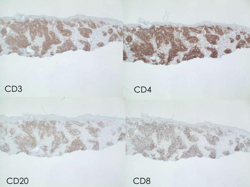
CD3 CD4
CD20 CD8
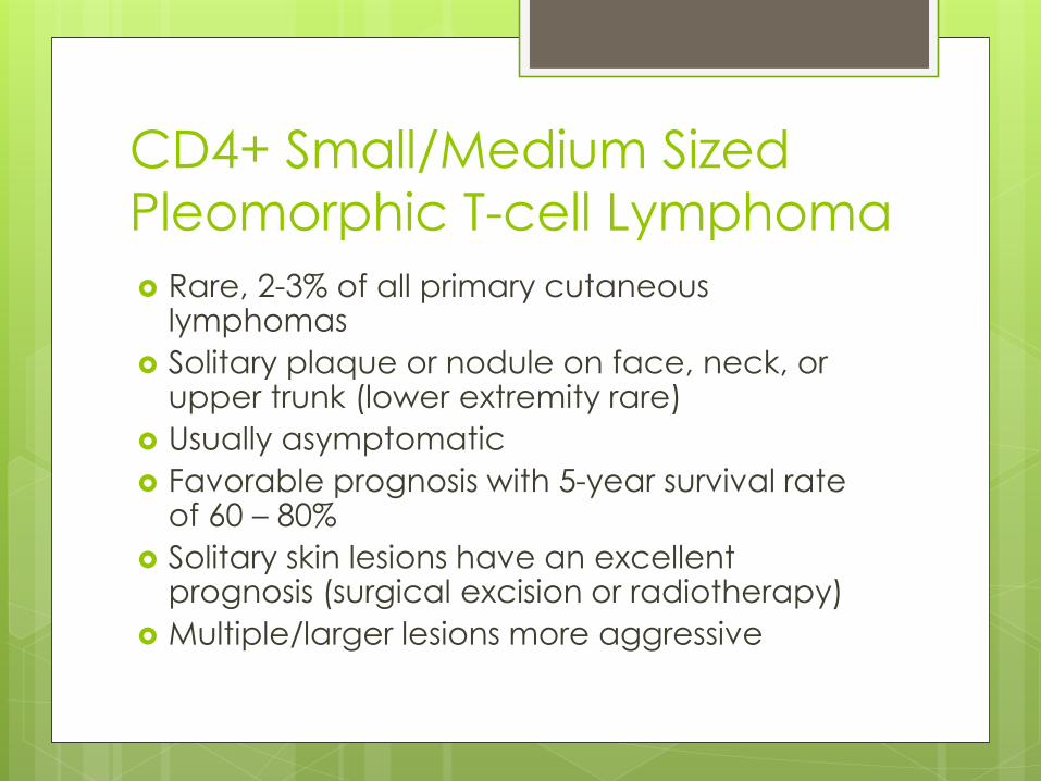
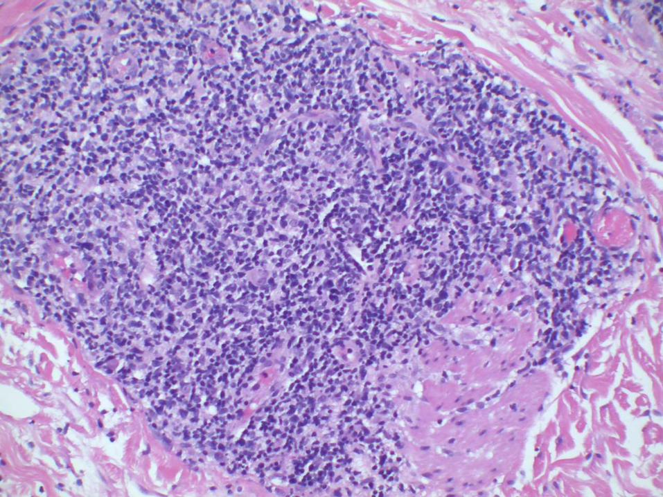
CD4+ Small/Medium Sized
Pleomorphic T-cell Lymphoma
Rare, 2-3% of all primary cutaneous lymphomas
Solitary plaque or nodule on face, neck, or upper trunk (lower extremity rare)
Usually asymptomatic
Favorable prognosis with 5-year survival rate of 60 – 80%
Solitary skin lesions have an excellent prognosis (surgical excision or radiotherapy)
Multiple/larger lesions more aggressive
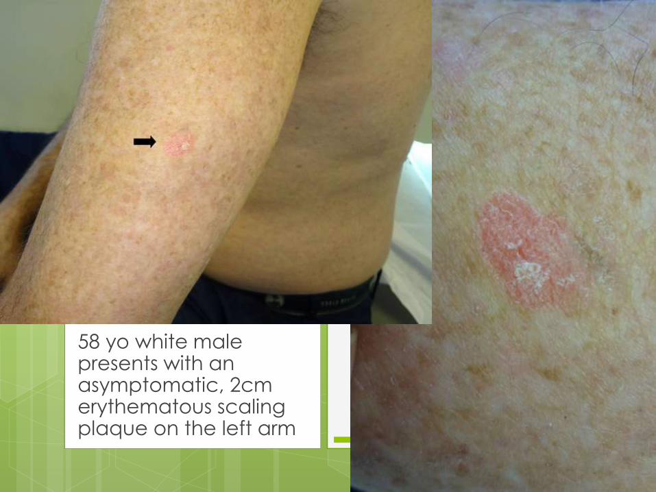
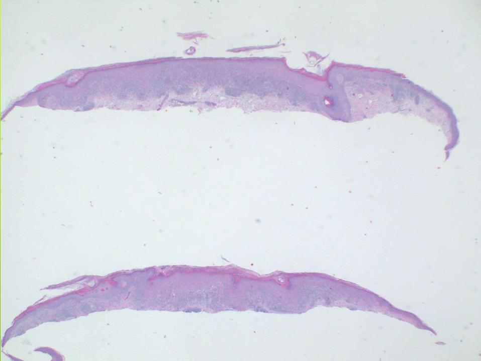
58 yo white male presents with an asymptomatic, 2cm erythematous scaling plaque on the left arm
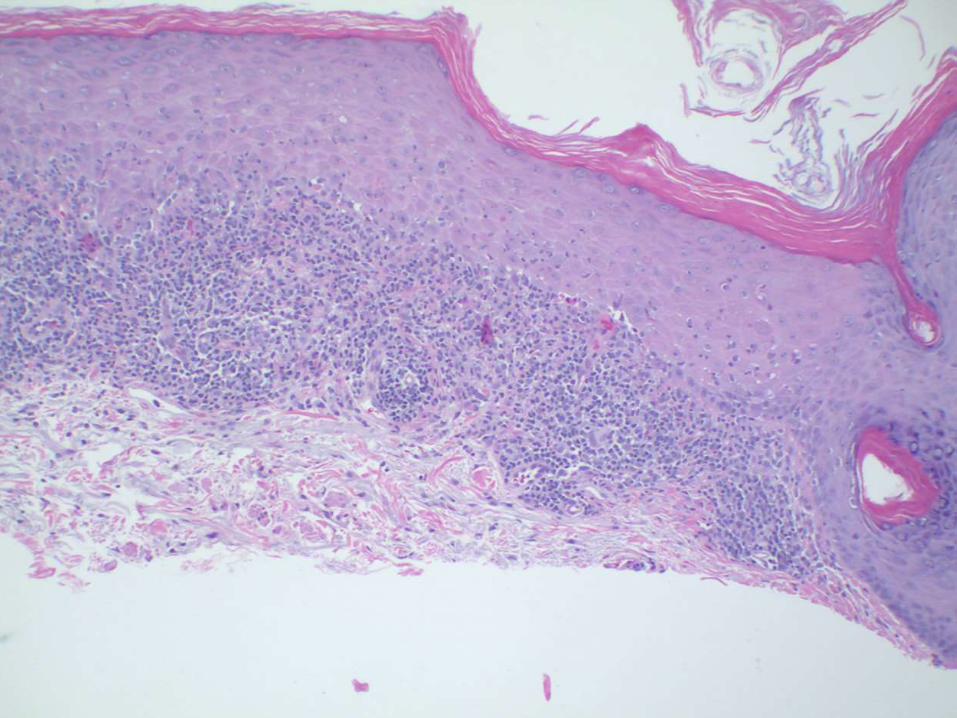
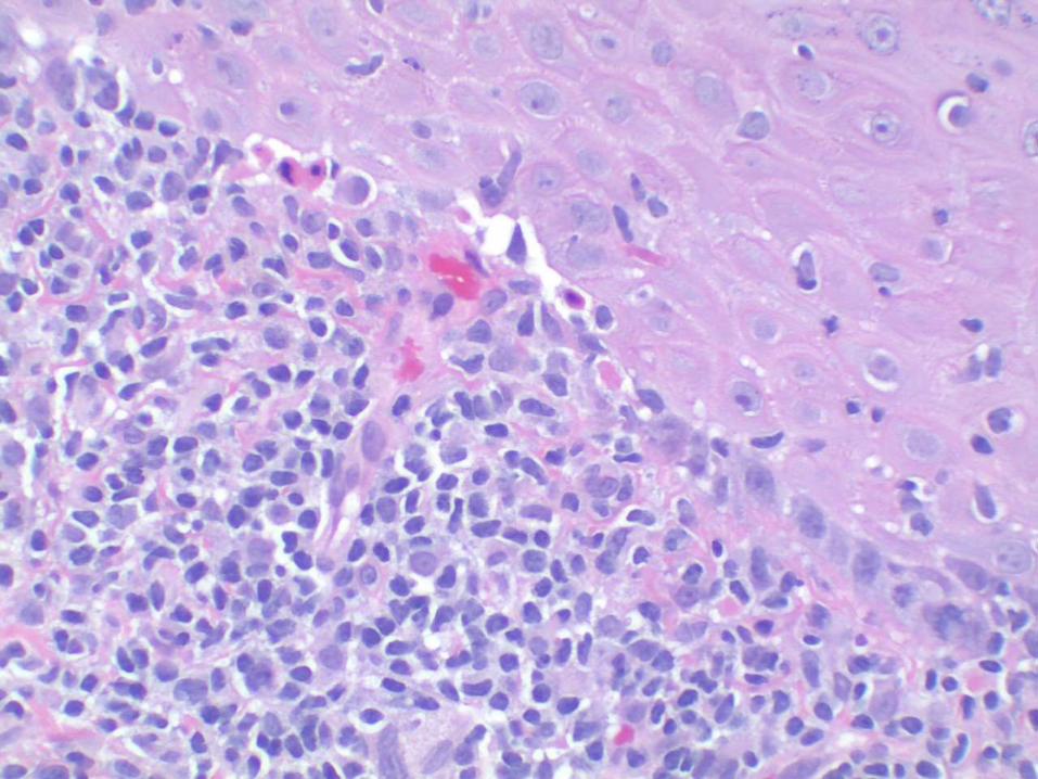
Benign Lichenoid Keratosis
Short duration
Predilection for face (cheeks and nose),
forearm and dorsal hand, upper trunk,
and neck
Predominately Caucasian
Females > Males
4 – 7th decade
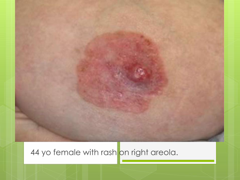
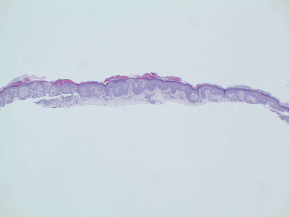
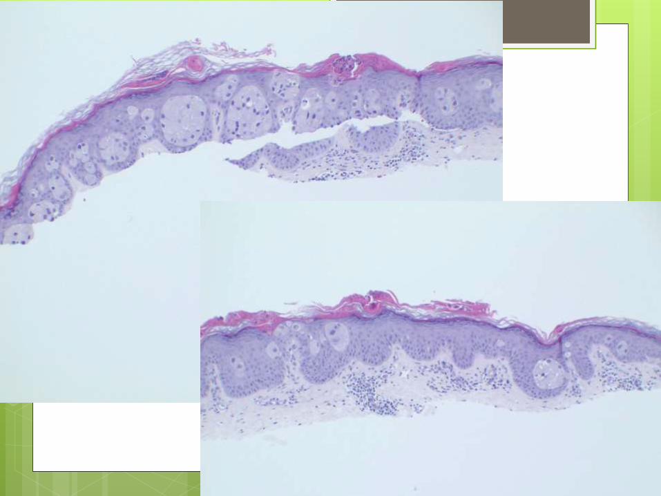
44 yo female with rash on right areola.
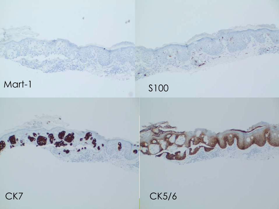
Mart-1 S100
CK7 CK5/6
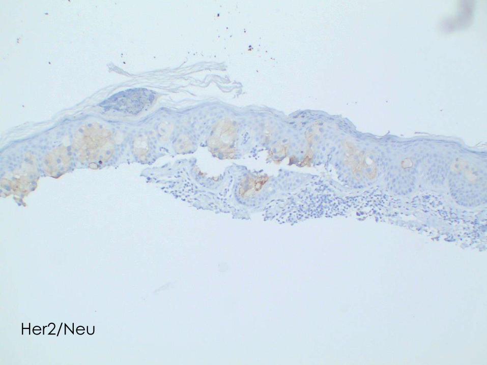
Her2/Neu
Paget’s Disease of the Nipple
Almost always associated with carcinoma
of the breast
Dermatosis results from spread of tumor
via the lactiferous ducts to the surface
epithelium
Breast carcinoma can be in situ or
invasive at time of presentation
Usually unilateral presentation
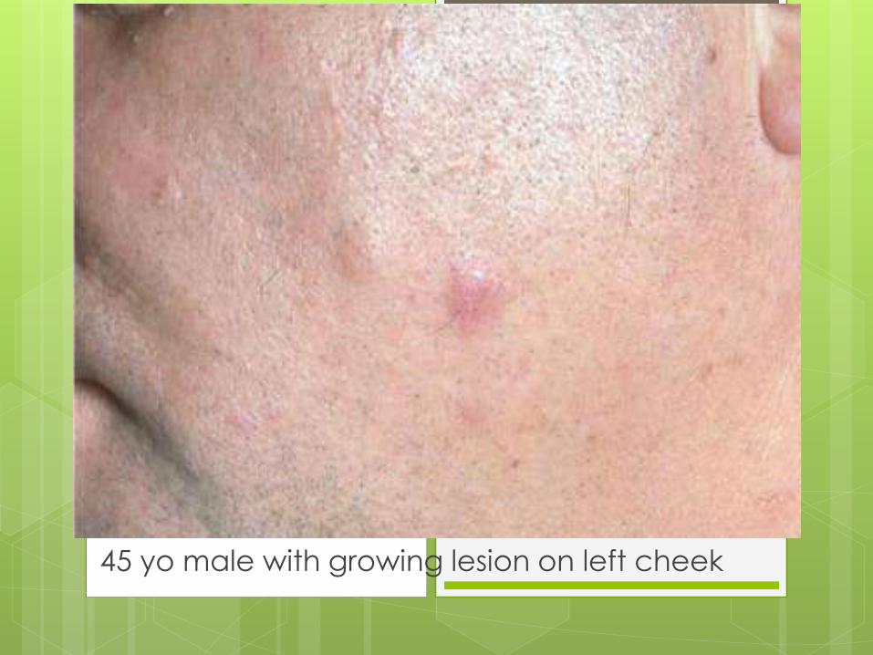
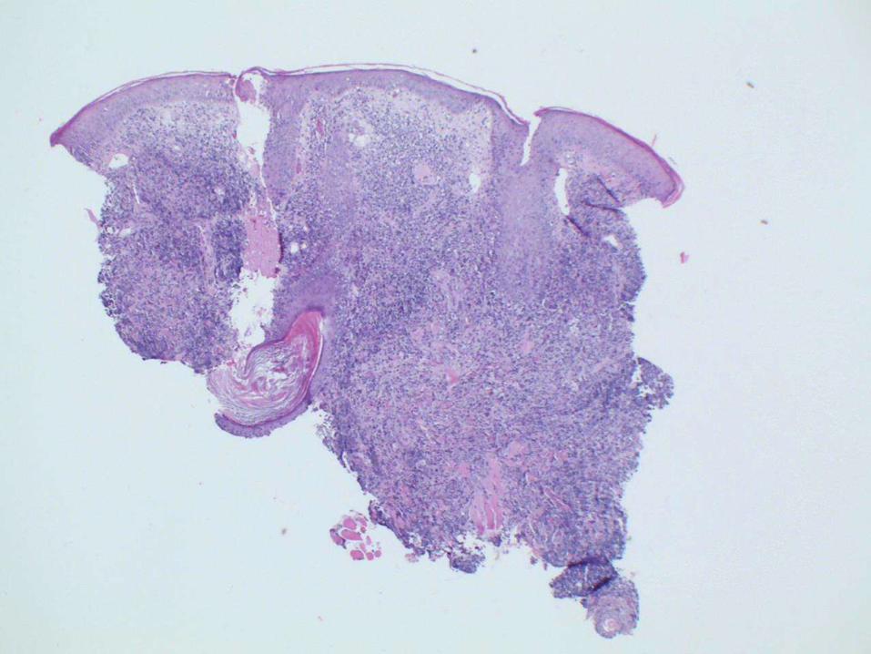
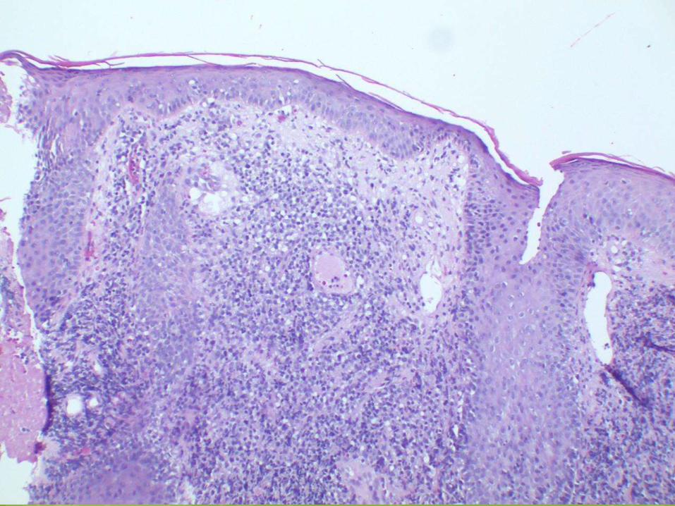
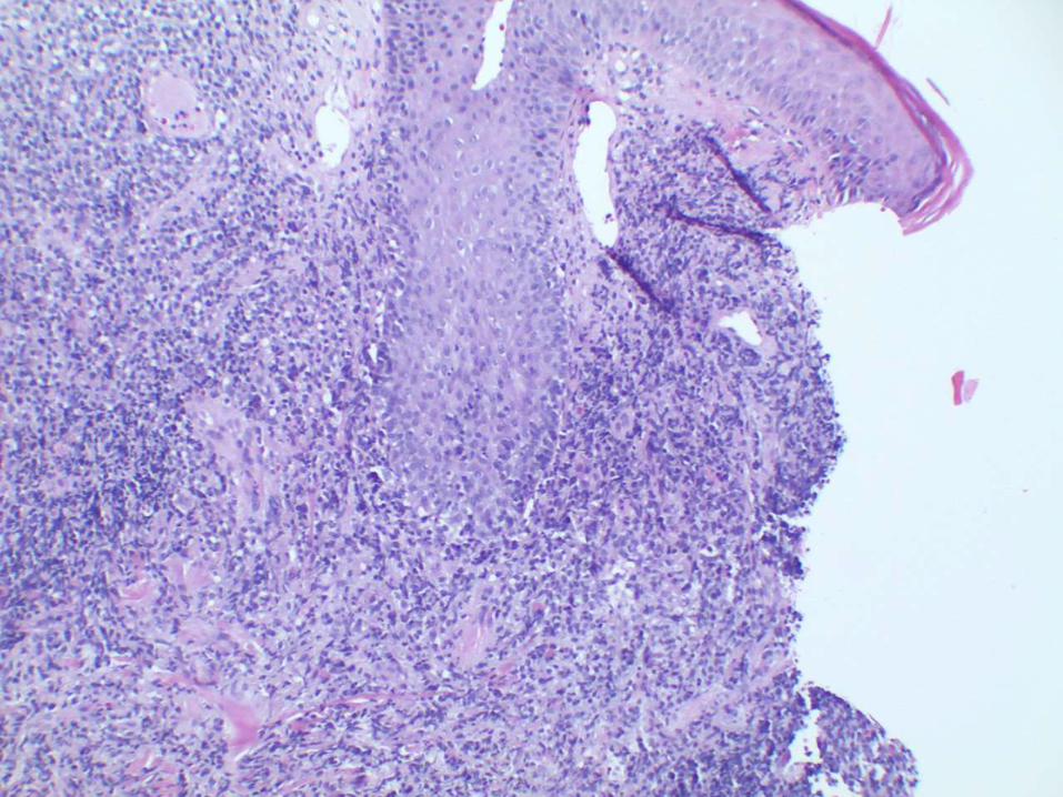
45 yo male with growing lesion on left cheek
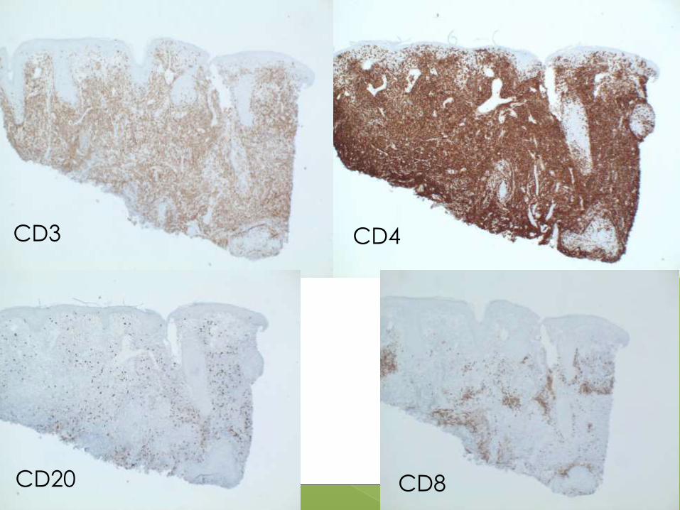
CD3 CD4
CD20 CD8
Folliculotropic Mycosis
Fungoides
Preferential location is head and neck
region
Follicular mucinosis often
Usually minimal epidermotropism
More refractory to treatment than classic
MF
Worse survival rates than classic MF (68%
at 5 years, 26% at 10 years)
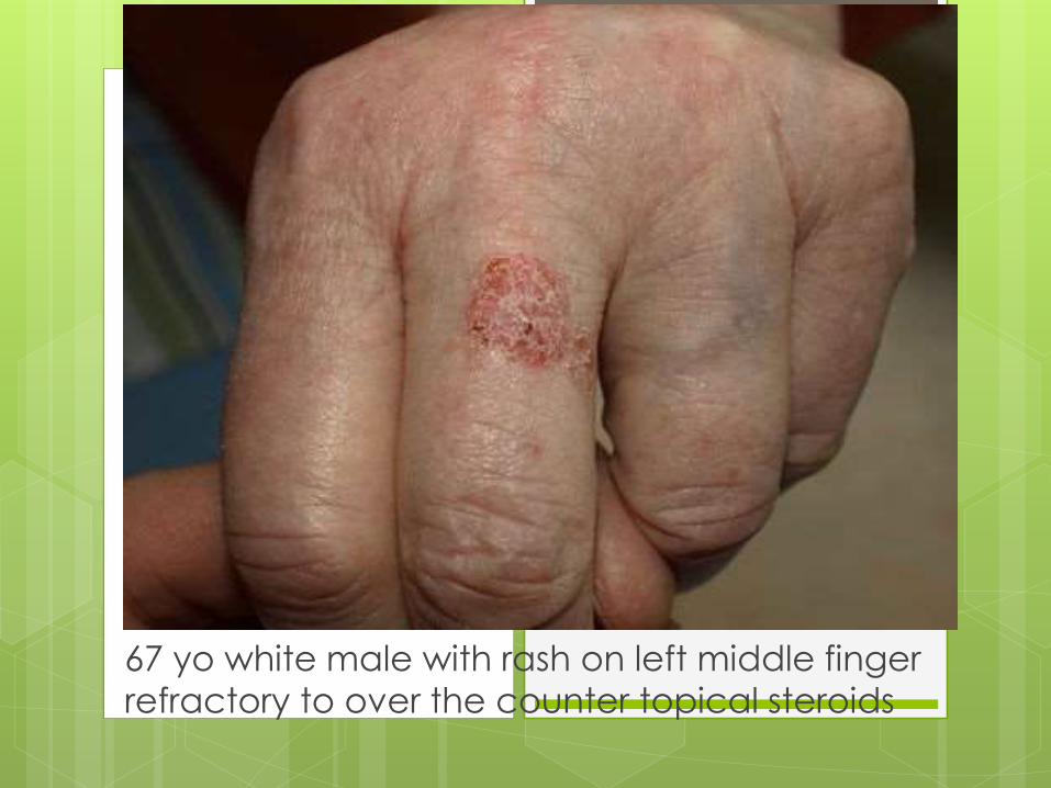
67 yo white male with rash on left middle finger
refractory to over the counter topical steroids
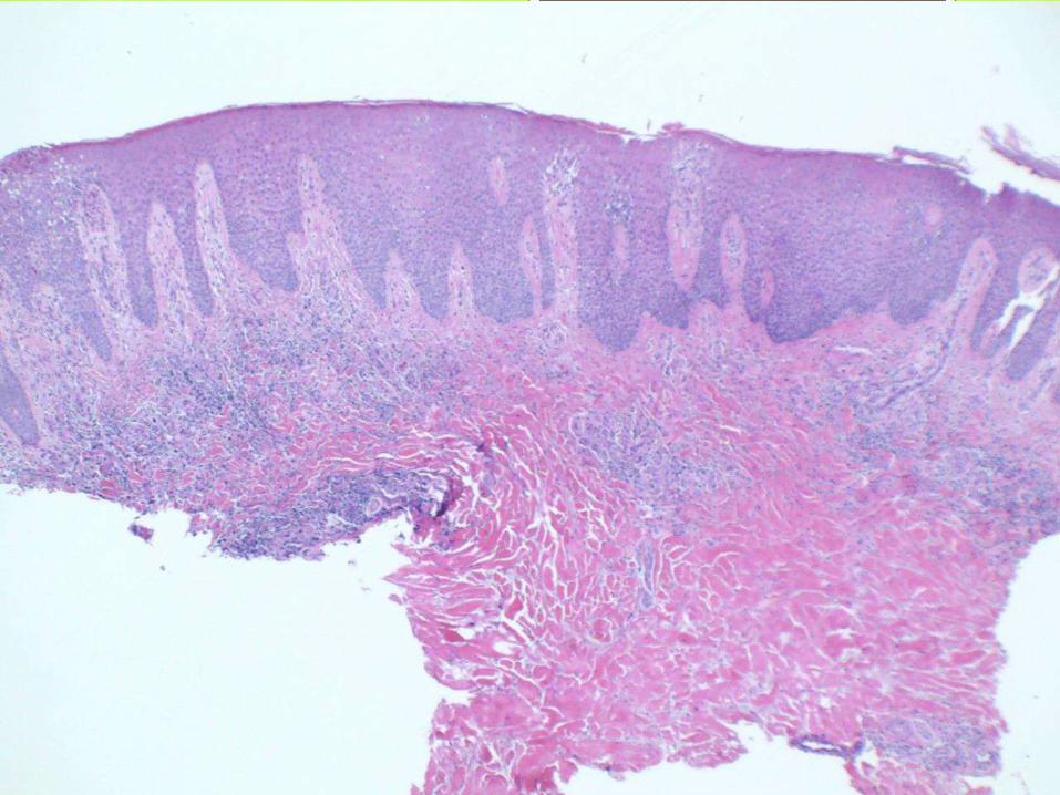
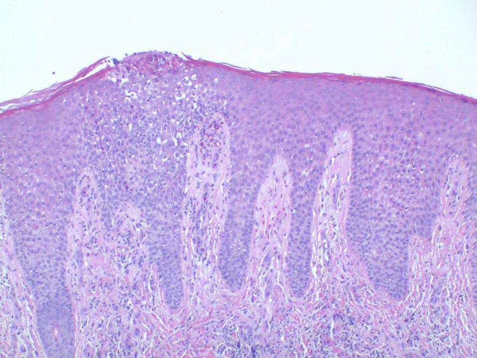
Psoriasis
Familial disease in 1-3% of the population
Most common on scalp, trunk, buttock,
elbows, and knees
Least common on the face (UV light
improves disease)
Nail dystrophy
Psoriatic arthritis in 1/3 of patients
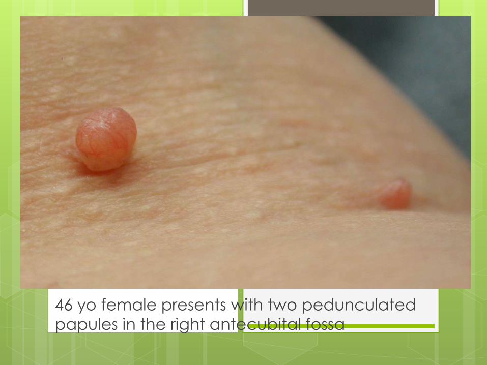
46 yo female presents with two pedunculated
papules in the right antecubital fossa
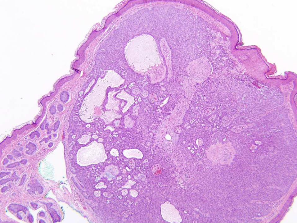
Basal Cell Nevus Syndrome
Autosomal dominant
Early onset, multiple basal cell carcinomas
Odontogenic keratocysts, palmoplantar pits,
falx cerebri calcifications, medulloblastomas,
hydrocephalus, cataracts
Mutation of chromosome 9 in the PTCH gene
Should consider biopsy of acrochordon-like
lesions in young patients
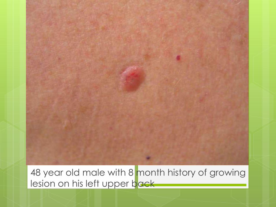
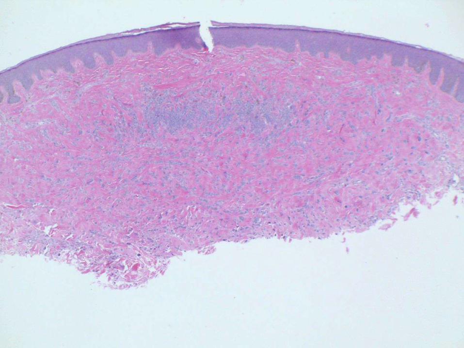
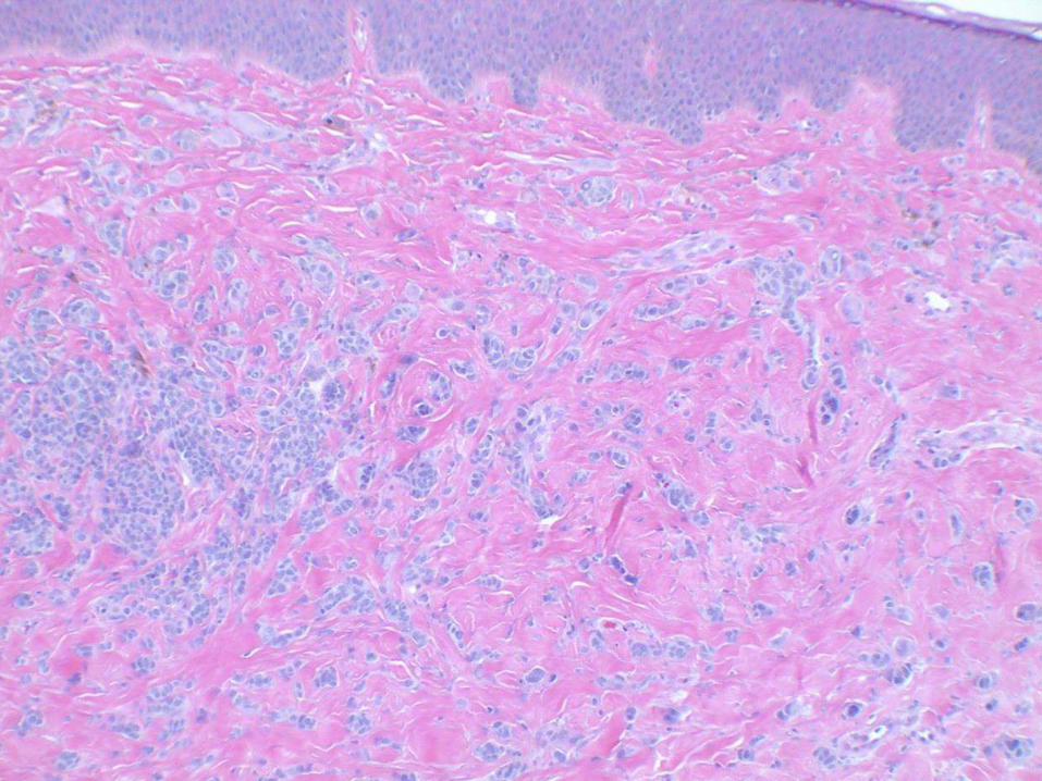
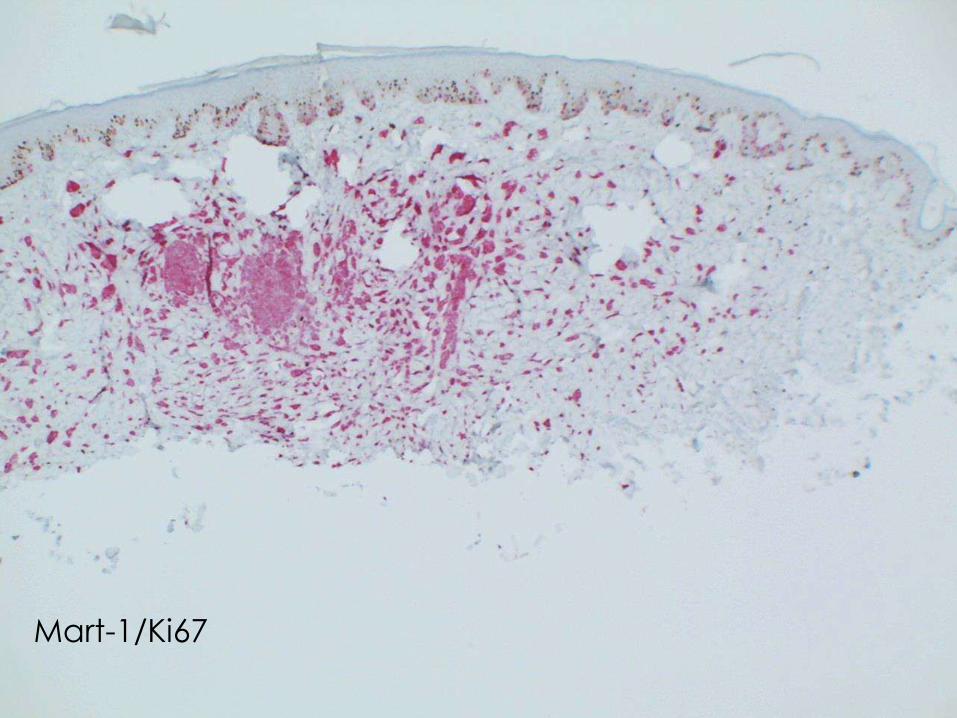
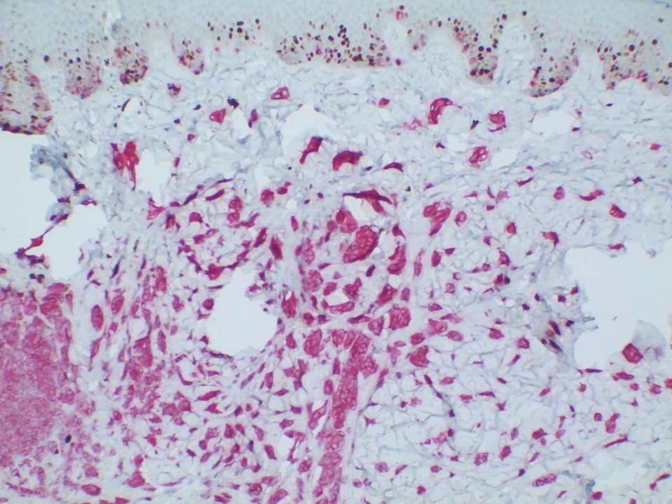
48 year old male with 8 month history of growing
lesion on his left upper back
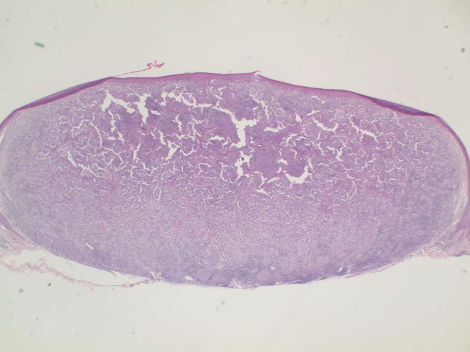
Amelanotic Melanoma
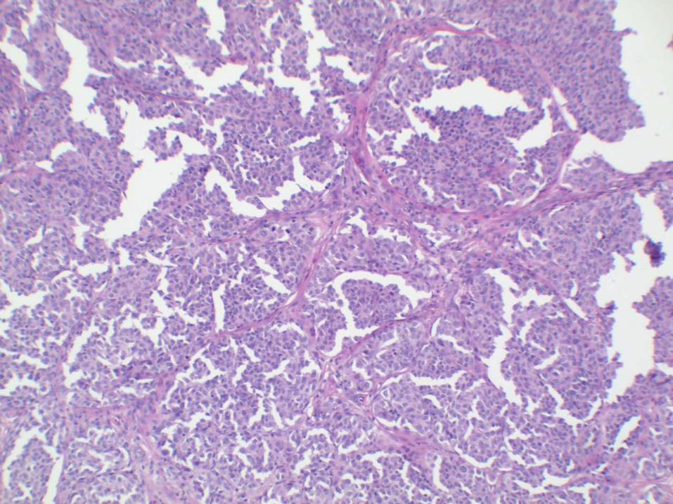
Amelanotic Melanoma
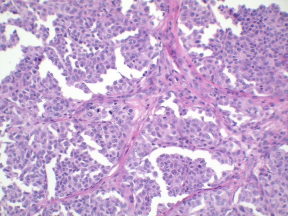
Amelanotic Melanoma
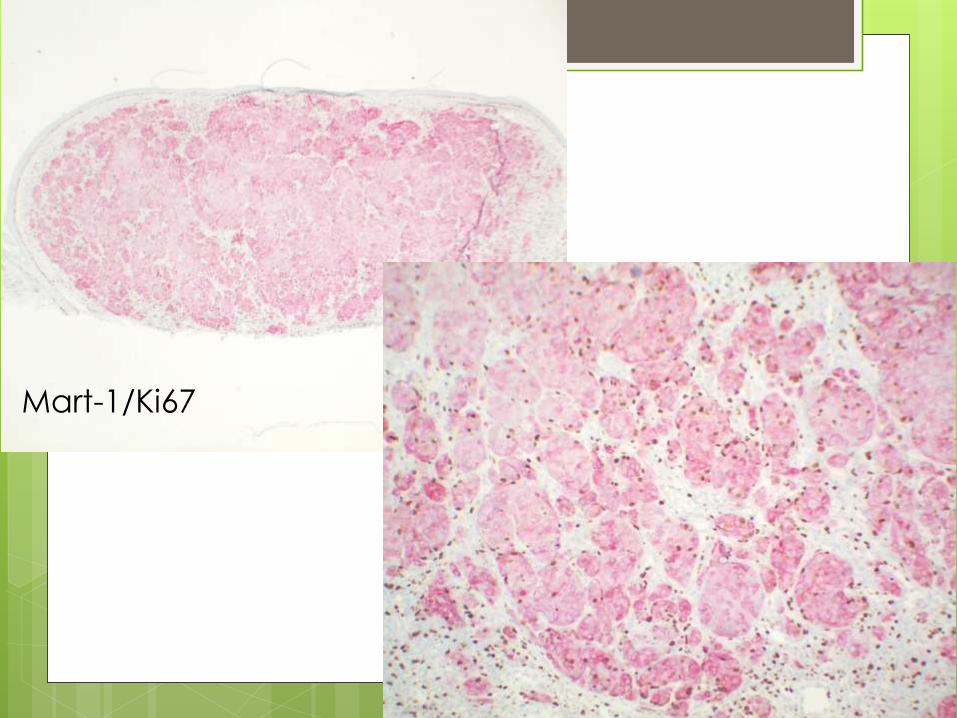
Mart-1/Ki67
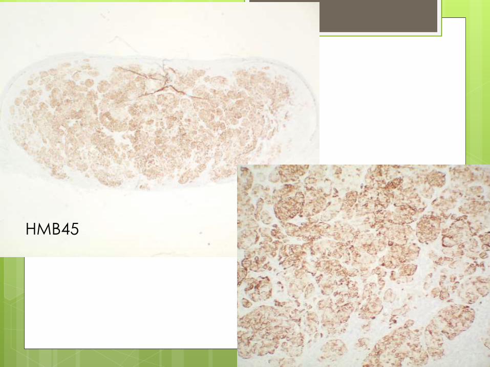
HMB45
Amelanotic Melanoma 5% of melanomas
Often misdiagnosed (eczema, seborrheic keratosis, Bowen’s disease, basal cell carcinoma, angiofibromas, etc)
Often leads to poor prognosis when diagnosed late
Breslow thickness (not Clark Level) and ulceration are the most dominant predictors of survival (same for all melanomas)
Mitotic rate also plays a role in staging

Melanoma Staging Tis – In-situ
T1a – Invasive but less than 1.0mm Breslow without ulceration and <1 mitosis/mm2
T1b – Less than 1.0mm Breslow but ulceration and/or >1 mitosis/mm2
T2a – 1.01-2.0mm thick without ulceration
T2b - 1.01-2.0mm thick with ulceration
T3a/b - 2.01-4.0mm thick with/out ulceration
T4a/b – Greater than 4.0mm thick with/out ulceration
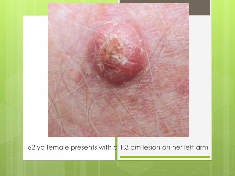
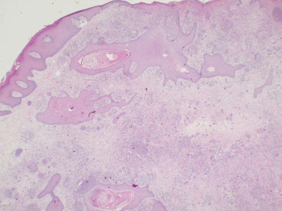
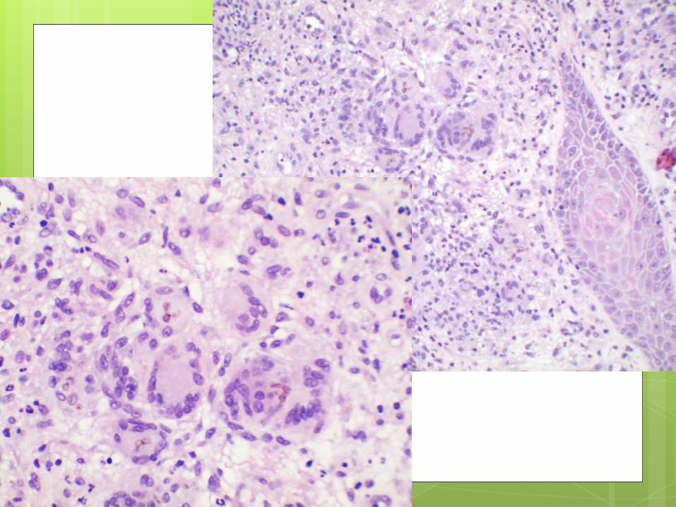
62 yo female presents with a 1.3 cm lesion on her left arm
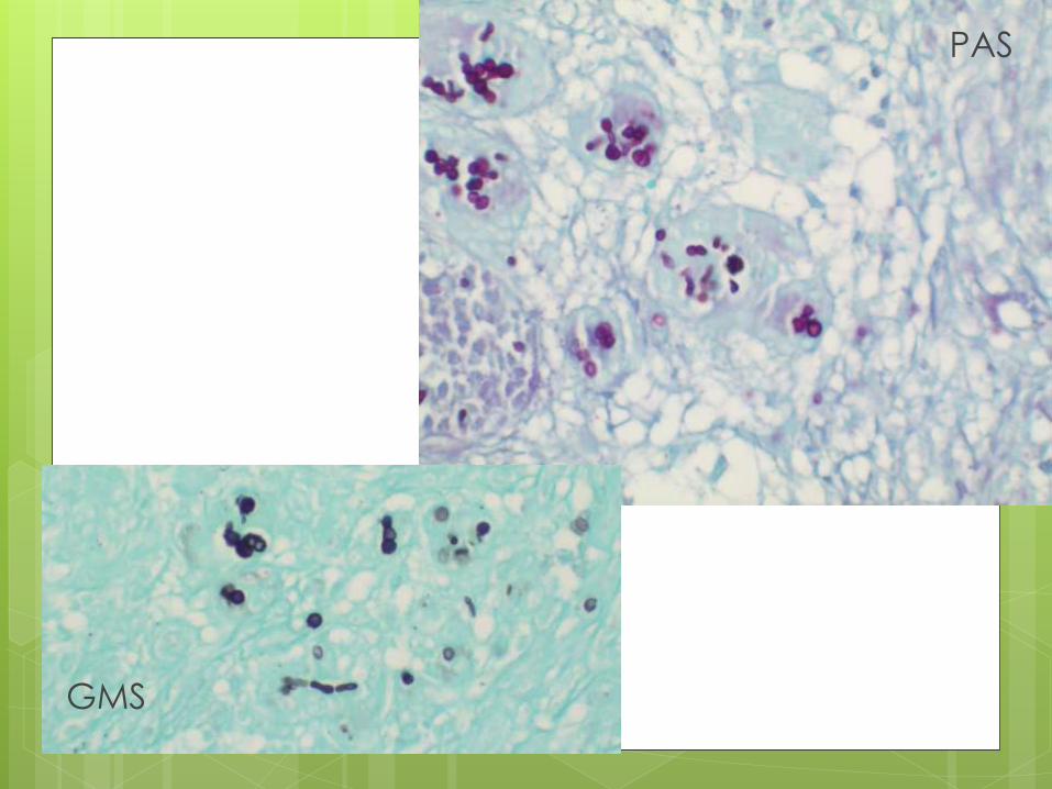
GMS
PAS

Chromohyphomycosis
Infection by fungal family Dematiaceae
(brown or black fungi)
Fungi with brown septated hyphae
Common saprophytic forms found in soil
and decomposing vegetation
Trauma is the gateway for infection
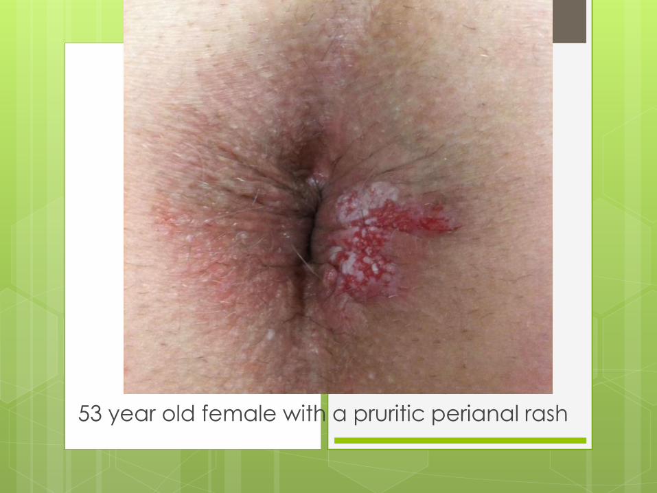
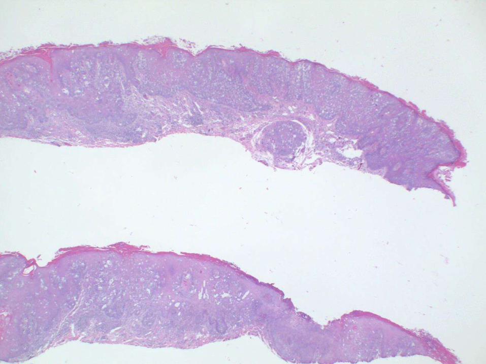
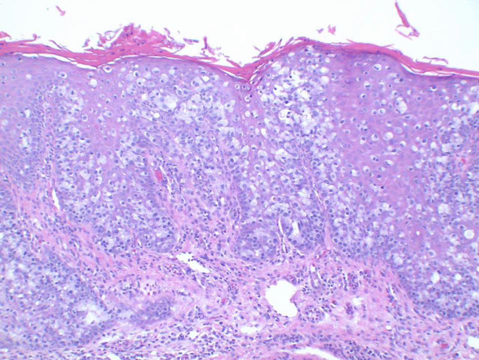
53 year old female with a pruritic perianal rash
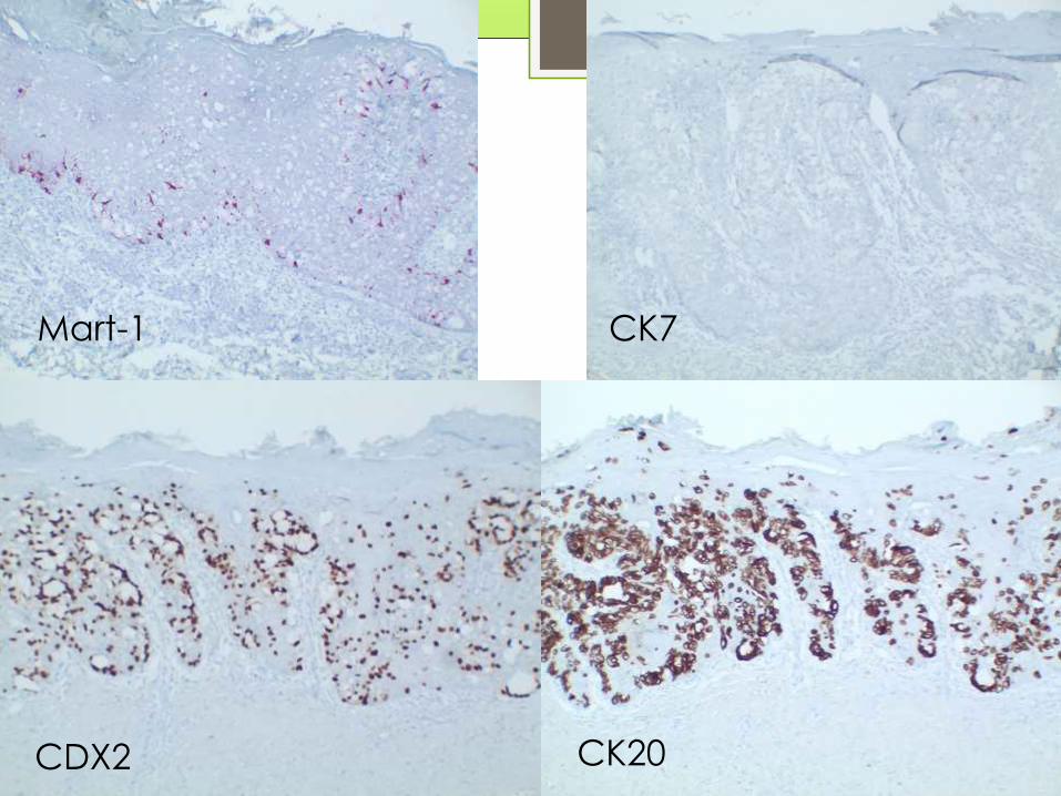
Mart-1 CK7
CK20CDX2
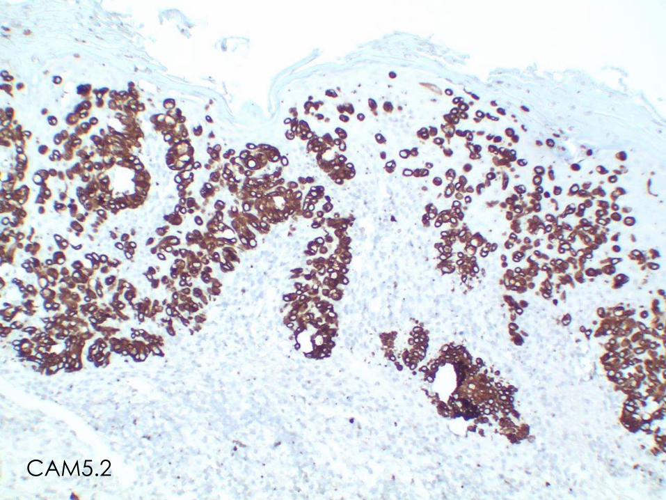
CAM5.2

Extramammary Paget’s
Disease
Majority of cases represent an in situ malignancy derived from intraepidermal sweat ducts
Minority of cases represent an epidermotropic metastasis from a distant malignant neoplasm (rectum, bladder, urethra, prostate or endocervix)
1/3 of perianal lesions are associated with a rectal adenocarcinoma
Overall association with an internal malignancy is 15%
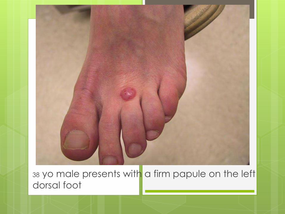
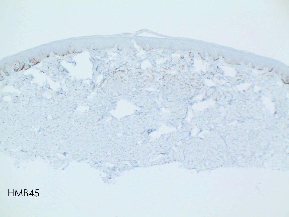
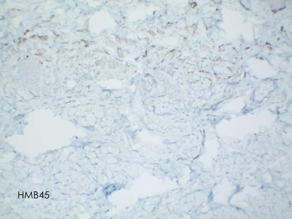
38 yo male presents with a firm papule on the left
dorsal foot
Mart-1/Ki67
HMB45
HMB45
Spitz Nevus
Benign melanocytic nevi
50% occur in children younger than 10yo
70% diagnosed during first 2 decades of
life
Differential diagnosis includes atypical
Spitz tumor and Spitz-type melanoma
If older patient, additional molecular tests
may be needed
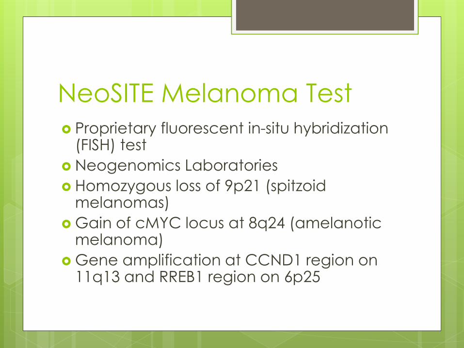
NeoSITE Melanoma Test
Proprietary fluorescent in-situ hybridization (FISH) test
Neogenomics Laboratories
Homozygous loss of 9p21 (spitzoid melanomas)
Gain of cMYC locus at 8q24 (amelanotic melanoma)
Gene amplification at CCND1 region on 11q13 and RREB1 region on 6p25
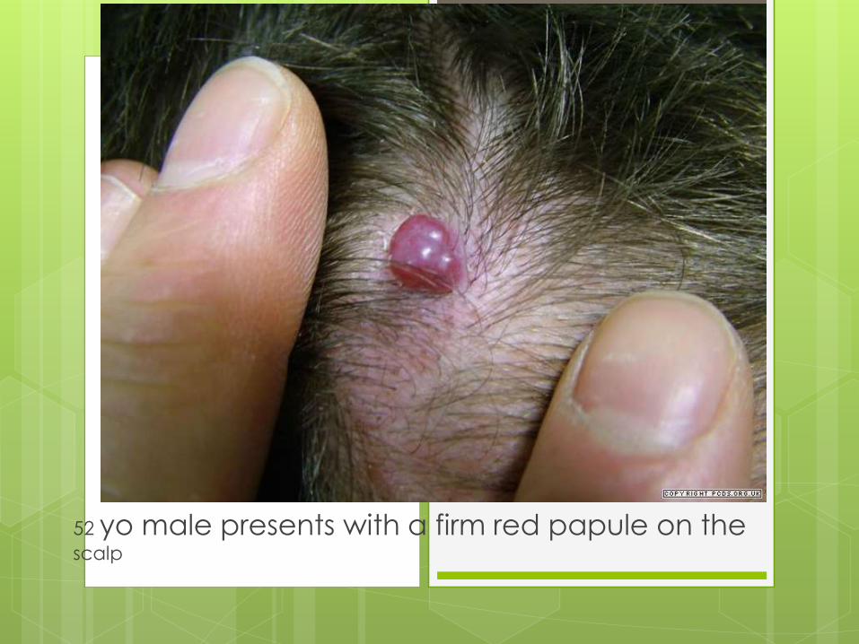
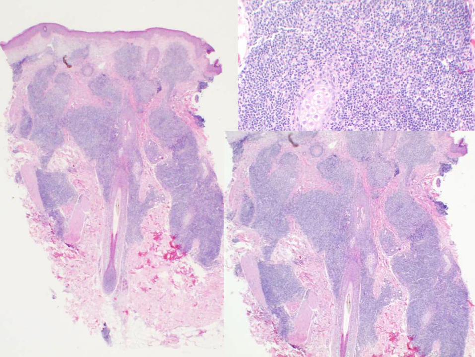
52 yo male presents with a firm red papule on the scalp
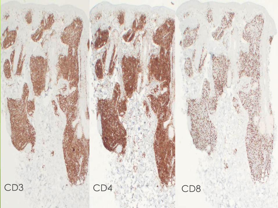
CD3 CD4 CD8
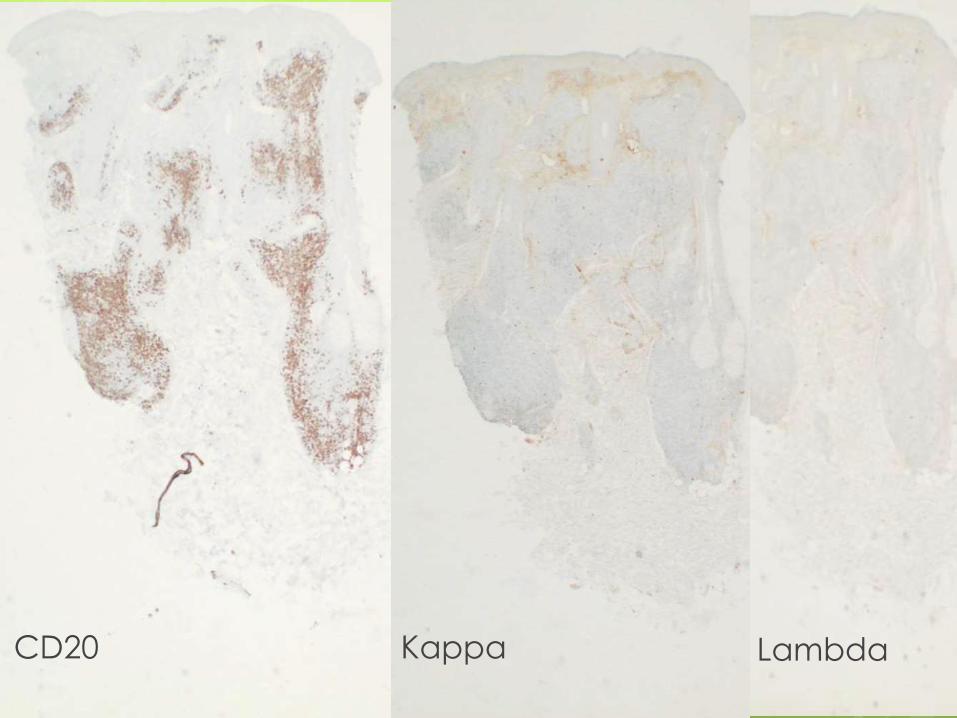
CD20 Kappa Lambda
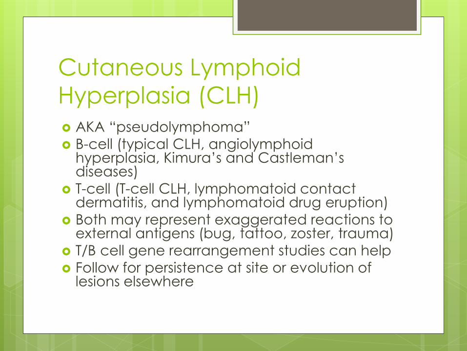
Cutaneous Lymphoid
Hyperplasia (CLH)
AKA “pseudolymphoma”
B-cell (typical CLH, angiolymphoid hyperplasia, Kimura’s and Castleman’s diseases)
T-cell (T-cell CLH, lymphomatoid contact dermatitis, and lymphomatoid drug eruption)
Both may represent exaggerated reactions to external antigens (bug, tattoo, zoster, trauma)
T/B cell gene rearrangement studies can help
Follow for persistence at site or evolution of lesions elsewhere
76 yo female with pearly
papule on left ala
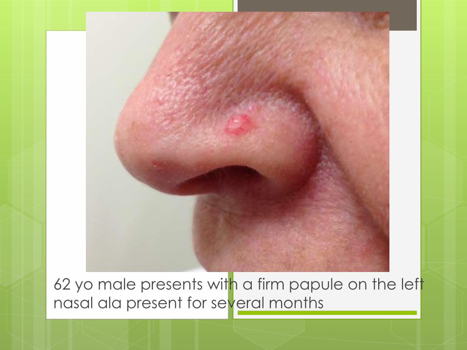
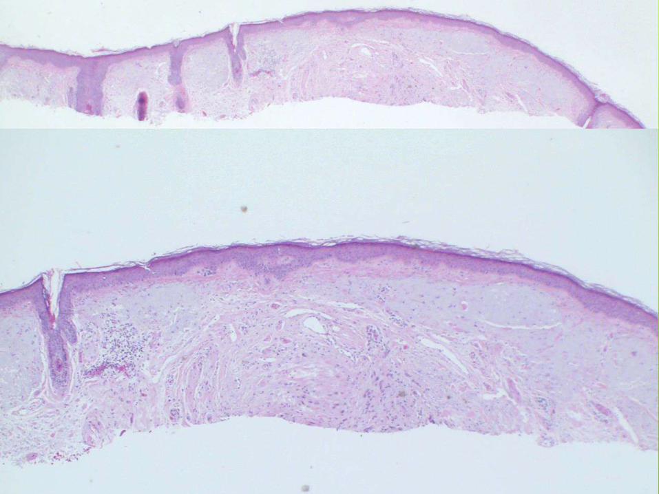
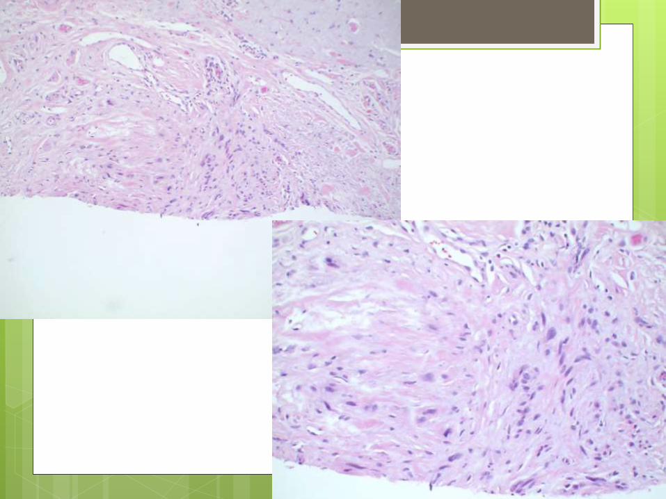
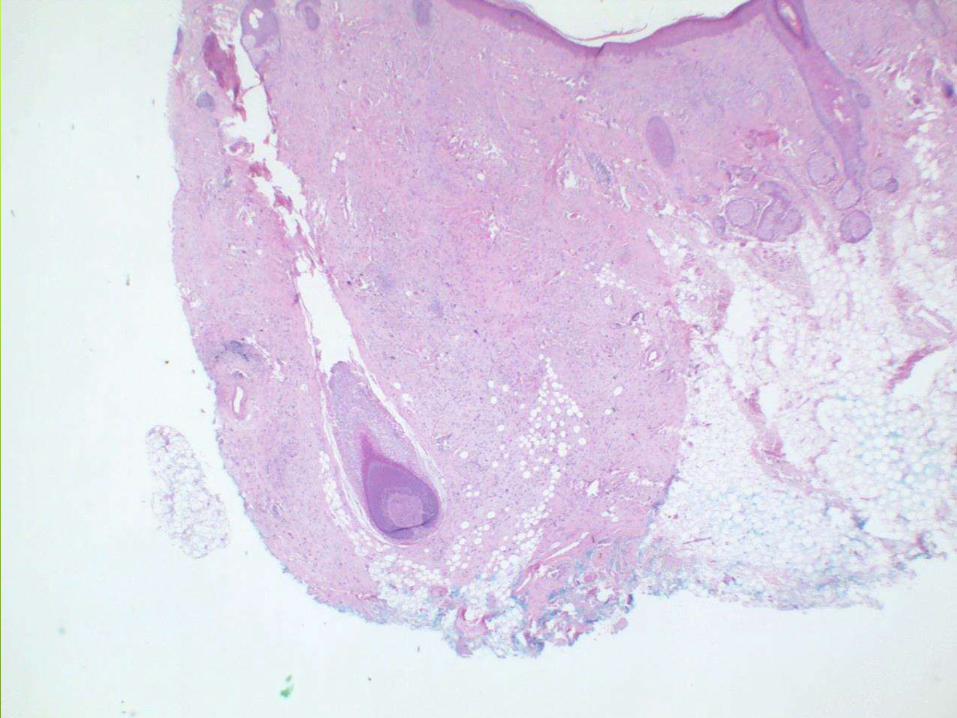
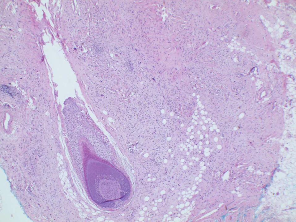
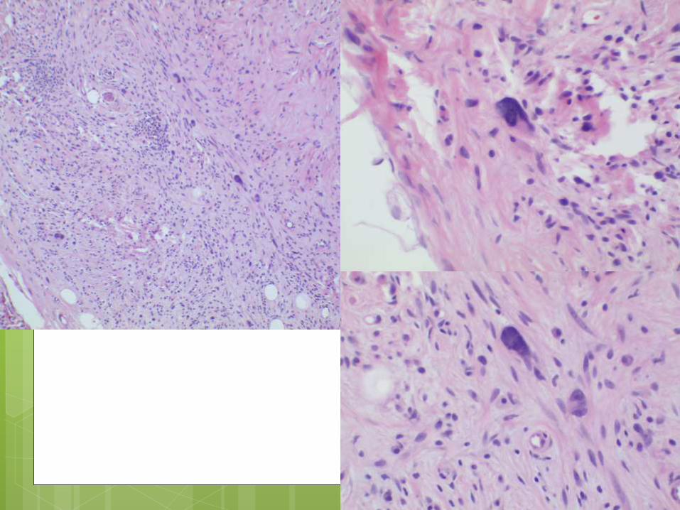
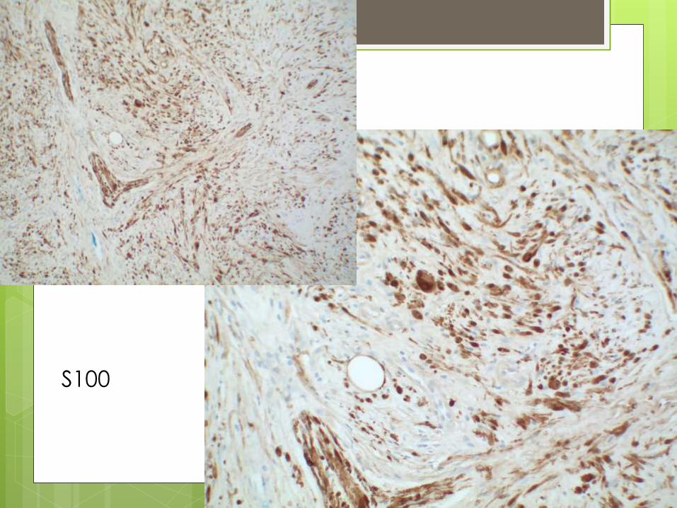
62 yo male presents with a firm papule on the left
nasal ala present for several months
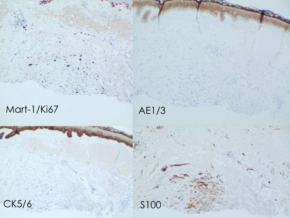
Mart-1/Ki67 AE1/3
CK5/6 S100
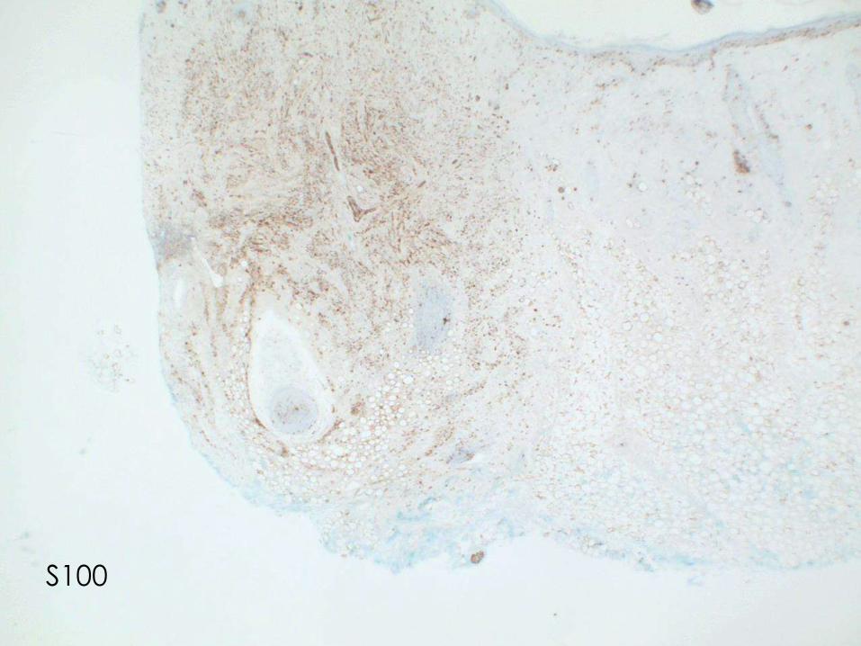
S100
S100
Desmoplastic Melanoma
Rare variant of spindle cell melanoma
Most frequently on sun damaged skin in the elderly
Uncommon, less than 4% of melanomas
Different clinical behavior than normal melanomas
Higher tendency for persistent local growth and less nodal metastasis
5 year survival from 70-90%