Embed Size (px)
Citation preview
Berberina inibe metástases do carcinoma nasofaringeal
Berberine Inhibits Metastasis of Nasopharyngeal
Carcinoma 5-8F Cells by Targeting Rho Kinase-mediated
Ezrin Phosphorylation at Threonine 567*
J Biol Chem. 2009 October 2; 284(40): 27456–27466.
Published online 2009 August 3. doi: 10.1074/jbc.M109.033795
PMCID: PMC2785675
Copyright © 2009 by The American Society for Biochemistry and Molecular Biology, Inc.
Faqing Tang,‡1 Dongsheng Wang,‡ Chaojun Duan,‡ Damao Huang,‡ Yuan Wu,‡ Yu Chen,‡
Weiwei Wang,‡ Chunlei Xie,‡ Jingjing Meng,‡ Lei Wang,‡ Bin Wu,‡ Shujin Liu,‡ Daofa Tian,‡
Feng Zhu,§ Zhiwei He,§ Fuliang Deng,¶ and Ya Cao¶
From the ‡Department of Clinical Laboratory, Xiangya Hospital, and
the ¶Cancer Research Institute, Central South University, Xiangya Road 87th, Changsha
410008, Hunan, China and
the §Hormel Institute, University of Minnesota, Austin, Minnesota 55912
1 To whom correspondence should be addressed. Tel.: Phone: 86-731-4327168; Fax: 86-731-
4327322; E-mail: [email protected] .
Received June 15, 2009; Revised July 23, 2009
This article has been cited by other articles in PMC.
Other Sections▼
Abstract
EXPERIMENTAL PROCEDURES
RESULTS
DISCUSSION
Supplementary Material
REFERENCES
Abstract
Ezrin is highly expressed in metastatic tumors and is involved in filopodia formation as well as
promotion of tumor metastasis. Thus, Ezrin may serve as a potential target for anti-metastatic
therapy. This study demonstrates that berberine reduces filopodia formation of a
nasopharyngeal carcinoma (NPC) cell line, 5-8F, at non-cytotoxic concentrations. Furthermore,
invasion and motility of 5-8F cells are decreased in a dose- and time-dependent manner,
resulting in 73.0% invasion and 67.0% motility inhibition at 20 μm. The inhibitory effects of
berberine on 5-8F cell metastasis were further confirmed in a mouse model of metastasis.
Berberine treatment in vivo resulted in a 51.1% inhibition of tumor metastasis to the lymph
nodes and decreased Ezrin phosphorylation at threonine 567 in metastatic samples. Berberine
suppressed the presence of phosphorylated Ezrin (phospho-Ezrin) in a dose- and time-
dependent manner but had no effect on total Ezrin protein expression at non-cytotoxic
concentrations. Furthermore, the inhibitory effects of berberine on phospho-Ezrin were
dependent on the suppression of Rho kinase activity. Reduction of Ezrin phosphorylation at
Thr567 by berberine was associated with its inhibitory effect on filopodia formation in 5-8F
cells. However, berberine did not effectively inhibit the motility and invasion of NPC cells
containing Ezrin Thr567 mutants. These results confirm that berberine inhibits Ezrin
phosphorylation at Thr567. Nonetheless, berberine reduces motility and invasion of cells and
inhibits tumor metastasis. The reduction of Rho kinase-mediated Ezrin phosphorylation
mediated by berberine may be a novel anti-metastatic pathway in NPC 5-8F cells.
Other Sections▼
Abstract
EXPERIMENTAL PROCEDURES
RESULTS
DISCUSSION
Supplementary Material
REFERENCES
Ezrin, a member of the ERM (ezrin-radixin-moesin) family of cytoskeletal proteins, has been
implicated in dynamic membrane-based processes, such as the formation and stabilization of
filopodia (1). DNA and protein sequencing indicate that human Ezrin is a highly charged protein
with an overall pI of 6.1 and a calculated molecular mass of 69 kDa (2, 3). It is also
evolutionarily conserved among widely divergent organisms. Within its N-terminal domain,
Ezrin has high amino acid sequence homology to the erythrocyte cytoskeleton protein band
4.1. Ezrin is involved in a variety of cellular functions, including cell adhesion, migration, and
organization of cell surface structures (4, 5). It may also contribute to the formation of the
scaffolding between the actin cytoskeleton and receptor retention (6) as well as filopodia
formation (1). Ezrin is overly expressed in various cancers and associated with cancer
metastasis (7–17).
One important mechanism of regulating the function of Ezrin is through phosphorylation at a
conserved threonine residue in the C terminus (Thr567) (18–21). Ezrin exists in a folded
conformation to mask its binding sites from other molecules, whereas phosphorylation of this
conserved threonine residue causes conformational changes exposing its binding sites (18, 21).
Therefore, phosphorylation of Ezrin at Thr567 keeps it open and active and prolongs its
lifetime (18).
2,3-Methylenedioxy-9,10-dimethoxyprotoberberine chloride (berberine),2 an isoquinoline
alkaloid present in plants of the genera Berberis and Coptis, possesses antimicrobial activity
against bacteria, fungi, viruses, Chlamydia, and protozoans (22–25). It also possesses an
antitumor effect on many types of malignancies, such as esophageal cancer (26), hepatoma,
lung carcinoma (27), leukemia (28), uterine cancer, prostate carcinoma (29), and gastric
carcinoma (30). Recently, the use of berberine has attracted great attention as an alternative
anti-metastasis therapy, considering its low toxicity and low cost. The mechanistic studies on
berberine antitumor effects showed that berberine inhibits NADH oxidase, reverse
transcriptase, and diaminooxidase (31), topoisomerase (32), nuclear transcript factor κ B
(NFκB) (33), activator protein 1 (27), cyclooxygenease-2 (34), and N-acetyltransferase activity
(35). Berberine induces apoptosis/necrosis in several human cancer cells (28, 29, 31, 36). Most
notably, berberine significantly inhibits the spontaneous mediastinal lymph node metastasis
produced by orthotopic implantation of Lewis lung carcinoma into the lung parenchyma in vivo
(27) and inhibits the motility and invasion of highly metastatic A549 cells at non-cytotoxic
concentrations in vitro (33). In a previous study, the Coptis chinensis compound containing
berberine was used to treat patients with metastatic nasopharyngeal carcinoma (NPC), and
NPC metastasis was inhibited (37). However, little is known about the molecular mechanisms
of these berberine anti-metastatic effects. This study demonstrates that Rho kinase activity is
suppressed by berberine, which leads to a reduction in Ezrin phosphorylation at Thr567 in NPC
5-8F cells. Therefore, a novel anti-metastatic mechanism of berberine is identified in this study.
Other Sections▼
Abstract
EXPERIMENTAL PROCEDURES
RESULTS
DISCUSSION
Supplementary Material
REFERENCES
EXPERIMENTAL PROCEDURES
Reagents and Antibodies
Berberine was purchased from Sigma. The compound was stored at 4 °C protected from
exposure to light. The stock solution of berberine was dissolved in DMSO. The final DMSO
concentration in the medium applied to cells was 0.1% (in both control and treated groups)
without affecting cell viability. Antibodies against Ezrin were purchased from Covance
(Berkeley, CA). Antibody against phosphorylated Ezrin at Thr567 (phospho-Ezrin Thr567) was
purchased from Cell Signaling Technology (Danvers, MA). Antibodies against Rho kinase, PKC,
Rac, Cdc42, GRK2 (G protein-coupled receptor kinase 2), myotonic dystrophykinase-related
Cdc42-binding kinase 2 (MRCK), and lymphocyte-oriented kinase (LOK) were purchased from
Santa Cruz Biotechnology, Inc. (Santa Cruz, CA). Antibodies against β-actin and normal mouse
IgG were purchased from Upstate Biotechnology, Inc. (Lake Placid, NY). The secondary
antibodies, horseradish peroxidase-linked anti-mouse IgG and anti-rabbit IgG, were purchased
from Santa Cruz Biotechnology, Inc. GST-Rhotekin-RBD protein-agarose beads were purchased
from Cytoskeleton Inc. (Denver, CO). Glutathione-Sepharose 4B was purchased from
Amersham Biosciences. The protein assay kit was purchased from Bio-Rad (Herndon, VA).
Immunoblotting detection reagents were purchased from Amersham Biosciences. Chemicals,
including DMSO, Tris-HCl, SDS, fluorescein isothiocyanate-phalloidin, 4′,6-diamidino-2-
phenylindole, and the MTT inner salt assay, were purchased from Sigma.
Cell Culture and Berberine Treatment
Human NPC cell lines, 5-8F and 6-10B, were purchased from the Cancer Research Institute of
Sun Yatsen University (Guangzhou, China). The 5-8F cell line is highly metastatic, and 6-10B
cells are non-metastatic (8). Cells were cultured as monolayers in RPMI 1640 medium
containing 10% fetal bovine serum, 2 mm l-glutamine, 100 μg/ml penicillin, 100 IU/ml
streptomycin (Invitrogen) and maintained in an incubator at 5% CO2 at 37 °C. For berberine
treatment, appropriate volumes of berberine stock solution were added to the cell cultures to
achieve the indicated concentrations and then incubated for the indicated amount of time.
After berberine treatment, cell viability was determined by the MTT assay. The dose response
of berberine-inhibited Ezrin phosphorylation was investigated in 5-8F cells treated with 2.5, 5,
or 10 μm berberine for 24 h. The time course of berberine inhibition was also investigated in 5-
8F cells treated with 5 μm berberine for 12, 24, and 48 h. After berberine treatments, cells
were harvested for protein extraction. The expression of Ezrin and phospho-Ezrin were
detected by immunoblotting.
Determination of Cell Viability (MTT Assay)
Berberine cytotoxicity was evaluated using the MTT assay to determine cell viability. Briefly, 5-
8F cells were seeded in 96-well plates at a density of 3.5 × 10³ cells/well and treated with
berberine at 0–100 μm concentrations at 37 °C for 24 or 48 h. After the exposure period,
medium was removed, cells were washed with phosphate-buffered saline (PBS), and fresh
medium was added. Cells were then incubated with 100 μl of MTT (5 mg/ml) in each well for 4
h. The absorbance of formazan produced by production of the MTT compound proportional to
the viable cells was recorded at 563 nm with a spectrophotometer.
Detection of Lactate Dehydrogenase (LDH)
Cytotoxicity of berberine on NPC cells was evaluated by detecting LDH in cell culture media
after berberine treatment. Briefly, 5-8F cells were seeded in 6-well plates at a density of 2 ×
104 cells/well and treated with berberine at 0–100 μm concentrations at 37 °C for 48 h. After
the exposure period, media were collected for the LDH assay. LDH activity was detected using
the LDH assay kit according to the manufacturer's instructions (Autec Diagnostica Co.).
Immunofluorescence Analysis
The 5-8F cells with or without berberine treatment were fixed with 2.0% formaldehyde in PBS
for 30 min, washed with PBS three times, and then treated with PBS containing 0.2% Triton X-
100 for 10 min. After being washed with PBS three times, cells were incubated with 0.5%
bovine serum albumin in PBS. After three PBS washes, cells were stained with 5 μg/ml
fluorescein isothiocyanate-phalloidin (Sigma) for 40 min and examined using a Zeiss
axiophotomicroscope (Carl Zeiss, Oberkochen, Germany). Cells stained with 4′,6-diamidino-2-
phenylindole served as a control. Random fields were counted for cells with filopodia.
Electron Microscopy
Cultures of 5-8F cells treated with berberine or transfected with plasmids were cultured on
CELLocate coverslips (Eppendorf, Hamburg, Germany). The cells were fixed with 2.5%
glutaraldehyde in 0.1 m cacodylate buffer (pH 7.4) for 2 h at room temperature. Samples were
then processed conventionally and observed under a scanning electron microscope (Hitachi
Co.). Cells with filopodia were counted.
Immunoblotting Analysis
After berberine treatment, cells were harvested and lysed in 1× lysis buffer (1× PBS, 1%
Nonidet P-40, 0.5% sodium deoxycholate, 0.1% SDS, and freshly added 100 μg/ml
phenylmethanesulfonyl fluoride, 10 μg/ml aprotinin, 1 mm sodium orthovanadate). The cell
lysates were then pelleted at 10,000 × g for 10 min at 4 °C. Resultant protein concentrations of
each sample were determined using the Bio-Rad protein assay. Equivalent amounts of protein
(40 μg/sample) were separated by 10% SDS-PAGE and transferred onto a nitrocellulose
membrane. The blot was blocked with 5% nonfat milk in PBS for 1 h, incubated with specific
antibody against Ezrin (Covance) or phospho-Ezrin (Cell Signaling Technology) for 2 h, and then
incubated with an appropriate peroxidase-conjugated secondary antibody for 1 h. All
incubations were carried out at room temperature, and intensive PBS washing was performed
after detection with each antibody. After washing three times with PBS, the signal was
developed using 4-chloro-1-napthol/3,3-o-diaminobenzidine, and relative photographic
density was quantified by a gel documentation and analysis system. β-Actin was used as an
internal control to verify basal expression levels and equal protein loading. The ratio of the
specific proteins to β-actin was calculated.
Cell Motility and Invasion Assay
For cell invasion assays, 5-8F cells were treated with the indicated concentrations of berberine
for indicated amounts of time. After berberine treatment, the cells were trypsinized, and their
invasiveness was tested using the in vitro Boyden chamber invasion assay (38). Matrigel
(Collaborative Biomedical Products, Bedford, MA) was diluted to 25 mg/50 ml with cold
filtered distilled water and applied to 8-μm pore size polycarbonate membrane filters in the
chamber. Treated cells were seeded into Boyden chambers (Neuro Probe Inc., Cabin John, MD)
at a density of 1.5 × 10⁴ cells/well in 50 μl of serum-free media and incubated for 12 h at 37 °C.
The bottom well contained standard medium with 20% fetal bovine serum. The cells invading
through the filter of the chamber were fixed with methanol and stained with hematoxylin and
eosin. Random fields were counted for invading cells under a light microscope.
The effects of berberine on cell motility were determined using cells seeded into Boyden
chambers without a Matrigel coating. Migration of cells in the presence or absence of
berberine was measured as described in the motility assay (38). Statistical analysis was
corrected with cell viability to clarify the effects of berberine.
Animals
Thirty female nude BALB/c mice (5–6 weeks old) were purchased from the Animal Center of
Central South University (Hunan, China). They were maintained in the Laboratory for
Experiments, Central South University under laminar air flow conditions. The studies were
conducted in accordance with the standards established by the Guidelines for the Care and
Use of Laboratory Animals by Central South University.
Evaluation of Anti-metastatic Activity of Berberine in Nude Mice
Analysis of metastatic tumors of 5-8F cells was performed as described previously with
modifications (27). Briefly, 100-μl aliquots of 5-8F, 6-10B-pcDNA3.1, 6-10B-pcDNA3.1- Ezrin
M(A), and 6-10B-pcDNA3.1-Ezrin cell suspensions (1 × 10⁴ cells) mixed with Matrigel were
injected into the tail vein of nude mice, 10 mice/group, respectively. Two groups of 5-8F cells
were given berberine once daily at a concentration of 15 or 30 mg/kg body weight (1 or 2
times the dosage of the human study), respectively. Groups of 6-10B-pcDNA3.1-Ezrin cells
were given 15 mg/kg berberine for 30 days starting on day 1 after the injection. Metastasis
was evaluated by measuring the weight of the metastasized tumor at mediastinal lymph nodes
on day 30 after the injection. Body weight of the nude mice was measured, and other adverse
effects of berberine were evaluated. The present study protocols were approved by the ethical
committee at Xiangya Hospital of Central South University.
Immunohistochemistry
Metastasized tumor samples in the nude mice were fixed in 4% paraformaldehyde, paraffin-
embedded, sectioned, and mounted on slides. Sections were stained with hematoxylin and
eosin for microscopic examination. Unstained sections were used for staining with antibodies
against Ezrin or phospho-Ezrin by immunohistochemistry. Immunohistochemistry followed
standard procedures with overnight exposure at room temperature to 1:500 Ezrin antibody or
1:200 phospho-Ezrin antibody diluted in 0.5% nonfat milk. After washing with PBS, the sections
were incubated sequentially with a secondary antibody against mouse, peroxidase enzyme
label, and diaminobenzidine (Sigma) and then stained with hematoxylin (Polysciences, Inc.,
Warrington, PA), dehydrated, and mounted under a glass coverslip. Sections stained with
normal mouse IgG served as a negative control.
Kinase Activity Assay
Cultures of 5-8F cells (1.0 × 10⁶) were grown in 100-mm dishes for 12–24 h. When cultures
were at 70–80% confluence, the cells were treated with berberine for 48 h in RPMI 1640
medium containing 10% fetal bovine serum. Treated cells were washed once with ice-cold PBS
and lysed in 250 μl of kinase lysis buffer (25 mm Tris-HCl (pH 7.5), 5 mm β-glycerophosphate,
0.1 mm Na3VO4, 10 mm MgCl2, 1 mm aprotinin, and 1 mm phenylmethanesulfonyl fluoride).
The clarified supernatant fractions containing 500 μg of protein were subjected to
immunoprecipitation using antibodies against Rho, PKC, GRK2, MRCK, or LOK, respectively. The
precipitation mediating Ezrin phosphorylation was determined by the kinase assay protocol
(Upstate Biotechnology, Inc.). Briefly, 20 μg of precipitated complex was added to 2.5 μl of 10×
kinase buffer (250 mm Tris-HCl (pH 7.5), 50 mm β-glycerophosphate, 20 mm dl-dithiothreitol,
1 mm Na3VO4, 100 mm MgCl2), 2.5 μl (2.5 μg) of a GST-Ezrin fusion protein, 10 μl of diluted
ATP mixture (Upstate Biotechnology, Inc.), 10 μCi of [32P]ATP, and H2O was added to 25 μl.
The reaction was incubated at 30 °C for 30 min and then subjected to separation by 12% SDS-
PAGE. The gels were stained with Coomassie Blue and then dried. Phosphorylated GST-Ezrin
was analyzed by autoradiography that determines kinase activity.
Kinetics of Berberine Inhibition of Rho Kinase
Various concentrations of berberine (0.1, 0.2, 0.4, 0.8, and 1.6 μm) were added to 5-8F cells,
and a Rho antibody was used to precipitate Rho from treated cells. The immunoprecipitates
were incubated with various concentrations of [32P]ATP and 10 μg of histone type 2 as
substrates at 30 °C for 30 min in a total volume of 30 μl of the kinase buffer. Incubation was
terminated by the addition of 10 μl of 4× Laemmli sample buffer. After boiling for 5 min, the
mixtures were subjected to 12% SDS-PAGE. The gels were stained with Coomassie Blue and
then dried. The bands corresponding to histone type 2 were excised, and the radioactivity was
measured. The dissociation constant for binding of the inhibitor to the enzyme (Ki) values were
calculated from the equation, Ki = IC50/(1 + S/Km), where S and Km were the concentration of
ATP and the Km value for ATP, respectively (39).
Construction of Expression Vectors
The pcDNA3.1 and pGEX-5x-1 vectors were purchased from Invitrogen. A DNA fragment
encoding the GTPase-binding domain of p21-activated kinase (PAK-PBD), comprising amino
acids 68–166, was generated by PCR and cloned into the BamHI/XhoI sites of pGEX-5x-1 and
expressed in Escherichia coli as GST-PAK-PBD fusion protein according to the manufacturer's
protocol. Ezrin and Rho DNA fragments were also generated by PCR and cloned into the
BamHI/XhoI sites of the pcDNA3.1 vector (Amersham Biosciences) to generate pcDNA3.1-Ezrin
and pcDNA3.1-Rho plasmids, respectively. For producing GST-Ezrin, the Ezrin DNA fragment
was cloned into the BamHI/XhoI sites of pGEX-5x-1 and named pGEX-5x-1-Ezrin. The mutant
pcDNA3.1-Ezrin plasmids were generated with the QuikChange II site-directed mutagenesis kit
and Ezrin mutant primers: Primer 1 (sense), 5′-CAGGGCAACGCCAAGCAGCGCAT-3′; Primer 2
(antisense), 5′-ATGCGCTGCTTGGCGTTGCCCTG-3′; Primer 3 (sense), 5′-
CAGGGCAACGACAAGCAGCGCAT-3′; Primer 4 (antisense), 5′-ATGCGCTGCTTGTCGTTGCCCTG-3′.
Primer 1 and Primer 2 were used for mutating Thr567 into Ala567 and generated pcDNA3.1-
Ezrin Ala567 (pcDNA3.1-Ezrin-M(A)). Primer 3 and Primer 4 were used for mutating Thr567
into Asp567 and generated pcDNA3. 1-Ezrin Asp567 (pcDNA3.1-Ezrin-M(D)). The pU6pro
vector was used to construct pU6pro-si-mock (si-mock) and pU6pro-si-Ezrin (si-Ezrin) following
the recommended protocol. For the si-mock and si-Ezrin, we synthesized primers for the si-
mock (general scramble: sense, 5′-
TTTGACTACCGTTGTTATAGGTGTTCAAGAGACACCTATAACAACGGTAGTTTTTT-3′; antisense, 5′-
CTAGAAAAAACTACCGTTGTTATAGGTGTCTCTTGAACACCTATAACAACGGTAGT-3′) and for si-Ezrin
(Set 1, 5′-CCCCAAAGAUUGGCUUUCC-3′ (position in the open reading frame, 704–722); Set 2,
5′-UCCACUAUGUGGAUAAUAA-3′ (open reading frame, 140–158)). All constructs were
confirmed by restriction enzyme mapping and DNA sequencing.
Rho Family GTPase Assay
Cultures of 5-8F cells (5.0 × 10⁶) were treated with berberine at 20 and 40 μm for 48 h as
described previously. After being washed with ice-cold PBS, they were lysed in 500 μl of kinase
lysis buffer. The clarified supernatant fractions containing 400 μg of protein were incubated
with 3 μg of GST-Rhotekin-RBD (Cytoskeleton Inc.) (40) or GST-PAK-PBD (41) for 12 h at 4 °C,
respectively. GST fusion proteins were collected by incubation with glutathione-Sepharose 4B
beads (Amersham Biosciences) for 3 h. Precipitates were washed five times in the lysis buffer,
resuspended in SDS sample buffer, and boiled for 10 min. After centrifugation for 10 min at
10,000 × g, the supernatants were subjected to immunoblotting with antibodies against Rho,
Rac, or Cdc42, respectively.
Gene Transfection and Generation of Stably Transfected Cell Lines
Cultures of 6-10B cells (5.0 × 10⁵) were seeded in 100-mm tissue culture dishes. After culturing
at 37 °C for 16–24 h, the cells were transfected with 4 μg of pcDNA3.1 (mock), pcDNA3.1-Ezrin,
pcDNA3. 1-Ezrin-M(A), or pcDNA3.1-Ezrin- M(D), respectively, using Lipofectamine 2000
reagent (Invitrogen) following the manufacturer's protocol. For establishing 5-8F-si-Ezrin cells,
5-8F cells were transfected with pU6pro-si-Ezrin. The stably transfected cell lines were
obtained by selection for G418 resistance (400 μg/ml) and further confirmed by assessing Ezrin
expression. To investigate whether the inhibitory effects of berberine on Rho activity could be
reversed with constitutively active Rho kinase, 5-8F cells were treated with berberine and then
transfected with pcDNA3.1-Rho. To confirm that berberine exerts an inhibition on metastasis
through Ezrin Thr567 phosphorylation, 5-8F-si-Ezrin cells were transiently transfected with
pcDNA3.1, pcDNA3.1-Ezrin, pcDNA3.1-Ezrin-M(A), or pcDNA3.1- Ezrin-M(D), and their motility
and invasion were detected using the in vitro Boyden chamber invasion assay (38).
Other Sections▼
Abstract
EXPERIMENTAL PROCEDURES
RESULTS
DISCUSSION
Supplementary Material
REFERENCES
RESULTS
Cytotoxic Effects of Berberine on NPC Cells
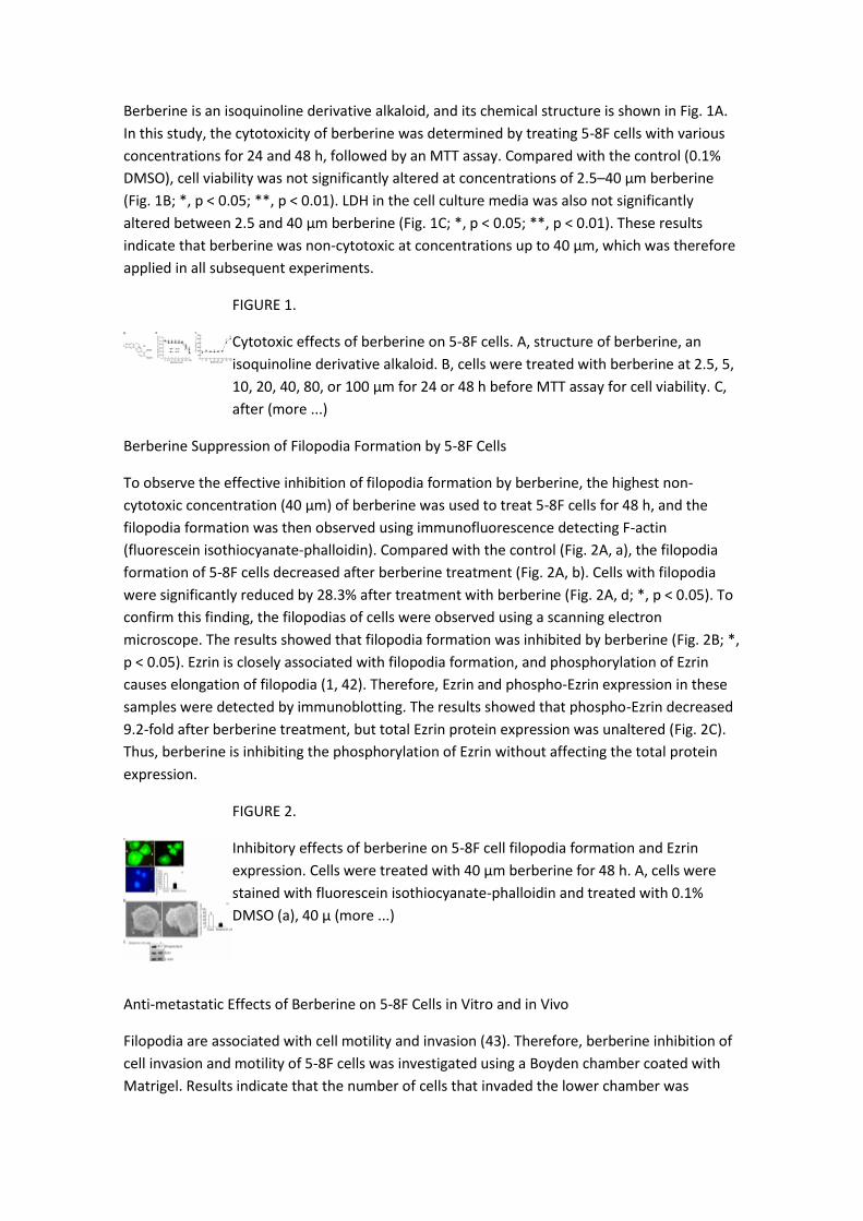
Berberine is an isoquinoline derivative alkaloid, and its chemical structure is shown in Fig. 1A.
In this study, the cytotoxicity of berberine was determined by treating 5-8F cells with various
concentrations for 24 and 48 h, followed by an MTT assay. Compared with the control (0.1%
DMSO), cell viability was not significantly altered at concentrations of 2.5–40 μm berberine
(Fig. 1B; *, p < 0.05; **, p < 0.01). LDH in the cell culture media was also not significantly
altered between 2.5 and 40 μm berberine (Fig. 1C; *, p < 0.05; **, p < 0.01). These results
indicate that berberine was non-cytotoxic at concentrations up to 40 μm, which was therefore
applied in all subsequent experiments.
FIGURE 1.
Cytotoxic effects of berberine on 5-8F cells. A, structure of berberine, an
isoquinoline derivative alkaloid. B, cells were treated with berberine at 2.5, 5,
10, 20, 40, 80, or 100 μm for 24 or 48 h before MTT assay for cell viability. C,
after (more ...)
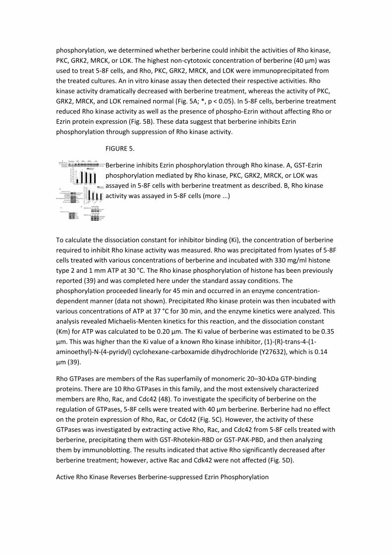
Berberine Suppression of Filopodia Formation by 5-8F Cells
To observe the effective inhibition of filopodia formation by berberine, the highest non-
cytotoxic concentration (40 μm) of berberine was used to treat 5-8F cells for 48 h, and the
filopodia formation was then observed using immunofluorescence detecting F-actin
(fluorescein isothiocyanate-phalloidin). Compared with the control (Fig. 2A, a), the filopodia
formation of 5-8F cells decreased after berberine treatment (Fig. 2A, b). Cells with filopodia
were significantly reduced by 28.3% after treatment with berberine (Fig. 2A, d; *, p < 0.05). To
confirm this finding, the filopodias of cells were observed using a scanning electron
microscope. The results showed that filopodia formation was inhibited by berberine (Fig. 2B; *,
p < 0.05). Ezrin is closely associated with filopodia formation, and phosphorylation of Ezrin
causes elongation of filopodia (1, 42). Therefore, Ezrin and phospho-Ezrin expression in these
samples were detected by immunoblotting. The results showed that phospho-Ezrin decreased
9.2-fold after berberine treatment, but total Ezrin protein expression was unaltered (Fig. 2C).
Thus, berberine is inhibiting the phosphorylation of Ezrin without affecting the total protein
expression.
FIGURE 2.
Inhibitory effects of berberine on 5-8F cell filopodia formation and Ezrin
expression. Cells were treated with 40 μm berberine for 48 h. A, cells were
stained with fluorescein isothiocyanate-phalloidin and treated with 0.1%
DMSO (a), 40 μ (more ...)
Anti-metastatic Effects of Berberine on 5-8F Cells in Vitro and in Vivo
Filopodia are associated with cell motility and invasion (43). Therefore, berberine inhibition of
cell invasion and motility of 5-8F cells was investigated using a Boyden chamber coated with
Matrigel. Results indicate that the number of cells that invaded the lower chamber was

significantly reduced by berberine in a dose-dependent manner with a maximal 73.6%
inhibition at 20 μm berberine (Fig. 3A; *, p < 0.01). Such inhibitory effects were also observed
in the mobility assay with 67.0% inhibition of mobility at 20 μm berberine (Fig. 3B; *, p < 0.01).
In a time course experiment, the lowest non-cytotoxic concentration (5 μm) of berberine was
used to treat cells for 24, 48, or 72 h. Our results revealed that berberine significantly inhibited
invasion (Fig. 3C) and motility (Fig. 3D) in a time-dependent manner. Anti-metastatic effects of
berberine were confirmed in vivo when 5-8F cells were injected into the tail veins of BABL/c
mice to establish a metastatic mouse model. These same mice were treated with berberine,
and the anti-metastatic effects were assessed. The metastasis of 5-8F cells to the mediastinal
lymph nodes was significantly inhibited by berberine (Fig. 3E). Berberine did not cause any
reduction of body weight or other adverse effects on the mice (Fig. 3F).
FIGURE 3.
Berberine inhibition of metastasis in 5-8F cells in vitro and in vivo. In the cell
motility and invasion assays, 5-8F cells were treated with berberine at 2.5, 5,
10, or 20 μm for 24 h for dose course assay. Cells were treated with berberine
at (more ...)
Berberine Suppression of Ezrin Phosphorylation
To determine if the anti-metastatic effects of berberine were associated with Ezrin
phosphorylation, phospho-Ezrin was detected in metastatic tumors using
immunohistochemistry. Phosphorylation of Ezrin decreased in metastatic tumors from mice
treated with berberine; however, total Ezrin expression was not changed (Fig. 4A). The
association between berberine treatment and decreased phospho-Ezrin suggests that the
reduction of Ezrin phosphorylation may mediate the anti-metastatic effects of berberine.
Furthermore, berberine decreased the phosphorylation of Ezrin in a dose-dependent (Fig. 4B)
and time-dependent (Fig. 4C) manner in 5-8F cells.
FIGURE 4.
Ezrin and phospho-Ezrin expression in metastatic tumors and 5-8F cells with
or without berberine treatment. A, Ezrin and phospho-Ezrin expression were
detected in metastatic tumor samples from the nude mice using
immunochemistry. Paraffin sections were (more ...)
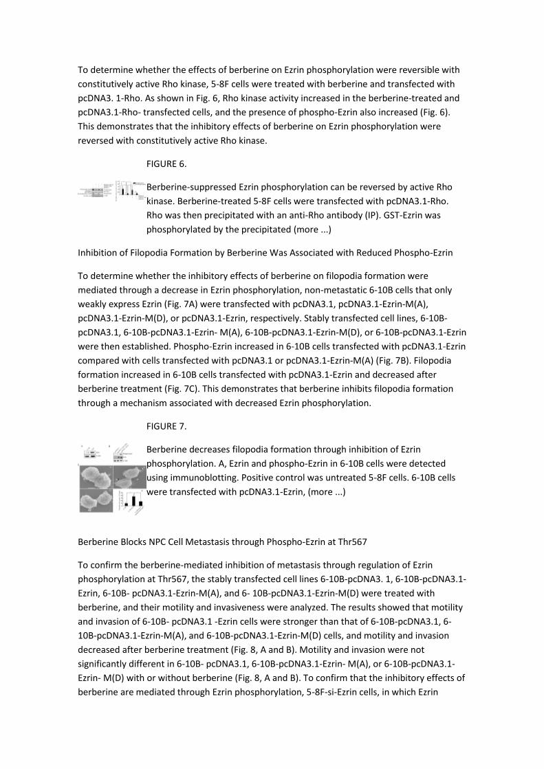
Berberine Reduces Ezrin Phosphorylation through Rho Kinase
Ezrin is known to be phosphorylated by several protein kinases, including Rho kinase, PKC,
GRK2, MRCK, and LOK (18, 44–47). To search for the upstream kinases of Ezrin
phosphorylation, we determined whether berberine could inhibit the activities of Rho kinase,
PKC, GRK2, MRCK, or LOK. The highest non-cytotoxic concentration of berberine (40 μm) was
used to treat 5-8F cells, and Rho, PKC, GRK2, MRCK, and LOK were immunoprecipitated from
the treated cultures. An in vitro kinase assay then detected their respective activities. Rho
kinase activity dramatically decreased with berberine treatment, whereas the activity of PKC,
GRK2, MRCK, and LOK remained normal (Fig. 5A; *, p < 0.05). In 5-8F cells, berberine treatment
reduced Rho kinase activity as well as the presence of phospho-Ezrin without affecting Rho or
Ezrin protein expression (Fig. 5B). These data suggest that berberine inhibits Ezrin
phosphorylation through suppression of Rho kinase activity.
FIGURE 5.
Berberine inhibits Ezrin phosphorylation through Rho kinase. A, GST-Ezrin
phosphorylation mediated by Rho kinase, PKC, GRK2, MRCK, or LOK was
assayed in 5-8F cells with berberine treatment as described. B, Rho kinase
activity was assayed in 5-8F cells (more ...)
To calculate the dissociation constant for inhibitor binding (Ki), the concentration of berberine
required to inhibit Rho kinase activity was measured. Rho was precipitated from lysates of 5-8F
cells treated with various concentrations of berberine and incubated with 330 mg/ml histone
type 2 and 1 mm ATP at 30 °C. The Rho kinase phosphorylation of histone has been previously
reported (39) and was completed here under the standard assay conditions. The
phosphorylation proceeded linearly for 45 min and occurred in an enzyme concentration-
dependent manner (data not shown). Precipitated Rho kinase protein was then incubated with
various concentrations of ATP at 37 °C for 30 min, and the enzyme kinetics were analyzed. This
analysis revealed Michaelis-Menten kinetics for this reaction, and the dissociation constant
(Km) for ATP was calculated to be 0.20 μm. The Ki value of berberine was estimated to be 0.35
μm. This was higher than the Ki value of a known Rho kinase inhibitor, (1)-(R)-trans-4-(1-
aminoethyl)-N-(4-pyridyl) cyclohexane-carboxamide dihydrochloride (Y27632), which is 0.14
μm (39).
Rho GTPases are members of the Ras superfamily of monomeric 20–30-kDa GTP-binding
proteins. There are 10 Rho GTPases in this family, and the most extensively characterized
members are Rho, Rac, and Cdc42 (48). To investigate the specificity of berberine on the
regulation of GTPases, 5-8F cells were treated with 40 μm berberine. Berberine had no effect
on the protein expression of Rho, Rac, or Cdc42 (Fig. 5C). However, the activity of these
GTPases was investigated by extracting active Rho, Rac, and Cdc42 from 5-8F cells treated with
berberine, precipitating them with GST-Rhotekin-RBD or GST-PAK-PBD, and then analyzing
them by immunoblotting. The results indicated that active Rho significantly decreased after
berberine treatment; however, active Rac and Cdk42 were not affected (Fig. 5D).
Active Rho Kinase Reverses Berberine-suppressed Ezrin Phosphorylation
To determine whether the effects of berberine on Ezrin phosphorylation were reversible with
constitutively active Rho kinase, 5-8F cells were treated with berberine and transfected with
pcDNA3. 1-Rho. As shown in Fig. 6, Rho kinase activity increased in the berberine-treated and
pcDNA3.1-Rho- transfected cells, and the presence of phospho-Ezrin also increased (Fig. 6).
This demonstrates that the inhibitory effects of berberine on Ezrin phosphorylation were
reversed with constitutively active Rho kinase.
FIGURE 6.
Berberine-suppressed Ezrin phosphorylation can be reversed by active Rho
kinase. Berberine-treated 5-8F cells were transfected with pcDNA3.1-Rho.
Rho was then precipitated with an anti-Rho antibody (IP). GST-Ezrin was
phosphorylated by the precipitated (more ...)
Inhibition of Filopodia Formation by Berberine Was Associated with Reduced Phospho-Ezrin
To determine whether the inhibitory effects of berberine on filopodia formation were
mediated through a decrease in Ezrin phosphorylation, non-metastatic 6-10B cells that only
weakly express Ezrin (Fig. 7A) were transfected with pcDNA3.1, pcDNA3.1-Ezrin-M(A),
pcDNA3.1-Ezrin-M(D), or pcDNA3.1-Ezrin, respectively. Stably transfected cell lines, 6-10B-
pcDNA3.1, 6-10B-pcDNA3.1-Ezrin- M(A), 6-10B-pcDNA3.1-Ezrin-M(D), or 6-10B-pcDNA3.1-Ezrin
were then established. Phospho-Ezrin increased in 6-10B cells transfected with pcDNA3.1-Ezrin
compared with cells transfected with pcDNA3.1 or pcDNA3.1-Ezrin-M(A) (Fig. 7B). Filopodia
formation increased in 6-10B cells transfected with pcDNA3.1-Ezrin and decreased after
berberine treatment (Fig. 7C). This demonstrates that berberine inhibits filopodia formation
through a mechanism associated with decreased Ezrin phosphorylation.
FIGURE 7.
Berberine decreases filopodia formation through inhibition of Ezrin
phosphorylation. A, Ezrin and phospho-Ezrin in 6-10B cells were detected
using immunoblotting. Positive control was untreated 5-8F cells. 6-10B cells
were transfected with pcDNA3.1-Ezrin, (more ...)
Berberine Blocks NPC Cell Metastasis through Phospho-Ezrin at Thr567
To confirm the berberine-mediated inhibition of metastasis through regulation of Ezrin
phosphorylation at Thr567, the stably transfected cell lines 6-10B-pcDNA3. 1, 6-10B-pcDNA3.1-
Ezrin, 6-10B- pcDNA3.1-Ezrin-M(A), and 6- 10B-pcDNA3.1-Ezrin-M(D) were treated with
berberine, and their motility and invasiveness were analyzed. The results showed that motility
and invasion of 6-10B- pcDNA3.1 -Ezrin cells were stronger than that of 6-10B-pcDNA3.1, 6-
10B-pcDNA3.1-Ezrin-M(A), and 6-10B-pcDNA3.1-Ezrin-M(D) cells, and motility and invasion
decreased after berberine treatment (Fig. 8, A and B). Motility and invasion were not
significantly different in 6-10B- pcDNA3.1, 6-10B-pcDNA3.1-Ezrin- M(A), or 6-10B-pcDNA3.1-
Ezrin- M(D) with or without berberine (Fig. 8, A and B). To confirm that the inhibitory effects of
berberine are mediated through Ezrin phosphorylation, 5-8F-si-Ezrin cells, in which Ezrin
expression was blocked, were transiently transfected with pcDNA3.1, pcDNA3. 1-Ezrin-M(A),
pcDNA3.1-Ezrin- M(D), or pcDNA3.1-Ezrin and then treated with berberine. The motility and
invasion analysis of these cells were similar to 6-10B cells transfected with the same constructs
(Fig. 9, C and D).
FIGURE 8.
Berberine inhibits motility and invasion in 6-10B cells through inhibition of
phosphorylation of Ezrin at Thr567. A, motility (A) and invasion (B) of the
stably transfected cell lines 6-10B-pcDNA3.1, 6-10B-pcDNA3.1-Ezrin, 6-10B-
pcDNA3.1-Ezrin-M(A), and (more ...)
FIGURE 9.
Berberine inhibits motility and invasion in 5-8F-si-Ezrin cells through inhibition
of Ezrin phosphorylation at Thr567. A, 5-8F cells were transfected with
pU6pro-si-Ezrin, and the 5-8F-si-Ezrin cell line was established by G418
selection. Ezrin expression (more ...)
To confirm the inhibitory effects of berberine on metastasis in vivo, 6-10B-pcDNA3.1, 6-10B-
pcDNA3.1- Ezrin, and 6-10B-pcDNA3.1-Ezrin- M(A) cells were injected into the tail veins of
BALB/c mice. These mice were also treated with berberine. The tumors that metastasized to
the mediastinal lymph nodes were detected. Similar to in vitro mobility and invasion assays, 6-
10B-pcDNA3.1 and 6- 10B-pcDNA3.1-Ezrin-M(A) cells displayed limited metastatic capabilities.
Transfection of 6-10B cells with the pcDNA3.1-Ezrin plasmid restored metastatic capability,
which was effectively inhibited by berberine (Fig. 8C). Levels of phospho-Ezrin were
determined from each of the stably transfected cells with or without berberine treatment.
Immunoblot results showed that levels of phospho-Ezrin from 6-10B-pcDNA3.1-Ezrin-
transfected cells dramatically decreased after berberine treatment; however, the total amount
of Ezrin did not change (Fig. 8D). Taken together, berberine exerts an anti-metastatic effect on
NPC cells through the reduction of Rho-kinase activity and subsequent Ezrin phosphorylation.
Other Sections▼
Abstract
EXPERIMENTAL PROCEDURES
RESULTS
DISCUSSION
Supplementary Material
REFERENCES
DISCUSSION
NPC cells are highly metastatic (49). In a previous clinical therapy study, a Coptic chinensis
compound containing berberine was used to treat patients with NPC and effectively inhibited
NPC metastasis (37). An NPC cell line, 5-8F, was used to investigate the anti-metastatic
mechanisms of berberine upon motility and invasion of NPC cells. Results from that study
indicated that berberine can inhibit the invasion and motility of NPC cells in vitro and in vivo. In
vivo, berberine can inhibit the metastasis of 5-8F cells into mediastinal lymph nodes, lungs,
and liver, with the mediastinal lymph nodes being the main sites of NPC metastasis (49).
Therefore, this study focused on the metastasis of NPC cells to mediastinal lymph nodes.
Metastasis is accompanied by various physiological alterations, such as filopodia formation,
allowing cancer cells to invade the blood or lymphatic system and to spread to other tissue or
organs. Thus, filopodia plays an important role in tumor metastasis (50). The present study
demonstrates that noncytotoxic concentrations of berberine exert an inhibitory effect on the
filopodia formation of 5-8F cells. Ezrin is a component of the filopodia of epithelial cells that
serves as a major cytoplasmic substrate for certain protein-tyrosine kinases. It has been
implicated in dynamic membrane-based processes, such as the formation and stabilization of
filopodia (1). Increased expression of Ezrin is associated with tumor metastasis in NPC (8, 15,
16, 51). In fact, it may be involved in multiple pathways and actually promote tumor
metastasis, making it a prospective therapeutic target (17). Therefore, the inhibitory effects of
berberine on Ezrin and Ezrin phosphorylation were investigated in the 5-8F NPC cell line. High
levels of Ezrin expression were detected in 5-8F cells, contrary to the lower levels of expression
detected in nonmetastatic 6-10B cells (Fig. 2B). This result is consistent with results from Yu et
al. (7), where Ezrin was highly expressed in metastatic murine rhabdomyosarcoma cells.
As a cytoskeleton organizer, Ezrin is involved in cell adhesion and migration (4, 5), which is also
associated with tumor metastasis. The phosphorylation at a conserved threonine residue in
the C terminus (Thr567) is an important mechanism for regulating the function of Ezrin (21).
This study demonstrated that berberine effectively inhibited the phosphorylation of Ezrin at
Thr567 without affecting Ezrin protein levels. Phosphorylation of Ezrin at Thr567 keeps it open
and active and prolongs its lifetime (18). Interestingly, the open conformation of phospho-
Ezrin may function as an actin filament/plasma membrane cross-linker (18–20). We
hypothesized that the anti-metastatic mechanisms of berberine may be acting through the
inhibition of Ezrin phosphorylation at Thr567. To test this hypothesis, plasmids were
constructed in which the hydrophilic threonine at position 567 was exchanged with a
hydrophobic alanine, and stable transfection of 6-10B cell lines with the construct was
achieved. The effects of berberine on the motility and invasion of this cell line were assayed.
Interestingly, 6-10B cells expressing wild type Ezrin displayed motility, invasion, metastatic
capabilities, and filopodia formation. Alternatively, the cells transfected with Ezrin mutants at
Thr567 behaved similarly to the controls. To further confirm berberine inhibition through Ezrin
phosphorylation at Thr567, we mutated threonine to aspartic acid and mocked 567
phosphorylation and observed cell motility and invasion. Data showed that Asp567 could not
elevate cell motility and invasion, and berberine had no effect on cells with Ezrin at Asp567.
Ezrin Asp567 may not mimic Thr567 phosphorylation. These results suggest that Ezrin
phosphorylation at Thr567 plays an important role in cell metastasis. The reduction of Ezrin
phosphorylation at Thr567 by berberine may be involved in the metastatic inhibition of NPC 5-
8F cells.
Ezrin is a known substrate for Rho kinase, PKC, GRK2, MRCK, and LOK (18, 44, 46, 47). In this
study, only Rho kinase activity was inhibited by berberine at non-cytotoxic concentration, and
repression of Rho kinase activity by berberine was critical to the inhibition of Ezrin
phosphorylation. It is therefore possible that Rho kinase may be an upstream target of the
berberine inhibition of Ezrin phosphorylation. A well known Rho kinase inhibitor named
Y27632 (39) was used as a comparator to the effects of berberine. Results here demonstrate
that the effects of berberine on Rho kinase were only slightly weaker than those of Y27632 (Ki
berberine = 0.35 μm; Ki Y27632 = 0.14 μm (39)).
Rho, Rac, and Cdc42 are the most extensively characterized members of the Rho GTPase family
(48). In actin organization, Rho induces the assembly of contractile actin-based filaments, such
as stress fibers. Rac regulates the formation of lamellipodia and membrane ruffles, whereas
Cdc42 is required for filopodia extension (52). However, our results indicate that berberine had
no effects on the protein expression or activity of Rho, Rac, or Cdc42. Therefore, reduction of
Rho kinase activity plays a central role in the anti-metastatic effects of berberine.
In this study, we provided three lines of evidence that berberine inhibits metastasis of NPC 5-
8F cells through the suppression of Ezrin phosphorylation. First, berberine inhibited the
motility and invasion of 5-8F cells, following the reduction of Ezrin phosphorylated at Thr567
(phospho-Ezrin). Second, phospho-Ezrin was expressed at low levels in lymph metastatic
samples after berberine treatment. Finally, reduction of the presence of phospho-Ezrin by
berberine was dependent on the repression of Rho kinase activity. In conclusion, berberine
inhibits Ezrin phosphorylation at Thr567 through the reduction of Rho kinase activity. This
effectively reduces filopodia formation, resulting in decreased motility and invasion and
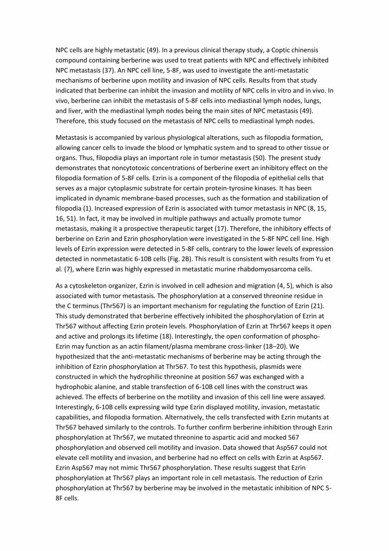
inhibition of NPC cell metastasis (Fig. 10).
FIGURE 10.
Schematic illustration of berberine-inhibited metastasis. Berberine-mediated
repression of Ezrin phosphorylation at Thr567 through suppression of Rho
kinase activity inhibits filopodia formation, resulting in the inhibition of
motility and invasion, which (more ...)
Supplementary Material
Supplemental Data
Click here to view.
Acknowledgments
We thank Professor Shanghui Wu and Hecheng Zhu for kindly providing cell lines. We
appreciate the contributions and helpful discussion of various members in the Ya Cao
laboratory. We thank Dr. Yanna Cao and Dr. Stephen Hampton of the University of Texas for
editing language.
*This work was supported in part by National Natural Science Foundation of China Grant
30572455, Program for New Century Excellent Talents in University Grant NCET-06-0685,
Traditional Chinese Medicine Foundation of Hunan Grant 2008039, Science and Technology
Foundation of Hunan Grant 08FJ3176, and Science Foundation of Central South University
Grant 08SDF07.
The on-line version of this article (available at http://www.jbc.org) contains supplemental
Fig. S1.
2The abbreviations and trivial names used are:
berberine 2,3-methylenedioxy-9,10-dimethoxyprotoberberine chloride
NPC nasopharyngeal carcinoma
MTT 3-(4,5-dimethylthiazol-2-yl)-2,5-diphenyltetrazolium bromide
LDH lactate dehydrogenase
MRCK myotonic dystrophykinase-related Cdc42-binding kinase 2
LOK lymphocyte-oriented kinase
RBD Rho-binding-domain of rhotekin
PAK-PBD GTPase-binding domain of p21-activated kinase
Ezrin M(A) Ezrin T567A
Ezrin
M(D) Ezrin T567D
GST glutathione S-transferase
PBS phosphate-buffered saline
Y27632 (1)-(R)-trans-4-(1-aminoethyl)-N-(4-pyridyl) cyclohexane-carboxamide
dihydrochloride
PKC protein kinase C.
Other Sections▼
Abstract
EXPERIMENTAL PROCEDURES
RESULTS
DISCUSSION
Supplementary Material
REFERENCES
REFERENCES
1. Furutani Y., Matsuno H., Kawasaki M., Sasaki T., Mori K., Yoshihara Y. (2007) J. Neurosci. 27,
8866–8876. [PubMed]
2. Miaczynska M., Pelkmans L., Zerial M. (2004) Curr. Opin. Cell Biol. 16, 400–406. [PubMed]
3. Gould K. L., Bretscher A., Esch F. S., Hunter T. (1989) EMBO J. 8, 4133–4142. [PMC free
article][PubMed]
4. Saotome I., Curto M., McClatchey A. I. (2004) Dev. Cell 6, 855–864. [PubMed]
5. Faure S., Salazar-Fontana L. I., Semichon M., Tybulewicz V. L., Bismuth G., Trautmann A.,
Germain R. N., Delon J. (2004) Nat. Immunol. 5, 272–279. [PubMed]
6. Roumier A., Olivo-Marin J. C., Arpin M., Michel F., Martin M., Mangeat P., Acuto O., Dautry-
Varsat A., Alcover A. (2001) Immunity 15, 715–728. [PubMed]
7. Yu Y., Khan J., Khanna C., Helman L., Meltzer P. S., Merlino G. (2004) Nat. Med. 10, 175–181.
[PubMed]
8. Yang X. Y., Ren C. P., Wang L., Li H., Jiang C. J., Zhang H. B., Zhao M., Yao K. T. (2005) Cell
Oncol. 27, 215–223. [PubMed]
9. Wang G., Mao W., Zheng S. (2008) FEBS Lett. 582, 3663–3668. [PubMed]
10. Palou J., Algaba F., Vera I., Rodriguez O., Villavicencio H., Sanchez-Carbayo M. (2008) Eur.
Urol., in press.
11. Kocher H. M., Sandle J., Mirza T. A., Li N. F., Hart I. R. (2009) Gut 58, 271–284. [PubMed]
12. Elzagheid A., Korkeila E., Bendardaf R., Buhmeida A., Heikkila S., Vaheri A., Syrjanen K.,
Pyrhonen S., Carpen O. (2008) Hum. Pathol. 39, 1737–1743. [PubMed]
13. Li Q., Wu M., Wang H., Xu G., Zhu T., Zhang Y., Liu P., Song A., Gang C., Han Z., Zhou J.,
Meng L., Lu Y., Wang S., Ma D. (2008) Cancer Lett. 261, 55–63. [PubMed]
14. Peng S., Fan S., Li X., Wang L., Liu H., Zhou M., Wang L., Shen S., Li G. (2007) Cancer Sci. 98,
341–349. [PubMed]
15. Zhang Y., Hu M. Y., Wu W. Z., Wang Z. J., Zhou K., Zha X. L., Liu K. D. (2006) J. Cancer Res.
Clin. Oncol. 132, 685–697. [PubMed]
16. Chuan Y. C., Pang S. T., Cedazo-Minguez A., Norstedt G., Pousette A., Flores-Morales A.
(2006) J. Biol. Chem. 281, 29938–29948. [PubMed]
17. Khanna C., Wan X., Bose S., Cassaday R., Olomu O., Mendoza A., Yeung C., Gorlick R.,
Hewitt S. M., Helman L. J. (2004) Nat. Med. 10, 182–186. [PubMed]
18. Matsui T., Maeda M., Doi Y., Yonemura S., Amano M., Kaibuchi K., Tsukita S., Tsukita S.
(1998) J. Cell Biol. 140, 647–657. [PMC free article][PubMed]
19. Chen J., Cohn J. A., Mandel L. J. (1995) Proc. Natl. Acad. Sci. U.S.A. 92, 7495–7499. [PMC
free article][PubMed]
20. Yonemura S., Matsui T., Tsukita S., Tsukita S. (2002) J. Cell Sci. 115, 2569–2580. [PubMed]
21. Zhu L., Zhou R., Mettler S., Wu T., Abbas A., Delaney J., Forte J. G. (2007) Am. J. Physiol. Cell
Physiol. 293, C874–C884. [PubMed]
22. Lee D. U., Kang Y. J., Park M. K., Lee Y. S., Seo H. G., Kim T. S., Kim C. H., Chang K. C. (2003)
Life Sci. 73, 1401–1412. [PubMed]
23. Lau C. W., Yao X. Q., Chen Z. Y., Ko W. H., Huang Y. (2001) Cardiovasc. Drug Rev. 19, 234–
244. [PubMed]
24. Küpeli E., Koşar M., Yeşilada E., Hüsnü K., Başer C. (2002) Life Sci. 72, 645–657. [PubMed]
25. Kuo C. L., Chi C. W., Liu T. Y. (2004) Cancer Lett. 203, 127–137. [PubMed]
26. Iizuka N., Miyamoto K., Okita K., Tangoku A., Hayashi H., Yosino S., Abe T., Morioka T.,
Hazama S., Oka M. (2000) Cancer Lett. 148, 19–25. [PubMed]
27. Mitani N., Murakami K., Yamaura T., Ikeda T., Saiki I. (2001) Cancer Lett. 165, 35–42.
[PubMed]
28. Letasiová S., Jantová S., Cipák L., Múcková M. (2006) Cancer Lett. 239, 254–262. [PubMed]
29. Mantena S. K., Sharma S. D., Katiyar S. K. (2006) Mol. Cancer Ther. 5, 296–308. [PubMed]
30. Lin J. P., Yang J. S., Lee J. H., Hsieh W. T., Chung J. G. (2006) World J. Gastroenterol. 12, 21–
28. [PubMed]
31. Slaninová I., Táborská E., Bochoráková H., Slanina J. (2001) Cell Biol. Toxicol. 17, 51–63.
[PubMed]
32. Kobayashi Y., Yamashita Y., Fujii N., Takaboshi K., Kawakami T., Kawamura M., Mizukami T.,
Nakano H. (1995) Planta Med. 61, 414–418. [PubMed]
33. Peng P. L., Hsieh Y. S., Wang C. J., Hsu J. L., Chou F. P. (2006) Toxicol. Appl. Pharmacol. 214,
8–15. [PubMed]
34. Kuo C. L., Chi C. W., Liu T. Y. (2005) In Vivo 19, 247–252. [PubMed]
35. Lin S. S., Chung J. G., Lin J. P., Chuang J. Y., Chang W. C., Wu J. Y., Tyan Y. S. (2005)
Phytomedicine 12, 351–358. [PubMed]
36. Hwang J. M., Wang C. J., Chou F. P., Tseng T. H., Hsieh Y. S., Lin W. L., Chu C. Y. (2002) Arch.
Toxicol. 76, 664–670. [PubMed]
37. Liu S. J., Sun Y. M., Tian D. F., He Y. C., Zeng L., He Y., Ling C. Q., Sun S. H. (2008) Br. J.
Cancer 98, 363–369. [PMC free article][PubMed]
38. Attiga F. A., Fernandez P. M., Weeraratna A. T., Manyak M. J., Patierno S. R. (2000) Cancer
Res. 60, 4629–4637. [PubMed]
39. Ishizaki T., Uehata M., Tamechika I., Keel J., Nonomura K., Maekawa M., Narumiya S. (2000)
Mol. Pharmacol. 57, 976–983. [PubMed]
40. Ishii S., Kihara Y., Shimizu T. (2005) J. Biol. Chem. 280, 9083–9087. [PubMed]
41. Park E. J., Ji K. A., Jeon S. B., Choi W. H., Han I. O., You H. J., Kim J. H., Jou I., Joe E. H. (2004)
J. Immunol. 173, 5697–5703. [PubMed]
42. Gallo G. (2008) Dev. Neurobiol. 68, 926–933. [PubMed]
43. Schafer C., Borm B., Born S., Mohl C., Eibl E. M., Hoffmann B. (2009) Exp. Cell Res. 313,
1212–1224. [PubMed]
44. Belkina N. V., Liu Y., Hao J. J., Karasuyama H., Shaw S. (2009) Proc. Natl. Acad. Sci. U.S.A.
106, 4707–4712. [PMC free article][PubMed]
45. Jiménez-Sainz M. C., Murga C., Kavelaars A., Jurado-Pueyo M., Krakstad B. F., Heijnen C. J.,
Mayor F., Jr., Aragay A. M. (2006) Mol. Biol. Cell 17, 25–31. [PMC free article][PubMed]
46. Cant S. H., Pitcher J. A. (2005) Mol. Biol. Cell 16, 3088–3099. [PMC free article][PubMed]
47. Ng T., Parsons M., Hughes W. E., Monypenny J., Zicha D., Gautreau A., Arpin M.,
Gschmeissner S., Verveer P. J., Bastiaens P. I., Parker P. J. (2001) EMBO J. 20, 2723–2741. [PMC
free article][PubMed]
48. Bishop A. L., Hall A. (2000) Biochem. J. 348, 241–255. [PMC free article][PubMed]
49. Leung T. W., Tung S. Y., Sze W. K., Wong F. C., Yuen K. K., Lui C. M., Lo S. H., Ng T. Y., O S. K.
(2005) Head Neck 27, 555–565. [PubMed]
50. Machesky L. M. (2008) FEBS Lett. 582, 2102–2111. [PubMed]

51. Deng X., Tannehill-Gregg S. H., Nadella M. V., He G., Levine A., Cao Y., Rosol T. J. (2007)
Clin. Exp. Metastasis 24, 107–119. [PubMed]
52. Ridley A. J., Allen W. E., Peppelenbosch M., Jones G. E. (1999) Biochem. Soc. Symp. 65,
111–123. [PubMed]


![Nasopharyngeal Carcinoma [Ind] - Fix 19](https://img.pdfslide.net/doc/110x75/55cf9043550346703ba47221/nasopharyngeal-carcinoma-ind-fix-19.jpg)